A STUDY ON NERVE CONDUCTION STUDY IN VIBRATORY TOOL USERS
Dissertation submitted to
THE TAMILNADU DR. M.G.R MEDICAL UNIVERSITY,
CHENNAI – 600032
In partial fulfillment of the requirement for the degree of Doctor of Medicine in Physiology ( Branch V )
M.D. ( PHYSIOLOGY ) APRIL 2019
DEPARTMENT OF PHYSIOLOGY COIMBATORE MEDICAL COLLEGE
CERTIFICATE
This dissertation entitled A STUDY ON NERVE CONDUCTION
STUDY IN VIBRATORY TOOL USERS” is submitted to The Tamil Nadu Dr.M.G.R Medical University, Chennai, in partial fulfillment of
regulations for the award of M.D. Degree in Physiology in the
examinations to be held during April 2019.
This dissertation is a record of fresh work done by the candidate
Dr.A. RAJKUMAR, during the course of the study ( 2016 – 2019 ). This work was carried out by the candidate himself under my supervision.
GUIDE:
Dr.D.SELVAM. M.D.,DCH,
Associate Professor,
Department of Physiology,
Coimbatore Medical College,
Coimbatore – 14.
PROFESSOR & HOD:
Dr.R.SHANMUGHAVADIVU. M.D.,
Professor,
Department of Physiology,
Coimbatore Medical College,
Coimbatore – 14.
DEAN:
Dr.B. ASOKAN M.S., M.Ch.,
Coimbatore Medical College & Hospital,
DECLARATION
I, Dr.A.RAJKUMAR solemnly declare that the dissertation entitled
“A STUDY ON NERVE CONDUCTION STUDY IN VIBRATORY TOOL
USERS” was done by me at Coimbatore Medical College, during the
period from July 2017 to June 2018 under the guidance and supervision
of Dr.D.SELVAM M.D.,DCH., Associate Professor, Department of
Physiology, Coimbatore Medical College, Coimbatore.
This dissertation is submitted to The Tamilnadu Dr. M.G.R. Medical
University towards the partial fulfillment of the requirement for the award
of M.D. Degree (Branch - V) in Physiology. I have not submitted this
dissertation on any previous occasion to any University for the award of
any degree.
Place:
Date:
ACKNOWLEDGEMENT
I express my sincere thanks to our respected Dean,
Dr.B.ASOKAN M.S., MCh., Coimbatore Medical College, Coimbatore for permitting me to conduct the study.
I thank Dr.P.KALIDAS M.D., Vice Principal, Coimbatore Medical
College, Coimbatore for his encouragement and suggestions in completing
the study.
I am extremely grateful to my beloved and respected Head of the
Department of Physiology, Professor Dr. R.SHANMUGHAVADIVU, M.D.,
for her encouragement in helping me to take up this study. I express my
heart - felt gratitude to her, for her moral support and encouragement
throughout the conduct of the study and also during my post graduate
course. I owe my sincere thanks to her.
I will ever remain in gratitude to Dr.D.SELVAM, M.D.,DCH.,
Associate Professor, Department of Physiology for his valuable support
and guidance for my study.
I am highly obliged to Dr.B. SUJATHA, M.D.,DA., Associate Professor, Department of Physiology, for her motivation to perform this
work.
I sincerely thank Dr.R.THENMOZHI, M.D., D.C.P., Associate
Professor, Department of Physiology for her valuable suggestions and
sparing her valuable time and patience, which helped me a lot to complete
this study under her expert guidance
My sincere thanks to beloved teachers Dr.S.Kavitha M.D., Dr.A.Abbas, M.D., Dr.A.Moorthy, M.D., Dr.E.S.Manikandan M.D., Dr.S.Kanchana Bobby M.D., Dr.P.Mohan.,M.D., Dr.S.Subhashini M.D., Mrs.D.Revathy M.sc., Dr.C.N.Angel Deepa, M.D., Dr.N.Latha M.D., Dr.K.Archanaa M.D., Assistant Professors, Department of Physiology for their valuable opinion and help to complete this study. I would like to
thank all my tutors for their support in completing this study.
I would grossly fail in my duty, if I do not mention here of my
subjects who gave full cooperation while doing my study.
My sincere thanks to all my fellow postgraduates for their involvement in helping me in this work.
CERTIFICATE - II
This is to certify that this dissertation work titled “A STUDY ON NERVE
CONDUCTION STUDY IN VIBRATORY TOOL USERS” of the
candidate Dr. A.Rajkumar with registration Number 201615253 for the
award of Doctor of Medicine in the branch of Physiology. I personally verified the urkund.com website for the purpose of plagiarism Check. I
found that the uploaded thesis file contains from introduction to conclusion
pages and result shows 3% percentage of plagiarism in the dissertation.
Guide sign with Seal.
CONTENTS
S.NO CONTENTS PAGE NO
1. INTRODUCTION 01
2. AIMS AND OBJECTIVES 03
3. REVIEW OF LITERATURE 04
4. MATERIALS & METHODS 24
5. RESULTS 31
6. DISCUSSION 51
7. SUMMARY 75
8. CONCLUSION 77
9. BIBLIOGRAPHY
ABBREVIATIONS
NCV : Nerve Conduction Velocity
MNCV : Motor Nerve Conduction Velocity
SNCV : Sensory Nerve Conduction Velocity
BMI : Body Mass Index
Ht : Height
1
INTRODUCTION
In this modern day, industrialization, consequent urbanisation and
the development of infrastructure have undergone mechanisation. To
reduce the manual labour due to scarcity of workers, save the time and
money, many industrial tools have been developed which had brought
in significant changes in the pattern of working in construction industry.
The construction of buildings involves massive work of cutting of
wood, timber and concrete into various shapes and sizes to be fit into
the buildings especially prefabricated structures. The cutting tools used
are chainsaws, hand drills, rock drill and the tamper which are hand
held. Among them the chainsaws are the commonly used instruments.
The chainsaws when held in the hand and operated, produce
vibration. When there is a change in the environmental stimulus, the living
cells get excited. The nerve is one such important excitable tissue1. In humans
conduction of nerve impulse is the specialised function of nerves2. The hands
and forearm are exposed to vibrations when the persons handle the
tools for many years and chronic vibration exposure occurs. This
vibrations can damage the muscles, tendons, joints , arteries, veins and
peripheral nerves. The vascular injury commonly seen is Raynauds
phenomenon3. The symptoms that are produced due to vibration
exposure is called vibration syndrome and it is related to vascular
2
As the chronic vibration exposure continues, the peripheral nerve
fibres can undergo damage. When a peripheral nerve gets damaged, the
myelination and axons are affected4. As the damage becomes significant,
the injury becomes irreversible and cumulative. When the nerve is
finally damaged, complete symptoms of peripheral neuropathy and its
complications develop5.
As the nerve injury starts occurring, axon and myelin sheath get
injured, and the nerve conduction is affected. When this nerve
conduction is studied electrophysiologically in the form of motor and
sensory nerve conduction velocities of peripheral nerves in upper limbs,
the results show changes in vibration exposed individuals, before the
development of established vibration syndrome6. Hence it will be of
immense use in early identification of nerve damage and plan for
alternate works for the individuals and to apply preventive measures in
the field of occupational vibration exposure.
The research reports on the nerve conduction velocity which is
an useful electrophysiological study to know the effect on peripheral
nerves are varying and less.
Hence this study is undertaken to study the motor and sensory
nerve conduction velocities of median, ulnar nerves and motor conduction
velocity of radial nerves of both upper limbs in chainsaw users and the
individuals not exposed to vibration and the data are compared to evaluate
3
AIM AND OBJECTIVES
AIM:To study the nerve conduction study in vibratory tool users.
OBJECTIVES:
Cross sectional study is done on the persons working with the
chainsaw which is a vibratory industrial tool. The nerve conduction
velocity study is conducted on median, ulnar, radial nerves on both
forearms. Nerve conduction study is conducted in individuals who are
not working with chainsaws. Both the data are compared to study the
4
REVIEW OF LITERATURE
Vibration induced nerve injury was initially reported in 1911 by
Loriga in the mines of Italy. Later in 1918 by Hamilton in Indiana7. In
due course , the musculo skeletal and neurological abnormalities became
established side effects of vibration. The latency of injury may vary
from less than a year to four decades depending on the degree of
vibration8.The vibration exposure is prevalent in construction industry,
forestry, mining, foundry, automobile and metal works.
The pathogenesis appears to be due to local endothelial damage
by a mechanical trauma and oxidative stress that can produce nerve
damage and vaso constriction by sympathetic discharge9. The vibration
damage occurs in large myelinated fibres and small unmyelinated and
myelinated fibres disproportionately . Local nerve damage can result in
muscle damage also.
High frequency vibration is produced by drills, milling machines,
chisels, sanding-cutting-polishing machines10. This can also produce
sensory neural damage and vascular damage. The low frequency vibration
is transmitted to the shoulders and arms which can cause musculo
skeletal abnormalities. The vibration is related to duration intensity, type
of the tools, work place, temperature, posture, rest breaks and grip
5
HISTORY
After the discovery of electricity, rapid advancement in the field
of electrical neurophysiology was made possible. Many researchers
specialised in the field of neurophysiology and had done animal
experiments and discovered the functions of nerves and muscles.
Benjamin Franklin (1745 – 1791), a scientist from Philadelphia
described positive and negative electrical charges12.
HISTORY OF NERVE CONDUCTION STUDY;
Galvani (in 1791) of Bologna University found out that the
nerves act as the conductors of electricity. He described the appearance of
spark during the handling of the amphibian nerve with a knife13. He
described that the electricity currents originate in the body and
channelled in the nerves. He also showed that when free nerve endings
were placed between 2 plates of metals, muscle contraction occurred in
amphibian muscle preparation. In 1838 Matteucci showed that following
the sciatic nerve stimulation, gastrocnemius contraction occurred in
sciatic nerve preparation14. In 1932 Charles Scott Sherrington explained
the Stretch reflex and its role in running and walking. In 1944, an
American Scientist Joseph Erlanger stated that the diameter of nerve
fibres too had contributed to the nerve conduction. Herber S. Gasser in
1944 classified the nerve fibres based on the diameter. Raymond found
that changes in the nerve potential, travel as impulse, down the nerve
Picture 1: Resting Membrane Potential
6
time documented the nerve conduction velocity by recording muscle
contraction. Tustin had described that median nerve conduction velocity
for motor component was 61 m/sec and sensory component was 60
m/sec16.
History of neurophysiology is a mixture of human intelligence,
advancements in technology and instrumentation with positive and
negative responses. Prior to a mechanical contraction response in a
muscle, a wave of electrical excitation occurs. This was first
demonstrated by Burden Sandarson in 189517. Duchenne undertook
studies on neuromuscular diseases based on this observation18.
When Erb in 1861 introduced the different patterns of current, it
became possible to conduct animal studies19. The patterns are two types.
One is Indirect or alternating current which is of high voltage and low
ampere and the second is galvanic current which is direct, having low
voltage and high ampere20.
Potential difference across the cell membrane was measured by
using the micropipette of 0.5µm in size which was developed by John
Eccles of Australia in 196321.
Squid and Cattle fish have giant axons. Utilising this fact, Alan
Lloyd Helghin and Andrews Huxley of United Kingdom studied the
nerve fibre electrical conduction22. They developed the voltage clamp
7
ions enter and same amount exit during the first half and second half
of voltage peak respectively per micrometer23.
They used radio active Na+ions and demonstrated that nearly
20,000 ions enter the cells per µm2 for each nerve impulse during first
half of voltage peak and same amount of ions come out in second half
of voltage peak24. They both described voltage clamp technique to
measure the potential difference across cell membrane. In 1970, British
Scientist Bernard Katz explained the synaptic transmission25.
NERVE CELL PHYSIOLOGY:
Nerve cell membrane is made up of lipid bilayer where protein
molecules are embedded. Electrically charged ions cannot pass through
it. The embedded proteins provide channels for the passage. The
proteins are called voltage gated ion channels. The most excitable part
in a nerve is axon hillock, the point at which axon leaves the cell body26.
The membrane potential in quiescent cells is called the resting membrane
potential( RMP).
RMP is the voltage difference between the two electrodes placed
inside and outside of the axon. Na+- K+ ATPase is the enzyme that
pumps 3 Na+ ions out of the cells and 2 K+ ions into the cells27.
Potassium leak channels also allow K+ions to move out of the cells.
This results in the negative charge inside the cell membrane compared
to exterior. When the concentration gradient which allows K+
8
balanced, equilibrium is achieved28. Now the potential is called
equilibrium potential. It is calculated by Nernst Equation29.
R – gas constant (1.987 cal/molK), T-Temperature, [X] out –
concentration outside, [X] in – concentration inside, Z- Charge of the ion,
F – Faradays constant. Resting membrane potential of axon is – 70Mv30.
NERVE FIBRE
As the nerve fibre leaves the cell body, it is covered by myelin
sheath. When the nerve fibre leaves the central nervous system, it is
covered by a second covering called neurolemma. As the fibre ends at
the periphery, the neurolemma is lost first, next the myelin sheath and
finally the axis cylinder. The axis cylinder ends as a naked process
without any covering31.
At the nerve ending, motor fibre splits into 150 branches which
end in muscle fibres. Motor unit is defined as the one nerve fibre along
with all the muscle fibres it supplies.
Myelination in peripheral nerve fibres.
Axon is the central core and the conducting membrane is the
surface of axon. The tissue fills the inter cellular area of axon. It is
called axoplasm32.
The myelin sheath is a lipid material that surrounds the axon.
The sheath is present in all somatic nerves. Myelin sheath of somatic
9
sheath. The nucleus of this cell is present near outer membrane of the
cells. This Schwann cells deposit myelin sheath33.
Physiological properties of nerve fibre are studied by Cathode
Ray Oscilloscope (CRO).
The study of action potential;
A micro electrode is placed inside the nerve fibre and at outside,
a differential electrode is placed. The micro electrode contains
concentrated potassium chloride solution34.
The two important properties of nerve fibres are; excitability and
conductivity.
Excitability:
The mechanical, thermal, chemical or electrical stimulus can
stimulate a nerve fibre. The stimulated part becomes electrically negative.
This can be detected by galvanometer or Cathode ray oscilloscope. When
this changes reach a threshold level, depolarisation occurs rapidly to
10
ERLANGER AND GASSER CLASSIFICATION OF NERVE FIBRES: MOTOR FIBRES Type Erlanger-Gasser Classification
Diameter Myelin Conduction
velocity
Associated muscle fibers
α Aα 13–20
µm Yes 80–120 m/s
Extrafusal muscle fibers
γ Aγ 5–8 µm Yes 4–24 m/s [2][3]
Intrafusal muscle fibers SENSORY FIBRES Type Erlanger-Gasser Classification
Diameter Myelin Conduction
velocity
Associated sensory receptors
Ia Aα 13–20
µm Yes
80–120 m/s[4]
Responsible for proprioception
Ib Aα 13–20
µm Yes 80–120 m/s Golgi tendon organ
II Aβ 6–12 µm Yes 33–75 m/s
Secondary receptors of muscle spindle. All
cutaneous mechanoreceptors
III Aδ 1–5 µm Thin 3–30 m/s
Free nerve endings of touch and pressure.
Nociceptors of neospinothalamic tract
old thermoreceptors
IV C 0.2–1.5
µm No 0.5–2.0 m/s
Nociceptors of paleospinothalamic tract. Warmth receptor
ACTION POTENTIAL
At resting stage; the negative potential exists inside the cell and
outside is positive. Na+ concentration is more outside and K+
11
cannot move in. the membrane pores are bound by calcium and Na+
cannot enter. When excited by an action potential, Ca+ moves out of the
binding site and tremendous Na+ conduction entry occurs. This is called
activation of membrane37. So, reversal of potential occurs with negative
outside. As the action potential reaches +35mV, calcium binds to the
pores and sodium ion entry is prevented. The stage of repolarisation
occurs37. The K+ conductance increases and K+ comes out of cell. At
the later stage, this K+ conductance is slowed down, and it is called
negative after potential. The active Na+ pump mechanism utilises ATP
for energy, Na+ is pumped out and K+ into the cell and resting
membrane potential is reached38.
Compound Action Potential
When the potential is recorded in a group of nerve fibres or
trunk of nerve, it is called compound action potential40. It is a
summated potential of nerve fibres with different conduction velocities.
Most of the nerves have myelinated nerve fibres with various diameter41.
This was studied in frog by Erlanger and Gasser. When
maximum shock is given, pressure is given between recording and
stimulating electrode, the pressure stops the conduction in thick fibres
initially. In an evoked potential, the earlier portion represents large
12
The conduction amplitude height represents all of the axon
bundles in the nerve .So first α wave appears then β, γ, δ and Β and
finally C wave of unmyelinated fibres occur serially43.
Conductivity:
This happens by the ubiquitously ATP utilising process of nervous
axons for the maintenance of metabolic integrity to provide nutrients to
the axons. The axons transport occur in anterograde and retrograde
fashion44. The velocity can be varying. The stages of axonal transport of
Signalling proteins are;
1) The synthesised new proteins are packed into organelles and sent
to the proximal axon.
2) The distally directed movement which is occur with the pauses
and transient reversal.
3) Arriving at the destination and get incorporated into axolemma.
4) Turn around
5) Dynein driven transport which is retrograde.
6) Lysosomes digest during transit or at arrival in cell body.
Nerve growth factors are transported in retrograde fashion from
nerve ending to cell bodies after entering by endocytosis45.
Propagation of Action Potential
At one point on the membrane , action potential occurs. The
action potential then excites the adjacent portion of membrane leading
Picture 3: Action Potential
13
permeability to sodium is increased46. The sodium ions diffuse in and
positive charges move in through depolarised membrane. Inside the
myelinated fibre, the voltage in increased by these positive charges
above threshold level and initiate action potential47.
Now in the new areas, sodium channels open and the spread of
action potential occurs. The new current flow and local circuit occurs
along the membrane. The depolarisation travels across the entire length.
This transmission is called Nerve Impulse48.
Direction
The action potential can travel in all directors in all branches of
nerve fibre till whole membrane is depolarised49.
All or none principle:
The process of depolarisation travels in the membrane if
environment is favourable, and does not travel if not favourable. When
action potential cannot generate voltage sufficient enough to stimulate
the next area, depolarisation process ceases. So, action potential to
threshold ratio should be more than 1 and it is the safety factor49.
The large nerve fibres are myelinated and small are unmyelinated.
A nerve trunk has 2 times of unmyelinated fibres than myelinated
fibres. In myelinated axon, the membrane conducts action potential.
Axon is filled with axoplasm. Axon is surrounded by myelin sheath.
14
The Schwann cell membrane rotates around the axon many times
by the lipid substance - sphingomyelin, which is an insulator which
reduces the ion flow by 5000 fold. Nodes of Ranvier occur at every
1-3 micrometer in the nerve which is uninsulated50.
SALTATORY CONDUCTION
In myelinated fibres, action potential occurs at the Nodes of
Ranvier only. The current flows in extracellular fluid outside and
axoplasm inside and reach the next node in an axon, exciting
subsequent nodes one by one. Hence impulse jumps from node to
another node51.
This mechanism increases the conduction velocity by 5-50 fold.
Also it causes conservation of energy by 100 times . So it requires low
metabolism for re-establishment of sodium and potassium concentration.
The velocity is 0.25 m/sec in unmyelinated fibres and 100 m/sec in
myelinated fibres51.
The impulse gets propagated in both the directions. When
diameter of nerve increases, velocity of conduction also increases.
Conductivity Hursh factor is 6 for humans, which is the ratio of
velocity to diameter. So when diameter of nerve is known , velocity can
be calculated52.
Conduction velocity of nerve bundle is calculated by giving
electrical stimulation at one end and recording the action potential at
15
Myelinated fibres have conduction velocity depending on their
diameter. In the unmyelinated nerve fibre, conduction is proportional to
the square root of diameter.
Other factors that influence conduction velocity are;
1. Temperature; cooling decreases the conduction,
2. Pressure; Increased pressure decreases conductivity,
3. Blood supply; if reduced , conductivity is reduced,
4. Chemicals; CO2 and narcosis diminish the conduction,
5. H+ ion Concentration; increased H+ ion, decreases conductivity,
6. O2; decreased O2, decreases the conductivity
Anatomy – Median Nerve
Median nerve has both motor and sensory components. It is
derived from the spinal nerve roots C5 to T1 through the lateral and
medial cords in brachial plexus.
It is motor to flexors of forearm and muscles of thenar
eminence. It provides sensory fibres to the palm on its lateral aspect,
terminal phalanges on dorsal surface along with thumb, index finger,
middle finger and half of the ring finger53.
Between the heads of pronator teres, it enters the forearm. It
16
flexor carpi radialis. Its anterior interosseous branch is a muscular
branch for flexor digitorum profundus, pronator quadratus and flexor
pollicis longus54.
Then it enters carpel tunnel. In the hand, first and second
lumbricals, flexor pollicis brevis, opponens pollicis and abductor pollicis
brevis are supplied55.
Before entering the carpel tunnel, a sensory branch called palmar
cutaneous branch is given that supplies thenar muscles56.
Ulnar Nerve
Brachial plexus gives medial cord from which the C7, C8, T1
root fibres form ulnar nerve. It lies close to brachial artery and median
nerve in the arm. Posterior to the epicondyle, it is located at condylar
groove. It enters cubital tunnel (Feindel and Straford 1958).
The cubital tunnel is formed by the medial ligament of elbow on the
floor and flexor carpi ulnaris aponeurosis on the roof56.
Here, flexor carpi ulnaris branch arises, then branches to flexor
digitorum profundus arise. In the Guyon’s Canal , between hook of
hamate and pisiform bone, it passes into the wrist57.
The fourth digit on its ulnar border and fifth digit receive
sensory supply. The hypothenar muscles that is; flexor digiti minimi,
abductor digiti minimi and opponens digiti minimi are supplied by the
17
At the lateral aspect of the hand, it supplies III and IV
lumbricals, interossei, flexor pollicis and adductor pollcis58.
The dorsal and palmar cutaneous branches do not pass through
the Guyon’s Canal58.
Radial Nerve
Brachial plexus gives posterior cord that continues as radial
nerve,. The spinal nerve roots are C5 to T1. All the three heads of the
triceps are supplied. Then it passes around spiral groove in humerus.
Posterior antibrachial branch is given in the spiral groove. It is
superficial distal to the deltoid insertion59.
It supplies extensor carpi radialis longus and brevis and brachio
radialis. Between branchioradials and brachialis, it enters forearm.
Posterior interossei branch in the forearm, gives branch to supinator
muscle. The nerve passes in between the superficial and deep parts of
muscle, piercing the arcade of Frohse. Then branches are given to
extensor indicis, extensor pollicis brevis, extensor pollicis longus,
extensor digiti minimi, extensor digitorum, extensor carpi ulnaris and
abductor pollicis longus. The cutaneous nerve passes on the lateral
aspect of forearm and supplies the hand on dorsal aspect59.
18
PERIPHERAL NERVES;
The peripheral nerves are surrounded by successive layers of
connective tissue. The axons are surrounded by perineurium.
Perineurium pack the axons into fascicles. The nerve is composed of
fascicles packed by epineurium. The blood vessels to the nerves are
called Vasa Vasorum. The nerves to the nerves are called Nervi
Nervorum. The axon is a cytoplasmic protrusion from the body of
neuron. The axon has a constant radius and longer than dendrites and
they transmit signals60.
The axolemma cover the axons. They are membranes. The
axoplasm is the cytoplasm. Telodendria are the end branches of axon.
The axon terminal is called telodendron which synapse with the other
cell body61.
When it forms synapse with the dendrite of same neuron, it is
called autapse. At synapse, the junction is formed with glands and
muscles. When synapse appear at the entire length of the axon it is
called en passant synapse61.
Discussion on nerves
The sensory nerves are the cable like bundles from the different
fibres that originate in sensory receptors of the peripheral nerves. The
fibres are usually paired with the efferent fibre of motor nerves and
put together to form the peripheral nerves. These different nerves leave
19
Some sensory neurons called pseudounipolar neurons, that
transmit warmth and touch, conduct the impulse from periphery to cell
body. Again the impulse conducted from cell body to spinal cord
through another branch of same axon. The myelinated nerve fibres are
group A and group B fibres. The unmyelinated nerve fibres are group
C fibres. The sensory fibres alone are separately grouped into type I,
type II, type III and type IV62.
BIOELECTRIC POTENTIALS
Bioelectric potentials are generated from the sources inside the
body that is peripheral nerves, muscles, and brain. They are recorded
by electrodes which are placed in same distance away. The potentials
originate from neuronal membranes which allows current flow from in
and out of the cell by capacitative effects and passive leakage. These
current lead to extracellular currents that flow in the conducting
medium in the body which is called as volume conductor. Volume
conduction is the transfer of potentials to a distance. In clinical
neurophysiology, the human body acts as nonhomogenous volume
conductor. These currents reach the surface in the skin. Hence potential
difference is created across the two electrodes placed over the skin. The
differential amplifier can be used for detection and amplification of
20
volume conductor, recording electrodes, propagation through volume
conductor, distance of propagation, all decide the effect of recording.
The potential in neurons is the sum of potentials generated by
individual neurons.
In a macroscopic level, the cortical pyramidal cells behave like
dipole layer and their synchronous activation generates potential field.
This is called open field configuration. But in the neurons with
dendritic arborizations, the fields generated are distributed radially
around cell body and called closed fields. Set of radially oriented
dipoles on the surface of the sphere produce fields that produce closed
field potentials. Such field is negligible at the distance because radial
and tangential current flow cancel each other.
Motor cortex or peripheral motor and sensory nerves are
stimulated and initiate peripheral evoked potentials. Motor cortex
potentials travel peripherally to anterior horn cells and muscles. These
potentials can be recorded in spinal cord, peripheral nerves and muscles.
Peripherally generated sensory nerve impulse travel in central direction
to cortex via dorsal nerve roots and spinal cord dorsal columns.
The peripheral nerve potentials have unique properties in volume
conductors. The nerve potentials are recorded from overlying skin
electrode as close to the generating source as possible. But the
21
The peripheral nerve fibres can be recorded individually, but the
synchronous volley of potentials are recorded from multiple grouped
parallel fibres which produce the nerve potentials that are recorded
clinically. The wave forms generate nerve action potential. The
travelling potential in a nerve fibre is represented in two dipoles placed
end to end. The configuration and size of the potential depends on the
recording electrode and generator and seen as positive wave forms.
Sensory evoked potentials at the cortex are the summated potentials
from cortical neurons.
A single source of current is called monopole. The magnitude of
current decreases with the distance away from current source can be
measured along the equipotential lines. Each such line represents a
constant potential along the line.
In a nervous system, adjacent monopoles of opposite polarity
define the dipole. Here, current flows from positive to negative pole.
Potential lines are generated away from dipole. The magnitude of
current falls off inversely in relation to distance from source.
The electrical activity in cortical neurons is contributed by the
excitatory and inhibitory post synaptic potentials in dendritic trees of
pyramidal neurons.
The potential of dipole fall of inversely with square of distance
22
The dipole records in relation to distant reference at points
perpendicular to dipole axis, appears as a single peak. This has sharps
that increases with decreasing distance from source.
In peripheral recordings in resistive-capacitive medium, the
volume conduction is a frequency dependent factor. The potentials are
out of phase. The latency recorded in a distance is shorter than that
recorded over nerve.
When potential difference is large, a high spatial gradient is
present. When it is high the potential is called a near field potential. In
motor conduction study, the typical recording G1 Montage over the
motor end plate and G2 over muscle tendon. It allows for initial
negative wave form with high amplitude. A negative positive biphasic
wave form is recorded.
Sensory nerve conduction studies produce biphasic or triphasic
wave forms. Triphasic wave forms are seen in sensory nerve
conduction. In motor conduction study, the electrodes are placed over
site of action potential generation. In sensory nerve conduction study
the action potential is always generated away from the recording
electrode.
Nerve conduction studies record the evoked response in response
to stimulation of peripheral nerves. The Nerve conduction velocity study
(NCV) are used as confirmatory tests in the suspicious neuropathy in
23
demyelination, acute, chronic or sub acute disease. The prediction of
prognosis of disease is also possible. Several factors determine the
accurate value of results.
CHAINSAW;
Chainsaw is a movable, mechanical instrument. It has a chain
with attached teeth. This can rotate on a guide bar. It is commonly
used in the construction industry for cutting of wood into various
shapes and sizes. At first , an instrument called Osteotome was invented
by Bernhar Heinean an orthopaedist in 1830. In 1783, hand saw with
serrated chain was invented. In 1927, first gasoline powered chain saw
was developed by EMIL LERP. It is a two stroke petrol engine using
internal combustion system. It has an elongated guide bar with alloy
steel which is 90 cm in length. It has tooth which is made up of
chromium plated steel. The instrument has chain brake and rear handle
guard. The chain saws produce vibrations and emit carbon monoxide63.
Now the electrically operated chainsaws and diesel operated chainsaws
24
MATERIALS AND METHODS
The design of the study is cross sectional study. The study was
conducted during the year 2017-2018. Ethical committee approval was
obtained from the Ethics committee of the Coimbatore Medical
College.
Cases / Exposed to vibration:
The subjects were selected from those working in the
construction sites in the Coimbatore area. These workers were selected
in their morning assembling area. The age range set was 20-35 years.
All the workers were males. Those workers who are on duty for 8
hours per day and using the hand held electrically operated chainsaw
for a minimum duration of 3 hours per day, working for atleast 5
days per week and handling the instrument for atleast 5 years of
duration were selected. Among them 50 cases were included for the
study. Written informed consent was obtained form each individual.
Their name, age, sex, height and weight were recorded. History
was taken and clinical examination was done .
Controls / Non exposed:
About 50 males in the age group of 20-35 years working in the
nearby areas not using the hand held vibratory tools in their profession
were selected. Written informed consent was taken. History was taken
25
Exclusion criteria:
In both the cases and controls, those persons having diabetes
mellitus, hypertension, neurological disorders, peripheral nerve diseases,
chronic smokers, chronic alcoholics, obesity, spinal cord diseases, cardio
vascular diseases, chronic respiratory diseases, fever were excluded.
RECORDING OF NERVE CONDUCTION:
Neuroperfect was placed in smooth levelled surface, it was kept
away from transformers, DC motors, Powers appliances to eliminate
electromagnetic interference. Proper grounding of AC outlets was done.
MOTOR CONDUCTION:
Electrode was applied at appropriate position. Nerve was
stimulated by pressing the foot switch corresponding to single. The
strength stimulus was increased or decreased by adjusting the control
provided on the stimulus electrode.
On getting satisfactory waves form, it was recorded.
SENSORY CONDUCTION;
Electrode was applied at appropriate position. Nerve was
stimulated by pressing the foot switch. The stimulus was adjusted by
adjusting the control provided on the stimulating electrode. The nerve
was stimulated till the averaged wave becomes smooth and sensory
nerve action potential became prominent.
Cursors for voltage and latency measurement get marked
26
Settings:
Motor nerve conduction velocity;
Sweep speed : 2ms/div,
Sensitivity : 3mV/div,
Hi filter : 10 khz ,
Lo filter : 2kh
Notch filter : One
Sensory nerve conduction velocity;
Sweep speed : 2 ms/div;
Sensitivity : 10µv/div;
Hi filter : 3khz;
Lo filter : 20 hz
Notch filter : On
Settings:
The interface between the hardware unit (neuroperfect) and
computer is done by using 9 pins D type connector. This connector can
be interfaced at any two of the serial ports available at PC side (com1,
and com2). In order to make the software settings, where the interface
connector is placed, port setting is done using the settings button.
Selecting this button, a new window pops up. The port where the
interface connector is placed must be appropriately selected for proper
Picture 8: Recording of Median Nerve Motor Conduction Velocity
27
Median nerve to the abductor pollicis brevis,(motor component);
Position: Subject was in supine position.
Active electrode(A): It was placed between the first metacarpo
phalangeal joint and the distal wrist crease in its midpoint , that is over
the abductor pollicis brevis.
Reference electrode: It was placed little distal to the first metacarpo
phalangeal joint that is over the tendon.
Ground electrode: It was placed near active electrode between the cathode
and active electrode.
Stimulation Point (1): The flexor carpi radialis tendon was located. A
point ulnar to the tendon was noted. It was the first point. Midpoint of
the distal wrist crease was the second point. A line was drawn between
the points. Isolated stimulator cathode was placed 3cm proximally from
active electrode in this line. Anode is placed proximally.
Stimulation point (2): In the anticubital region, brachial pulse was felt.
Medial to this pulse, cathode was placed.
Instrument settings: 3mV division is the sensitivity, 2-3 Hz is low
frequency filter, 10kz is high frequency filter. 2m sec/division is the
sweep speed.
Tested fibres: Brachial plexus medial cord - anterior division in lower
trunk- C8 to T1 nerve roots.
28
Median nerve; Sensory Nerve Conduction study;
Ring electrode nerve conduction study was performed. Recording
electrode was placed at first interphalangeal joint in the second digit.
Stimulating electrode was placed at 3cm from distal wrist crease
proximally. Reference electrode was placed 3cm proximally. The distant
latency, conduction velocity and action potential were measured.
Ulnar nerve: motor nerve conduction velocity:
It is motor nerve to the muscle abductor digiti minimi.
Position: The arm was kept abducted, externally rotated at 45˚. The
elbow was flexed to 90˚. Thumb pointing to the ear was the neutral
position of forearm.
Active Electrode(A): A midpoint was marked between the 5th metacarpo
phalangeal joint and pisiform bone. Electrode was placed over
hypothenar eminence in this point.
Reference Electrode (R): It was placed near 5th metacarpo phalangeal
joint distally.
Ground Electrode: It was kept between the active electrode and cathode.
Stimulation Point (S1): Cathode was placed near active electrode 8cm
proximally over a line that is radially running near the tendon of
flexor carpi ulnaris. Anode was located proximal to it.
Stimulation Point(S2): Near the medial epicondyle, about 4cm distally,
Picture 10: Recording of Ulnar Nerve Motor Conduction Velocity
29
Settings: 3mV/division was sensitivity, 2-3Hz is low frequency filter,
10Hz was the high frequency filter, 2mS division was sweep speed.
Tested Nerve: C8, T1 roots in lower trunk and anterior division in the medial cord of brachial plexus.
ULNAR NERVE Sensory Nerve Conduction Study
Antidromic nerve conduction study was carried out. Cathode was
placed near distal wrist crease 3cm proximally. Nerve conduction was
recorded from fifth digit inter phalangeal ring electrode
RADIAL NERVE Motor nerve conduction study Motor nerve to the extensor carpi ulnaris
Position: The subject was kept in supine position.
Active Electrode(A): A midpoint was marked between the lateral
epicondyle and ulnar styloid process in the midforearm. Active electrode
was placed here.
Reference Electrode: It was placed over thumb.
Stimulation Point: The electrode was unipolar cathode and kept 6cm
proximal to the lateral epicondyle in the lateral upper arm. Anode was
placed 2cm proximally.
Machine setting: 3 mV/division was the sensitivity, sweep speed was
2msec/division.
Fibres Tested: C6, C7, C8 roots through lower middle, upper trunks in
30
International tobacco association has classified smokers as;
(i) Smokers who has used more than 100 cigarettes in his life time
and is continuing smoking.
(ii) Non smokers who has not smoked 100 cigarettes in his life time
and currently not smoking for 6 months.
(iii) Ex-smokers, who has smoked 100 cigarettes in his life time and
currently not smoking for 6 months.
In this study only non smokers have been included to prevent
the nicotine related nerve injury that can interfere with the study
results.
STATISTICAL ANALAYSIS
To test the mean value difference between the two groups, paired
‘t’ test and unpaired ‘t’ test were used. The data were compared by
chi-square(x2)test. The linear regression analysis was done to calculate
the correlation between the conduction velocities. The significance of
statistical value was considered when ‘p’ value was less than 0.05 (5%).
The descriptive statistics for nerve conduction and study
population are given as means and standard deviations, ranges or
31
RESULTS
Among the selected individuals, 100 subjects willing to participate
were included in the study. Results are depicted in the tables 1 to 10.
Anthropometric particulars are given in Table:9. In the results, the data
is depicted for each nerve and each side.
Cases : Exposed / Chainsaw workers
[image:58.595.109.531.339.480.2]Controls : Non exposed / Non workers
Table:1. Right ulnar motor nerve Nerve
Involved
Worker Category
N Mean
Velocity
Standard Deviation
P value
Right Ulnar
Motor Nerve
Chainsaw
Workers
50 49.71 6.05 0.34
(>0.05)
Non
Workers
50 50.90 6.30
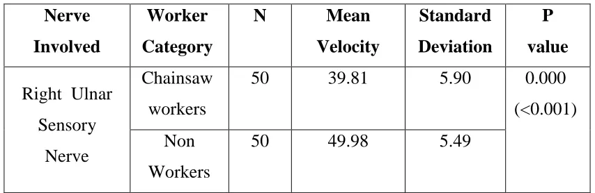
Table:2. Right ulnar sensory nerve Nerve
Involved
Worker Category
N Mean
Velocity
Standard Deviation
P value
Right Ulnar
Sensory
Nerve
Chainsaw
workers
50 39.81 5.90 0.000
(<0.001)
Non
Workers
[image:58.595.105.532.565.704.2]32
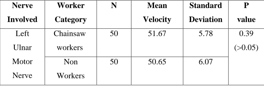
Table: 3.Left ulnar motor nerve Nerve
Involved
Worker Category
N Mean
Velocity Standard Deviation P value Left Ulnar Motor Nerve Chainsaw workers
50 51.67 5.78 0.39
(>0.05)
Non
Workers
[image:59.595.106.530.326.466.2]50 50.65 6.07
Table: 4. Left ulnar sensory nerve Nerve
Involved
Worker Category
N Mean
Velocity
Standard Deviation
P value
Left Ulnar Sensory Nerve Chainsaw Workers
50 37.57 4.97 0.000
(<0.001)
Non
Workers
50 50.25 6.47
Table: 5. Right median motor nerve Nerve
Involved
Worker Category
N Mean
Velocity Standard Deviation P value Right Median Motor Nerve Chainsaw Workers
50 49.71 6.51 0.86
(>0.05)
Non
Workers
[image:59.595.108.530.522.663.2]33
Table: 6. Right median sensory nerve Nerve
Involved
Worker Category
N Mean
Velocity
Standard Deviation
P value
Right Median Sensory Nerve Chainsaw Workers
50 40.58 6.17 0.000
(<0.001)
Non
Workers
[image:60.595.106.531.320.463.2]50 50.30 4.79
Table: 7. Left median motor nerve Nerve
Involved
Worker Category
N Mean
Velocity Standard Deviation P value Left Median Motor Nerve Chainsaw Workers
50 50.76 6.02 0.87
(>0.05)
Non
Workers
50 50.95 5.59
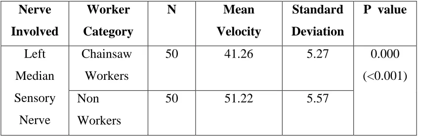
Table: 8. Left median sensory nerve Nerve
Involved
Worker Category
N Mean
Velocity
Standard Deviation
P value
Left Median Sensory Nerve Chainsaw Workers
50 41.26 5.27 0.000
(<0.001)
Non
Workers
50 51.22 5.57
[image:60.595.107.533.518.657.2]34
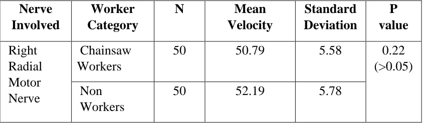
Table: 9. Right radial motor nerve Nerve
Involved
Worker Category
N Mean
Velocity Standard Deviation P value Right Radial Motor Nerve Chainsaw Workers
50 50.79 5.58 0.22
(>0.05)
Non Workers
[image:61.595.111.529.304.429.2]50 52.19 5.78
Table: 10. Left radial motor nerve
Nerve Involved
Worker Category
N Mean
Velocity Standard Deviation P value Left Radial Motor Nerve Chainsaw Workers
50 51.79 5.43 0.12
(>0.05)
Non Workers
50 50.19 4.99
Table: 11. Anthropometric particulars
Cases Control
Mean Range Mean Range
Age (Years) 31.0 20-35 33 20-35
Height (cm) 164 154-176 162.5 151-171
Weight (kg) 55 42-72 52 46-72
No.of years in present employment
[image:61.595.107.530.494.685.2]35
Right Ulnar Nerve; Motor Nerve Conduction Velocity
In the 50 volunteers from the chain saw users, the motor nerve
conduction velocities, fall within the range of 45.05 to 57.32 m/sec.
The mean value of the resulting data shows 49.7 m/sec. with standard
deviation of 6.05 with 97% confidence limits.
For the 50 healthy volunteers from controls, the motor conduction
results lie in the range between 42.08 to 60.01 and the mean of all the
fifty values is 49.98 with standard deviation of 5.49 with confidence
limits 97%.
Left Ulnar Nerve; motor Nerve Conduction Velocity
The controls who are not exposed to vibration have velocity
range 46.05 to 58.34 m/sec. The mean value is 50.67 with 6.07
standard deviation falling within the 97% confidence limits.
The exposed have a velocity range of 44.44 to 57.33 m/sec. the
average conduction velocity is 51.67 m/sec.
Right Median Nerve; Motor Conduction Velocity
The conduction velocity for right upper limb for median nerve is
distributed in between the values 42.04 and 61.04 m/sec with a mean
of 49.93 m/sec for controls.
The left upper limb motor velocity of median nerve has the
values in the range of 41.44 to 58.33 m/sec. The mean value lies at
36
Left Median nerve; Motor conduction velocity
Motor conduction velocity mean value is 50.76 m/sec with
standard deviation 6.02 for exposed and for non exposed 50.95±5.9 m/sec.
‘p’ value is >0.05.
Right Radial Motor Nerve conduction velocity;
In the non exposed, the mean is 50.79 with standard deviation
5.58. In the exposed, the mean is 52.19 with standard deviation 5.78.
Left Radial Motor Nerve conduction velocity;
For cases the mean falls in 49.15 with standard deviation 6.04 for
exposed and mean is 49.61with standard deviation 6.09 for unexposed.
Sensory Conduction Velocity
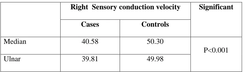
Right Ulnar Nerve: The mean value is 39.81±5.90 m/sec for exposed and 49.98±5.49 m/sec for non exposed with ‘p’ value < 0.001.
Left Ulnar Nerve: The mean value is 31.57±4.97 m/sec in non exposed individuals and 50.25±6.47 m/sec in exposed with ‘p’ value <0.001.
Right Median Nerve: Mean nerve conduction velocity is 40.58 ±6.17 m/sec for exposed and 50.30±4.79 m/sec for non exposed with ‘p’ value <0.001.
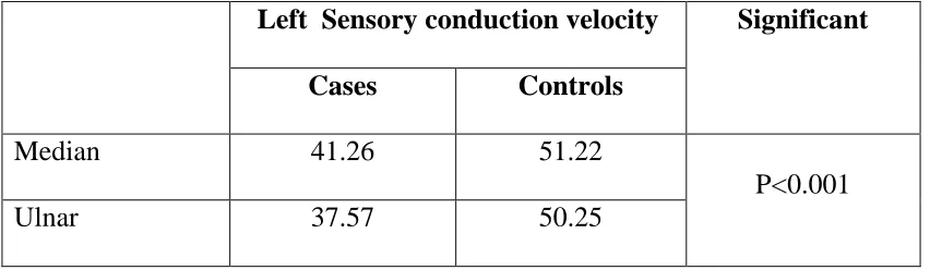
Left Median Nerve: Mean value is 41.26±5.27 m/sec in exposed and 51.22±5.57 m/sec in non exposed with ‘p’ value <0.001.
37
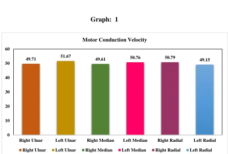
TABLE ; 12. VIBRATION EXPOSED (Cases) Motor Conduction Velocity
Mean Velocity Standard Deviation
Right Ulnar 49.71 6.05
Left Ulnar 51.67 5.78
Right Median 49.61 6.51
Left Median 50.76 6.02
Right Radial 50.79 5.58
Left Radial 49.15 6.08
Graph: 1
49.71 51.67 49.61 50.76 50.79 49.15
0 10 20 30 40 50 60
Right Ulnar Left Ulnar Right Median Left Median Right Radial Left Radial
Motor Conduction Velocity
38
TABLE : 13
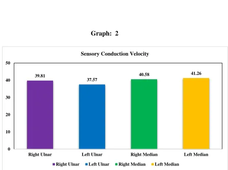
VIBRATION EXPOSED Sensory Conduction Velocity
Mean Velocity Standard Deviation
Right Ulnar 39.81 5.9
Left Ulnar 37.57 4.97
Right Median 40.58 6.17
Left Median 41.26 5.27
Graph: 2
39.81
37.57
40.58 41.26
0 10 20 30 40 50
Right Ulnar Left Ulnar Right Median Left Median
Sensory Conduction Velocity
39
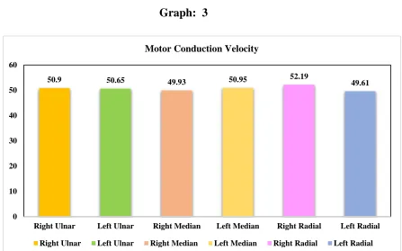
Controls (Non Exposed) Motor Conduction Velocity
Table: 14
Mean Velocity Standard Deviation
Right Ulnar 50.9 6.3
Left Ulnar 50.65 6.07
Right Median 49.93 6.13
Left Median 50.95 5.59
Right Radial 52.19 5.78
Left Radial 49.61 6.09
Graph: 3
50.9 50.65 49.93 50.95 52.19 49.61
0 10 20 30 40 50 60
Right Ulnar Left Ulnar Right Median Left Median Right Radial Left Radial
Motor Conduction Velocity
[image:66.595.114.561.429.708.2]40
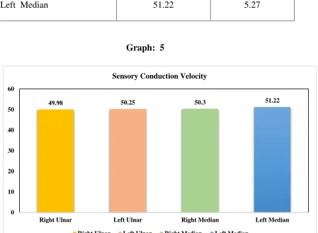
Controls (Non Exposed) Sensory Conduction Velocity
Table: 15
Mean Velocity Standard Deviation
Right Ulnar 49.98 5.9
Left Ulnar 50.25 4.97
Right Median 50.30 6.17
Left Median 51.22 5.27
Graph: 5
49.98 50.25 50.3 51.22
0 10 20 30 40 50 60
Right Ulnar Left Ulnar Right Median Left Median
Sensory Conduction Velocity
[image:67.595.113.562.315.644.2]41
Results in motor nerve conduction velocity Table: 16
Right Motor Conduction velocity Significant
Cases Controls
Median 49.74 49.93
p>0.05
Ulnar 49.71 50.90
Radial 50.79 52.19
Results in motor nerve conduction velocity Table: 17
Left Motor nerve conduction velocity Significant
Cases Controls
Median 50.76 50.95
p>0.05
Ulnar 51.67 50.65
Radial 49.15 49.61
Results in sensory conduction velocity Table: 18
Right Sensory conduction velocity Significant
Cases Controls
Median 40.58 50.30
P<0.001
[image:68.595.106.532.132.285.2] [image:68.595.106.531.366.522.2] [image:68.595.107.534.599.725.2]42
Results in sensory conduction velocity Table: 19
Left Sensory conduction velocity Significant
Cases Controls
Median 41.26 51.22
P<0.001
Ulnar 37.57 50.25
From the table 14, the motor nerve conduction velocities in
median, ulnar and radial nerves on the right upper limb showed no
significant difference between the exposed and non exposed. All the
comparative values have ‘p’ value of >0.05 which shows statistically no
significant difference.
From the table 14, the left upper limb motor conduction velocities
for median, ulnar and radial nerves show little difference that too is
statistically insignificant with ‘p’ value of >0.05.
From the table 15, the conduction velocities for sensory
component of right median nerve and right ulnar nerves show a gross
difference. When the values of exposed are compared with non
exposed, the ‘p’ value is <0.001 which is statistically more significant.
Hence, the sensory conduction velocities of median and ulnar nerves are
delayed.
From the table 18 and 19, the sensory conduction velocity on left
side, and right side of median and ulnar nerves are compared. Both the
[image:69.595.107.533.126.250.2]43
individuals with more significant ‘p’ value of <0.001. Hence sensory
nerve conduction velocities are delayed.
Sensory Conduction Velocity Table: 20
Right Left Significant
Median Ulnar Median Ulnar
Exposed 40.58 39.81 41.26 37.57
p>0.05
Non-exposed 50.30 49.98 51.22 50.25
Graph: 6
In the vibration exposed subjects , right median sensory conduction
velocity is little more delayed than left median nerve but not
significant. In case of ulnar nerve, left side sensory conduction is more
[image:70.595.110.550.229.622.2]