The immune evasion protein Sbi of
Staphylococcus aureus
occurs both extracellularly and anchored to the cell envelope
by binding lipoteichoic acid
mmi_7966 789..804Emma Jane Smith,1Rebecca M. Corrigan,2 Tetje van der Sluis,1†Angelika Gründling,2 Pietro Speziale,3Joan A. Geoghegan1and Timothy J. Foster1*
1Microbiology Department, Moyne Institute of Preventive
Medicine, Trinity College, Dublin 2, Ireland. 2Section of Microbiology, Imperial College London,
Armstrong Rd, London SW7 2AZ, UK.
3Department of Molecular Medicine, Viale Taramelli 3/b,
27100 Pavia, Italy.
Summary
The Sbi protein ofStaphylococcus aureuscomprises two IgG-binding domains similar to those of protein A and a region that triggers the activation of comple-ment C3. Sbi is expressed on the cell surface but its C-terminal domain lacks motifs associated with wall or membrane anchoring of proteins in Gram-positive bacteria. Cell-associated Sbi fractionates with the cytoplasmic membrane and is not solubilized during protoplast formation.S. aureusexpressing Sbi trun-cates of the C-terminal Y domain allowed identifica-tion of residues that are required for associaidentifica-tion of Sbi with the membrane. Recombinant Sbi bound to purified cytoplasmic membrane materialin vitroand to purified lipoteichoic acid. This explains how Sbi partitions with the membrane in fractionation experi-ments yet is partially exposed on the cell surface. An LTA-defective mutant ofS. aureushad reduced levels of Sbi in the cytoplasmic membrane.
Introduction
Staphylococcus aureuspermanently colonizes the moist squamous epithelium of the anterior nares of approxi-mately 20% of the population while the remainder carry
the organism intermittently (Williams, 1963; Peacock
et al., 2001). Colonization is an established risk factor for development of infection both in the hospital and in the
community (Lowy, 1998).S. aureuscan cause a variety of
infections ranging from superficial skin lesions such as boils and abscesses to invasive and potentially life-threatening infections such as osteomyelitis, septic arthri-tis and endocardiarthri-tis (Petti and Fowler, 2003; Fowleret al., 2005).
The ability ofS. aureusto cause infections is in part due to proteins that are anchored to the cell surface and to those that are secreted into the medium. Among the latter are cytolytic toxins, enzymes and proteins with immune evasion functions that interfere with neutrophil migration and complement fixation (Foster, 2005). While a major function of surface-anchored proteins is to act as adhes-ins and invasadhes-ins (Foster, 2005), it is also clear that several can also help the bacterium evade innate immune responses. Thus protein A binds to the Fc region of IgG and coats the cell with antibody that cannot be recognized by Fc receptors on neutrophils and cannot catalyse complement fixation. Clumping factor A binds fibrinogen
and fibrin (McDevitt et al., 1997) but it can also capture
and activate the complement regulatory protease factor I which results in enhanced degradation of C3b (Cunnion
et al., 2004; Hairet al., 2008).
Proteins can be anchored to the cell envelope of
Gram-positive bacteria by several mechanisms (Cabaneset al.,
2002). (i) Covalent linkage to cell wall peptidoglycan occurs by the action of sortase on the LPXTG motif that is part of a C-terminal wall-anchoring domain (Mazmanian
et al., 2001). The sorting signal also comprises a hydro-phobic membrane-spanning domain followed at the extreme C-terminus by positively charged residues. (ii) Lipoproteins are possibly anchored to the outer face of the
cytoplasmic membrane (Inouye et al., 1977; Bubeck
Wardenburget al., 2006). The signal peptide is typically
shorter than that of proteins that are secreted into the medium and is followed by a cysteine residue. Lipoprotein diacylglycerol transferase catalyses transfer of diacylgly-cerol from phosphatidylglydiacylgly-cerol in the outer face of the membrane to the sulphydryl moiety of the cysteine followed by cleavage of the signal peptide by signal
peptidase II (Tokunaga et al., 1982; Choi et al., 1986;
Accepted 28 December, 2011. *For correspondence. E-mail
[email protected]; Tel. (+353) 1 8962014; Fax (+353) 1 6799295.
†Present address: Department of Immunohematology and Blood
Transfusion, Leiden University Medical Center, Albinusdreef 2, 2333 ZA, Leiden, the Netherlands.
Re-use of this article is permitted in accordance with the Terms and Conditions set out at http://wileyonlinelibrary.com/onlineopen# OnlineOpen_Terms
Gan et al., 1993). (iii) Proteins may be anchored non-covalently to the cell wall components peptidoglycan and
teichoic acids. Internalin B (InlB) ofListeria
monocytoge-neshas C-terminal ‘GW’ repeat domains of~80 residues
that bind to lipoteichoic acid (LTA) (Jonquieres et al.,
1999). Thus InlB is associated with the cytoplasmic mem-brane in cell fractionation experiments but can also occur
extracellularly (Braun et al., 1997). Furthermore,
cell-bound InlB can be displaced by soluble LTA and by highly
negatively charged heparin sulphate proteoglycan
(Jonquieres et al., 1999). Autolysins such as Alt from
S. aureusand AtlE fromStaphylococcus epidermidisare also attached to the cell envelope via GW repeats (Oshida
et al., 1995; Heilmannet al., 1997). (iv) The extracellular adherence protein Eap (also known as Map) has repeated domains that can bind to several different ligands
(Cha-vakis et al., 2002). The protein can also bind to the
bacterial cell surface by an unknown mechanism and promote attachment to and invasion of mammalian cells, most likely by binding to fibronectin and forming a bridge to thea5b1 integrin (Harraghyet al., 2003; Hussainet al., 2008).
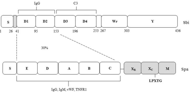
The second binding protein for immunoglobulins (Sbi) comprises four N-terminal ligand-binding repeats. Its C-terminus comprises the proline-rich region Wr and the tyrosine-rich region Y that is assumed to be involved in Sbi association with the cell envelope (Fig. 1; Zhang
et al., 1998). The first two N-terminal repeats (D1 and D2) have sequence similarity to the IgG-binding domains of protein A. Indeed the predicted structures show that resi-dues on the faces of helices I and II of Spa and Sbi are conserved, allowing Sbi to bind to IgG in a similar fashion to the archetypal LPXTG-anchored immune evasion protein. Domains D3 and D4 are also separately folded
and contribute to the elongated structure of the protein (Burmanet al., 2008; Upadhyayet al., 2008). They bind to complement protein C3. It has been argued that binding to and promoting the conversion of C3 to C3b would only be an effective immune evasion mechanism if the protein
were extracellular (Burmanet al., 2008).
Previously we reported a detailed and systematic analysis of the cellular location of Sbi (Smithet al., 2011). By analysing mutants lacking Sbi and protein A we were able to demonstrate that Sbi occurs both extracellularly and bound to the cell envelope and that both contribute to immune evasion. By expressing truncated Sbi in
S. aureus and using purified recombinant Sbi we show that the C-terminal Y domain is required for attachment to the membrane. This is likely to be mediated by its inter-action with lipoteichoic acid.
Results
Surface expression of Sbi D3D4 ligand-binding domains
Previously we reported the surface exposure of the IgG
binding D1D2 domains of Sbi (Smithet al., 2011). In order
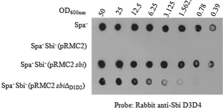
to determine if the C3-binding domains of Sbi were exposed on the cell surface whole cells were immobilized on membranes and probed with rabbit antibodies raised against the non-IgG binding D3D4 domains of Sbi. Cells expressing wild-type Sbi could bind antibodies via domains D1 and D2 by a non-immune reaction as well as by a specific immune reaction with domains D3 and D4. The same reactivity was seen with cells expressing wild-type Sbi from the chromosomal gene and when induced
from pRMC2sbi+(Fig. 2). In contrast cells expressing a
[image:2.595.119.480.53.228.2]mutant of pRMC2sbi+ which expressed a truncate that
lacked the IgG binding D1D2 domains reacted 16- to 32-fold less. Given that D1 and D2 can each bind to a single Fc region each whereas D3D4 most likely has several epitopes for polyclonal IgG Fab it is possible that the majority of D3D4 are buried within the cell wall and are not exposed on the cell surface.
Sbi binding to the cytoplasmic membrane
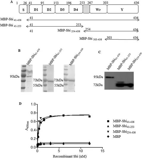
To address the importance of the C-terminal domain of Sbi in membrane anchoring, three maltose-binding protein (MBP) fusion proteins were constructed (Fig. 3A). These comprised the entire Sbi protein (residues 41–436), the N-terminal ligand-binding domains (residues 41–253) and the C-terminal domains Wr and Y (residues 253–436). The
proteins were expressed inEscherichia coliand purified by
affinity chromatography. Their purity and integrity were verified by SDS-PAGE (Fig. 3B) and Western blotting with anti-MBP antiserum (Fig. 3C).
Cytoplasmic membrane material purified fromS. aureus
Newman Spa- Sbi- was incubated in microtitre plates
and coating of the surface was verified with antibodies recognizing the integral membrane protein EbpS (data not shown). The membranes were incubated with
MBP–Sbi41–436and MBP–Sbi254–436which were able to bind
in a dose-dependent and saturable manner with half
maxima of 0.54⫾0.1 nM and 0.57⫾0.1 nM,
respec-tively, while MBP–Sbi41–253 and the MBP control were
unable to bind (Fig. 3D). These results indicate that the C-terminal WrY domain of Sbi binds to purified cytoplasmic membrane mimicking precisely the results seen with
frac-tionatedS. aureuscells expressing Sbi truncates.
Recombinant Sbi binds to whole cells and fractionates with the cytoplasmic membrane
Recombinant MBP–Sbi binds to purified cytoplasmic membrane material with high affinity. To address whether
this mode of association is similar to that of Sbi expressed by S. aureus, recombinant proteins MBP–Sbi41–436,
MBP–Sbi41–253 and MBP–Sbi254–436were separately
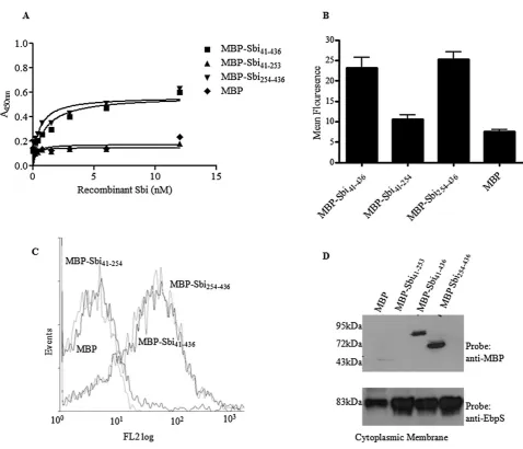
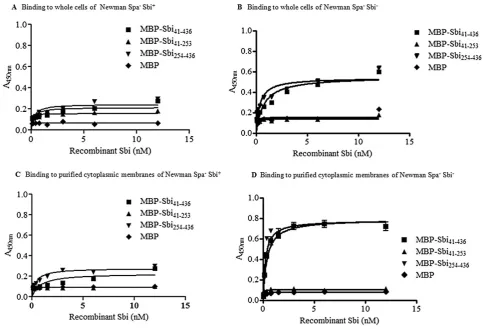
incu-bated with whole cells of Newmanspa sbi. MBP–Sbi41–436
and MBP–Sbi254–436 both bound to whole cells
dose-dependently and saturably with similar half-maximal
binding concentrations (0.55⫾0.03 nM and 0.57⫾
0.02 nM respectively) (Fig. 4A). Binding of recombinant MBP–Sbi41–436, MBP–Sbi41–253, MBP–Sbi254–436and MBP to
whole cells of strain Newman Spa-Sbi-was also
investi-gated by flow cytometry. Recombinant Sbi derivatives were
separately incubated with whole cells of Newman Spa
-Sbi-and binding was detected with monoclonal mouse
anti-MBP followed by FITC-labelled rabbit anti-mouse IgG.
MBP–Sbi41–436 and MBP–Sbi254–436 both bound to whole
cells confirming the enzyme-linked immunosorbent assay (ELISA) result (Fig. 4B and C).
In order to determine if recombinant Sbi that bound to the bacterial cells fractionated with the cell wall or membranes during protoplast formation, and to determine if a receptor
for Sbi is exposed on the cell surface, Newman Spa-Sbi
-was incubated with MBP–Sbi41–436 and MBP–Sbi254–436,
washed and then fractionated to isolate the cytoplasmic membrane. Figure 4D shows the purified cytoplasmic membrane fraction probed with HRP-conjugated anti-MBP
antiserum. The ~86 kDa band present in lane 3
corre-sponds to recombinant MBP–Sbi41–436while the~63 kDa
band in lane 4 corresponds to recombinant MBP–Sbi254–436
(Fig. 4D). These data show that a receptor for the C-terminal domain of Sbi is exposed on the cell surface and yet the added recombinant protein is associated with the membrane following fractionation of protoplasts
Sbi binds to lipoteichoic acid
The C-terminal region of Sbi has neither a sequence of hydrophobic residues sufficient to span the cytoplasmic membrane typical of integral or membrane-spanning pro-teins nor a lipoprotein consensus sequence that could be involved in anchoring the protein to the cytoplasmic mem-brane, raising the possibility that the association of Sbi with the cytoplasmic membrane could be due to binding to a membrane-associated component. Lipoteichoic acid (LTA) is an anionic polymer linked to a glycolipid anchor in the outer face of the cytoplasmic membrane and with a poly(g-lycerophosphate) chain that extends across the cell wall (Neuhaus and Baddiley, 2003). LTA remains associated with the protoplast after removal of the cell wall peptidogly-can by lysostaphin (Neuhaus and Baddiley, 2003). To determine if LTA could be the target for the C-terminal WrY domain of Sbi, wells of microtitre plates were coated
with a constant amount of purified S. aureus LTA and
incubated with increasing concentrations of
[image:3.595.56.284.60.174.2]recombin-ant MBP–Sbi41–436, MBP–Sbi41–253 and MBP–Sbi254–436.
Fig. 2. Surface expression of Sbi domains D3D4. Serial dilutions of cells were applied to a nitrocellulose membrane and probed with rabbit anti-Sbi D3D4WrY IgG followed by HRP-conjugated goat anti-rabbit IgG.
Fig. 3. Binding of MBP–Sbi41–436, MBP–Sbi41–253and MBP–Sbi254–436to purified cytoplasmic membrane.
A. Schematic diagram of Sbi showing the residues present in each recombinant MBP-tagged protein. B. Coomassie stain of an SDS-PAGE gel of MBP–Sbi recombinant proteins.
C. Western immunoblot of MBP–Sbi recombinant proteins probed with HRP-conjugated anti-MBP IgG.
D. Binding of MBP–Sbi41–436, MBP–Sbi254–436MBP–Sbi41–253and MBP to wells coated with cytoplasmic membrane material isolated from
Proteins containing the C-terminal domain WrY (Sbi41–436
and Sbi254–436) were able to bind LTA in a dose-dependent
and saturable manner with half maximal concentrations of
0.86⫾0.2 nM and 0.84⫾0.2 nM respectively (Fig. 5A).
Furthermore, pre-incubation of Sbi with different
concen-trations ofS. aureusLTA inhibited binding to immobilized
LTA and to purified cytoplasmic membranes in a dose-dependent manner (Fig. 5B and C).
To investigate the specificity of the interaction between Sbi and LTA, MBP–Sbi was pre-incubated with different
concentrations of heparin sulphate, an anionic glycosami-noglycan which consists of a repeating disaccharide unit of glucosamine and uronic acid residues and which occurs on the surface of mammalian cells (Li and Vlo-davsky, 2009). Heparin sulphate (HS) was able to
dis-place InlB from the surface ofL. monocytogenesbut was
[image:5.595.57.536.46.456.2]not able to inhibit the interaction of MBP–Sbi with immo-bilized LTA or purified cytoplasmic membrane material (Fig. 5B and C), suggesting that the interaction between LTA and Sbi is specific and not simply due to the positively
Fig. 4. Binding of MBP–Sbi41–436, MBP–Sbi41–253and MBP–Sbi254–436to whole cells of Newman Spa-Sbi-.
A. Binding of MBP–Sbi41–436, MBP–Sbi254–436MBP–Sbi41–253and MBP to wells coated with whole Newman Spa-Sbi-cells. Recombinant protein
binding was detected with HRP-conjugated anti-MBP IgG. Binding assay was preformedn=3 times with similar results. The graph shown is a representative of one experiment with each plot in the graph representing the average of duplicate wells.
B. Washed whole cells of Newman Spa-Sbi-were incubated with 0.5mM MBP–Sbi
41–436, MBP–Sbi254–436,MBP–Sbi41–253and MBP followed by
mouse anti-MBP antiserum and FITC-labelled rabbit anti-mouse IgG. Fluorescence intensity was measured by flow cytometry. The assay was preformedn=3 times. Each plot on the graph represents the average value for all three replicas. Error bars show the standard deviation. C. A representative flow cytometry trace of recombinant Sbi derivatives binding to Newman Spa-Sbi-.
D. Recombinant MBP–Sbi41–436, MBP–Sbi254–436MBP–Sbi41–253and MBP were incubated with whole cells of Newman Spa-Sbi-and
fractionated. Cytoplasmic membrane fractions were analysed by Western blotting with HRP-conjugated anti-MBP IgG or rabbit anti-EbpS IgG followed by HRP-conjugated protein A. All immunoblotting experiments were repeatedn=3 times.
charged protein binding to negatively charged residues in surface polymers.
Can Sbi bind to purified cell wall?
The cell wall contains wall teichoic acid, a polymer of ribitol phosphate that is covalently anchored to the
pepti-doglycan (Neuhaus and Baddiley, 2003; Xiaet al., 2010)
as well as covalently anchored wall-associated proteins
such as ClfA and Spa. The cell wall fraction ofS. aureus
Newman Spa-Sbi-was purified following disruption of the
cells by mechanical shearing and by boiling with deter-gent to solubilize the cytoplasmic membrane.
The coating affinity and purity of the cell wall fraction was assessed by ELISA using (i) polyclonal ClfA anti-bodies and (ii) polyclonal anti-EbpS antianti-bodies, with maximum absorbencies at saturation 0.76 and 0.22 respectively. This showed that the wall fraction bound to the ELISA wells efficiently and that the level of
contami-nation with membrane was low. MBP–Sbi41–436 and
MBP–Sbi254–436was added to the cell wall-coated ELISA
wells but did not bind detectably. This shows that Sbi cannot bind to the purified cell wall fraction (Fig. 5D).
Displacement of Sbi from purified cytoplasmic membranes by soluble LTA
In order to determine if soluble LTA could displace Sbi
from the membrane ofS. aureuscells, purified
cytoplas-mic membranes of the Sbi+ strain Newman Spa- were
incubated with different concentrations of LTA. Some Sbi was displaced into the supernatant in a concentration-dependent manner while the majority remained associ-ated with the membrane (Fig. 6A). This indicates that Sbi is attached to the membrane with high affinity at least in part by binding to LTA and suggests the possibility of a second ligand.
Detection of Sbi–LTA interaction by Far Western blotting
The experiment described above shows that recombinant Sbi can bind to LTA. In order to determine if Sbi expressed byS. aureusbinds LTA a Far Western blotting approach
was taken. The membrane fraction ofS. aureusNewman
Spa-Sbi-lacking the ability to express the IgG-binding
proteins Spa and Sbi from chromosomal genes but
expressing SbiDD1D2from pRMC2 was purified. The
mem-Fig. 5. Interaction of MBP–Sbi with LTA.
A. Binding of MBP–Sbi41–436, MBP–Sbi254–436MBP–Sbi41–253and MBP to LTA-coated wells.
B and C. (B) Inhibition of binding of MBP–Sbi41–436and MBP–Sbi254–436to LTA and (C) to the purified cytoplasmic membrane fraction from
S. aureusNewman Spa-Sbi-. Recombinant proteins were pre-incubated with increasing concentrations of eitherS. aureusLTA or heparin sulphate (0–200mg ml-1) before being added to coated microtitre plates.
D. Binding of MBP–Sbi proteins to purified cell wall-coated wells. In each assay recombinant protein binding was detected with
[image:6.595.61.534.52.329.2]brane fraction was separated by SDS-PAGE, transferred to a nitrocellulose membrane and probed with LTA fol-lowed by a mouse monoclonal antibody to LTA and HRP-conjugated rabbit anti-mouse IgG. The reactive band at
~35 kDa corresponds to SbiDD1D2demonstrating that Sbi
expressed fromS. aureusbinds LTA (Fig. 6B). An
immu-noreactive band of 17–24 kDa was detected in both
samples which presumably corresponds to LTA that is present in the membrane fraction (Fig. 7B). The Far Western blot was subsequently stripped, probed with anti-Sbi serum and overlaid on the original blot (Fig. 6B).
A reactive band at ~35 kDa was detected which
corre-sponds to SbiDD1D2demonstrating that it is Sbi expressed
[image:7.595.117.478.59.278.2]fromS. aureuswhich binds LTA in the Far Western blot.
Fig. 6. Analysis of the Sbi–LTA interaction.
A. Displacement of Sbi from the cytoplasmic membrane by soluble LTA. Newman Spa-cytoplasmic membrane was incubated with increasing amounts ofS. aureusLTA (0–400mg ml-1). Sbi bound to the cytoplasmic membrane or released into the supernatant was detected using rabbit
anti-Sbi D3D4WrY IgG followed by HRP-conjugated goat anti-rabbit IgG.
B. Far Western blotting ofS. aureuscytoplasmic membrane fractions with LTA. Newman Spa-Sbi-and Newman Spa-Sbi-(pRMC2-sbiD
D1D2)
cytoplasmic membrane material was fractionated by SDS-PAGE, transferred to a nitrocellulose membrane incubated with purified LTA and bound LTA was detected with anti-LTA monoclonal antibody followed by HRP-linked rabbit anti-mouse IgG. All immunoblotting experiments were repeatedn=3 times.
Fig. 7. Sbi cellular location in LTA-negative strains.
A. Whole-cell lysate fractions of RN4220 Spa-, 4S5 and 4S5 (pCN34-ltaS) analysed by Western immunoblotting with monoclonal mouse anti-LTA antibodies followed by HRP-conjugated rabbit anti-mouse IgG.
B. Cytoplasmic membrane and culture supernatant fractions of RN4220 Spa-, 4S5 and 4S5 (pCN34-ltaS) analysed by Western
immunoblotting with rabbit anti-Sbi D3D4WrY IgG and HRP-conjugated goat anti-rabbit IgG (i and ii) and rabbit anti-EbpS IgG followed by HRP-conjugated goat anti-rabbit IgG (iii). Blots shown are representative of three independent experiments. Densitometric analysis was carried out using ImageJ software. Integrated band densities were measured with correction of background. Values given are the mean⫾the standard deviation ofn=3 experiments
[image:7.595.140.452.482.621.2]An LTA defective mutant has decreased levels of Sbi in the cytoplasmic membrane
Staphylococcus aureus mutants that lack LTA can only grow under osmotically stabilizing conditions or by the
acquisition of compensatory mutations (Okuet al., 2009;
Corrigan et al., 2011). The RN4220 Spa--derived
LTA-deficient strain 4S5 contains a complete deletion of the
LTA synthase geneltaSand has acquired two additional
mutations that permit this strain to grow and divide in the
absence of LTA similar to the wild type (Corriganet al.,
2011). To assess the role of LTA in the localization of Sbi to the cell membrane, cytoplasmic membrane and culture
supernatant fractions of wild-type RN4220 Spa-and 4S5
cells grown to mid-exponential phase were isolated and analysed by SDS-PAGE and Western immunoblotting using the rabbit anti-Sbi D3D4WrY serum. An
immunore-active band of~50 kDa was detected in the membrane
and supernatant fractions of both the wild-type and LTA-negative strain [Fig. 7B(i)]. However, densitometric analy-sis of band intensity indicated that strain 4S5 exhibited
3.4⫾0.4-fold lower levels of Sbi in its cytoplasmic
mem-brane but contained a 1.8⫾0.2-fold higher level of Sbi in
the supernatant fraction as compared with the wild-type strain [Fig. 7B(ii)].
To confirm the role of LTA in Sbi localization and to rule out any involvement of the suppressor mutations present in
4S5, the complementation vector pCN34-ltaS (Corrigan
et al., 2011), which expressesltaSfrom its native promoter, was electroporated into 4S5. This resulted in the restora-tion of LTA synthesis as judged by Western immunoblotting
(Fig. 7A). Complementation with pCN34-ltaSalso restored
Sbi expression in the cytoplasmic membrane to wild-type levels (Fig. 7B). This shows that the suppressor mutations are not responsible for the reduction in Sbi in the mem-brane fraction and directly implicates LTA in attaching Sbi to the cell envelope. Furthermore, the level of the integral membrane protein EbpS in the cytoplasmic membrane of all strains was the same. This indicates that the inhibition of LTA expression does not affect another membrane-associated protein [Fig. 7B(iii)].
Association of Sbi with the membrane by its C-terminal Y domain
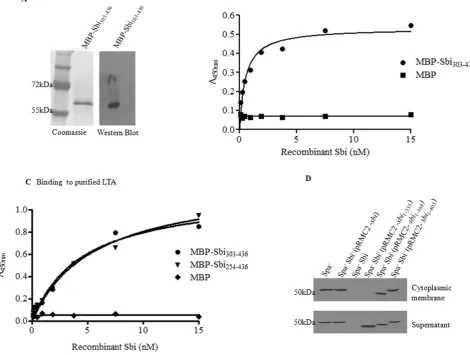
It seemed likely that the C-terminal domain Y is involved in attaching Sbi to the cytoplasmic membrane. To test this, an MBP-fusion protein was constructed that comprised the C-terminal Y domain (residues 303–436) (Fig. 3A).
The protein was expressed inE. coliand purified by
affin-ity chromatography. The protein’s puraffin-ity and integraffin-ity were verified by SDS-PAGE and Western blotting with anti-MBP serum (Fig. 8A).
Cytoplasmic membrane material purified fromS. aureus
Newman Spa-Sbi-was incubated in microtitre plates and
coating of the surface verified with antibodies recognizing the integral membrane protein EbpS (data not shown). The
membranes were incubated with MBP–Sbi303–436 which
was able to bind in a dose-dependent and saturable
manner with half maxima of 0.5⫾0.04 nM while the MBP
control was unable to bind (Fig. 8B). These results indicate that the C-terminal Y domain of Sbi binds to purified
cytoplasmic membrane. MBP–Sbi303–436also bound
puri-fied LTA in a dose-dependent and saturable manner with
half maxima of 1⫾0.15 nM (Fig. 8C).
To localize the residues involved, the full-lengthsbigene and a series of deletions were cloned into the expression
vector pRMC2 so that thesbi gene and truncates were
expressed from the anhydrotetracycline-inducible pro-moter on the vector. The plasmids expressing full-length
Sbi and the C-terminal truncates Sbi1–335, Sbi1–368 and
Sbi1–403were introduced into Newman Spa-Sbi-, bacteria
were grown in the presence of the inducer and cytoplasmic membrane and culture supernatant fractions analysed by SDS-PAGE and Western blotting. A 50 kDa band corre-sponding to Sbi was detected in the membrane and super-natant fractions of the strain expressing the wild-type Sbi. The three truncates were detected in the culture superna-tant, but in the case of the shortest, Sbi1–335, the protein was
not present in the membrane fraction and was only found in the supernatant (Fig. 8D). This suggests that the C-terminal domain Y is required to anchor the protein to the membrane and that residues 335–368 are required for efficient membrane anchoring but are not solely responsible.
Why does Sbi occur extracellularly?
Given that recombinant MBP–Sbi protein binds to LTA exposed on the bacterial cell surface it is perhaps surpris-ing that Sbi can occur extracellularly unless the cell-bound form saturates the surface-exposed LTA or the C-terminus of the secreted form is modified so that it cannot
recog-nize LTA. To address this issue Newman Spa-Sbi+and
Newman Spa- Sbi-cells were coated onto the surface
of ELISA wells and incubated with MBP–Sbi41–436 and
MBP–Sbi254–436. A reduction in absorbance of 2.3- and
3-fold occurred, respectively, when binding of MBP–Sbi to
the Sbi+ strain was compared with the Sbi- mutant
(Fig. 9A and B). This suggests that a significant fraction of
the LTA on the surface of Sbi+ cells is unavailable for
binding by the secreted form of Sbi.
Similar results were obtained when purified membranes
from Newman Spa- Sbi+ and Newman Spa- Sbi- cells
were immobilized and incubated with MBP–Sbi proteins, with a 2.4-fold and 2.7-fold reduction in binding of
MBP–Sbi41–436 and MBP–Sbi254–436 respectively (Fig. 9C
the Sbi+ cells. To rule out the possibility that increased
binding by Sbi-cells and cytoplasmic membranes may be
due to increased production of LTA the amount of LTA in
Sbi+and Sbi-cells was investigated by Western blotting
with anti-LTA serum. No difference in LTA expression was
seen between Sbi+and Sbi-bacteria (data not shown).
Can the Sbi Y domain support binding to the cytoplasmic membranes of other type I LTA expressing strains?
The ability of Sbi to bind to membranes (presumably via LTA) of other Gram-positive bacteria was investigated.
Cytoplasmic membrane material purified fromS. aureus,
S. epidermidis, S. lugdunensis, L. monocytogenes and
the Gram-negative bacteria E. coli was incubated in
microtitre plates. The membranes were incubated with
either MBP–Sbi303–436 or MBP. MBP–Sbi303–436bound the
cytoplasmic membranes of all five Gram-positive bacteria with a similar affinity while the MBP control was unable to bind (Fig. 10A). All five Gram-positive bacterial strains tested express type I LTA which has a poly(glycerophos-phate) backbone (Neuhaus and Baddiley, 2003). In
con-trast MBP–Sbi303–436 did not bind to the cytoplasmic
membrane material ofE. coliwhich does not express LTA.
These results indicate that the C-terminal Y domain of Sbi binds strongly to purified cytoplasmic membranes of strains expressing LTA which has a
poly(glycerophos-Fig. 8. Cellular localization of Sbi C-terminal truncates.
A. Coomassie stain of an SDS-PAGE gel of MBP–Sbi303–436. Western immunoblot of MBP–Sbi303–436probed with HRP-conjugated anti-MBP
IgG.
B. Binding of MBP–Sbi303–436and MBP to plates coated in Newman Spa-Sbi-cytoplasmic membrane material.
C. Binding of MBP–Sbi254–436, MBP–Sbi303–436and MBP to Newman Spa-Sbi-LTA-coated wells. Recombinant protein binding was detected with
HRP-conjugated anti-MBP IgG. Each assay was preformedn=3 times with similar results. The graphs shown are representatives of one experiment with each plot in the graph representing the average of duplicate wells.
D. Cytoplasmic membrane and culture supernatant fractions of Newman Spa-Sbi-(pRMC2-sbi) C-terminal truncates analysed by Western immunoblotting with HRP-labelled rabbit IgG.
[image:9.595.60.531.65.420.2]phate) backbone. Furthermore MBP–Sbi303–436binding to
the cytoplasmic membranes of all four Gram-positive
bac-teria was inhibited by pre-incubating MBP–Sbi303–436with
soluble purified LTA fromS. aureus(Fig. 10B).
Discussion
The notion that the function of the Sbi protein is to help
protect S. aureus from innate immune defences of the
host was initially based on the in vitro activities of the
recombinant D1 and D2 domains that bind to the Fc region of IgG and the D3 and D4 domains which can bind to complement factor C3 in serum and can promote its
futile consumption (Zhang et al., 1998; 1999; Burman
et al., 2008). If Sbi protects cells in the same manner as protein A by binding IgG at the Fc region so that the immunoglobulin cannot act as an opsonin or promote complement fixation, the protein must be bound to the cell envelope with domains D1 and D2 exposed on the cell surface. However, if domains D3 and D4 are to promote
futile consumption of C3 in the fluid phase they must do so at a distance from the cell otherwise they would actually promote opsonin formation. This requires the protein to be secreted from the cell and for the D3D4 domains of the cell-associated Sbi to be inactive.
We have recently shown that Sbi both is associated with the cell envelope (where it is at least partly displayed on the surface) and is also present extracellularly and that both forms of the protein contribute to immune evasion
(Smithet al., 2011). Examination of the level of exposure
of Sbi on the surface of whole cells indicates that the N-terminal D1 and D2 domains are available to bind IgG. In contrast only a small proportion of the D3 and D4 domains are recognized by the Fab regions of anti-D3D4WrY IgG.
[image:10.595.56.541.53.382.2]The Sbi protein is attached to the cell envelope by an unusual mechanism. When cells were converted to pro-toplasts the cell-associated protein was not solubilized like the LPXTG-anchored protein A or ClfA. Instead it was attached to the cytoplasmic membrane fragments
Fig. 9. Binding of MBP–Sbi to whole cells and purified cytoplasmic membrane. Binding of MBP–Sbi41–436,MBP–Sbi254–436MBP–Sbi41–253and
following lysis of protoplasts and sedimentation. Purified recombinant MBP–Sbi bound to immobilized membrane
fragments prepared from an Sbi- mutant. Finding that
recombinant Sbi bound to purified lipoteichoic acid both dose-dependently and saturably suggests that LTA is the ligand in the cell envelope bound by Sbi when it is secreted across the membrane of growing cells. Also, reaching saturation in the binding assays is important because it is indicative of a specific interaction. Further-more MBP–Sbi could not bind to purified cell wall material.
In support of our contention that LTA is a ligand for attaching Sbi to the cell envelope is (i) the specific binding of rSbi both to purified LTA and to the membrane fraction, (ii) inhibition of binding of rSbi to the membrane fraction by soluble LTA, (iii) partial displacement of Sbi from the mem-brane fraction by soluble LTA, (iv) binding of rSbi to the surface of whole cells where LTA is known to be exposed, (v) a reduction in the level of Sbi in the cytoplasmic mem-brane of an LTA-negative mutant, and (vi) the ability to bind to the membranes of other Gram-positive bacteria with type I LTA in their cytoplasmic membranes. However, LTA might not be the only ligand for Sbi in the membrane. High concentrations of soluble LTA could only partially displace bound Sbi. Figure 11 summarizes our current understanding of the association of Sbi with the cell enve-lope and its role in immune evasion. It shows membrane-associated Sbi partially exposed on the cell surface with the N-terminal D1D2 domains able to bind IgG. Extracel-lular Sbi cannot attach to the cell because most of the LTA
is already occupied ensuring that at least some Sbi is available and possibly triggers C3 metabolism as
described by Burmanet al. (2008). The secreted form of
Sbi can also bind IgG but this does not provide any
protection from opsonophagocytosis (Smithet al., 2011).
The behaviour of Sbi in cell fractionation experiments and its ability to bind to LTA resembles internalin B (InlB) of
L. monocytogenes(Jonquiereset al., 1999). InlB binds to the human growth hormone receptor Met on mammalian cells and triggers bacterial internalization by
receptor-mediated endocytosis (Mengaudet al., 1996; Braunet al.,
1998; Shenet al., 2000). In order to act as an invasin InlB must be able to promote clustering of the Met receptor in the host cell membrane and trigger the signalling that leads to cytoskeletal rearrangements and endocytosis (Niemann, 2011). The ability of a secreted protein to perform this seems paradoxical until one considers the role of heparin sulphate proteoglycans that are bound on
the surface of mammalian cells (Marinoet al., 2002). HSP
is able to displace cell-associated InlB and to promote its release from the cell surface. It does so by binding to the GW repeats that contain the binding sites for LTA. It is likely that HSP–InlB complexes cooperate to trigger Met receptor clustering.
A major difference between the association of InlB and Sbi with LTA is that the latter does not bind to HSPsin vitro
and cannot be displaced easily from the membrane. This is likely to be due to the highly specific interaction that occurs between Sbi and LTA whereas the binding of InlB to LTA appears to be much weaker and is probably
non-Fig. 10. Binding of MBP–Sbi303–436to the cytoplasmic membranes of Gram-positive bacteria.
A. Cytoplasmic membrane material purified fromS. aureus,S. epidermidis,S. lugdunensis,L. monocytogenesandE. coliwere incubated with MBP–Sbi303–436and MBP in microtitre plates. Recombinant protein binding was detected with HRP-conjugated anti-MBP IgG.
B. Recombinant MBP–Sbi303–436was pre-incubated with increasing concentrations of eitherS. aureusLTA or bovine serum, albumin (BSA)
(0–200mg ml-1) before being added to cytoplasmic membrane-coated microtitre plates. Recombinant protein binding was detected with
HRP-conjugated anti-MBP IgG.
Each assay was preformedn=3 times with similar results. The graphs shown are representatives of one experiment with each plot in the graph representing the average of duplicate wells.
[image:11.595.61.537.57.243.2]specific and based on binding of GW repeats to highly negatively charged polymers.
In conclusion Sbi is an immune evasion protein that
helpsS. aureusto evade neutrophil-mediated
phagocyto-sis in human blood. It is present both in the medium and in the cell envelope where it is displayed on the surface of the bacterium. Both the secreted and cell-bound forms of Sbi are required for full protection. The association of Sbi with the cell envelope involves a specific interaction with LTA and possibly another component of the membrane.
Experimental procedures
Bacterial strains, plasmids and growth conditions
Bacterial strains and plasmids used in this study are listed in Table S1.S. aureuswas routinely grown on tryptic soy agar (TSA) or broth (TSB) at 37°C with shaking. E. coli strains were grown on L agar (Difco) or in L broth at 37°C with shaking. Antibiotics (Sigma) were added as required: chloramphenicol (Cm) at 10mg ml-1, erythromycin (Em) at 10mg ml-1, tetracycline (Tc) at 2mg ml-1, kanamycin (Ka) at 50mg ml-1and ampicillin (Ap) at 100mg ml-1.
Isolation of ansbimutant by allelic replacement
To inactivate thesbigene, DNA fragments comprising 900 bp upstream and 740 bp downstream ofsbiwere amplified by PCR from genomic DNA and cloned together with anermC cassette (from pTSermC) between the HindIII and BamHI sites of plasmid pBluescript. The construct was then ligated to pTStetK, a plasmid that is temperature sensitive for replica-tion in S. aureus, and the resulting chimeric plasmid was designated pES2. pES2 was electroporated into S. aureus strain RN4220 then transferred into S. aureus Newman to achieve integration of the ermC gene into the genome by homologous recombination by selecting on agar containing Em at 43°C. After several cycles of growth in broth at 28°C and at 43°C colonies were selected on Em agar and tested for loss of Tcr. Thesbimutation was validated by PCR and Western immunoblotting.
Cell fractionation
[image:12.595.118.483.53.371.2]in 1/20 volume of protoplast buffer [50 mM Tris-HCl, 20 mM MgCl2, 30% (w/v) raffinose, pH 7.5] containing complete mini EDTA-free protease inhibitors (Roche). Cell wall proteins were solubilized by digestion with lysostaphin (200mg ml-1) at 37°C for 15 min. Protoplasts were harvested by centrifugation at 6000gfor 15 min and the supernatant was retained as the cell wall fraction. Protoplasts were sedimented by centrifugation at 6000gand resuspended in protoplast buffer with protease inhibitors. Protoplast pellets were washed once and resus-pended in ice-cold 50 mM Tris-HCl, pH 7.5 containing pro-tease inhibitors and DNase (80mg ml-1). Protoplasts were lysed on ice by vortexing. The membrane fraction was obtained by centrifugation at 40 000g for 1 h at 4°C. The supernatant was retained as the cytoplasm fraction. The pellet was washed once with ice-cold lysis buffer and finally resus-pended in 50 mM Tris-HCl, pH 7.5. The culture supernatant was filtered through a 0.45mm filter and proteins were precipi-tated by addition of a 1:20 volume of ice-cold 100% w/v trichloroacetic acid (TCA).
Listeria monocytogenes cytoplasmic membranes were isolated by the same method as described above with the following additions. Bacterial cultures were harvested by cen-trifugation at 2000gfor 10 min and cells were washed twice in PBS. An OD600of 10 was resuspended in 250ml of digestion buffer (20 mM Tris-HCl, 10 mM MgCl2, 500 mM sucrose, pH 7.5). Complete EDTA-free protease inhibitor cocktail (70ml of a 10¥ stock), mutanolysin (1000 U ml-1) and lysozyme (1 mg ml-1
) were added to the cells and incubated at 37°C for 20 min. Protoplasts were harvested by centrifugation at 3500gfor 15 min. Cell envelope material from E. coliwas isolated as follows. Cultures were harvested by centrifugation at 2000gfor 10 min and cells were washed twice in PBS. An OD600 of 10 was resuspended in 10 ml of PBS containing protease inhibitors (Roche), lysozyme (200mg ml-1) and DNase (3mg ml-1
) and allowed to stand on ice for 1 h. Cells were lysed by repeated passage through a French Pressure Cell. The lysate was centrifuged at 20 000gfor 15 min at 4°C in a Sorvall SS-34 rotor and the pellet was retained as the cell envelope fraction. The pellet was washed once with ice-cold lysis buffer and the pellet containing the cell envelope fraction was finally resuspended in 1 ml of ice-cold lysis buffer.
SDS-PAGE
Protein samples were diluted in final sample buffer [125 mM Tris-HCl, pH 6.8, 4% (w/v) SDS, 20% (v/v) glycerol, 10% (v/v)
b-mercaptoethanol and 0.002% (w/v) bromophenol blue] and boiled for 5 min. Samples were loaded onto acrylamide gels (3% stacking and 12% separating gel) and separated by electrophoresis (Laemmli, 1970) at 120 V after which proteins were visualized by Coomassie blue staining or electroblotted onto PVDF membranes (Roche) for Western immunoblotting.
Western immunoblotting
Proteins were electroblotted onto PVDF membranes (Roche) for 1 h at 100 V using a wet transfer cell (Bio-Rad). Mem-branes were incubated for 1 h at 4°C in TS buffer (10 mM Tris-HCl, pH 7.4, 150 mM NaCl) containing 10% (w/v) skimmed milk (Marvel) (Marvel TS). Next, horseradish
peroxidise-conjugated antibodies or primary antibodies diluted in Marvel TS were incubated with the membranes for 1 h at room temperature with shaking. Unbound antibody was removed by three 10 min washes with TS buffer containing 0.01% Tween. Where necessary secondary antibodies (HRP-conjugated) diluted in Marvel TS were then incubated with the membranes for 1 h at room temperature with shaking. Unbound secondary antibody was removed by washing three times with 0.05% Tween/TS buffer and developed with chemi-luminescent substrate LumiGlo (New England Biolabs). Blots were exposed to X-Omat autoradiographic film (Kodak).
Whole-cell immunoblots
Cells were washed twice in PBS and adjusted to an OD600of 1. Doubling dilutions (5ml) were dotted onto a nitrocellulose membrane (Protran). The membrane was blocked for 1 h with Marvel TS. Specific anti-Sbi D3D4WrY serum was diluted in Marvel TS buffer and incubated with the membrane for 1 h at room temperature with shaking and washed three times with TS buffer to remove unbound antibody. The secondary anti-body was HRP-labelled goat anti-rabbit IgG. The membrane was developed in the dark using the chemiluminescent sub-strate LumiGlo (New England BioLabs)
Anti-Sbi serum
Antibodies were raised in specific pathogen free rabbits to recombinant Sbi153–436and the immunoglobulin fraction was purified. Antibodies to recombinant Sbi153–436 were affinity-purified to remove antibodies that cross-reacted with other S. aureusproteins.
Construction of Sbi C-terminal truncates
pRMC2 plasmids that expressed N-terminal and C-terminal truncates of Sbi were generated by PCR. Amplimers were cloned between the KpnI and BglII sites in pRMC2 to create pRMC2-sbi1–335, pRMC2-sbi1–368, pRMC2-sbi1–403and pRMC2-sbiDD1D2.
Expression and purification of recombinant MBP-tagged Sbi
For expression of recombinant Sbi, pMAL-c2G constructs were purified from E. coli XL1-Blue and transformed into E. coliTB1 cells. Overnight cultures (20 ml) were inoculated into fresh medium (1:50) and grown to an OD600of 0.5. IPTG was added to a concentration of 1.5 mM and the culture was grown for a further 3 h. Cells were harvested by centrifuga-tion at 7000gfor 10 min at 4°C. The pellet was resuspended in PBS containing protease inhibitor (Roche), lysozyme (200mg ml-1) and DNase I (3mg ml-1) and allowed to stand on ice for 1 h. Cells were lysed by repeated passage through a French Pressure Cell. Cell debris was removed by centrifu-gation at 7000gfor 30 min at 4°C and the supernatant fil-tered through a 0.45mm filter. The MBP-fusion proteins were purified using an amylose column (Bio-Rad). Proteins were eluted using 10 mM maltose in amylose column buffer and
samples were analysed by SDS-PAGE for presence of the recombinant protein. Positive fractions were pooled and dialysed against PBS. Recombinant MBP-fusion proteins had approximate Mws ranging from 86 kDa (Sbi41–436) to 66 kDa (Sbi41–253and Sbi254–436). Protein concentrations were deter-mined using the BCA protein assay kit (Pierce) according to the manufacturer’s protocol.
Enzyme-linked immunosorbent assay (ELISA)
Nunc Maxisorp Immunoplates were coated with OD60010 of purified cytoplasmic membrane or whole cells ofS. aureus overnight at 4°C in 50 mM sodium carbonate buffer pH 9.6. Coating was verified with anti-EbpS and anti-ClfA antibodies respectively. Wells were blocked with 5% BSA in PBS for 2 h at 37°C. Between each, wells were washed 3¥ with PBS. Increasing concentrations (0–12mM) of recombinant Sbi41–436, Sbi41–253and Sbi254–436proteins were added to coated wells for 2 h at 37°C and detected with HRP-conjugated anti-MBP IgG (Bio Labs) and 3,3′, 5,5′-tetramethylbenzidine (TMB), the reaction was stopped with 2 M H2SO4and the results read at 450 nm.
Plates were coated with 5mM LTA (Sigma) overnight at 4°C in 50 mM sodium carbonate buffer pH 9.6. Coating A was verified with an LTA-specific monoclonal antibody. Wells were blocked with 5% BSA in PBS for 2 h at 37°C. Between each step wells were washed 3¥ with PBS. Increasing concen-trations (0–12 nM) of recombinant Sbi41–436, Sbi41–253 and Sbi254–436proteins were added to coated wells for 2 h at 37°C and detected as described above. Half maximum binding concentrations were calculated using GraphPad Prism version 4.00 for Windows, GraphPad Software, San Diego, California, USA. ELISA-type binding graphs shown through-out this article are graphs of individual experiments that are representative of three independent experiments. Each plot represents the average of duplicate or triplicate wells. Half maxima values represent the mean of three independent experiments⫾standard deviation.
Cell wall preparation
Cells from a stationary-phase culture of strain Newman Spa
-Sbi-were adjusted to an OD
600100 in PBS and were resus-pended in 1.5 ml of lysis buffer containing protease inhibitors, DNase and RNase (80mg ml-1). The cell suspension was transferred to a blue cap FastRNA tube and shaken in a Fastprep™ cell disrupter at speed 6 for 40 s. This was repeated 12 times with cooling on ice for 1 min between cycles. Cell lysis was monitored by phase-contrast microscopy. Lysates were then centrifuged for 2 min at 3000gto pellet any remaining whole cells. The supernatant containing the cell wall fragments was boiled in 4% SDS for 2 h to remove cytoplasmic and membrane contaminates and then washed in deionized water to remove SDS. The result-ing lysates were centrifuged for 15 min at 15 000gto sedi-ment the cell wall fragsedi-ments.
Inhibition of Sbi binding to purified cytoplasmic membrane and immobilized LTA with LTA
Nunc Maxisorp Immunoplates were coated with OD60010 of purified cytoplasmic membrane or LTA (5mM) overnight at
4°C in 50 mM sodium carbonate buffer pH 9.6. Wells were blocked with 5% BSA in PBS for 2 h at 37°C. Between each step wells were washed 3¥ with PBS. Recombinant MBP Sbi41–436, Sbi41–253 and Sbi254–436 proteins were incubated with increasing concentrations of LTA or heparin sulphate (0–200mg ml-1) for 1 h at 37°C before being added to coated wells for 2 h at 37°C. MBP protein binding was detected as described above.
Fractionation of exogenously added recombinant Sbi with the cytoplasmic membrane
Newman Spa- Sbi-cells (OD
600=5) were washed twice in PBS and incubated in PBS with 5mg ml-1of each recombi-nant protein Sbi41–436, Sbi41–253and Sbi254–436for 1 h at 37°C. Bacteria were pelleted by centrifugation for 5 min at 14 000g. Cell fractionation was repeated as described above. Equiva-lent amounts of material separated by 10% SDS-PAGE and probed with HRP-conjugated anti-MBP antibodies and detected as described above.
Displacement of Sbi from the cytoplasmic membrane with LTA
Cytoplasmic membrane fractions of Newmanspawere pre-pared as described above and a sample (OD6001) was incu-bated in PBS with various concentrations ofS. aureus LTA (0–400mg ml-1) for 1 h at 37°C and then pelleted by centrifu-gation 40 000gfor 1 h at 4°C. The pellets were resuspended in 50 mM Tris-HCl, pH 7.5. The supernatants were filtered through a 0.45mm filter and proteins were concentrated by addition of a 1:20 volume of ice-cold 100 w/v TCA.
Far Western blotting ofS. aureuscytoplasmic membrane fractions with LTA
Cytoplasmic membrane fractions of Newmanspawere pre-pared as described above. Proteins were electroblotted onto PVDF membranes (Roche) for 1 h at 100 V using a wet transfer cell (Bio-Rad). Membranes were blocked with Marvel TS and incubated with 2.5mg ml-1LTA in TS/Marvel for 1 h at room temperature. Between each subsequent step mem-branes were washed 3¥in TS/with Tween 0.05%. Bound LTA was detected with an LTA (polyglycerolphosphate)-specific monoclonal antibody (clone 55, HyCult Biotechnology) fol-lowed by HRP-linked anti-mouse IgG (Cell Signalling).
Purification of lipoteichoic acid fromS. aureus
Lipoteichoic acid was isolated fromS. aureusstrain RN4220 as described previously (Grundling and Schneewind, 2007). Purity was confirmed by NMR analyses.
Densitometric analysis
Acknowledgements
The Health Research Board (RP/2007/3) and Science Foun-dation Ireland (08/IN.1/B1854) provided financial support for E.J.S., J.A.G. and T.J.F. P.S. acknowledges Fondazione CARIPLO for a grant ‘Vaccines 2009-3546’. Wellcome Trust Grant WT084483 supported R.M.C. and A.G.
References
Braun, L., Dramsi, S., Dehoux, P., Bierne, H., Lindahl, G., and Cossart, P. (1997) InlB: an invasion protein of Listeria monocytogeneswith a novel type of surface association. Mol Microbiol25:285–294.
Braun, L., Ohayon, H., and Cossart, P. (1998) The InIB protein ofListeria monocytogenesis sufficient to promote entry into mammalian cells. Mol Microbiol 27: 1077– 1087.
Bubeck Wardenburg, J., Williams, W.A., and Missiakas, D. (2006) Host defenses against Staphylococcus aureus infection require recognition of bacterial lipoproteins.Proc Natl Acad Sci USA103:13831–13836.
Burman, J.D., Leung, E., Atkins, K.E., O’Seaghdha, M.N., Lango, L., Bernado, P.,et al. (2008) Interaction of human complement with Sbi, a staphylococcal immunoglobulin-binding protein: indications of a novel mechanism of complement evasion by Staphylococcus aureus. J Biol Chem283:17579–17593.
Cabanes, D., Dehoux, P., Dussurget, O., Frangeul, L., and Cossart, P. (2002) Surface proteins and the pathogenic potential ofListeria monocytogenes.Trends Microbiol10:
238–245.
Chavakis, T., Hussain, M., Kanse, S.M., Peters, G., Bretzel, R.G., Flock, J.I.,et al. (2002)Staphylococcus aureus extra-cellular adherence protein serves as anti-inflammatory factor by inhibiting the recruitment of host leukocytes.Nat Med8:687–693.
Choi, D.S., Yamada, H., Mizuno, T., and Mizushima, S. (1986) Trimeric structure and localization of the major lipo-protein in the cell surface ofEscherichia coli.J Biol Chem
261:8953–8957.
Corrigan, R.M., Abbott, J.C., Burhenne, H., Kaever, V., and Grundling, A. (2011) c-di-AMP is a new second messenger inStaphylococcus aureuswith a role in controlling cell size and envelope stress.PLoS Pathog7:e1002217.
Cunnion, K.M., Hair, P.S., and Buescher, E.S. (2004) Cleav-age of complement C3b to iC3b on the surface of Staphy-lococcus aureusis mediated by serum complement factor I.Infect Immun72:2858–2863.
Foster, T.J. (2005) Immune evasion by staphylococci. Nat Rev Microbiol3:948–958.
Fowler, V.G., Jr, Miro, J.M., Hoen, B., Cabell, C.H., Abrutyn, E., Rubinstein, E., et al. (2005) Staphylococcus aureus endocarditis: a consequence of medical progress.JAMA
293:3012–3021.
Gan, K., Gupta, S.D., Sankaran, K., Schmid, M.B., and Wu, H.C. (1993) Isolation and characterization of a temperature-sensitive mutant of Salmonella typhimurium defective in prolipoprotein modification. J Biol Chem 268: 16544– 16550.
Grundling, A., and Schneewind, O. (2007) Genes required for
glycolipid synthesis and lipoteichoic acid anchoring in Sta-phylococcus aureus.J Bacteriol189:2521–2530. Hair, P.S., Ward, M.D., Semmes, O.J., Foster, T.J., and
Cunnion, K.M. (2008) Staphylococcus aureus clumping factor A binds to complement regulator factor I and increases factor I cleavage of C3b.J Infect Dis198:125– 133.
Harraghy, N., Hussain, M., Haggar, A., Chavakis, T., Sinha, B., Herrmann, M., and Flock, J.I. (2003) The adhesive and immunomodulating properties of the multifunctional Sta-phylococcus aureusprotein Eap.Microbiology149:2701– 2707.
Heilmann, C., Hussain, M., Peters, G., and Gotz, F. (1997) Evidence for autolysin-mediated primary attachment of Staphylococcus epidermidisto a polystyrene surface.Mol Microbiol24:1013–1024.
Hussain, M., Haggar, A., Peters, G., Chhatwal, G.S., Her-rmann, M., Flock, J.I., and Sinha, B. (2008) More than one tandem repeat domain of the extracellular adherence protein ofStaphylococcus aureusis required for aggrega-tion, adherence, and host cell invasion but not for leukocyte activation.Infect Immun76:5615–5623.
Inouye, S., Wang, S., Sekizawa, J., Halegoua, S., and Inouye, M. (1977) Amino acid sequence for the peptide extension on the prolipoprotein of theEscherichia coliouter membrane.Proc Natl Acad Sci USA74:1004–1008. Jonquieres, R., Bierne, H., Fiedler, F., Gounon, P., and
Cossart, P. (1999) Interaction between the protein InlB of Listeria monocytogenes and lipoteichoic acid: a novel mechanism of protein association at the surface of gram-positive bacteria.Mol Microbiol34:902–914.
Laemmli, U.K. (1970) Cleavage of structural proteins during the assembly of the head of bacteriophage T4.Nature227:
680–685.
Li, J.P., and Vlodavsky, I. (2009) Heparin, heparan sulfate and heparanase in inflammatory reactions. Thromb Haemost102:823–828.
Lowy, F.D. (1998)Staphylococcus aureusinfections.N Engl J Med339:520–532.
McDevitt, D., Nanavaty, T., House-Pompeo, K., Bell, E., Turner, N., McIntire, L.,et al. (1997) Characterization of the interaction between theStaphylococcus aureusclumping factor (ClfA) and fibrinogen.Eur J Biochem247:416–424. Marino, M., Banerjee, M., Jonquieres, R., Cossart, P., and Ghosh, P. (2002) GW domains of theListeria monocytoge-nesinvasion protein InlB are SH3-like and mediate binding to host ligands.EMBO J21:5623–5634.
Mazmanian, S.K., Ton-That, H., and Schneewind, O. (2001) Sortase-catalysed anchoring of surface proteins to the cell wall of Staphylococcus aureus.Mol Microbiol 40: 1049– 1057.
Mengaud, J., Ohayon, H., Gounon, P., Mege, R.M., and Cossart, P. (1996) E-cadherin is the receptor for internalin, a surface protein required for entry ofL. monocytogenes into epithelial cells.Cell84:923–932.
Neuhaus, F.C., and Baddiley, J. (2003) A continuum of anionic charge: structures and functions of D -alanyl-teichoic acids in gram-positive bacteria.Microbiol Mol Biol Rev67:686–723.
Niemann, H.H. (2011) Structural insights into Met receptor activation.Eur J Cell Biol90:972–981.
Oku, Y., Kurokawa, K., Matsuo, M., Yamada, S., Lee, B.L., and Sekimizu, K. (2009) Pleiotropic roles of polyglycerol-phosphate synthase of lipoteichoic acid in growth of Sta-phylococcus aureuscells.J Bacteriol191:141–151. Oshida, T., Sugai, M., Komatsuzawa, H., Hong, Y.M.,
Sugi-naka, H., and Tomasz, A. (1995) AStaphylococcus aureus autolysin that has anN-acetylmuramoyl-L-alanine amidase domain and an endo-beta-N-acetylglucosaminidase domain: cloning, sequence analysis, and characterization. Proc Natl Acad Sci USA92:285–289.
Peacock, S.J., de Silva, I., and Lowy, F.D. (2001) What deter-mines nasal carriage ofStaphylococcus aureus? Trends Microbiol9:605–610.
Petti, C.A., and Fowler, V.G., Jr (2003)Staphylococcus aureus bacteremia and endocarditis.Cardiol Clin21:219–233. Shen, Y., Naujokas, M., Park, M., and Ireton, K. (2000)
InIB-dependent internalization ofListeriais mediated by the Met receptor tyrosine kinase.Cell103:501–510.
Smith, E.J., Visai, L., Kerrigan, S.W., Speziale, P., and Foster, T.J. (2011) The Sbi protein is a multifunctional immune evasion factor ofStaphylococcus aureus.Infect Immun79:
3801–3809.
Tokunaga, M., Tokunaga, H., and Wu, H.C. (1982) Post-translational modification and processing of Escherichia coli prolipoprotein in vitro. Proc Natl Acad Sci USA 79:
2255–2259.
Upadhyay, A., Burman, J.D., Clark, E.A., Leung, E., Isenman,
D.E., van den Elsen, J.M., and Bagby, S. (2008) Structure– function analysis of the C3 binding region of Staphylococ-cus aureusimmune subversion protein Sbi.J Biol Chem
283:22113–22120.
Williams, R.E. (1963) Healthy carriage of Staphylococcus aureus: its prevalence and importance.Bacteriol Rev27:
56–71.
Xia, G., Kohler, T., and Peschel, A. (2010) The wall teichoic acid and lipoteichoic acid polymers of Staphylococcus aureus.Int J Med Microbiol300:148–154.
Zhang, L., Jacobsson, K., Vasi, J., Lindberg, M., and Fryk-berg, L. (1998) A second IgG-binding protein in Staphylo-coccus aureus.Microbiology144:985–991.
Zhang, L., Jacobsson, K., Strom, K., Lindberg, M., and Fryk-berg, L. (1999)Staphylococcus aureus expresses a cell surface protein that binds both IgG and beta2-glycoprotein I.Microbiology145:177–183.
Supporting information
Additional supporting information may be found in the online version of this article.