Dissertation On
A STUDY ON THYROID FUNCTION TESTS IN DECOMPENSATED LIVER DISEASE AND IMPLICATION OF SERUM FREE
T3 AS A PROGNOSTIC INDICATOR
Submitted In Partial Fulfilment of Requirements for
M.D. DEGREE BRANCH I GENERAL MEDICINE
OF
THE TAMILNADU DR.M.G.R. MEDICAL UNIVERSITY CHENNAI
INSTITUTE OF INTERNAL MEDICINE
MADRAS MEDICAL COLLEGE & GOVERNMENT GENERAL HOSPITAL CHENNAI – 600 003
CERTIFICATE
This is to certify that this dissertation entitled “A STUDY ON THYROID FUNCTION TESTS IN DECOMPENSATED LIVER DISEASE AND IMPLICATION OF SERUM FREE T3 AS A PROGNOSTIC INDICATOR” submitted by Dr.S.SABARISH appearing for M.D. Branch I - General Medicine Degree examination in 2019 is a bonafide record of work done by him under my direct guidance and supervision in partial fulfilment of regulations of the TamilNadu Dr. M.G.R. Medical University, Chennai. I forward this to the TamilNadu Dr.M.G.R. Medical University, Chennai, Tamil Nadu, India.
Prof.Dr.P.VASANTHI, M.D. Prof.Dr.S.TITO, M.D.
Professor of Medicine, Director and Professor of Medicine, Institute of Internal Medicine, Institute of Internal Medicine, MMC and RGGGH, MMC and RGGGH,
Chennai – 600 003. Chennai – 600 003.
Prof.Dr.R.JAYANTHI, M.D., FRCP (GLAS). The Dean,
DECLARATION
I solemnly declare that the dissertation titled “A STUDY ON THYROID FUNCTION TESTS IN DECOMPENSATED LIVER DISEASE AND IMPLICATION OF SERUM FREE T3 AS A PROGNOSTIC INDICATOR” is done by me at Madras Medical College & Rajiv Gandhi Govt. General Hospital, Chennai during 2017 under the guidance and supervision of Prof.Dr.P.VASANTHI., M.D. The dissertation is submitted to The Tamilnadu Dr.M.G.R. Medical University towards the partial fulfilment of requirements for the award of M.D. Degree (Branch I) in General Medicine.
Place: Dr.S.SABARISH, Date: M.D - General Medicine, Post Graduate student,
ACKNOWLEDGEMENT
I would like to thank our respected Dean, Madras Medical College, Prof.Dr.R.JAYANTHI, M.D, FRCP(GLAS), for her kind permission to use the hospital resources for this study.
I would like to express my sincere gratitude to my beloved Professor and Director, Institute of Internal Medicine Prof.Dr.S.TITO,M.D., for his guidance and encouragement.
With extreme gratitude, I express my indebtedness to my respected Chief and teacher Prof. Dr.P.VASANTHI.,M.D., for her motivation, advice and valuable criticism, which enabled me to complete this work.
I would like to express my gratitude to my former Chief and teacher Prof.Dr.R.SABARATNAVEL., M.D and former Director, Institute Of Internal Medicine, Prof.Dr.S.MAYILVAHANAN.,M.D.
I am extremely thankful to Assistant Professors of Medicine Dr.J.JACINTH PREETHI, M.D., and Dr.A.PRIYATHARICINI, M.D., for their co-operation and guidance.
I would always remember with extreme sense of thankfulness, the co-operation and criticism shown by my Postgraduate colleagues.
ABBREVIATIONS
NAFLD - Non Alcoholic Fatty Liver Disease PBC - Primary Biliary Cirrhosis
PSC - Primary Sclerosing Cholangitis
MPGN - Membrano Proliferative Glomerulo Nephritis HHT - Hereditary Hemorrhagic Telangiectasia PDGF - Platelet Derived Growth Factor
MAP - Mean Arterial Pressure
INR - International Normalized Ratio AST - Aspartate Amino Transferase ALT - Alanine Amino Transferase ALP - Alkaline phosphatase
GGT - Gamma Glutamyl Transferase NT - Nucleotidase
HCC - HepatoCellular Carcinoma
TIPS - Transjugular Intrahepatic PortoSystemic Shunt RAAS - Renin Angiotensin Aldosterone System
ADH - Anti Diuretic Hormone
BRTO - Balloon Occluded Retrograde Transvenous Obliteration HPS - HepatoPulmonary Syndrome
POPH - Porto Pulmonary Hypertension HRS - Hepato Renal Syndrome
BMI - Body Mass Index T3 - Tri Iodo thyronine T4 - Thyroxine
CONTENTS
SERIAL NO :
TITLE PAGE NO:
1 INTRODUCTION 1
2 AIMS AND OBJECTIVES 3
3 REVIEW OF LITERATURE 4
4 MATERIALS AND METHODS 51
5 OBSERVATION AND RESULTS 53
6 DISCUSSION 83
7 CONCLUSION OF THE STUDY 87
8 LIMITATIONS OF THE STUDY 88
9 BIBLIOGRAPHY 89
10
ANNEXURES
i. PROFORMA
ii. ETHICAL COMMITTEE
APPROVAL
iii. PLAGIARISM SCREENSHOT iv. PLAGIARISM CERTIFICATE
v. INFORMATION SHEET
vi. CONSENT FORM
vii. MASTER CHART
1
INTRODUCTION
Chronic liver disease refers to a disease of the liver characterized by progressive destruction associated with regeneration of the liver parenchyma ultimately resulting in fibrosis and cirrhosis. Chronic refers to disease process which lasts for over six months.18
Cirrhosis of liver is a histopathological diagnosis. It results in numerous complications such as
1. Ascites
2. Spontaneous bacterial peritonitis
3. Portal hypertension with variceal bleeding 4. Hepatic encephalopathy
5. Hepatorenal syndrome 6. Hepatopulmonary syndrome 7. Hepatocellular carcinoma 8. Coagulation disorder 9. Endocrine dysfunction
Endocrine dysfunction includes derangements in the functions of adrenal gland, disturbances in the gonadal axis, bone diseases and thyroid dysfunction3.
2
binding thyroid hormones in circulation and delivering them to various body tissues. Chronic liver disease is associated with a low T3 syndrome.
Low T3 syndrome is characterized by low levels of T3, increased rT3 and decreased T3:T4 ratio. The reduced levels of T3 serves as an adaptive response to reduce the basal metabolic rate of hepatocytes and preserve liver function. T4 levels tend to be in the lower limits of normal range whereas TSH tends to remain in the normal range.
There appears to be an inverse correlation between the levels of T3 and the Child Pugh score and class. The levels of T3 are found to be significantly lower as the level of decompensation worsens. Thus serum free T3 levels can be used as a reliable prognostic marker in patients with cirrhosis. The levels of T3 tends to improve as the patient’s liver function improves.
3
AIMS AND OBJECTIVES
4
REVIEW OF LITERATURE
DEFINITION
The term chronic liver disease in clinical context refers to the disease process of the liver that causes progressive destruction and regeneration of liver parenchyma that leads to cirrhosis and fibrosis. Chronicity denotes disease process which lasts over six months. It encompasses a wide spectrum of disease process which includes inflammation (chronic hepatitis), liver cirrhosis and hepatocellular carcinoma.40
Common causes of cirrhosis include:
1. Chronic viral hepatitis (hepatitis B and hepatitis c) 2. Alcoholic liver disease
3. Nonalcoholic fatty liver disease
The word cirrhosis is derived from the greek word ‘kirrhos” meaning “yellow/tawny” and the suffix “osis” meaning “condition”. The term was coined by Rene Laennec. The term cirrhosis refers to the final common pathway for the wide array of chronic liver diseases which results in replacement of the normal liver architecture by nodules. The rate of progression of chronic liver disease to cirrhosis varies from patient to patient.
variety of complications and this is resu
expectancy with a 10 year mortality of 34 to 66%, depending on the cause of cirrhosis.
ETIOLOGIES OF CIRRHOSIS
5
variety of complications and this is results in marked reduction in their life expectancy with a 10 year mortality of 34 to 66%, depending on the cause of
ETIOLOGIES OF CIRRHOSIS
COMPLICATIONS OF CIRRHOSIS
PATHOGENESIS OF CIRRHOSIS
It is the final common result of various chronic liver diseases. Fibrosis is the precursor of cirrhosis. Various types of cells, cytokines and miRNA are involved in the initiation and progression of liver fibrosis and cirrhosis. Hepatic stellate cell activation is the main event in fibrosis. There is defenestration and capillarization of liver sinusoidal endothelial cells. The kupffer cells cause destruction of hepatocytes and activates
of apoptosis and regeneration o
In recent decades NAFLD is emerging as a leading cause of chronic liver disease with prevalence as high as 30% in general population.
hepatic stellate cells resemble pericytes that lies in the
6 COMPLICATIONS OF CIRRHOSIS
PATHOGENESIS OF CIRRHOSIS
It is the final common result of various chronic liver diseases. Fibrosis is the precursor of cirrhosis. Various types of cells, cytokines and miRNA are involved in the initiation and progression of liver fibrosis and cirrhosis. Hepatic ation is the main event in fibrosis. There is defenestration and capillarization of liver sinusoidal endothelial cells. The kupffer cells cause destruction of hepatocytes and activates hepatic stellate cells
of apoptosis and regeneration of hepatocytes leads to established cirrhosis. In recent decades NAFLD is emerging as a leading cause of chronic liver disease with prevalence as high as 30% in general population.
7
sinusoidal endothelial cell in the space of Disse. Stimulus for activation of hepatic stellate cells leads to transformation into a myofibroblast. This transformation is characterized by increased expression of smooth muscle actin, motility and contractility. The activated stellate cells lay down various forms of matrix proteins such as fibronectin, collagen 1. This matrix generation leads to further activation of HSCs and alteration in hepatic angioarchitecture. These pathways are mediated by kinase activation pathways that are mediated by PDGF, TGF-B and integrin.18,40
Other cell type implicated in cirrhosis include portal fibroblast. It resides closer to portal tract and is considered to be responsible for liver fibrosis that develops in portal based pathologies such as PBC and PSC. Epithelial injury in periportal region leads to transformation of portal fibroblasts to myofibroblasts. Cells other than myofibroblasts also play a pivotal role in pathogenesis of cirrhosis.Macrophages are also implicated in development of cirrhosis. They release inflammatory cytokines which cause HSC activation.
CLINICAL FEATURES OF CIRRHOSIS
10
CLINICAL AND PATHOLOGICAL ASSOCIATIONS
1: Gastrointestinal
The presence of splenomegaly and venous collaterals signifies portal hypertension. About 11% of patients with cirrhosis tend to have peptic ulceration. Duodenal ulcers are more frequent than gastric ulcers. Patients with cirrhosis tend to have greater prevalence of helicobacter pylori on serology.
Bacterial overgrowth occurs in about 30% of patients with cirrhosis and concomitant ascites. There is a positive correlation between administration of H2 blockers/PPIs and prevalence of bacterial overgrowth. The prevalence increases with age of the patient.
In patients with ascites there is an increased incidence of abdominal hernia. Surgical correction of hernia is usually deferred unless there is danger to life or in the presence of well compensated cirrhosis.
Pigment type gall stones are common in cirrhotics. Patients are poor candidates for surgical management of gall stones. Pancreatic calcifications and relapsing pancreatitis are usually associated with chronic liver disease. Parotid enlargement is usually noticed.
2: Renal
11 3:Foetor Hepaticus
The presence of dimethyl sulphide and ketones in alveolar air gives rise to a slightly fecal smell of breath which complicates severe hepatocellular disease and signifies existence of extensive collateral circulation. It can serve as a useful diagnostic sign in patients presenting with coma.
4:Vascular Spiders
5: Palmar Erythema
A blanching erythematous rash can be observed in the thenar and hypothenar eminences with sparing of the centre of the palm. They are also appreciated in the soles. There can be associated throbbing pain and tingling of the hands. It is not a very common manifestation in cirrhotics
The vascular spiders and palmar erythema occur estrogen in circulation that results in vasodilatation.
6:Leuconychia
The presence of white finger nails are related to hypoalbuminemia
12 Palmar Erythema
A blanching erythematous rash can be observed in the thenar and eminences with sparing of the centre of the palm. They are also appreciated in the soles. There can be associated throbbing pain and tingling of the hands. It is not a very common manifestation in cirrhotics.
The vascular spiders and palmar erythema occur as a result of excess estrogen in circulation that results in vasodilatation.
The presence of white finger nails are related to hypoalbuminemia
A blanching erythematous rash can be observed in the thenar and eminences with sparing of the centre of the palm. They are also appreciated in the soles. There can be associated throbbing pain and tingling of
.
as a result of excess
7:Clubbing
Clubbing though uncommon in cirrhosis, can be seen in cases of cystic fibrosis or hepato pulmonary syndrome. They are primarily due to platelet aggregation that passes peripherally through pulmonary AV shunts and causing release of PDGF.
8:Dupuytren’s Contracture
It is seen in alcoholic cirrhosis as a result of thickening of the palmar fascia in the hand.
9:Malnutrition
Reduced intake of food with increased energy expenditure results in protein-calorie malnutrition and predicts shortened survival in cirrhotics. Patient experience reduced muscle mass and depleted fat stores with resultant muscle weakness and muscle wasting. Reduced hepatic bile salt production can give rise to steatorrhoea. Triceps skin fold thickness, BMI, mid arm muscle circumference can be used for bedside assessment of nutritional status
13
Clubbing though uncommon in cirrhosis, can be seen in cases of cystic ato pulmonary syndrome. They are primarily due to platelet aggregation that passes peripherally through pulmonary AV shunts and causing
8:Dupuytren’s Contracture
It is seen in alcoholic cirrhosis as a result of thickening of the palmar
Reduced intake of food with increased energy expenditure results in calorie malnutrition and predicts shortened survival in cirrhotics. Patient experience reduced muscle mass and depleted fat stores with resultant muscle weakness and muscle wasting. Reduced hepatic bile salt production can give rise to steatorrhoea. Triceps skin fold thickness, BMI, mid arm muscle circumference can be used for bedside assessment of nutritional status
Clubbing though uncommon in cirrhosis, can be seen in cases of cystic ato pulmonary syndrome. They are primarily due to platelet aggregation that passes peripherally through pulmonary AV shunts and causing
It is seen in alcoholic cirrhosis as a result of thickening of the palmar
10:Hyperglycemia
Upto 80% of c
10 – 20% developing diabetes.
11:Hypogonadism
Males tend to experience reduced libido and impotence with loss of secondary sexual characters and testicular atrophy. Females have ovulation failure, can develop infertility, irregular menstrual cycles. Tender gynaecomastia can develop in men as a result of drugs or alcohol induced enlargement of glandular elements of breast or in chronic autoimmune hepatitis. End organ sensitivities to sex hormones is
Feminization can also signal primary liver cancer occasionally. pituitary dysfunction is also present.
MECHANISM OF HYPOGONADISM IN CIRRHOSIS
14
Upto 80% of cirrhotics tend to develop impaired glucose tolerance with 20% developing diabetes.
Males tend to experience reduced libido and impotence with loss of secondary sexual characters and testicular atrophy. Females have ovulation can develop infertility, irregular menstrual cycles. Tender gynaecomastia can develop in men as a result of drugs or alcohol induced enlargement of glandular elements of breast or in chronic autoimmune hepatitis. End organ sensitivities to sex hormones is altered in cirrhotics. Feminization can also signal primary liver cancer occasionally.
pituitary dysfunction is also present.3,5,15,18
MECHANISM OF HYPOGONADISM IN CIRRHOSIS
irrhotics tend to develop impaired glucose tolerance with
Males tend to experience reduced libido and impotence with loss of secondary sexual characters and testicular atrophy. Females have ovulation can develop infertility, irregular menstrual cycles. Tender gynaecomastia can develop in men as a result of drugs or alcohol induced enlargement of glandular elements of breast or in chronic autoimmune altered in cirrhotics. Feminization can also signal primary liver cancer occasionally.
15 12:Eye signs
The incidence of lid retraction and lid lag is significantly increased in cirrhotics compared to control population.
13:Muscle cramps
They correlate with presence of ascites, low MAP and plasma renin activity. They can be managed with oral quinine sulphate
14:Drug metabolism
There is reduced hepatic elimination of drugs with resultant increase in drug levels. This can be explained by reduced functional hepatic mass and shunting of blood past the liver. The dosage of drugs should be adjusted according to severity of the disease. There is also altered drug absorption, drug distribution, reduced protein binding, altered biliary secretion, enterohepatic circulation and unresponsiveness of target organs.
DIAGNOSIS OF CIRRHOSIS
16 LIVER FUNCTION TESTS
The battery of investigations that are useful in evaluating the functions of the liver include
1. Total bilirubin and direct bilirubin 2. Albumin
3. Prothrombin time/ INR 4. AST/ALT
5. ALP
6. GGTP/5’NT
The liver function tests can give an insight in establishing the etiology of the disease, differentiating the type of liver disorder, in assessing the severity of the disease as well as monitoring the disease progression and response to therapy.
I - TESTS BASED ON EXCRETORY FUNCTION
1. Serum bilirubin 2. Urine bilirubin
3. Urine and fecal urobilinogen 4. Urine bile salts
17
II - TESTS BASED ON DETOXIFICATION FUNCTION
1. Determination of blood ammonia 2. Hippuric acid test
III - TESTS BASED ON SYNTHETIC FUNCTION
1. Plasma proteins 2. Prothrombin time
IV - TESTS BASED ON METABOLIC FUNCTION
1. Galactose tolerance test (carbohydrate metabolism) 2. Serum cholesterol (lipid metabolism)
3. Serum proteins and aminoaciduria (protein metabolism)
V - ENZYMES IN DIAGNOSIS OF LIVER DISEASE
1. Serum transaminases
2. Serum alkaline phosphatase
VI – TESTS TO DETECT HEPATIC FIBROSIS
1. Liver biopsy
2. Hyaluronan measurement 3. Fibrotest
VII – QUANTITATIVE LIVER FUNCTION TESTS 1. Indocyanin green clearance and
2. Galactose elimination capacity and Lidocaine metabolite formation 3. Aminopyrine breath test
FUNCTIONS OF LIVER
Liver participates in storage, degradation of hormones, metabolism, synthesis. Derangement of liver function can manifest with
abnormalities in anyone of these aspects. Laboratory investigations focused at these functions can throw light on the extent of liver damage, the probable etiology and the prognosis. Serial evaluation the liver function tests can help predict response to treatment and recovery from illness.
18
QUANTITATIVE LIVER FUNCTION TESTS Indocyanin green clearance and caffeine clearance
Galactose elimination capacity and Lidocaine metabolite formation Aminopyrine breath test
FUNCTIONS OF LIVER
Liver participates in storage, degradation of hormones, metabolism, synthesis. Derangement of liver function can manifest with
abnormalities in anyone of these aspects. Laboratory investigations focused at these functions can throw light on the extent of liver damage, the probable etiology and the prognosis. Serial evaluation the liver function tests can help
response to treatment and recovery from illness.
Galactose elimination capacity and Lidocaine metabolite formation
APPROACH TO A PATIENT WITH ELEV
19
CAUSES OF HYPERBILIRUBINEMIA
CAUSES OF ELEVATED TRANSAMINASES
20 CAUSES OF HYPERBILIRUBINEMIA
INTERPRETATION OF ABNORMAL LFT
21
LFT PATTERNS IN HEPATOBILIARY DISORDERS
Establishing a pattern of liver disease at the end of liver function testing can help the clinician in further evaluation of the disease process. These tests need to be repeated on several instances over days to week to embark upon a diagnostic pattern. Th
from region to region. Infectious diseases tend to cause more cases of chronic liver disease in developing nations when compared with developed nations. The most effective way to increase the specifi
function testing is to combine a battery of investigations namely bilirubin, aminotransferases, prothrombin time, albumin, alkaline phosphatase along with judicious use of other investigations to establish the etiology.
22
LFT PATTERNS IN HEPATOBILIARY DISORDERS
Establishing a pattern of liver disease at the end of liver function testing can help the clinician in further evaluation of the disease process. These tests need to be repeated on several instances over days to week to embark upon a diagnostic pattern. The causes of derangement of liver function tests varies from region to region. Infectious diseases tend to cause more cases of chronic liver disease in developing nations when compared with developed nations. The most effective way to increase the specificity and sensitivity of the liver function testing is to combine a battery of investigations namely bilirubin, aminotransferases, prothrombin time, albumin, alkaline phosphatase along with judicious use of other investigations to establish the etiology.
NATURAL HISTORY OF THE DISEASE
Cirrhosis can be classified as either compensated or decompensated depending upon the development of complications.
Decompensation refers to the development of complications in the form of variceal hemorrhage, ascites, hepatic
hepatocellular carcinoma. These complications are not present in compensated cirrhosis. 4 clinical stages of cirrhosis has been defined
Stage 1 – absence of both ascites and varices
Stage 2 – presence of varices without bleeding
Stage 3 – ascites with or without esophageal varices
Stage 4 – variceal bleeding with or without ascites
The most common cause of death in cirrhosis is due to development of hepatic decompensation namely portal hypertension, HCC an
of compensated cirrhosis death occurs due to cardiovascular cause or stroke or malignancy or renal disease.
23 TURAL HISTORY OF THE DISEASE
Cirrhosis can be classified as either compensated or decompensated depending upon the development of complications.
Decompensation refers to the development of complications in the form of variceal hemorrhage, ascites, hepatic encephalopathy, jaundice or hepatocellular carcinoma. These complications are not present in compensated cirrhosis. 4 clinical stages of cirrhosis has been defined
absence of both ascites and varices
presence of varices without bleeding and absence of ascites
ascites with or without esophageal varices
variceal bleeding with or without ascites
The most common cause of death in cirrhosis is due to development of hepatic decompensation namely portal hypertension, HCC an
of compensated cirrhosis death occurs due to cardiovascular cause or stroke or malignancy or renal disease.
Cirrhosis can be classified as either compensated or decompensated
Decompensation refers to the development of complications in the form encephalopathy, jaundice or hepatocellular carcinoma. These complications are not present in compensated
and absence of ascites
24 I – ASCITES
The term ascites refers to the presence of free fluid within the peritoneal cavity. It can be caused due to cirrhotic or non cirrhotic causes. Cirrhosis remains the commonest cause of ascites. Non cirrhotic causes of ascites include peritoneal malignancy, cardiac failure and peritoneal tuberculosis.
Though there are different mechanisms of ascites formation presence of portal hypertension and renal sodium retention remains universal. It progresses through stages of diuretic responsiveness to dilutional hyponatremia, refractory ascites and can terminate in Hepato renal syndrome.
The transition from simple ascites to the stage of diuretic resistant ascites leads to decline in 1 year survival rate from 85% to 25%. The treatment of ascites is important in improving the quality of life, to avoid development of spontaneous bacterial peritonitis (SBP). Though newer treatments such as TIPS are being evaluated for treatment of refractory ascites Liver transplantation remains the ultimate therapy for ascites and should also always be considered when the patient presents for the first time with ascites.
Theories Of ascites formation:
25
The development of ascites requires a minimal portal pressure gradient of 12mm Hg. Clinically significant portal hypertension refers to portal pressure gradient of 10mm Hg. Urinary sodium excretion is usually less than 5mmol/day. There is avid sodium retention even in the absence of ascites.
26
reduced effective arterial blood volume and fall in systemic pressure which results in the activation of RAAS and sympathetic system. There is downstream release of aldosterone from adrenal gland. The aldosterone acts upon the renal tubules and results in reabsorption of sodium. Angiotensin 2 also leads to release of ADH and activation of adrenergic system.
27
Ascites due to cirrhosis and elevated portal pressure gradient gives rise to a high SAAG (serum ascites albumin gradient) more than or equal to 1.1g/dl. The first line of management of ascites is salt restricted diet upto 2g/day. If this is ineffective then patient is started on oral diuretics. Frusemide and spironolactone are usually used. They can be titrated to achieve a maximum dose of 160mg for frusemide and 400mg for spironolactone. The term refractory ascites is given to free fluid in the abdomen despite optimal salt restriction and maximal dose of diuretics.
28
paracentesis should be accompanied by albumin infusions to prevent circulatory dysfunction.
Complications of ascites include spontaneous bacterial peritonitis, hepatic hydrothorax and hepatorenal syndrome.
II – PORTAL HYPERTENSION
Capillary blood from the esophagus, stomach, small and large intestine, pancreas, gall bladder and spleen are carried via the portal venous system to the liver. It a high compliance and low pressure system. There is dual blood supply to the liver from mesenteric venous circulation (75%) and the remainder from hepatic artery. Liver consists of an inherent auto regulatory mechanism which helps in maintaining the total hepatic blood flow at a constant rate.
Portal hypertension arises as a result of change in portal resistance and change in portal blood flow. These changes can arise as a result of mechanical and vascular factors. Fibrosis and regenerative nodules constitute the mechanical factors. There are numerous mediators that result in intrahepatic vasoconstriction with splanchnic and peripheral vasodilatation.
There are four zones of venous drainage in the distal esophagus that is important to the understanding of the development of esophageal varices. This includes
1. The gastric zone 2. The palisade zone
29
There are four zones of venous drainage in the distal esophagus that is important to the understanding of the development of esophageal varices. This
The gastric zone – draining into short gastric and left gastric veins palisade zone – do not communicate with periesophageal veins There are four zones of venous drainage in the distal esophagus that is important to the understanding of the development of esophageal varices. This
30
3. The perforating zone – connects esophageal submucosal veins and periesophageal veins
4. The truncal zone – longitudinal veins in lamina propria.
The palisade zone is most prone for bleeding since there is no perforating veins connecting the submucosal veins with periesophageal veins.40
Patients with intrahepatic cause of portal hypertension most commonly present with esophageal/gastric varices in continuity with esophageal varices. This requires a portal pressure gradient of 10mm Hg to develop. Increase in this gradient to 12mm Hg is required for the varices to bleed. Hepatic vein pressure gradient(HVPG) is an indirect method of measuring portal pressure. It is the difference between wedged hepatic venous pressure(WHVP) and free hepatic vein pressure(FHVP). It measures the gradient between portal vein and the intra abdominal inferior vena cava. 3 measurements are done in order to ensure reproducibility. Its uses include
1. Monitoring of portal pressure in patient taking drugs to prevent variceal bleeding
2. For prognostification
3. In clinical trials to asses drug efficacy
Other less common methods of
system includes splenic pulp pressure, portal vein pressure,endoscopic variceal pressure.
CLASSIFICATION OF PORTAL HYPERTENSION
31
Other less common methods of assessing the pressure in the portal system includes splenic pulp pressure, portal vein pressure,endoscopic variceal
LASSIFICATION OF PORTAL HYPERTENSION
32
Approximately one third of cirrhotics have varices and 5 to 15% of cirrhotics per year develop varices. One third of patients with varix develop bleeding. Portal hypertension is ameliorated by reducing the blood flow or decreasing intrahepatic resistance.
Pharmacologic agents act either by decreasing the splanchnic blood flow or by decreasing the intrahepatic vascular resistance.
1. Vasopressin analogs can be used for acute reduction of splanchnic blood flow. There can be systemic adverse effects resulting in vasoconstriction, negative inotropic/chronotropic effects on the myocardium. There is a possibility of increased afterload which can precipitate an episode of myocardial infarction. It can induce diuresis by acting on the kidney and can result in hyponatremia. Terlipressin can be used in combination with nitrates to reduce the systemic adverse effects. 2. Somatostatin and its analogs can cause reduction in portal blood
pressure and reduce the collateral blood flow by inhibiting the release of glucagon. It can also bring about a decrease in portal pressure by reducing the splanchnic blood flow.
33
disease and also in cases of refractory ascites it tends to increase the mortality.
4. Combined alpha and beta blockade. Carvedilol has non selective beta blockade along with a weak alpha receptor blocker activity. Carvedilol can cause hypotension and renal sodium retention. It has added antioxidant and anti proliferative properties. It reduces the incidence of rebleeding with significantly better adverse drug reaction profile. Increase in doses are heralded by the development of hypotension. 5. Nitrates result in venodilataion and brings down the portal pressure by
reducing the portal venous blood flow. It is usually used in combination with a beta blocker to prevent episodes of rebleeding.
6. Other agents used are prazosin, losartan, simvastatin, ET blockers and NO donors.
7. Other treatment modalities include sclerotherapy, variceal ligation, detachable snares, balloon tamponade, stents, TIPS, BRTO.
III – HEPATIC ENCEPHALOPATHY
It is a state of reversible neuropsychiatric symptoms that primarily results from accumulation of waste products especially ammonia by the liver. It has important implications on the patient survival. There can be either mild neurocognitive disturbance or frank coma. It is usually precipitated by an inciting event and elimin
for removal of excess ammonia is the mainstay treatment. Liver can also reverse HE.
34 HEPATIC ENCEPHALOPATHY
It is a state of reversible neuropsychiatric symptoms that primarily esults from accumulation of waste products especially ammonia by the liver. It has important implications on the patient survival. There can be either mild neurocognitive disturbance or frank coma. It is usually precipitated by an inciting event and elimination of the inciting event along with pharmacotherapy for removal of excess ammonia is the mainstay treatment. Liver
PATHOPHYS
There are 3 major categories of HE
1. Type A – associated
2. Type B – associated with portosystemic shunts without liver disease 3. Type C – associated with chronic liver disease
Type C is the most common and can be further divided based on west haven criteria. Treatment of hepatic encephalopathy includes the use of non absorbable disaccharides (lactulose or lactitol).
35
PATHOPHYSIOLOGY OF HEPATIC ENCEPHALOPATHY
There are 3 major categories of HE
associated with acute liver failure
associated with portosystemic shunts without liver disease associated with chronic liver disease
Type C is the most common and can be further divided based on west haven criteria. Treatment of hepatic encephalopathy includes the use of non absorbable disaccharides (lactulose or lactitol).
IOLOGY OF HEPATIC ENCEPHALOPATHY
associated with portosystemic shunts without liver disease
GRADING OF HEPATIC ENCEPHALOPATHY
Oral antibiotics are als
intestinal bacterial flora and lowering of stool pH to promote excretion of ammonia. Rifaximin 550mg BD is widely used. Other agents include use of neomycin, metronidazole, vancomycin.
Sodium benzoate, sodium ph
enhance the excretion of ammonia in the urine. Zinc can also help in improvement of hepatic encephalopathy. MARS (molecular adsorbent recirculating system), albumin dialysis can also be tried. The role of LOLA has also been validated in many clinical trials and can alleviate the degree of hepatic encephalopathy.
36
GRADING OF HEPATIC ENCEPHALOPATHY
Oral antibiotics are also used to treat HE. The aim is to modify the intestinal bacterial flora and lowering of stool pH to promote excretion of ammonia. Rifaximin 550mg BD is widely used. Other agents include use of neomycin, metronidazole, vancomycin.
Sodium benzoate, sodium phenylbutyrate and sodium phenylacetate can enhance the excretion of ammonia in the urine. Zinc can also help in improvement of hepatic encephalopathy. MARS (molecular adsorbent recirculating system), albumin dialysis can also be tried. The role of LOLA has also been validated in many clinical trials and can alleviate the degree of hepatic encephalopathy.
o used to treat HE. The aim is to modify the intestinal bacterial flora and lowering of stool pH to promote excretion of ammonia. Rifaximin 550mg BD is widely used. Other agents include use of
IV – HEPATOPULMONARY SYNDROME AND PORTOPULMONARY HYPERTENSION
Hepatopulmonary syndrome results from alterations in the pulmonary microvasculature that lea
vasoconstriction and remodeling in resistance vessels. They indicate increased mortality rates in affected patients. HPS is associated with increased levels of NO in the pulmonary circulation that results in v
in the pulmonary microvasculature. POPH histologically resembles other forms of pulmonary arterial hypertension. It is more common in women. The classic manifestations of HPS includes platypnea and orthodeoxia. Presence of clubbing and arterial hypoxemia in the absence of other cardiopulmonary disease should lead to a suspicion of HPS in patients with cirrhosis. The symptoms worsen over time. There is marked hypoxemia during sleep. IV contrast echocardiography can be used to d
shunting of blood.
37
HEPATOPULMONARY SYNDROME AND PORTOPULMONARY
Hepatopulmonary syndrome results from alterations in the pulmonary microvasculature that leads to impaired gas exchange. POPH results from vasoconstriction and remodeling in resistance vessels. They indicate increased mortality rates in affected patients. HPS is associated with increased levels of NO in the pulmonary circulation that results in vasodilatation and angiogenesis in the pulmonary microvasculature. POPH histologically resembles other forms of pulmonary arterial hypertension. It is more common in women. The classic manifestations of HPS includes platypnea and orthodeoxia. Presence of ubbing and arterial hypoxemia in the absence of other cardiopulmonary disease should lead to a suspicion of HPS in patients with cirrhosis. The symptoms worsen over time. There is marked hypoxemia during sleep. IV contrast echocardiography can be used to detect presence of intrapulmonary HEPATOPULMONARY SYNDROME AND PORTOPULMONARY
POPH is characterized by exertional dyspnea. Physical examination can reveal signs suggesting pulmonary arterial hypertension. Presence of peripheral edema out of proportion to ascites should lead
superadded right ventricular dysfunction resulting from POPH. Initial echocardiographic evaluation suggesting right ventricular dysfunction should be subjected to pulmonary artery catheterization.
HPS reversed in u
the treatment of choice. Liver transplantation i
of POPH because of the presence of right ventricular dysfunction. Pre treatment with medications to lower pulmonary vasc
transplantation in such patients. Diuretics are used in POPH to reduce preload. Prostacyclin analogs, ET antagonist, PDE 5 inhibitors can be tried.
38
POPH is characterized by exertional dyspnea. Physical examination can reveal signs suggesting pulmonary arterial hypertension. Presence of peripheral tion to ascites should lead to suspicion of the presence of a superadded right ventricular dysfunction resulting from POPH. Initial echocardiographic evaluation suggesting right ventricular dysfunction should be subjected to pulmonary artery catheterization.
HPS reversed in upto 80% of patients with liver transplantation and is the treatment of choice. Liver transplantation is contraindicated in the presence of POPH because of the presence of right ventricular dysfunction. Pre treatment with medications to lower pulmonary vascular resistance can permit transplantation in such patients. Diuretics are used in POPH to reduce preload. Prostacyclin analogs, ET antagonist, PDE 5 inhibitors can be tried.
POPH is characterized by exertional dyspnea. Physical examination can reveal signs suggesting pulmonary arterial hypertension. Presence of peripheral to suspicion of the presence of a superadded right ventricular dysfunction resulting from POPH. Initial echocardiographic evaluation suggesting right ventricular dysfunction should
39 V – HEPATORENAL SYNDROME
Hepatorenal syndrome is linked to the cascade of altered hemodynamics that is present in patients with cirrhosis of liver. There is intense renal vasoconstriction which leads on to development of renal failure with anatomically normal kidneys. It is essential to recognize and treat hepatorenal syndrome as it is linked to reduced survival in patients with established liver disease.
The 3 events which has been postulated in the initiation and progression of hepatorenal syndrome are
1. Splanchnic and systemic arterial vasodilatation 2. Renal vasoconstriction
3. Cardiac dysfunction
PATHOPHYSIOLOGY OF HRS
The diagnosos of hepatorenal syndrome requires exclusion of other causes of renal dysfunction. The patients are asymptomatic from renal failure. The raising BUN can precipitate encephalop
40 PATHOPHYSIOLOGY OF HRS
The diagnosos of hepatorenal syndrome requires exclusion of other causes of renal dysfunction. The patients are asymptomatic from renal failure. The raising BUN can precipitate encephalopathy.
DIAGNOSTIC CRITERIA FOR HRS
There are two types of HRS
Type 1:
Usually triggered by an inciting event SBP is the most common inciting event Rapidly progressive renal dysfunction
Serum creatinine levels are usually above 2.5mg/dl
Type 2:
Slower compared to type 1 HRS
Usually observed in patients with severe diuretic resistant ascites Serum creatinine levels are usually less than 2.5mg/dl
41 DIAGNOSTIC CRITERIA FOR HRS
There are two types of HRS
Usually triggered by an inciting event SBP is the most common inciting event Rapidly progressive renal dysfunction
Serum creatinine levels are usually above 2.5mg/dl
compared to type 1 HRS
Usually observed in patients with severe diuretic resistant ascites Serum creatinine levels are usually less than 2.5mg/dl
CRITERIA FOR DIAGNOSIS OF KIDNEY DYSFUNCTION IN PATIENTS WITH CIRRHOSIS
Management of HRS includes
Correction of intravascular volume depletion Treating underlying infections
Avoidance of nephrotoxic drugs
Therapeutic options include medical therapy, TIPS and liver transplantation
The therapies aim at reversing the systemic and splanchnic vasodilatation with
circulating volume with use of colloid followed by definitive treatment –
42
CRITERIA FOR DIAGNOSIS OF KIDNEY DYSFUNCTION IN PATIENTS WITH CIRRHOSIS
Management of HRS includes
ction of intravascular volume depletion Treating underlying infections
Avoidance of nephrotoxic drugs
Therapeutic options include medical therapy, TIPS and liver transplantation
The therapies aim at reversing the systemic and splanchnic vasodilatation with vasoconstrictors and increasing the effective circulating volume with use of colloid followed by definitive
– liver transplantation
CRITERIA FOR DIAGNOSIS OF KIDNEY DYSFUNCTION IN
Therapeutic options include medical therapy, TIPS and liver
Extracorporeal albumin dialysis with MARS, use of dopamine and use of non selective endothelin receptor antagonis
demonstrated benefits on a large scale
MANAGEMENT OF HRS
43
Extracorporeal albumin dialysis with MARS, use of dopamine and use of non selective endothelin receptor antagonis
demonstrated benefits on a large scale
MANAGEMENT OF HRS
44 VI - COAGULATION DISORDERS
Loss of functional hepatocytes results in deficiency of procoagulant factors that are dependent on vitamin K (2,7,9,10), factor 5 and factor 11. There is a prolonged prothrombin time with increased risk of bleeding. This can be managed by transfusion of fresh frozen plasma, vitamin K and factor VIIa. INR is used in prediction of survival in cirrhotics by used of MELD scoring and also in prioritizing patients with liver disease for transplantation.
Presence of thrombocytopenia can indicate portal hyptertension leading on to hypersplenism. The platelet counts can also be decreased as a result of reduced thrombopoietin synthesis and bone marrow toxicity. There is also impaired platelet thrombin generation and as result there is impaired clot formation. However there is no direct increase in the risk of bleeding and hence routine transfusion of platelets are not indicated.
Cirrhosis leads on to dysfibrinogenimia that manifests either as hyper or hypofibrinolysis. There is also impaired production of endogenous anticoagulant proteins resulting in a state of hypercoagulability and risk of thrombosis. However there are no definite guidelines regarding the role of anticoagulation in preventing portal vein thrombosis in case of chronic liver disease.
45 VII – ENDOCRINE DYSFUNCTION
Cirrhosis is associated with numerous endocrine abnormalities such as5 1. Adrenal insufficiency
2. Gonadal dysfunction 3. Bone disease
4. Thyroid dysfunction 1. Adrenal insufficiency
Both compensated and decompensated liver disease is associated with a state of relative adrenal insufficiency. This results in greater hemodynamic instability with increased mortality. The proposed mechanisms of adrenal insufficiency in liver disease are as follows
- Inadequate hepatic cholesterol production resulting in insufficient release of adrenal cortisols during periods of stress.
- Adrenal insufficiency due to increased levels of endotoxins and circulating cytokines.
There is no sufficient data regarding the benefit of glucocorticoid supplementation in patients with liver disease.
2. Gonadal dysfunction
46
synthesis of dehydroepiandrosterone sulfate which is the cause of the feminization syndrome seen in male patients with cirrhosis.5,16,19
Alcohol has direct effects on the leydig cells and also on the hypothalamo – pituitary – gonadal axis. This leads to a decrease in the serum levels of luteinizing hormone and reduced responsiveness to gonadotropin releasing hormone.
Spironolactone which is used in treatment of ascites in cirrhotics displaces androgen from its receptor and binding protein and also causes increased levels of estradiol with increased clearance of testosterone from the circulation thus leading to the development of painful gynecomastia. The role of testosterone supplementation in cirrhotics is not validated.37
3. Bone disease
47 4. Thyroid disease.
Liver plays an important role in metabolism of thyroid hormones and also in producing circulating thyroid hormone binding globulin. Thyroid hormones are produced from a precursor glycoprotein Thyroglobulin (TG). The TG gets iodinated after secretion into the thyroid follicles. The TG gets iodinated on the tyrosine residues and are coupled via an ether linkage. There is reuptake of TG into the follicular cells and proteolysis with resultant release of T3 and T4.18,39
TSH is responsible for regulation of thyroid gland function. It exerts its action via TSH receptor which is a G protein coupled receptor. Other factors regulating thyroid hormone synthesis includes insulin like growth factor – I, epidermal growth factor, transforming growth factor B, endothelins and various cytokines.18,39,40
T4 is produced 20 times more than T3. binding globulin (TBG),
increase the circulation pool of thyroid
helps in delivery of thyroid hormones to various tissues.
TBG carries 80% affinity for T4.
48
T4 is produced 20 times more than T3. They are bound to thyroxine binding globulin (TBG), Transthyretin (TTR), and albumin. Binding proteins increase the circulation pool of thyroid hormone, prolongs their half life and helps in delivery of thyroid hormones to various tissues.
TBG carries 80% of circulating pool of thyroid hormones with greater They are bound to thyroxine Transthyretin (TTR), and albumin. Binding proteins hormone, prolongs their half life and
Albumin has relatively lower affinity for thyroid hormones and binds 10% of T4 and 30% of
approximately 99.98% of T4 and 99.7%
PROPERTIES OF THYROID HORMONES
T4 acts as precursor for T3. T4 is converted to T3 by the deiodinase enzymes. Type 1 deiodinase enzyme is located in thyroid gland, liver and kidneys. It has low affinity for T4. Type 2 deiodinase enzyme has higher affinity towards T4 and is found in pitui
gland. Type 3 deiodinase enzyme is responsible for inactivation of T4 and T3 and is the most important source for rT3. Liver plays a pivotal role in metabolism and circulation of thyroid hormone by producing Thyroi
globulin. It also plays an important role in producing T3 by action of the enzyme 5’deiodinase which
rT3. Low free T3 syndrome is frequently found in patients with cirrhosis and it
49
Albumin has relatively lower affinity for thyroid hormones and binds 10% of T4 and 30% of T3. TTR carries 10% T4 and very little T3. In toto approximately 99.98% of T4 and 99.7% T3 are protein bound.
PROPERTIES OF THYROID HORMONES
T4 acts as precursor for T3. T4 is converted to T3 by the deiodinase enzymes. Type 1 deiodinase enzyme is located in thyroid gland, liver and kidneys. It has low affinity for T4. Type 2 deiodinase enzyme has higher affinity towards T4 and is found in pituitary gland, brain, brown fat and thyroid gland. Type 3 deiodinase enzyme is responsible for inactivation of T4 and T3 and is the most important source for rT3. Liver plays a pivotal role in metabolism and circulation of thyroid hormone by producing Thyroi
globulin. It also plays an important role in producing T3 by action of the enzyme 5’deiodinase which is a selenium dependent enzyme a
rT3. Low free T3 syndrome is frequently found in patients with cirrhosis and it Albumin has relatively lower affinity for thyroid hormones and binds T3. TTR carries 10% T4 and very little T3. In toto
protein bound.
50
is characterized by low levels of T3, increased rT3 and decreased T3:T4 ratio. The low levels of T3 is an adaptive response to reduce the basal metabolic rate of hepatocytes and preserve the liver function. Low T3 levels are also associated with low normal levels of fT4 and TSH levels in the normal or high normal range. There is an inverse correlation between the levels of T3 and the Child Pugh class and score. Loss of peripheral deiodination is the principal cause of decrease T3 levels.2,3,10,22
Poor nutrition has also been linked to the low levels of fT3 in circulation. Release of cytokines like IL6, alcohol consumption have also been linked to the low levels of fT3 in blood. Free T4 and TSH levels are not found to be significantly related to the Child Pugh class or scoring.19,24,32
51
MATERIALS AND METHODS
Study Centre : Institute of Internal Medicine, Madras Medical College and Rajiv Gandhi Government General hospital.
Study Design : Single centre observational prospective study.
Venue : Rajiv Gandhi Government General Hospital, Chennai.
Duration : Study conducted from June 2017 to November 2017.
52
A thorough physical examination was done and the degree of ascites and encephalopathy were quantified. Child Pugh score was calculated. Patients were treated as per established protocols.
Inclusion criteria:
Patients with a diagnosis of decompensated chronic liver disease admitted in medical wards.
Exclusion criteria:
Patients with cardiac failure, patients with chronic kidney disease, patients with pre existing thyroid dysfunction (hypo/hyperthyroidism) and patients who are terminally ill.
Statistical analysis plan:
Data analysed using statistical package – SPSS software
Consent:
Written informed consent was obtained from the participating patients/attenders.
Ethical committee approval:
OBSERVATION AND RESULTS
Table
AGE_GROUP 30-40 YEARS 41-50 YEARS ABOVE 50
YEARS TOTAL
26.7%
53
OBSERVATION AND RESULTS
Table 1 : Age groups in the study population
AGE_GROUP FREQUENCY PERCENT
40 YEARS 15 25.0
50 YEARS 29 48.3
ABOVE 50
YEARS 16 26.7
TOTAL 60 100.0
25%
48.3% 26.7%
AGE IN YEARS
30
41
ABOVE 50 YEARS : Age groups in the study population
PERCENT 25.0 48.3 26.7 100.0
30-40 YEARS
41-50 YEARS
54
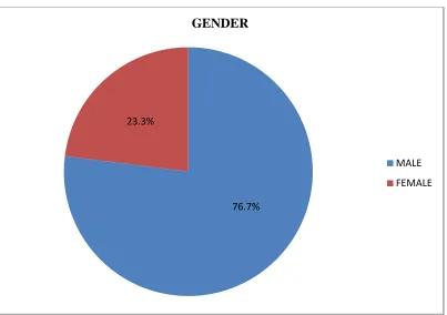
Table 2 : Gender distribution in the study population
GENDER FREQUENCY PERCENT
MALE 46 76.7
FEMALE 14 23.3
TOTAL 60 100.0
76.7% 23.3%
GENDER
MALE
55
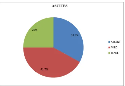
Table 3 : Ascites in the study population
ASCITES FREQUENCY PERCENT
ABSENT 20 33.3
MILD 25 41.7
TENSE 15 25.0
TOTAL 60 100.0
33.3%
41.7% 25%
ASCITES
ABSENT
MILD
56
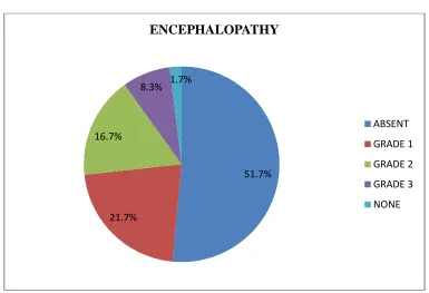
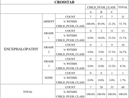
Table 4 : Encephalopathy in the study population
ENCEPHALOPATHY FREQUENCY PERCENT
ABSENT 31 51.7
GRADE 1 13 21.7
GRADE 2 10 16.7
GRADE 3 5 8.3
NONE 1 1.7
TOTAL 60 100.0
51.7%
21.7% 16.7%
8.3% 1.7%
ENCEPHALOPATHY
ABSENT
GRADE 1
GRADE 2
GRADE 3
57
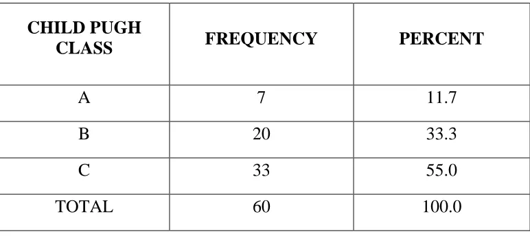
Table 5 : Child Pugh class in the study population
CHILD PUGH
CLASS FREQUENCY PERCENT
A 7 11.7
B 20 33.3
C 33 55.0
TOTAL 60 100.0
11.7%
33.3% 55%
CHILD PUGH CLASS
A
B
58
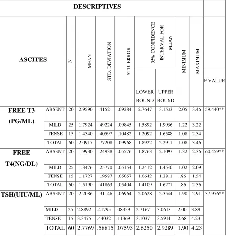
Table 6: Analysis of thyroid profile in patients with ascites in the study
DESCRIPTIVES
ASCITES N
M E A N S T D . D E V IA T IO N S T D . E R R O R 9 5 % C O N F ID E N C E IN T E R V A L F O R M E A N M IN IM U M M A X IM U M LOWER BOUND UPPER BOUND F VALUE FREE T3 (PG/ML)
ABSENT 20 2.9590 .41521 .09284 2.7647 3.1533 2.05 3.46 59.440**
MILD 25 1.7924 .49224 .09845 1.5892 1.9956 1.22 3.22
TENSE 15 1.4340 .40597 .10482 1.2092 1.6588 1.08 2.34
TOTAL 60 2.0917 .77208 .09968 1.8922 2.2911 1.08 3.46
FREE T4(NG/DL)
ABSENT 20 1.9930 .24938 .05576 1.8763 2.1097 1.32 2.36 60.459**
MILD 25 1.3476 .25770 .05154 1.2412 1.4540 1.02 2.09
TENSE 15 1.1727 .19587 .05057 1.0642 1.2811 .86 1.54
TOTAL 60 1.5190 .41863 .05404 1.4109 1.6271 .86 2.36
TSH(UIU/ML) ABSENT 20 2.2086 .31146 .06964 2.0628 2.3544 1.90 2.91 37.976** MILD 25 2.8892 .41795 .08359 2.7167 3.0618 2.00 3.89
TENSE 15 3.3475 .44032 .11369 3.1037 3.5914 2.68 4.23
59
Free T3 levels in different clinical grades of ascites in the study
Free T4 levels in different clinical grades of ascites in the study 2.96
1.79
1.43
0.00 0.50 1.00 1.50 2.00 2.50 3.00 3.50
ABSENT MILD TENSE
FREE T3 (PG/ML)
1.99
1.35
1.17
0.00 0.50 1.00 1.50 2.00 2.50
ABSENT MILD TENSE
60
Serum TSH levels in different clinical grades of ascites in the study
(Continued) 2.21
2.89
3.35
0.00 0.50 1.00 1.50 2.00 2.50 3.00 3.50 4.00
ABSENT MILD TENSE
61
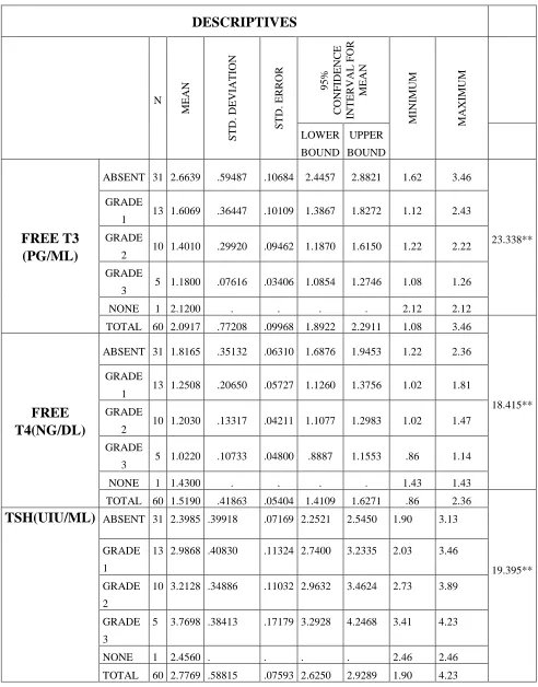
Table 7: Analysis of thyroid profile in patients with encephalopathy in the study
DESCRIPTIVES N M E A N S T D . D E V IA T IO N S T D . E R R O R 9 5 % C O N F ID E N C E IN T E R V A L F O R M E A N M IN IM U M M A X IM U M LOWER BOUND UPPER BOUND FREE T3 (PG/ML)
ABSENT 31 2.6639 .59487 .10684 2.4457 2.8821 1.62 3.46
23.338** GRADE
1 13 1.6069 .36447 .10109 1.3867 1.8272 1.12 2.43
GRADE
2 10 1.4010 .29920 .09462 1.1870 1.6150 1.22 2.22
GRADE
3 5 1.1800 .07616 .03406 1.0854 1.2746 1.08 1.26
NONE 1 2.1200 . . . . 2.12 2.12
TOTAL 60 2.0917 .77208 .09968 1.8922 2.2911 1.08 3.46
18.415**
FREE T4(NG/DL)
ABSENT 31 1.8165 .35132 .06310 1.6876 1.9453 1.22 2.36
GRADE
1 13 1.2508 .20650 .05727 1.1260 1.3756 1.02 1.81
GRADE
2 10 1.2030 .13317 .04211 1.1077 1.2983 1.02 1.47
GRADE
3 5 1.0220 .10733 .04800 .8887 1.1553 .86 1.14
NONE 1 1.4300 . . . . 1.43 1.43
TOTAL 60 1.5190 .41863 .05404 1.4109 1.6271 .86 2.36
19.395**
TSH(UIU/ML) ABSENT 31 2.3985 .39918 .07169 2.2521 2.5450 1.90 3.13 GRADE
1
13 2.9868 .40830 .11324 2.7400 3.2335 2.03 3.46
GRADE
2
10 3.2128 .34886 .11032 2.9632 3.4624 2.73 3.89
GRADE
3
5 3.7698 .38413 .17179 3.2928 4.2468 3.41 4.23
NONE 1 2.4560 . . . . 2.46 2.46
62
Free T3 levels in different grades of encephalopathy in the study
Free T4 levels in different grades of encephalopathy in the study 2.66
1.61
1.40
1.18
2.12
0.00 0.50 1.00 1.50 2.00 2.50 3.00
ABSENT GRADE 1 GRADE 2 GRADE 3 NONE
FREE T3 (PG/ML)
1.82
1.25
1.20
1.02
1.43
0.00 0.20 0.40 0.60 0.80 1.00 1.20 1.40 1.60 1.80 2.00
ABSENT GRADE 1 GRADE 2 GRADE 3 NONE
63
Serum TSH levels in different grades of encephalopathy in the study
(Continued) 2.40
2.99
3.21
3.77
2.46
0.00 0.50 1.00 1.50 2.00 2.50 3.00 3.50 4.00
ABSENT GRADE 1 GRADE 2 GRADE 3 NONE
64
Table8: Analysis of thyroid profile and serum bilirubin, albumin and INR in the study
FREE T3
(PG/ML)
FREE
T4(NG/DL)
TSH(UIU/ML)
TOTAL
BILIRUBIN
(MG/DL)
PEARSON CORRELATION -.777 -.729 .732
SIG. (2-TAILED) .000 .000 .000
N 60 60 60
ALBUMIN
(G/DL)
PEARSON CORRELATION .840 .819 -.801
SIG. (2-TAILED) .000 .000 .000
N 60 60 60
INR
PEARSON CORRELATION -.825 -.777 .782
SIG. (2-TAILED) .000 .000 .000
N 60 60 60
CHILD PUGH
SCORE
PEARSON CORRELATION -.964 -.923 .879
SIG. (2-TAILED) .000 .000 .000
65
Correlation between Free T3 levels and Bilirubin levels in the study
Correlation between Free T3 and Albumin levels in the study 0 2 4 6 8 10 12 14
0 0.5 1 1.5 2 2.5 3 3.5 4
T O T A L B IL IR U B IN ( M G /D L)
FREE T3 (PG/ML)
TOTAL BILIRUBIN (MG/DL)
0 0.5 1 1.5 2 2.5 3 3.5 4
0 0.5 1 1.5 2 2.5 3 3.5 4
A LB U M IN ( G /D L)
FREE T3 (PG/ML)
66
Correlation between Free T3 and INR levels in the study
Correlation between Free T3 levels and the Child Pugh score in the study 0 0.5 1 1.5 2 2.5
0 0.5 1 1.5 2 2.5 3 3.5 4
IN
R
FREE T3 (PG/ML)
INR 0 2 4 6 8 10 12 14 16
0 0.5 1 1.5 2 2.5 3 3.5 4
C H IL D P U G H S C O R E
FREE T3 (PG/ML)
67
Correlation between Free T4 levels and Bilirubin levels in the study
Correlation between Free T4 levels and Albumin levels in the study -2 0 2 4 6 8 10 12 14
0 0.5 1 1.5 2 2.5
T O T A L B IL IR U B IN ( M G /D L)
FREE T4 (NG/ML)
TOTAL BILIRUBIN (MG/DL)
0 0.5 1 1.5 2 2.5 3 3.5 4
0 0.5 1 1.5 2 2.5
A LB U M IN ( G /D L) )
FREE T4 (NG/ML)
68
Correlation between Free T4 levels and INR in the study
Correlation between Free T4 levels and Child Pugh score in the study 0 0.5 1 1.5 2 2.5
0 0.5 1 1.5 2 2.5
IN
R
FREE T4 (NG/ML)
INR 0 2 4 6 8 10 12 14 16
0 0.5 1 1.5 2 2.5
C H IL D P U G H S C O R E
FREE T4 (NG/ML)
69
Correlation between serum TSH levels and Total Bilirubin in the study
Correlation between serum TSH levels and Albumin levels in the study 0 2 4 6 8 10 12 14
0 0.5 1 1.5 2 2.5 3 3.5 4 4.5
T O T A L B IL IR U B IN ( M G /D L) TSH(uIU/ML)
TOTAL BILIRUBIN (MG/DL)
0 0.5 1 1.5 2 2.5 3 3.5 4
0 0.5 1 1.5 2 2.5 3 3.5 4 4.5
70
Correlation between serum TSH levels and Child Pugh score
Correlation between serum TSH levels and INR in the study 0 2 4 6 8 10 12 14 16
0 0.5 1 1.5 2 2.5 3 3.5 4 4.5
C H IL D P U G H S C O R E TSH(uIU/ML)
CHILD PUGH SCORE
0 0.5 1 1.5 2 2.5
0 0.5 1 1.5 2 2.5 3 3.5 4 4.5
IN
R
TSH(uIU/ML)
71
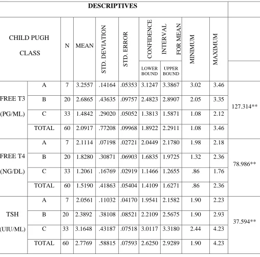
Table 9: Analysis of Thyroid profile and the Child Pugh classes in the study
DESCRIPTIVES
CHILD PUGH CLASS
N MEAN
S T D . D E V IA T IO N S T D . E R R O R C O N F ID E N C E IN T E R V A L F O R M E A N M IN IM U M M A X IM U M LOWER BOUND UPPER BOUND FREE T3 (PG/ML)
A 7 3.2557 .14164 .05353 3.1247 3.3867 3.02 3.46
127.314** B 20 2.6865 .43635 .09757 2.4823 2.8907 2.05 3.35
C 33 1.4842 .29020 .05052 1.3813 1.5871 1.08 2.12 TOTAL 60 2.0917 .77208 .09968 1.8922 2.2911 1.08 3.46
FREE T4 (NG/DL)
A 7 2.1114 .07198 .02721 2.0449 2.1780 1.98 2.18
78.986** B 20 1.8280 .30871 .06903 1.6835 1.9725 1.32 2.36
C 33 1.2061 .16769 .02919 1.1466 1.2655 .86 1.76 TOTAL 60 1.5190 .41863 .05404 1.4109 1.6271 .86 2.36
TSH (UIU/ML)
A 7 2.0561 .11032 .04170 1.9541 2.1582 1.90 2.23
37.594** B 20 2.3892 .38108 .08521 2.2109 2.5675 1.90 2.93
72
Free T3 levels and Child Pugh class in the study
Free T4 levels and Child Pugh class in the study 3.26
2.69
1.48
0.00 0.50 1.00 1.50 2.00 2.50 3.00 3.50
A B C
FREE T3 (PG/ML)
1.99
1.35
1.17
0.00 0.50 1.00 1.50 2.00 2.50
A B C
73
Serum TSH levels and Child Pugh class in the study
(Continued) 2.21
2.89
3.35
0.00 0.50 1.00 1.50 2.00 2.50 3.00 3.50 4.00
A B C
74
Table 10: Analysis of Free T3 levels and Bilirubin, Albumin, Child Pugh scores in the study
CORRELATIONS IN R T O T A L _ B IL IR U B IN _ M G D L A L B U M IN _ G D L C H IL D _ P U G H _ S C O R E FREE_T3_ PGML PEARSON
CORRELATION -.825
** -.777** .840** -.964**
P VALUE P<0.01 P<0.01 P<0.01 P<0.01
N 60 60 60 60
75
Correlation of Free T3 levels with total bilirubin in the study
Correlation of Free T3 levels with INR in the study 0
2 4 6 8 10 12 14
0 0.5 1 1.5 2 2.5 3 3.5 4
FREE T 3 (PG/ML)
CORRELATION BETWEEN T3 WITH TOTAL BILIRUBIN (MG/DL)
0 0.5 1 1.5 2 2.5
0 0.5 1 1.5 2 2.5 3 3.5 4
FREE T 3(PG/ML)