0022-538X/90/104767-09$02.00/0
Copyright C) 1990, American Society for Microbiology
Transforming Growth
Factors Beta
1and
2
Transcriptionally
Regulate Human Papillomavirus
(HPV)
Type
16
Early Gene
Expression in HPV-Immortalized
Human
Genital Epithelial Cells
C. D.
WOODWORTH,'*
V. NOTARIO,2 ANDJ. A. DiPAOLO1Laboratory of Biology, Division ofCancerEtiology, National Cancer Institute, Bethesda, Maryland20892,1and
Department
ofRadiationMedicine,
Georgetown University,
Washington,
D.C.200072
Received11 May1990/Accepted 29June1990Human papillomavirustype 16 (HPV16) early proteinsE6 and E7 have beenimplicatedinmaintenance of themalignantphenotype in cervicalcancer.Transforminggrowthfactorsbetaoneandtwo(TGFI3s 1and 2), polypeptides thatregulate cellular growth and differentiation, reversibly inhibited expression oftheHPV16E6 andE7genesinseveralimmortal genitalepithelial cell lines. Loss of E6 and E7 protein expression followeda
dramatic time-anddose-dependentdecrease in E6 and E7 RNA levels andwasaccompanied by cessation of cell
proliferation. TGF,s 1 and 2inhibited HPV16 RNA expression atthe transcriptional level; inhibition was
dependent uponongoing protein synthesis. TGFj3s 1 and2 also inducedasix-tosevenfold increase inTGFi 1RNA.Cellsbecamepartiallyresistanttotheinhibitory effectsofTGF,3 1oncell growth and HPV earlygene
expressionafter prolonged cultivationin vitroorafter malignant transformation. Thus, TGF( 1mayfunction asanautocrineregulator of HPVgeneexpressionininfected genital epithelial cells.
Human papillomaviruses (HPVs) are a group of small DNA virusesthatinduce papillomas in mucosal and epider-malepithelia (35, 57). DNAsfrom several HPV types have been detected in intraepithelial neoplasias of the cervix,
vulva, and penis, as well as in carcinomas (4, 6, 15, 29),
implyingthatthe virusmaybeafactor in thedevelopment of these tumors.Integrationof the HPV genome usually occurs in cervical cancers and carcinoma cell lines in a specific manner that ensures that the E6 and E7 early genes are
selectively retainedandtranscribed(43, 45, 46, 57). The E6 and E7 gene products regulate cell proliferation and gene
expression,astransfection ofprimaryhumangenital epithe-lial cells withrecombinantE6and E7 DNAconfers immor-tality(19, 32) and resistanceto terminal differentiation(23). The HPV type 16(HPV16) E7 protein cooperates with an
activated cellular Ha-ras gene to transform primary rodent cells (47), but continued expression of E7 is required to maintain the transformed phenotype (13). Normal genital epithelial cells possess an intracellular control mechanism directed againstHPV genetranscription (56, 57); however, cellular functions downregulating HPV expression are ab-sentingenital carcinomacells (40),suggestingthat this loss
represents animportant stepin thedevelopment ofcancer. The betatransforming growthfactors (TGF,s) are mem-bers ofa family ofpolypeptides (TGF,s 1 to 5, inhibins, activins; reviewed in reference 39)thatmodulate cell prolif-eration and gene expression in diverse cells. The cellular response to
TGFps
varies depending on the specific cells, growth conditions, and the presence ofadditional growth factors (2, 20, 21, 31, 33, 49). Generally, TGF,s inhibit proliferation ofepithelial cells, but they can either inhibit (24) or stimulate (31, 33, 48) cellular differentiation.Alter-ations in the expressionorresponsiveness toTGF,s often
occur in malignancy (7,
20-22,
44, 49). In cultured normalgenital epithelial cells,
TGFP
1inhibitsproliferation (11, 44)andinduces expressionof itsownRNA(2), suggestingthat
*Correspondingauthor.
it might function as an autocrine regulator ofgrowth and geneexpression in normal genital epithelium. Limited infor-mation is available concerning the effect of TGF,s on expression of viral genes in infected cells (3).
Inthe presentstudy,theeffect ofTGF, on papillomavirus gene expression was determined in a series of immortal genital epithelial cell lines containing integrated and
tran-scriptionally active HPV16 DNA (54, 55). TGFfis 1 and 2 dramatically down regulate expression of RNAs encoding the HPV16 E6 and E7 genes in several independently
derived cell lines. Continuous serial passage in culture or malignant transformation by oncogenes results in partial
resistancetotheinhibitory effects of
TGFO
1oncellgrowth and HPVearly gene expression.MATERIALS ANDMETHODS
Cell culture. Primary cultures of human genital epithelial cells were isolated from foreskin and cervical tissue as described before(54, 55). The derivation and characteriza-tion of cell lines immortalizedbytransfection with recombi-nantHPV16 DNAhave also beendescribed in detail(37,54, 55). Normal foreskin or cervical cells and all HPV-immor-talized cell lines were maintained in serum-free MCDB153-LB medium (37). In some experiments, cells were maintained inmediumlacking growth factors (insulin,
hydro-cortisone, transferrin,
triiodothyronine, epidermal
growth factor,and bovinepituitary
extract)toinhibitproliferation.Cervical carcinoma cell lines QGU, QGH
(45),
and SiHa(American Type Culture Collection,
Rockville, Md.)
were grownina1:1mixture of Dulbecco modifiedEagle medium(DMEM) and F12 medium supplemented with 5% fetal
bovineserum. Earlyand late passages ofHPV-immortalized cell lines were used. By
definition,
early passage refers to cells used within 60 populationdoublings
after transfection withHPVDNA,and late passagereferstothose maintained for more than 250population
doublings (each
passage rep-resentsapproximately
four to fivepopulation doublings).
Rapidly
proliferating
culturesat60to80% confluencewere 4767on November 10, 2019 by guest
http://jvi.asm.org/
4768 WOODWORTH ET AL.
used. In some experiments cellular protein synthesis was inhibited by treating cultures with cycloheximide (10 ,ug/ml
ofmedium) (Sigma Chemical Co., St. Louis, Mo.), and the extent of inhibition of protein synthesis was monitored as described before (30). To measure RNA stability, dactino-mycin (Fluka Chemical Corp., Ronkonkoma, N.Y.) was
added to cultures at 10 ,ug/ml of medium.
Cloning experiments. Approximately 500 to 1,000cells in 5.0 ml of medium were added to duplicate 60-mm tissue culture dishes and allowed to attach overnight. TGF1 1
obtained from human platelets (Collaborative Research, Bedford, Mass.) or porcine TGF,B 2 (R and D Systems, Minneapolis, Minn.) was added in 5.0 ml of medium the next day, and cultures were incubated for 10 days. Cultures were fixed with 3% Formalin and stained with Giemsa, and colonies containing more than 20 cells were counted.
Immunoprecipitation and gel electrophoresis. After a 30-min incubation of subconfluent cell cultures in MCDB153 without methionine or cysteine, 4.0 ml of medium containing 100,uCi of[35S]methionine and 100 SuCiof[35S]cysteine per ml was added. Cells wereincubated at 37°C for an additional 4 h. Labeling was terminated by washing cells with cold phosphate-buffered saline. Cells were lysed in 1.0 ml per 10-cm plate of RIP buffer (1% Triton X-100, 1.0% sodium dodecyl sulfate, 0.5% deoxycholate, 0.1 NaCl, 0.1 M phen-ylmethylsulfonyl fluoride, and 20 ,ug of aprotinin per ml). Samples (200 ,ul) were incubated with rabbit polyclonal anti-HPV16 E6 (provided by J. Schiller), mouse monoclonal
anti-HPV16 E7 (Triton Biosciences Inc., Alameda, Calif.), orrabbit polyclonalantiinvolucrin (Biomedical Technologies Inc., Stoughton, Mass.) for 2 h on ice before being
precip-itated with protein A-Sepharose. Immunoprecipitates were washed several times in RIP buffer and analyzed by sodium
dodecyl sulfate-polyacrylamide gel electrophoresis. Parallel immunoprecipitations were performed with lysate samples with either equal total protein concentration (400 ,ug) or
equal counts (75 x 106 cpm) to control for differences in
amino aciduptake. Markers were purchased from Bethesda Research Laboratories (Gaithersburg, Md.). Normal cervi-cal cells were used as anegative control.
Recombinant plasmid DNAs. Plasmid DNAs included pD5DD1 (a0.63-kilobase-pair [kbplDdeI fragment
contain-ing HPV16 E6 and partial E7 open reading frames from nucleotides 25 to 653; provided by J. Doniger), a 0.7-kbp SacI-PvuII fragment of porcine TGF,3 1 cDNA (26), a 1.2-kbp HindIll fragment of simian TGF,B 2 cDNA (18), human c-myc gene (Oncor Incorp, Gaithersburg, Md.), a human ,B-actin gene fragment (28), a cDNA encoding the humanlaminin
,13
chain (provided by Y. Yamada), and an 18S rRNA probe (provided by F. Mushinski). Plasmid DNAs weredigested with the appropriate restriction endonucleases tocleave vector sequences, except for the 18S RNA probe, which was used as complete plasmid DNA. Insert DNAs were separated on agarose gels, and the purified fragments wereused as probes for nick translation or for slot blots.Northern (RNA blot) analysis. Total cellular RNA was
purified from subconfluent cultures by lysis in guanidine thiocyanate (8), followed by centrifugation through cesium trifluoroacetate. RNA (10 to 15 ,ug) was separated by elec-trophoresis in 1.5% agarose gels containing formaldehyde, transferred to nylon membranes (Schleicher and Schuell,
Keene, N.H.) by capillary blotting, baked at 80°C for 2 h, andhybridized to
32P-labeled
DNAs under stringentcondi-tions (50% formamide, 10% dextran sulfate, 5x Denhardt solution (30), 1% sodium dodecyl sulfate) at 42°C for 16 to 24 h. Filterswerewashed twice with 2x SSC (lx SSC is 0.15
MNaClplus 0.015 M sodium citrate) containing 1% sodium dodecylsulfate for 30 minatroomtemperature and then with O.1x SSC containing 0.1% sodium dodecyl sulfate at 50to 65°C for 30 min. Filters were exposed to Kodak XAR-2 film with anenhancing screen at -72°C. RNA molecular weight standards were obtained from Bethesda Research Laborato-ries.
Runoff transcriptional analysis. Nucleiwereisolated from subconfluent cell cultures as described before (42) and maintained at -70°Cinfreezingbuffer(50%glycerol, 20 mM Trishydrochloride, 75 mM NaCl, 0.5 mM EDTA, 0.85 mM
dithioerythritol, 100 UofRNaseinhibitor per ml) for 2to 3 weeks before use. Runoff transcription analysis was per-formed basically as described before (42). The in vitro elongation reactions were performed by the method of
Gariglio et al. (16) in a mixture containing 100 mM Tris
hydrochloride (pH 7.9), 200 mM NaCl, 0.4 mM EDTA, 0.1 mM phenylmethylsulfonyl fluoride, 0.3 M (NH4)2SO4, 4.0 mMeachMgCl2 and MnCl2, 1.2 ,uM dithioerythritol, 1.0 mM each GTP, ATP, and CTP, 250 jiCi of 32P-labeled UTP (3,000Ci/mmol), 10 mMcreatinephosphate, 20 U of
placen-tal RNase inhibitor perml, 30% glycerol,andapproximately 2 x 10⁷ nuclei. a-Amanitin was added to some reaction mixes as anegative control. The reaction mixes were incu-batedfor 30 min at 27°C. Labeled RNA was extracted from the reaction mixture and purified by gel filtration with Sephadex G-50 (Pharmacia LKB Biotechnology, Inc., Pis-cataway, N.J.), followed by precipitation with ammonium
acetate. Nitrocellulose filters containing 2
jig
of purifiedinsert DNA from eachrecombinantplasmid were prehybrid-ized for24to 48 h in 50 mM HEPES (N-2-hydroxyethylpi-perazine-N'-2-ethanesulfonicacid, pH 7)-0.75MNaCl-50% formamide-0.5% sodium dodecyl sulfate-2.0 mM EDTA-lOx Denhardt solution-200 ,ug of salmon sperm DNA per ml-10
jig
ofpolyadenylic acid per ml. Equal amounts of labeled RNA (approximately 107 cpm/ml) were added to eachfilter,andhybridization was allowed to proceed at 42°C for 48 to 72 h. The intensity of hybridization signals was measuredby scanning laser densitometry.RESULTS
Inhibition of cell proliferation byTGF13 1. TGF,B 1 at 3.0
ng/mlcompletely inhibited clonal growth in secondary cul-tures of normal genital epithelial cells derived from either
foreskin(HK) orcervix (CX) (Table 1). Cells flattened and
proliferation ceased approximately 24 to 48 h after treat-ment. Growth inhibition was partially reversible when fore-skin-derived keratinocyte cultures that were exposed to TGF,B 1 for 48 h were washed and then refed with fresh medium lacking
TGF1
1 for anadditional 8 days (data not shown); however, cervicalepithelial cells were more suscep-tible and clonal growth was irreversibly inhibited by 3.0 ng ofTGF1
1 per ml by 48 h. Different cell lines derived by transfection and immortalization of normal cells with HPV16 DNAvaried significantly in their response toTGF1
1, with some being more resistant than normal cells. Cell lines of late passage (>250population doublings) often exhibited a further increase in resistance.Two immortalized cell lines that were malignantly trans-formed after transfection with the v-Ha-ras oncogene (14) or the herpes simplex virus type 2 (HSV-2) BglII N fragment (13a)werealso examined. Both cell lines formed colonies in
TGF1
1-containing medium with a frequency greater than the parental lines. Three cervical carcinoma-derived tumor lines grew clonally in medium containingTGF13 1, although J. VIROL.on November 10, 2019 by guest
http://jvi.asm.org/
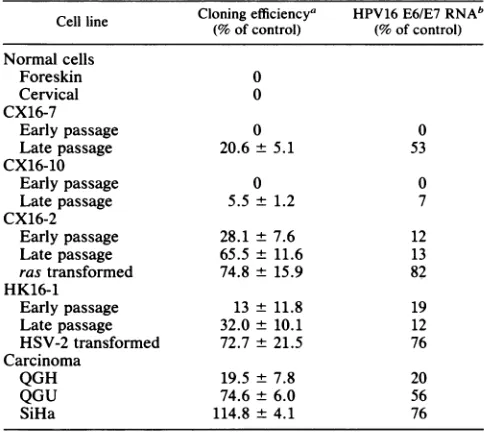
TGF, REGULATION OF HPV GENE EXPRESSION 4769 TABLE 1. Effects ofTGF, 1 on clonal growth and
HPV16E6/E7RNAexpression
Cloning efficiencya HPV16E6/E7RNAb Cell line
(%
ofcontrol)(%
of control)Normal cells
Foreskin 0
Cervical 0
CX16-7
Early passage 0 0
Late passage 20.6 + 5.1 53
CX16-10
Earlypassage 0 0
Latepassage 5.5 ± 1.2 7
CX16-2
Earlypassage 28.1 ± 7.6 12
Latepassage 65.5 ± 11.6 13
rastransformed 74.8 ± 15.9 82
HK16-1
Early passage 13 ± 11.8 19
Latepassage 32.0 ± 10.1 12
HSV-2 transformed 72.7± 21.5 76
Carcinoma
QGH 19.5 ± 7.8 20
QGU 74.6± 6.0 56
SiHa 114.8 ± 4.1 76
aClonal growthwasmeasuredinthepresence ofTGFP1 (3.0 ng/ml) for 10
days. Data representthe mean of four dishes ± standard error. Untreated cultures servedasthe control.
b Northern blotswereanalyzed byscanningdensitometry, and values are
expressed asapercentageof the RNAin cultures grownin the absence of
TGF,B1(control).
A
CX16-2
+TGFB 1 _
0o
x It
0 "4.co OD
t e; C
r
I
'TM
nc'Io v A So
Ir
g1 ,l--&
I
S,
- 43Z
_*
I.i ~~~-29
E6- *
B
x
0
I
CX16-2 +TGFB1
co C
04 ( a:
- 68
- 43
- 29
- 18.4 - 14.3
E7
- 18.4
- 14.3
FIG. 1.
TGFP
1 effects on expression of HPV16 E6 and E7 proteins. CX16-2 cells were treated with TGFI 1 (3.0 ng/ml) for variousintervals (4, 24, 48, and 96 h), andexpression of HPV E6 (A) and E7 (B) proteins was assessed afterimmunoprecipitation and polyacrylamide gel electrophoresis. Secondary cultures ofnormal cervical cells (HCX), which do not express HPV16 E6 or E7 proteins,wereused as anegative control.Thespecificityofthe E6 antiserumhas beendescribed previously(1). TodeterminewhetherTGFP
1 effects on HPVexpression werereversible,TGFP
1-con-taining medium wasreplaced with freshmediumafter48h(48/48). Numbersattherightof panels indicate molecularmass(in kilodal-tons).growth of one line (QGH) was reduced significantly with respect to untreated cultures. Another tumor line (SiHa) exhibited aslightly greater cloning efficiency in the presence
of
TGFP
1.Inhibition of HPV16 geneexpression by
TGFOi
1and 2.TheeffectofTGF,1ontheexpression of the HPV16 early genes E6 and E7 was examined in the CX16-2
HPV16-immortal-izedcervical cell line that was partially resistant to TGF, 1
in clonal growth assays. All experiments were done with early-passage cultures (<60 population doublings) of
sub-confluent, proliferating cells.
TGFP
1 treatment for 24 hmarkedlydecreased levels ofE6 and E7protein expression (Fig. 1). When cultures were maintained in the presence of
TGFP
1, E7expressionremainedlow to undetectable for 48 h, but expression waspartially restored when cultureswere switched after 48 hto fresh mediumeither with or withoutTGF, 1 for another 48 h. In contrast, E6 expression re-mained undetectable evenafter TGF, 1 wasremoved.DNA
synthesis, measuredbyincorporationof tritiatedthymidine,
was decreased approximately 60% after treatment with
TGF3
1 for 24 or 48 h. TGF, did not alter expression of involucrin(datanotshown),amarkerofsquamousdifferen-tiationincervicalepithelium (52).
RNAanalyses wereperformedtofurther define the level atwhichTGF,3 1regulated
papillomavirus
geneexpression.The CX16-2 cell line expressed three different RNAs that
hybridized to the HPV16 E6 and E7 open reading frames
(Fig. 2A). RNAs of 1.7 and 4.2 kilobases (kb) were
ex-pressed in each of seven HPV16-immortalized cell lines
examined,anda3.4-kbspecieswasdetected infour ofseven lines (54). This impliesthat thesetranscripts begin and end within virus sequences andare notfusion RNAscontaining
both virus and cellular sequences. Exposure to TGF, 1 resulted in time-dependentreductions in both cell
prolifera-tion and steady-state levels of E6 and E7 RNAs (Fig. 2A).
The 1.7-kb RNA decreased within 6 to8 h after treatment andremained lowtoundetectable by24to48h. In contrast, the 4.2-and 3.4-kb RNAswerereduceddramaticallywithin 2 to 4 h, but later were reexpressed at reduced levels
comparedwith the untreated cells. Thepattern ofinhibition of HPV RNAexpression by
TGFP
1wasreproducibleintwoexperiments with the CX16-2 cell line. These cells also
expresseda2.4-kbTGF, 1 RNA(Fig.2A).TGF, 1 induced
atime-dependentincrease inTGF, 1RNAthatwasmaximal
(sevenfold)after 24 h. Rehybridizationof the filterto acDNA
for
glyceraldehyde
phosphate dehydrogenase (GAPDH), ahousekeepinggene,demonstrated thatequalamountsofRNA were presentin each lane.
Toexamine whether downregulationof virus gene expres-sion by
TGFP
1mightoccursecondarilytocessationofcellproliferation, cellgrowthwas inhibited inreplicate cultures
by twodifferent methods. CX16-2 cellsweremaintainedfor 2 or 4 days in basal medium lacking insulin, epidermal growth factor,transferrin,triiodothyronine,bovine
pituitary
extract, and
hydrocortisone.
This treatment induced cell flattening and inhibited celldivisionby approximately72to83%
in replicate experiments, as determined bydirect cell counts.These cells could be stimulatedtodivideby
replac-ingthebasal mediumwith
complete
medium.Alternatively,
culturesweregrowntoconfluence,toinhibit
proliferation by
contactwithadjacentcells. In both instances the CX16-2 cell line continued to express normal levels of all three virus RNAs (Fig. 2B). Maintaining cells in medium deficient in
growthfactorsdidnotalter
TGFP
1RNA levels but ledto aslightdecrease inexpression of GAPDH RNA.
TGF,B 1 and 2 were equally effective in
reducing
HPV16 E6/E7 RNA levels after a 24-h treatment(Fig.
2C). BothTGF, types inhibited virus RNA
expression
in adose-dependentmanner,andaslittleas0.3
nglml
waseffective.In VOL.64, 1990on November 10, 2019 by guest
http://jvi.asm.org/
[image:3.612.61.303.100.317.2]4770 WOODWORTH ET AL.
B
GFD CONFL
CD
-IID)
CYst°O cocr >->. >- >- ) ECF >- >-< >-< <0
CCr r r
<0
0 a 0Cr 2: (D a 0 N 0 0
N Kr OCO ,r- ,- \N 'i N4 ( N .; LI)
C
0.3 1.0 3.0 10.0
-4
0
in co m m
Z LL LL U_ U.
o C LI) I)
o
-N cN
m m a
LL a, LL LL
an co cv
}
F-HPV16 W
E6/E17.
wF
......
- 28S
£ w w ~~-18S
HPV 16 E6/E7
I' **
TGFBI ..*
I0
* - t8185
- 18S
TGFB1 *to @*t - l8S
- 18S
GAPDH
0
a
0 40
40 46
4w
JtGAPDH * *
W
,*.FIG. 2. Northernanalysesof cells treated withTGF,s1 and 2 andcells arrested ingrowth. (A)CX16-2 cellsweretreated with TGFI 1
(3.0 ng/ml) for various intervalsorfor 2days followed by removal of
TGFP
1 for 2 days (2/2 days). (B)Growth arrest wasinduced by maintaining CX16-2 cellsinmediumlacking growthfactors(GFD) (seeMaterialsand Methods)orby maintainingcultures for 5 to 10days asconfluentmonolayers(CONFL). (C)CX16-2 cellsweretreatedfor 24 h with various concentrations(0.3, 1.0, 3.0,and 10.0,ug/ml)ofTGF,1 and 2. Three RNAs of4.2, 3.4, and 1.7 kbhybridizedto the HPV16 E6/E7probe. Theprobes used forhybridization were pD5DD1(a
0.63-kbpDdeIfragmentof HPV16containingE6 andpartialE7openreading frames)andaportionof theTGF,B1 cDNA(0.7-kbpSacI-PvuII
fragment).To ascertain that each lane containedequalamountsof totalRNA,blotswererehybridizedtoacDNAfor GAPDH. Numbersat
theright of panels indicatethepositionsof the 18S and 28S rRNAs. addition, both
TGFP
1 and 2 induced a six- to sevenfold increase inTGF,1 RNA.However,TGFP
2 RNAswerenot detected in this cell lineevenwhen thefilterwasexposedfor aslongas 1 week.To determine whether inhibition of HPV16 RNA
expres-sionorinduction ofTGFi1 RNAby
TGFP
1wasdependenton continued protein synthesis, cultures of CX16-2 cells were treated withcycloheximide (10 ,ug/ml) in thepresence
orabsence ofTGF, 1. This concentration ofcycloheximide inhibited incorporation of [35S]methionine into proteins in CX16-2 cells by approximately 80%. Cycloheximide
treat-ment prevented the reduction in the 1.7-kb HPV RNA causedby
TGFP
1 (Fig.3),butcycloheximide alonedid not alter the level of thistranscript. In contrast, cycloheximide treatment caused steady-state levels of the 4.2- and 3.4-kb RNAsto decline to undetectable levels by 8 h. The differ-entialeffects ofcycloheximideonthe HPV16 1.7- and 4.2-kb RNAs indicate that these RNAs areregulated differently.The effect ofcycloheximideon autoregulationof TGFI 1 RNAwascomplex. Cycloheximidetreatmentfor 8 h didnot affectTGF,B1 RNAlevelsbut enhanced the autoinduction of TGF,B 1 RNAapproximately fourfold. However, cyclohex-imidetreatmentfor24 hinhibitedTGF,B1RNAexpression.
Nuclear runoffexperiments were performed to examine whether reductions in steady-state levels ofHPV16 RNAs by TGF,s 1 and 2 were due to changes in transcription. Rapidly proliferating CX16-2 cell culturesweretreated with TGFI3 1 or2 for 24h, and the elongation of transcripts for several genes was compared. Results were normalized to GAPDHexpression, asprevious experiments demonstrated that TGF,s 1 and 2 do not altersteady-state levels of this RNA. Transcription of HPV16 RNAwas reduced
dramati-callyincells treated with eitherTGF, 2 or 1(Fig. 4;Table
8 HR 24 HR
m
L
cr I
Z LA
m
c) 0 0
0 CC
z
0 C)
em
HPV16 E6/E7
LLCO
aD
x i.;0 x
V C)
- 28S
- 18S
,. i
TGFBl1 4.45 0* - l8S
- 18S
GAPDH D D D D D w
FIG. 3. Effect ofcycloheximideonTGFI 1-inducedchangesin
geneexpression. ProliferatingCX16-2 cellsweretreated with 10 p.g
ofcycloheximide (CHX)perml,3.0ngof TGFf3 1perml,orboth for either 8or42 h. Filterswerehybridizedtotheprobesdescribed in thelegendtoFig. 2.
A
TGFB1
-4
0
z
0
28S
W*
-18SJ.VIROL.
on November 10, 2019 by guest
http://jvi.asm.org/
[image:4.612.63.549.75.304.2] [image:4.612.341.532.419.650.2]C
a:
z 0
04
U-a (m
LL HL
HPV16 _ -
-BETA ACTIN
C-MYC
GAPDH _ - _
LAMININ INVOLUCRIN
FIG. 4. Nuclear runoff transcription analysis of CX16-2 cells treatedwithTGF( 1 or 2. Nuclei were isolated from proliferating cultures ofCX16-2 cells that were treated with TGF( 1 or 2 (3.0 ng/ml) for 24 h or from untreated cultures (control). Then, 2.5 ,ug of purified insert DNA from plasmids containing HPV16, (-actin, c-myc,GAPDH, laminin (31 chain, or involucrin DNA was immo-bilizedonnitrocellulosefilters and hybridized to an equal amount of radioactivity(107cpm/ml)for 3days at 42°C.
2). In contrast, TGF,Bs 1 and 2 increased transcription of RNAs for the laminin (1 chain and ,B-actin. This increase wasapparentafter normalization of signal intensity to that of GAPDH (i.e., slightly more counts of nascent RNAs were present in the control lane). TGF,Bs 1 and 2 did not affect c-myctranscription in this immortalizedcell line.
Todetermine whether the inhibition of virus gene expres-sion by TGF(3 1 might be further regulated by posttran-scriptional mechanisms, the stability of HPV16 early-gene transcripts was measured. Cultures were treated with dacti-nomycin (10 ,ug/ml) to inhibit RNA polymerase II activity, andthe ratesof decay ofexistingtranscriptswerecompared in control cultures and those treated for 2 h withTGF( 1. When the hybridization intensity of the 1.7-kb RNA was plotted against time, itshalf-life was interpolated as 14.5 h. Treatment withTGF( 1didnotalterits stability(Fig. 5). In contrast,thehalf-life of the 4.2- and 3.4-kb RNAswasmuch shorter. When similar experiments were conducted over shorterintervals,the 4.2-kb RNA decreased within 1 to2h aftertreatmentwithdactinomycin, andTGF,B1did not alter this decay (datanot shown).
Influence of
TGFOi
1on HPV16 geneexpression in immor-talizedandtumorigenic cell lines. The biological significanceof the ability ofTGF( 1 to downregulate papillomavirus early gene expression was examined with a series of cell
TABLE 2. Changes intranscription induced byTGF(s 1and2a Transcriptionalactivity Gene (%of control)
TGF,B1 TGF, 2
HPV16 12 17
(-Actin 114 134
c-myc 91 98
GAPDH 100 100
Laminin
(31
chain 353 237Involucrin 46 70
aCX16-2 cells were treated for 24 h with TGF,B 1 or 2 at 3.0 ng/ml. Autoradiographsfromtranscriptionalrunoffanalyseswereanalyzedby
scan-ningdensitometry,and values were normalized to levels of GAPDH
transcrip-tion. Values are expressed as apercentage oftranscriptional activity
un-treated cultures.
ACTINOMYCIN D
-J
0
a: c Er a:
Z x II
0 Co '-t C)
L) co - N Cl
HPV 16
E6lE7 9 I ,
TGFB1 +
ACTINOMYCIN D
-J
0
cc z Er:r m :
0CO U CW)
- 28S
t
- 18S
RIBOSOMAL - 18S
FIG. 5. Stability of HPV16E6/E7 RNAs. Rapidly proliferating CX16-2 cells were treated with dactinomycin (10 ,ug/ml) or incu-bated for 2 h withTGFI 1 (3.0ng/ml) before addition of dactino-mycin. Total cellular RNA was purified at various intervals and hybridizedtotheHPV16E6/E7probedescribedin thelegend toFig. 2.Filterswererehybridizedto an18SrRNA probe to ascertain that alllanescontained equalamountsofRNA.
lines derived from human genital epithelium. The primary objectivewasto determine whether inhibition of virus gene expression byTGF,B 1 occurred inareproduciblemannerin differentHPV16-immortalized cell lines. Inaddition, TGF,3 1responsivenesswas assessed incell lines maintained for an extendedinterval inculture,incellsmalignantly transformed by subsequent transfection with the v-Ha-ras oncogene or the HSV-2BglII N fragment, and incarcinoma-derivedcell lines.
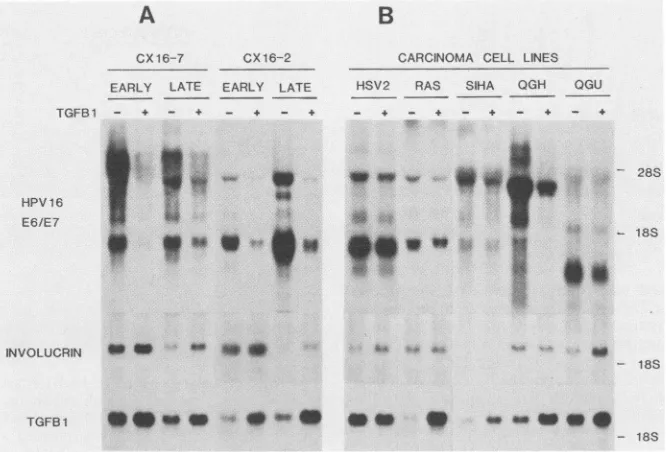
Treatment with TGF,B 1 for 24 h dramatically reduced steady-statelevelsof HPV16 RNA inearly-passagecultures of all three immortal lines (CX16-2, CX16-7, and CX16-10) derived from cervical epithelium and one (HK16-1) from neonatal foreskin (Table 1; Fig. 6A). When the same cell lines were examined after extended maintenance in culture (>250population doublings), down regulation of HPV gene expression by TGF,B 1 was often less pronounced but still detectable. One line (CX16-2) developed increased expres-sion of HPV16 E6/E7 RNAs at latepassage,whereas expres-sioninanother was decreased(datanot shown). Thesecell linesoriginallywerepolyclonalbut becamemonoclonalwith increasing passage in culture, as reflected by their chromo-somal constitution (38). Therefore, changes in steady-state levels of HPV16 RNA that occur in cells of late passage mighthave resulted fromselection of asubpopulationwhich differsin the level of virus gene expression.
HPV16E6/E7RNA was decreasedonlyminimally (56to 76% ofcontrols) by TGF,B 1 inthe two cervical carcinoma cell lines QGU and SiHa (Fig. 6B); however, virus gene expression was down regulated significantly (80%) in an-other tumorline, QGH. Furthermore,virus RNAexpression decreased only slightly after TGF( 1 treatmentofimmortal cell lines that had been malignantly transformed with the HSV-2BglIINfragmentorthe V-Ha-rasoncogene. Immor-talized nontumorigenic and malignantly transformed cell lines varied widely in their ability to express RNA for
involucrin, a structural protein thatrepresents a marker of squamous differentiation in anogenital epithelium (52). TGF(3 1 treatmentslightlyenhancedexpressionofinvolucrin
on November 10, 2019 by guest
http://jvi.asm.org/
[image:5.612.319.559.74.256.2] [image:5.612.113.245.76.207.2] [image:5.612.59.297.586.681.2]4772 WOODWORTH ET AL.
A
B
CX 16-7
EARLY LATE
TGFB1 - 4 - +
CX16-2 EARLY LATE
- -.44.
CARCINOMA CELL LINES HSV2 RAS SIHA QGH QGU
_ 4 _ 4 _ 4 _ 4 - 4
*-~~~~~~~~~21~~~~ 8
HPV 16
E6/E7 ii iiwi 188
*Zt**i! ' 18S
N*LCN-m - W*.1*
INVOLU URN * 4:F*n . ,X _
TGFB1 4 e.O '-4* * *a *
- 18S
- 18S
FIG. 6. Effect ofcellpassage ormalignanttransformationon
TGFP
1-mediated changesin geneexpression. (A)TwoHPV16-immortalized celllines derived from cervicalepithelium(CX16-7 andCX16-2)weretested atearly(<60population doublings) and late (>250doublings) passage.Cellswereeither exposedtoTGF, (3.0ng/ml) for24h(+) orleft untreated (-). (B)In areplicate experiment,twoimmortalcell lines malignantly transformed by additionof a v-Ha-ras oncogene(RAS)ortheHSV-2BglIINfragment (HSV2)and threecervical carcinoma cell lines that express HPV16 RNA(SiHa, QGH, andQGU)werealsotested. Filters werehybridizedtoprobesforHPV16, involucrin,orTGFP
1.RNA in several lines. A direct correlation was observed
betweenthe basal level of involucrinexpression ina partic-ular cell line and the sensitivity of the line to inhibition of HPV16 RNAexpression. Celllines expressing higherlevels
of involucrin were more sensitiveto
TGFP
1.TGFP
1 alsoinduced its own expression in most of the cell lines
exam-ined,butthe level of
TGFP
1autoinduction did not correlate with sensitivity to inhibition of HPV early gene expression. Furthermore, some cell lines (SiHa andRAS) weresuscep-tibletoautoinduction of
TGFP
1RNA but wererefractory to the effects of TGF, 1 on growth and HPV16 gene expres-sion. Therefore, the resistance of these lines to TGFi1 was notduesolely to an absence of TGF, 1receptors atthe cellsurface.
DISCUSSION
The E6 and E7 proteins of the oncogenic HPVs are
implicated in maintenance of the malignant phenotype in
cervicalcancerandcancer-derived cell lines (40, 43, 46,51, 57). Transcription of the HPV16 E6 and E7 genes was dramatically inhibited by picomolar concentrations of
TGF,s 1 and 2 in immortalized cell lines derived from human cervical and foreskin epithelium. Down regulation of virus gene expression was accompanied by inhibition of cell
proliferation.Inaddition,
TGFP
1induced the expression of its own RNA, thereby providing the potential to amplify and sustain inhibitory effects on HPV gene expression. Thus, these results suggest an autocrine function forTGFP
1 in down regulating HPV gene expression in infected anogenitalepithelium.
A host intracellular control mechanism that down regu-lates HPV gene expression in infected anogenital epithelial cellshas been described previously (56, 57). Our observation thatTGF,s1and 2 decrease HPV16 transcription in
immor-talized, nontumorigenic cervical cellshas similarities to the reportof Rosletal. (40), whoexamined HPV gene expres-sion in hybrids of normal keratinocytes and HeLacervical carcinoma cells expressing HPV18. 5-Azacytidine, a de-methylating agent, inhibited HPV18 gene transcription in nontumorigenic hybrid cells but not in the tumorigenic segregantsthatsubsequentlyarose. Furthermore, inhibition of HPV gene expression was reversible and dependent on
ongoing protein synthesis, suggesting that5-azacytidine in-duceda cellularfactor that controls virus gene expression. Incontrast,5-azacytidine did not inhibit HPV gene expres-sion in cervical carcinoma cell linesortumorigenic hybrids,
implying that loss of responsiveness represents a step in
malignant progression (40). The similarities in the mode of action of5-azacytidine and
TGFP
suggest that both may inhibit HPV geneexpression by a similarmechanism, such as interaction with a transcription factor. Furthermore, autocrine regulation of HPV16 gene expression by TGF, 1 could represent one component ofan intracellular surveil-lance systemdirectedagainstHPVtranscription. Incontrast to 5-azacytidine, theeffects ofTGF, 1 onHPV expressionvaried significantly in different cell lines, and often the resistance that developed in tumorigenic cells was only
partial.
Apositive correlation was apparent between the
magni-tude of reductions in both HPV gene expression and cell
growth in mostcelllines (Table 1), suggestingthat the two processes were related. To examine whether the effects of
TGFP
on virus gene expression occurred secondarily togrowth inhibition,cellswereincubated underconditions that
preventedcellreplication priorto assessmentof HPV RNA levels. Cultures maintained in growth factor-deficient me-diumoratconfluentdensity expressedHPV RNAsatlevels similar to rapidly proliferating cultures. Thus, HPV gene J. VIROL.
on November 10, 2019 by guest
http://jvi.asm.org/
[image:6.612.144.477.72.298.2]expression is not necessarily inhibited by growth arrest. Furthermore, the results suggest but do not prove an alter-native, that cell growth mightdepend on continued papillo-mavirusgeneexpression. Downregulation of the
papilloma-virus E6 and E7 gene products inhibits the growth rate of
C4-1 cervical carcinoma cells (51), and continuous
expres-sion of the E7 gene is required for maintenance of the
transformed phenotypeinbabyratkidneycells transformed
by EJ ras and HPV16 E7 (13). The current results are
consistent with these observations.
TGFP
regulates expression ofa wide variety of cellular genes(39), andregulationmayoccur atmultiplelevels(2, 11,25, 26, 33, 34, 41, 50). Our results are the first report that
TGFps
1 and 2 inhibit transcription ofviral genes.Papillo-mavirustranscriptionisregulated byseveral distinct enhanc-erslocatedwitha5'longcontrolregion (9, 12, 17, 27)which respond to glucorticoids (17), keratinocyte-specific factors (12), andautoregulation by products ofthe HPV E2 gene(5,
27). Sequenceanalysis ofthelong controlregionshows that thekeratinocyte-specificenhancercontainsbinding sitesfor
nuclearfactor 1 and AP-1 (9), and mutational studies have shown that these sites are required forfull enhancement of
virus geneexpression (9).
TGFP
1stimulatestranscriptionof the collagen type 1 promoter via nuclear factor 1binding
(41), andautoinduction of
TGFP
1RNAismediatedby
the AP-1 complex (25). Therefore, these factorsmight
also beinvolved in the TGF, 1-mediated
downregulation
ofpapil-lomavirus transcription.
Cells infected with
papillomaviruses
in vivo harbor virus DNA in episomal form, andtranscriptional regulation
iscomplex (10, 27).
Analysis
oftranscription
of thecancer-associatedtypesHPV16 and HPV18 ishindered
by
thelack of an animal or cell culture model forstudying
productive
infection. Much of the work on
transcription
ofoncogenic
HPV typeshas been donewith cervicalcancercell lines
(43,
45, 46). In carcinoma cell
lines,
the viral DNA characteris-tically integrates inaspecific pattern,interrupting
theE1IE2
genes and preservingthe E6/E7 genesin a
transcriptionally
active form(43, 57). All oftheHPV-immortalized cell lines used in thisstudy containoneor more
complete
copies
oftheviral genome (54, 55); however, these
copies
areintegrated
within the host chromosome and therefore
partially
rear-ranged. Thus, thesecells
might
resemblecarcinoma-derivedcell lines more than
productively
infected cells in their pattern ofvirustranscriptional
regulation.
Alterations in the
ability
of cells tosynthesize
orrespond
toTGFP
occurduring
thedevelopment
ofneoplasia (7,
20-22, 44, 49),implying
that thesechanges
represent animportantstepinmalignant
progression. TGF,
1completely
inhibited clonal
growth
ofnormalgenital
epithelial cells,
but the resistance of HPV16-immortalized cell lines varied sig-nificantly. Some immortal cell lines werepartially
resistantto TGFi 1,
particularly
when the cells werecontinually
maintained in culture. Inaddition,
themajority
of thetumorigenic
cell lines examined(four
offive)
also becameresistant.
Thus,
loss ofresponsiveness
toTGF,
1 often precedesoraccompanies
malignant
development
in culturedgenital
epithelial
cells. Differenttumorigenic
cell lines alsovaried significantly in their resistance to
TGFP,
and onecarcinoma-derived
line,
QGH,
waspartially
sensitive.Therefore, while
acquisition
ofresistanceto TGFI 1might
contributetothecarcinogenesis
process, thisstudy
aswell asothers(20,48, 49,53)
indicates that resistancetoTGFP
is not aprerequisite.The
variability
inresponsiveness
ofdifferent cell lines toTGF,B 1 was
directly
related to theirability
to expressinvolucrin RNA.
Involucrin
isamarker of squamous differ-entiation in normal cervicalepithelium
andis either absent orweakly
expressed
inpoorly differentiated,
dysplastic
epithelium (52).
Cellsexpressing high
levels of involucrinwere more
susceptible
to theeffects ofTGFPi
1 ongrowth
and HPV16 gene
expression
than were cells that failed to expressinvolucrin
orexpressed
low levels ofit.Thus,
thedegree
ofdifferentiation ofgenital epithelial
cells influencestheir
susceptibility
toTGF,B
1. The current results areconsistent with the report of Hooseien et al.
(20),
who demonstrated that resistance of colon carcinoma cells togrowth
inhibitionby
TGFP
1wasrelatedtothedegree
of celldifferentiation. The mechanisms
by
whichpoorly
differenti-ated cellsbecome resistant isnotknown.
However,
TGF,B
1 maintained itsability
toupregulate
itsownRNA intwocell lines(RAS
andSiHa)
thatremainedrefractory
totheeffectsof
TGF,
1ongrowth
and HPV RNAexpression.
Therefore,
the
primary
defectin these resistant cells doesnotappearto be an absence ofTGF,
1receptors,
indicating
that otherchanges
in thesignal
transductionpathway
must be in-volved.ACKNOWLEDGMENTS
We thank Anita Roberts for
helpful suggestions
and critical review ofthemanuscript,
John Schiller forproviding
antiserumtoHPV16 E6, Frederic Mushinski forthe 18S rRNA
plasmid,
and Yoshihiko Yamadafor thelaminin P1chainplasmid.
ADDENDUM
During
review of themanuscript, Pietenpol
et al.(36)
reported
that humangenital
epithelial
cells become resistant togrowth
inhibitionby
TGF,
1 after transformation withpapillomavirus
or simian virus 40 DNA.LITERATURE CITED
1. Androphy, E.J.,N. L.Hubbert,J.T.Schiller,and D. R.Lowy.
1987. Identification ofthe HPV16 E6
protein
from transformed mousecellsand humancervicalcarcinoma cell lines.EMBOJ. 6:989-992.2. Bascom, C.C.,J. R. Wolfshohl,R.J.
Coffey,
Jr.,L. Madisen,N. R.Webb,A. R.Purchio,R.
Derynck,
and H. L. Moses.1989.Complex
regulation
oftransforming
growth
factorp1,
P2,
andP3mRNA
expression
inmousefibroblastsandkeratinocytes by
transforming growth
factorsp1
andP2.Mol. Cell. Biol.9:5508-5515.
3. Bauer, G., U. Birnbaum, P. Hofler, and C. H. Heldin. 1985.
EBV-inducing
factorfromplatelets
exhibitsgrowth
promoting
activity
forNIH 3T3 cells. EMBOJ. 4:1957-1961.4. Beaudenon, S., D. Kremsdorf, 0. Croissant, S. Jablonska, S. Wain-Hobson, and G. Orth. 1986. A novel type of human
papillomavirus
associatedwithgenital neoplasias.
Nature(Lon-don)321:246-249.
5. Bernard,B.A.,C.
Bailly,
M.C.Lenoir,M.Darmon,F.Thierry,and M. Yaniv. 1989. The human
papillomavirus
type 18(HPV18)E2 gene
product
isarepressorofthe HPV18regula-tory
region
in humankeratinocytes.
J. Virol.63:4317-4324.6. Boshart, M., L. Gissmann, H. Ikenberg, A. Kleinheinz, W. Scheurlen,andH. zurHausen.1984. Anewtypeof
papilloma-virus DNA, its presence ingenital
cancerbiopsies
and incell lines derived from cervical cancer.EMBO J. 3:1151-1157. 7. Braun, L., P. Gruppuso, R. Mikumo, and N. Fausto. 1990.Transforming
growth
factorp1in livercarcinogenesis:
messen-ger RNA
expression
andgrowth
effects. Cell Growth Differen-tiation 1:103-111.8.
Chirgwin,
J. M., A. E.Przybyla,
R. J. MacDonald, andW.J.Rutter. 1979. Isolation of
biologically
active ribonucleic acidfromsourcesenriched inribonuclease.
Biochemistry
18:5294-5299.
on November 10, 2019 by guest
http://jvi.asm.org/
4774 WOODWORTH ET AL.
9. Chong, T., W. K. Chan, and H. U. Bernard. 1990. Transcrip-tionalactivationof human papillomavirus 16by nuclearfactor1, AP1, steroidreceptorsandapossibly novel transcriptionfactor, PVF, a model for the composition of genital papillomavirus
enhancers. Nucleic Acids Res. 18:465-470.
10. Chow, L. T., M. Nasseri, S. M. Wolinsky, and T. R. Broker.
1987. Human papillomavirus types 6 and 11 mRNAs from genital condylomata acuminata. J. Virol. 6:2581-2588.
11. Coffey, R. J., Jr., C. C. Bascom, N.J. Sipes, R. Graves-Deal,
B.E.Weissman,and H.L. Moses. 1988. Selective inhibitionof growth-related gene expression in murine keratinocytes by transforminggrowth factorP. Mol. Cell. Biol. 8:3088-3093.
12. Cripe, T. P., T. H. Haugen, J. P. Turk, F. Tabatabai, P. G.
SchmidIII,M.Durst, L. Gissmann, A. Roman, and L. P. Turek.
1987.Transcriptionalregulation of the human papillomavirus 16 E6-E7 promoterbyakeratinocyte-dependent enhancer, and by
viral E2 transactivator and repressor gene products:
implica-tionsforcervical carcinogenesis. EMBO J. 6:3745-3753.
13. Crook, T., J. P.Morgenstern,L. Crawford, and L. Banks. 1989. Continued expression of HPV16 E7 protein is required for
maintenance of the transformed phenotype of cells
co-trans-formedby HPV16 plus EJ-ras. EMBOJ. 8:513-519.
13a.DiPaolo, J. A., C.D. Woodworth, N. C. Popescu, D. L. Koval,
J. V.Lopez,and J. Doniger. 1990. HSV-2 induced tumorigenic-ityinHPV16-immortalized human genital keratinocytes.
Virol-ogy177:777-779.
14. DiPaolo,J. A., C. D.Woodworth, N. C. Popescu, V. Notario, and
J. Doniger. 1989. Induction of human cervical squamous cell
carcinomaby sequentialtransfection with human
papillomavi-rus16DNA and viralHarveyras. Oncogene4:395-399.
15. Durst,M., L.Gissmann,H. Ikenberg, and H.zurHausen.1983. A papillomavirus DNA from a cervical carcinoma and its
prevalence incancerbiopsy samplesfrom differentgeographic
regions. Proc. Natl. Acad. Sci. USA 80:3812-3815.
16. Gariglio, P., M. Beliard, and P.Chambon. 1981. Clusteringof RNA polymerase B molecules in the 5' moiety of the adult
p-globin geneof hen erythrocytes. Nucleic AcidsRes. 9:2589-2598.
17. Gloss, B., H.U. Bernard, K.Seedorf, and G. Klock. 1987. The
upstream regulatory region of the human papillomavirus 16 contains an E2 protein-independentenhancer whichis specific
for cervical carcinoma cells and regulated by glucocorticoid hormones. EMBOJ. 6:3735-3743.
18. Hanks, S. K., R. Armour, J. H. Baldwin, F. Maldonado, J.
Spiess, and R. W. Holley. 1988. Amino acid sequence ofthe
BSC-1cellgrowth inhibitor(polyergin) deduced fromthe nucle-otide sequence of the cDNA. Proc. Natl. Acad. Sci. USA 85:79-82.
19. Hawley-Nelson, P., K.H. Vousden, N.L. Hubbert, D. R. Lowy,
andJ. T. Schiller.1989. HPV16E6andE7proteinscooperateto
immortalize human foreskin keratinocytes. EMBO J.
8:3905-3910.
20. Hoosein, N. M., M.K. McKnight, A. E.Levine,K. M.Mulder,
K. E. Childress, D. E. Brattain, and M. G. Brattain. 1989.
Differentialsensitivityof subclassesof human coloncarcinoma
cell lines to growth inhibitory effects of transforming growth
factorp1. Exp. Cell Res. 181:442-453.
21. Hosobuchi, M., and M. R. Stampfer.1989. Effectsof transform-ing growth factor P on growth of human mammary epithelial
cellsinculture. In VitroCell. Dev. Biol. 25:705-713.
22. Hubbs,A. F., F.F. Hahn,and D. G. Thomassen. 1989. Increased
resistance to transforming growth factor beta accompanies neoplastic progressionofrattrachealepithelial cells. Carcino-genesis 10:1599-1605.
23. Hudson,J. B., M.A. Bedell,D. J.McCance, and L. A. Laimins.
1990. Immortalizationand altered differentiationof human
ke-ratinocytes in vitroby the E6 and E7 openreading frames of
human papillomavirustype 18.J. Virol. 64:519-526.
24. Ignotz, R. A., and J. Massague. 1985. Type P transforming growth factor controls the adipogenic differentiation of 3T3
fibroblasts. Proc. Natl. Acad. Sci. USA 82:8530-8534.
25. Kim, S. J.,P. Angel,R. Lafyatis,K. Hattori, K. Y. Kim,M.B. Sporn, M. Karin, and A. B. Roberts. 1990. Autoinduction of
transforminggrowth factor
p1
ismediatedbythe AP-1complex. Mol. Cell. Biol. 10:1492-1497.26. Kondiah, P., E. Van Obberghen-Schilling, R. L. Ludwig, R. Dhar,M. B.Sporn,and A. B. Roberts. 1988. cDNA cloningof porcine transforming growth factor
P1
mRNAs: evidence foralternate
splicing and polyadenylation. J. Biol. Chem. 263:18313-18317.
27. Lambert, P. F., C. C. Baker, and P. M. Howley. 1988. The genetics of bovine papillomavirus type 1. Annu. Rev. Genet. 22:235-258.
28. Leavitt, J., P. Gunning, P. Porreca,S. Y. Ng, C. S. Lin, and L. Kedes. 1984. Molecularcloningand characterization ofmutant
and wild-type human P-actin genes. Mol. Cell. Biol. 4:1961-1969.
29. Lorincz, A. T., W. D. Lancaster, and G. F. Temple. 1986. Cloning and characterization of the DNA of a new human papillomavirus from a woman with dysplasia of the uterine cervix. J. Virol. 58:225-229.
30. Maniatis, T., E. F. Fritsch, and J. Sambrook. 1982. Molecular cloning: alaboratory manual. ColdSpring HarborLaboratory, Cold Spring Harbor, N.Y.
31. Masui,T., L. M.Wakefield, J. F. Lechner, M. A.Laveck, M. B. Sporn, and C. C. Harris. 1986. Type P transforming growth factor is the primary differentiation-inducing serum factor for normal humanbronchial epithelialcells. Proc.
Natl.
Acad. Sci. USA 83:2438-2442.32. Munger, K., W. C. Phelps, V. Bubb, P. M. Howley, and R. Schlegel. 1989. The E6 and E7 genesof the human papillomavi-rus type16together are necessaryand sufficientfor transforma-tionof primary humankeratinocytes. J. Virol. 63:4417-4421. 33. Noda, M., and G. A. Rodan. 1987. Type
I8
transforminggrowthfactor (TGFB) regulation of alkaline phosphatase expression and otherphenotype-related mRNAs in osteoblastic rat
osteo-sarcomacells. J. Cell. Physiol. 133:426-437.
34. Penttinen, R. P., S. Kobayashi, and P. Bornstein. 1988. Trans-forming growth factor p increases mRNA for matrix proteins both inthe presence and absence ofchangesin RNAstability. Proc. Natl. Acad. Sci. USA 85:1105-1108.
35. Pfister, H. 1987. Human papillomaviruses and genital cancer. Adv. CancerRes. 48:113-147.
36. Pietenpol, J. A., R. W. Stein, E. Moran, P. Yaciuk, R. Schlegel, R. M. Lyons, M. R. Pittelkow, K. Munger, P. M. Howley, and H. L. Moses. 1990.
TGFp1
inhibitionof c-myc transcription and growth in keratinocytes is abrogated by viral transforming proteins with pRB binding domains. Cell 61:777-785.37. Pirisi, L.,S. Yasumoto, M.
Feller,
J. Doniger, and J. A.DiPaolo. 1987. Transformation of human fibroblasts and keratinocytes with human papillomavirus type 16 DNA. J. Virol. 61:1061-1066.38. Popescu, N. C., and J. A. DiPaolo. 1990. Integration ofhuman papillomavirus 16 DNA and genomic rearrangements in immor-talizedhuman keratinocyte lines. Cancer Res. 50:1316-1323. 39. Roberts, A. B., and M. B. Sporn. 1990. Thetransforminggrowth
factor-p's. Handb. Exp. Pharmacol. 45:419-472.
40. RosI, F., M. Durst, and H. zurHausen. 1988. Selective suppres-sion of human papillomavirus transcription in non-tumorigenic cells by 5-azacytidine. EMBO J. 7:1321-1328.
41. Rossi, P., G. Karsenty, A. B. Roberts, N. S. Roche, M. B. Sporn, and B. de Crombrugghe. 1988. A nuclear factor 1 binding site mediates the transcriptional activation of a type 1 collagen promoter by transforming growth factorP. Cell 52:405-414. 42. Schibler, U., 0.Hagenbuchle, P. K.
WelHauer,
and A. C. Pittet.1983. Two promoters of different strengths control the transcrip-tion of the mouse alpha-amylase gene Amy-la in the parotoid gland and the liver. Cell 33:501-508.
43. Schneider-Gadicke, A., and E. Schwarz. 1986. Different human cervical carcinoma cell lines show similar transcription patterns of human papillomavirus type 18 early genes. EMBO J.
5:2285-2292.
44. Shipley, G. D., M. R. Pittelkow, J. J. Wille, R. E. Scott, and H. L. Moses. 1986. Reversible inhibition of normal human prokeratinocyte proliferation by type P transforming growth J. VIROL.
on November 10, 2019 by guest
http://jvi.asm.org/
factor inhibitor in serum-free medium. CancerRes. 46:2068-2071.
45. Shirasawa, H., Y. Tomita, S. Sekiya, H. Takamizawa, and B. Simizu.1987. Integration and transcription of human papilloma-virus type 16 and 18 sequences in cell lines derived from cervical carcinomas. J. Gen. Virol. 68:583-591.
46. Smotkin, D., and F. 0Wettstein. 1986. Transcription of human papillomavirus type 16 early genes in a cervical cancer and cancer-derived cell line and identification of the E7 protein. Proc. Natl. Acad. Sci. USA 83:4680-4684.
47. Storey, A., D. Pim, A. Murray, K. Osborn, L. Banks, and L. Crawford. 1988.Comparisonof the invitrotransforming activ-ities of human papillomavirus types. EMBO J. 7:1815-1820. 48. Twardzik, D. R., J. E.Ranchalis,J.M. McPherson, Y. Ogawa,
L. Gentry, A. Purchio, E. Plata, and G. J. Todaro. 1989. Inhibition and promotion ofdifferentiated-like phenotypeofa human lung carcinoma in athymicmiceby natural and recom-binant forms oftransforming growth
factor-P.
J. Natl. Cancer Inst. 81:1182-1185.49. Valverius, E. M., D. Walker-Jones, S. E. Bates, M. R. Stampher, R.Clark, F. McCormick, R. B. Dickson, and M. E. Lippman. 1989. Production and responsiveness to transforming growth factor-a innormal andoncogene-transformedhuman mammary epithelialcells. Cancer Res. 49:6269-6274.
50. VanObberghen-Schilling,E., N. S.Roche, K. C. Flanders, M. B. Sporn, and A. B. Roberts. 1988.Transforminggrowthfactor ,B1 positively regulates its own expression in normal and trans-formedcells. J. Biol. Chem.263:7741-7746.
51. von Knebel-Doeberitz, M., T. Oltersdorf, E. Schwarz, and L.
Gissmann. 1988. Correlation ofmodifiedhumanpapillomavirus early gene expression with altered growth properties of C4-1 cervical carcinomacells. Cancer Res.48:3780-3786.
52. Warhol, M. J., G.S. Pinkus, R. H. Rice, G. H. El-Tawil, W. D. Lancaster, A. B. Jenson, and R. J. Kurman. 1984. Papillomavi-rusinfection of the cervix. III. Relationship of the presence of viralstructural proteins to theexpression of involucrin. Int. J. Gynecol.Pathol. 3:71-81.
53. Wollenberg, G.K., E. Semple, B. A. Quinn, and M. A.Hayes. 1987. Inhibition of proliferation of normal, preneoplastic, and neoplastic rat hepatocytes by transforming growth factor-P. CancerRes. 47:6595-6599.
54. Woodworth, C. D., P. E. Bowden, J. Doniger, L. Pirisi, W. Barnes, W. D. Lancaster, and J. A. DiPaolo. 1988. Characteri-zation of normal humanexocervical cells immortalized in vitro by papillomavirus type 16 and 18 DNA. Cancer Res. 48:4620-4628.
55. Woodworth, C. D., J. Doniger, andJ. A. DiPaolo. 1989. Immor-talization ofhuman foreskin keratinocytes by various human papillomavirus DNAs corresponds to their association with cervical cancer.J. Virol. 63:159-164.
56. zurHausen, H. 1986. Intracellular surveillance ofpersisting viral infections. Lancet ii:489-491.
57: zurHausen, H. 1989.Papillomavirusesinanogenitalcancer asa model to understand the role of viruses in human cancer.
CancerRes. 49:4677-4681.