0022-538X/83/110521-13$02.00/0
Copyright C1983, AmericanSociety forMicrobiology
Synthesis
and
Processing of Glycoprotein
D
of
Herpes
Simplex Virus Types 1 and 2 in
an
In
Vitro System
JAMES T.MATTHEWS,123t GARY H. COHEN,'2 ANDROSELYN J.
EISENBERG'
3*CenterforOral Health Research' andDepartment of Microbiology,2 SchoolofDentalMedicine, and DepartmentofPathobiology, SchoolofVeterinary Medicine,3 Universityof Pennsylvania, Philadelphia,
Pennsylvania 19104
Received 27May 1983/Accepted 9 August 1983
We carriedoutstudies of invitro translation and processing of glycoprotein D (gD) of herpessimplex virustypes1and 2 byusing mRNA from cells infected for 6 h and areticulocyte lysate translation system. Polypeptides of 49,000 daltons
wereimmunoprecipitated with anti-gD-1sera. Eachinvitro-synthesized molecule
had the same methionine tryptic peptide profile as the respective in vivo precursors, pgD-1 and pgD-2. In addition, the polypeptides synthesized in vitro were larger than the corresponding molecules synthesized in the presence of
tunicamycin. This suggested that each of the gD polypeptides synthesized in vitro containeda transient N-terminal signal sequence. When the translation mixture was supplemented with pancreatic microsomes, each of the gD polypeptideswas
converted cotranslationally to a larger-molecular-weight form. Processing
in-volved addition of three N-asparagine-linkedoligosaccharides and removal of the signal peptide. When trypsinwas added after invitroprocessing, a polypeptide
which was 3,000 daltons smaller than the in vitro-modified form of gD was
immunoprecipitated. Experiments with endo-,-N-acetylglucosaminidase H showed that this polypeptide still contained the three N-asparagine-linked oligo-saccharides. Two monoclonal antibodies, 57S
(group
V) and 170 (groupVII),
were used to further orient gD in microsomes. The group V determinant waslocated in the trypsin-sensitive 3,000-daltonfragment, and thegroupVII
determi-nant was located in the portion of gD which was protected from trypsin. We
concluded thatgD is oriented with the threeglycosylation sites inside the vesicles and that 3,000 daltons containing the group V determinant are located outside.
Immunofluorescence studies indicated that the group V determinant ofgD is inside the plasma membrane of
herpes
simplex virus-infected cells and that thegroup VII determinant is outside. This cellular orientation is consistent with
predictions basedonthe in vitroexperiments.
Glycoprotein
D(gD) of
herpes
simplex
virus
(HSV) is
a type-common componentof
thevirion
envelope which stimulates
production
of
high titers of
virus-neutralizing antibody
and is
believed
tobe
important in the initial
stagesof
viral
infection (5, 6, 8, 12, 34).
Inprevious
studies
(5, 6, 10, 11,
20, 33,
35,
36), attention
wasfocused
onthedetails of
synthesis
and
process-ing of gD
inHSV
type1(HSV-1)-
andHSV
type 2(HSV-2)-infected cells.
Thesestudies
indicat-ed that
gD
isprocessed from
alower-molecular-weight
precursor (pgD-1for
HSV-1 orpgD-2 forHSV-2)
to ahigher-molecular-weight product
(gD-1
orgD-2) in virus-infected
cells. This in-crease is due to the additionof
threeN-aspara-gine-linked oligosaccharides
toboth
gD-1
andt Present address:DepartmentofPathology, Harvard Uni-versity School ofMedicine, Boston, MA 02115.
gD-2
and to thesubsequent modification of
theseoligosaccharides,
without
significant
alterationof the
polypeptides
(6,
7,
10, 12).
Extensive
analysis
of
gD-1
and
gD-2
struc-tures wascarried
out,including: tryptic peptide
analysis of the
precursorandproduct forms (10,
12),
analysis
of the number and characteristics
of
N-asparagine-linked
oligosaccharides
(7),
anddetermination
of
the amino acidcomposition of
the
purified
glycoproteins (13)
andpartial
N-terminal
sequences (R. J.Eisenberg,
D.Long,
R.
Hogue-Angeletti,
and G. H.Cohen,
submit-tedfor
publication).
Theseinvestigations
ledus toconclude thatgD-1
andgD-2
areverysimilar,
but not
identical,
in structure. Studiesemploy-ing both polyclonal
and monoclonalantibodies
have shown that
gD-1
andgD-2
alsodisplay
antigenic similarities (11, 12).
However,
there are also a numberof
type-specific
antigenic
521
on November 10, 2019 by guest
http://jvi.asm.org/
522 AND EISENBERG
determinants in addition to the type-common
determinants
present ineachprotein.
Recently,
the genefor gD-1
wasmapped (19),
and its nucleotide sequence was determined (41). The
deduced
amino acid sequence shows thatgD-1contains
twohydrophobic
regions,
one at theaminoterminus, presumed
tobe asignal
peptide
(1) ormembrane
insertion sequence(18,
29), and the other near the
carboxy terminus,
postulated
tobe
amembrane-anchoring
se-quence(29).
Inaddition,
threecarbohydrate
acceptance sequences
(Asn-X-Thr
orSer)
have beenidentified.
Studies in ourlaboratory
have shown thatpgD
andgD
isolated from infectedcells
lack the first 25
residues of the deducedamino
acid
sequence.Thus,
anearly processing
event appears to involve removal ofa
putative
signal
peptide from
theN-terminus. In the pres-entstudy,
weused acoupled
in vitrotranslation-processing
system toexamine
specific
eventsin theprocessing of gD.
These eventsnormally
occurred
toorapidly
tobe
examined
in infected cells. Inaddition,
weused
the in vitrotransla-tion-processing
system toexamine
theorienta-tion of
thegD
molecule in membrane vesicles.
Our results indicatethat the structure and orien-tation of gD-1 and gD-2 are very similar and agree with the predictions made from the pri-mary sequence.
(A portion ofthis work has been submittedby J.T.M. in
partial
fulfillment of therequirements
for the
degree
of Doctor ofPhilosophy
at the UniversityofPennsylvania.)MATERIALS AND METHODS
Cell cultures.Conditions forthegrowthand mainte-nance of KB and BHK cells have been described previously (4, 8).
Virus preparation and titration. The procedures used for thepreparationof virus stocks of HSV-1(HF) and HSV-2 (SAVAGE), as well as for the plaque assay, have beenpreviously described (4, 8). Forall experiments, a multiplicityofinfection of 20 per cell was used for HSV-1, and 10 per cell was used for HSV-2.
Pulse-labeling procedures. The protocols for pulse-labeling with [2, 3-3H]arginine, [35S]methionine, and
[3H]methionine,
as well as for the preparation of cytoplasmic extracts, havebeen describedelsewhere (5, 6, 10, 12). Briefly, after absorption of virus (2h at 37°C), monolayers (KBorBHKcells for HSV-1 and BHKcellsforHSV-2)wereoverlaid withEagle mini-malessential mediumcontaining 10%thenormal con-centration ofmethionineorarginine.At6 h postinfec-tion,the cells(35-mmplates)wereincubatedin 0.5 ml of Hanks saltscontaining 100 ,uCi of[35S]methionine (1,200 Ci/mmol), 125 ,uCi of [2, 3-3H]arginine (15 Ci/mmol),or 1mCi of[3H]methionine(80Ci/mmol)for 15min.Theradiolabeledaminoacidswerepurchased fromNewEnglandNuclearCorp. Forexperimentsin whichtunicamycin (TM) (2 ,ug/ml) (Calbiochem) wasused, BHK cells were used for both HSV-1 and HSV-2 infections (24).
Extraction of total cytoplasmic RNA from HSV-infected cells. At6h postinfection, monolayers were rinsed withcoldsaline, suspended in saline, and then washed three times by centrifugation. Cytoplasmic extracts were prepared, andRNA was extractedby the procedure of Preston (25). RNA recovered by ethanolprecipitationwasdried, suspended in water (5 to 10mg/ml), divided into equal portions, and stored at
-100°C.
Invitro translation. Rabbit reticulocyte lysateswere prepared from New Zealand white rabbits and were digested with micrococcal nuclease (22, 40). Alternate-ly, we used a lysate preparation from New England Nuclear. Translation of total cytoplasmic RNA was optimized by adjusting the final concentrations of magnesiumacetate (0.5 mM), potassiumacetate (100 mM), andspermidine (0.05 mM) and by adjusting the temperature of incubation (28°C). Translation was assessed qualitatively with either late adenovirus mRNA(New EnglandNuclear)ormRNAfrom Rous sarcomavirus-infected cells(kindly supplied by Susan Weiss, University of Pennsylvania School of Medi-cine, Philadelphia). No differences were discerned between thetwolysate preparations. For both HSV-1 andHSV-2, we used 0.5 to 1.0,ugof heat-denatured RNA and 50,uCi of[35S]methionine (1,200 Ci/mmol) per 25 p.1 of assay. For translation in the presence of pancreatic vesicles, weuseda protein processing kit (New England Nuclear). The microsome suspension wasdiluted1to4in 20 mM HEPES (N-2-hydroxyeth-ylpiperazine-N-2-ethanesulfonic acid) buffer (pH 7.5), and1 ,ulwasadded per25,u1of assay. Conditions for incubationwereexactly as described above.
Isolation of microsomes after in vitro translation. Aftertranslation, the cell-free system was adjusted to 500mMKCl-10mMEDTA-10mMTris (pH 7.5), and the microsomes were sedimented through a 15% (wt/vol) sucrose cushion at 34,000 rpm at 20°C for 90 min inanSW50.1 rotor(BeckmanInstruments, Inc.) (9, 21). The supernatantwasaspirated, and the pellet wassolubilized in the 1x detergent solution usedfor immunoprecipitation (see below).
Post-translational proteolysis. Proteolysis studies of the in vitro translation products were carried out essentiallyasdescribedby Scheeleetal.(30). Briefly, after translation, the lysates were incubated in the presenceof 100p.gof RNase per ml for 15 min at 28°C. Tetracaine-hydrochloride (Sigma Chemical Co.) was added at a final concentration of 2 mM, and the mixture was incubated for 10 min at 28°C and then chilledat0°C. Trypsin (50,ug/ml), chymotrypsin (100 ,ug/ml), or both were addedtosamples either beforeor afterthe addition of0.5% TritonX-100. The samples were incubated for 2 h at 0°C, trasylol was added (1,500 KIU),and thesampleswere held at0°Cforan additional10minbefore furtherprocessing.
Immunoprecipitation. The preparation and charac-terization of gD-specific polyclonal (mouse) and monoclonal antibodies used in this study have been describedpreviously (5, 8, 11, 13, 23, 32). Anti-gD-1 serumandgroupVII (170) antibody reactwith both gD-1 and gD-2 (11, 12, 23). Group V (57S) antibody reacts only with gD-1 (11, 32). Protein A-Sepharose (Pharmacia FineChemicals) was used to collect the antigen-antibody complexes. Briefly, the gel was sus-VIROL.
on November 10, 2019 by guest
http://jvi.asm.org/
pended in NET buffer (140 mM NaCl, 1 mMEDTA, 10 mMTris-hydrochloride [pH 7.5]) containing 10% su-croseand0.5% Nonidet P-40, washed three times, and resuspendedin NETbufferat aconcentration of 10% (wt/vol). After in vitro translation,anequal volume of 2x detergent solution (100 mM NaCl-1% sodium deoxycholate-1% nonidet P-40-0.2% sodium dodecyl sulfate (SDS)-20 mM L-methionine in 40 mM Tris-hydrochloride, pH 7.5)was added to the translation mixture. Polyclonal anti-gD-1 serum or monoclonal antibody (170 or 57S) was added, and the mixtures were incubated for 1 h at 25°C. Protein A-Sepharose was added, and the mixtures were incubated for 30 min at 25°C and then sedimented through a 1-ml cushionof1Msucrose(in 1xdetergent solution) for 5 minat 15,000 x g.Thepellets were washed 10 times with 10 mMTris hydrochloride (pH 7.4) containing 140 mMNaCl,10 mML-methionine, 0.1% SDS, and 0.2% Nonidet P-40 and suspended in disrupting buffer for SDS-polyacrylamidegel electrophoresis (SDS-PAGE) (5, 6,12).
Preparation of samples for tryptic peptide analysis. Proteins were eluted from Protein A-Sepharose by boiling for 5 min in 3% SDS andwereprecipitated with 25%(vol/vol) cold trichloroacetic acid in the presence of carrierbovineserumalbumin (6, 11).Trypsinization andion-exchange chromatographyonChromabeadsP (TechniconCorp., Inc.)werecarried outaspreviously described (6, 11, 12).
Digestion with Endo H. Polypeptides were eluted from Protein A-Sepharose by boiling for5min in 1% SDS-1%,B-mercaptoethanol andwereadjustedtopH 6.0with 0.25Mcitratecontaining1 mM phenylmethyl-sulfonyl fluoride (3, 7). One-half of this sample was treated with 0.5 mU of endo-,-N-acetylglucosamini-dase H(Endo H) (perp.lofeluate), and the other half servedas acontrol(3, 7). Both sampleswere incubat-edat37°Cfor 20 h. Bovineserumalbuminwasadded, and the reaction was terminated with 25% trichloro-acetic acid. Theprecipitatewascollectedby centrifu-gation, washed successively with 95% ethanol and ether,anddissolved in SDS-PAGEdisrupting buffer.
Immunofluorescenceanalysis. Amodification of the indirect procedure described by Reed et al. (26) was employed for fixed cells. Briefly, monolayers of KB cells grown on Lab-Tek slides (Miles Laboratories, Inc.) were infected with HSV-1 (HF) (multiplicity of infection, 10). After2hof absorption, the cells were overlaid with complete medium and incubated foran additional 8 h. Theslideswerefixed in 3.7% (vol/vol) formaldehyde,washed withphosphate-bufferedsaline (PBS), dehydrated with acetone, and washed again withPBS. The monolayers were overlaid with 50 ,ul of theappropriately diluted antiserum (prepared in mice) or monoclonal antibody, incubated for 1 h at 37°C, washedwith PBS, and then incubated for 1 h with a mixture of fluoresceinisothiocyanate-conjugated goat anti-mouse immunoglobulin G (Cappel Laboratories) and rhodamine-conjugated albumin (Microbiological Associates). Forunfixed cells, a suspension culture of KB cellswasinfected with HSV-1(HF) (multiplicity of infection, = 20). At 12 h postinfection, the cells were washed with cold PBSandthenincubated with 100
p.l
of theappropriately diluted antibody for 1 h at 37°C. The cells were washed with cold PBS and incubated with 100p.l
of thefluorescein isothiocyan-ate-rhodamine staining mixture described above for 1-w -~pgD
[image:3.491.290.408.73.145.2]1 2 3 4 5 6
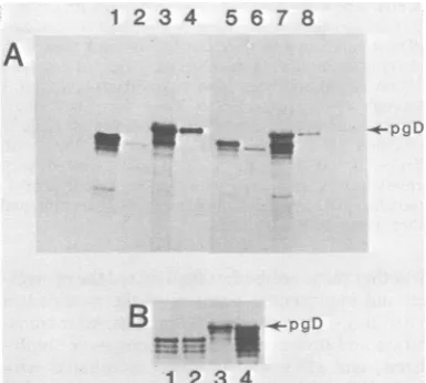
FIG. 1. SDS-PAGE analysis of gD-1 and gD-2 polypeptidessynthesized in vivo and in vitro. Autora-diogram ofa9 to 12%gradient SDS-polyacrylamide gel. Cytoplasmic RNA from cells infected with HSV-1 (lane 1) or HSV-2 (lane 2) was extracted at 6 h postinfection and translated in vitro inareticulocyte lysate system in the presence of[35S]methionine. The polypeptides wereimmunoprecipitated with anti-gD-1 serum.BHKcellswere infected with HSV-1 (lane 3) or HSV-2 (lane 4) in the presence of TM. At 6 h postinfection, the cells werepulse-labeled for 15 min with[35S]methionine. Cytoplasmic extracts were pre-pared and immunoprecipitated with anti-gD-1 serum. BHKcells wereinfected withHSV-1 (lane 5)or HSV-2(lane 6) andwerepulse-labeled with[35S]methionine for15min at 6 hpostinfection. Cytoplasmicextractsof these cells were immunoprecipitated with anti-gD-1 serum.Lanes1and2ofthis gel were exposedtoX-ray film for 14 days. Lanes 3 through 6 were exposed for7 days.
h at 37°C. The cells were washed with cold PBS, resuspended incoldPBS, and placed on fluorescent-antibody testslides.
Electrophoresis of polypeptide products. Proteins were subjected to electrophoresis on slabs of either 10% or9 to12%linear gradient SDS-polyacrylamide, cross-linked with 0.4% N,N'-diallytartardiamide (6, 11, 13, 34). After electrophoresis, the gels were stained, dried, and exposed to Kodak XAR-5 film.For fluorography, the procedure of Bonner and Laskey was followed (2). Protein molecular weight markers ranging from 15,000 to 130,000 daltons (15 to 130K) were included on each gel.
RESULTS
Comparison
of HSV-1 and HSV-2polypeptides
translated invitro and in infected cells
(in vivo).
Earlier studies showed
thatsynthesis
andproc-essing
of
gD-1 and
gD-2 in vivo occurred
maxi-mally from
5to8 hpostinfection (6, 12).
Conse-quently,
we used totalcytoplasmic
RNAextracted from cells infected
at6 hpostinfection
as the source of
gD-specific
mRNA(hereafter
called HSV-1 mRNA or HSV-2
mRNA)
for translation in vitro. A 49Kpolypeptide
wasimmunoprecipitated
from invitro translations
programmed with HSV-1 mRNA
(Fig.
1, lane1)
or HSV-2 mRNA
(Fig.
1,
lane2).
These 49K polypeptides were not immunoprecipitated bypreimmune
serum orfromtranslations in which RNA from mock-infected cells was used (data notshown). These 49K moleculeswerealsoca. 1,500 to 2,000 daltons larger than theon November 10, 2019 by guest
http://jvi.asm.org/
sponding polypeptides synthesized in vivo in the
presenceof TM (24),asshown in Fig. 1, lanes 3
(gD-1) and 4 (gD-2). In addition, TM-gD-1 (48K) was slightly larger than TM-gD-2
(47.5K). This molecular weight difference was
consistent with that seen between the in vivo precursorspgD-1 (Fig. 1, lane 5;53K) and pgD-2
(Fig. 1, lane 6; 52K). Due to the increased resolution of polypeptides with gradient gels, the
apparent molecular weights of the gD polypep-tides differed somewhat from those previously reported (7, 8, 12, 24). It should also be noted that the apparent molecular weight of the 49K polypeptide synthesizedin vitro wassomewhat
greater than the molecular weight of 43,291 predicted from the deduced amino acid se-quence (41). A similar molecular weight for gD
synthesized in vitro was reported by Lee et al. (19) and by Inglis and Newton(15).
Tryptic peptide analysis ofinvitro-synthesized products.To establish theauthenticity of the gD polypeptides synthesized in vitro, we analyzed
thetryptic peptides bycation-exchange chroma-tography. The profile of [35S]methionine-labeled tryptic peptides of the gD-1 polypeptide synthe-sized in vitro (Fig. 2A) was identical to that obtained previously for pgD-1 and
gD-1
(6, 10, 11). The profile consisted ofaflow-through (FT)fraction, a peak which eluted at pH 3.33 (frac-tion 95, presumably peptide f in reference 11), and a minorpeak termed the basic wash
(frac-tions 171 to180). The proportions of label
recov-ered in the FT fraction(75%) and infraction 95 (25%)werecomparable tothosefound in
previ-ous results (6, 10-13). Apparently, the gD-1 polypeptide synthesized in vitro contains no
additional methionine-labeled tryptic peptides. The FT fraction was examined furtherby
high-pressure liquid chromatographyandby Bio-Gel P6 filtration (data not shown) and was foundto
containthetwo methioninepeptides previously found (7) in the Chromabeads P FTfraction of TM-gD-1. Peptide fwaspreviously showntobe
anarginine-labeled tryptic peptide (11). To
con-firm that the methioninepeakelutingatpH 3.33
was peptide f, we cochromatographed the
[35S]methionine-labeled tryptic peptidesderived from the in vitro gD-1 polypeptide with [3H]ar-ginine-labeled tryptic peptides derived from pgD-1 (10-12)onChromabeads P(Fig. 2B). The
[35S]methionine-labeled tryptic peptidecoeluted with the [3H]arginine-labeled tryptic peptide of pgD-1 previously identified aspeptidef(11).
Each of the [35S]methionine-labeled tryptic peptides of TM-gD-2coeluted from the Chroma-beads P column with the[3H]methionine-labeled tryptic peptides of pgD-2 (Fig. 3A and
refer-ences 12 and 13). The elution profile of
[35S]methionine-labeled
tryptic peptidesderivedfrom gD-2 synthesized in vitro (Fig. 3B) was
very similar to
profiles of
pgD-2and
TM-gD-2methionine-labeled
tryptic peptides (Fig. 3A).
No
additional
methionine-containing
tryptic
peptides of
thein vitro
product
wereresolved
by
this
technique; however,
alarger fraction of the
recovered
radioactive label
wasfound in the
FTfraction
(seeFig.
3A andreferences
12and
13).
Subsequent
analysis of the
FTfraction
by
high-pressure
liquid chromatography
andgel
filtra-tion showed that it
contained
onetryptic peptide
with
the samemolecular
size and
hydrophobic-ity
as amethionine-containing
tryptic peptide
found in
the FTfraction of
TM-gD-2 (7). In
addition,
asignificant
amountof free methionine
was
found (data
notshown). Thus,tryptic
pep-tide
analysis confirmed that
the 49Kpolypep-tides
aretheauthentic in
vitro-synthesized
pre-cursormolecules
of pgD-1 and
pgD-2.In vitro processing of
translated
products. When microsomes (isolated from dogpancre-as)
areadded
toin
vitro translation
mixtures,
glycoprotein
precursors canbe
processed by
removal
of signal peptide
sequencesand
addi-tion of
N-asparagine-linked oligosaccharides
(1, 16,17, 21, 31, 39).
Theprocessed
molecules
areusually similar in molecular size
to the precursorforms of the
proteins which
aresynthesized in
vivo.
When mRNA fromHSV-infected
cells wastranslated in the
presenceof
microsomes (Fig.
4),
polypeptides (Fig.
4A, lanes 3and
7)which
comigrated with the corresponding in
vivo-syn-thesized
precursorspgD-1 and
pgD-2(Fig.
4A,lanes
4and 8)
wereimmunoprecipitated.
Acomparison of the in vitro-translated
andin
vitro-processed forms is
shown inFig.
4B, lanes 1through
4. Itshould be noted
thatmodified
gD-2 wasslightly smaller
thanmodified gD-1.
These size
comparisons suggested that in vitro
processing might be comparable
toin
vivo
proc-essing. Recently (7), it
wasshown that
treatmentof
pgD-1 and pgD-2 with Endo
Hgenerated
four
polypeptides from each pgD molecule, three of
which
wereglycosylated.
Todetermine
whetherthe
invitro-modified
gDforms contained
N-asparagine-linked
oligosaccharides,
wetreated
themolecules with Endo H underconditions of partial
digestion.
Inpreliminary
experiments,
we found that
modified
gD-1and
gD-2 were Endo Hsensitive.
However, thesecleavage
products
had to be detectedagainst
aback-ground
of unmodified
gD polypeptides present in thetranslation mixture.
To overcomethis
problem,
themicrosomes (containing
processedgD
molecules)
werecollected
by
centrifugation
through
asucrosecushion after
in vitrotransla-tion.
Thepellets
weresolubilized,
immunopreci-pitated,
and then treatedwith Endo
H(Fig.
5).
The
resultsin
Fig.
5 wereobtained
withmodi-fied
gD-2. Identical
results wereobtained with
gD-1 (data
notshown).
Inthe control
(noEndo
on November 10, 2019 by guest
http://jvi.asm.org/
IN VITRO SYNTHESIS AND PROCESSING OF HSV gD 525 35s
500
C.
a.)
450
400 350 300 250 200 150 100
50
0
250 238 213 188 163 138 1 13 88 63 38 13
225
200
175 150 125 100 75 50
25
A
A A
.-I
L
LI
1.
0 19 38 57 76 95 114 133 152
B
LA
.6-1
I
UI E
5.00
4.40
3.80
2:
0.
3.20
2.60
. 2.00
171 190
5 .00
4. 40
3.80
3.20
I
0.
2.60
0 .__ . 2.00
0 20 39 59 78 98 118 137 157 176 196
FRACTION NUMBER
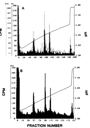
FIG. 2. Tryptic peptide analysis of gD-1polypeptides synthesizedin vitro and in vivo. (A) [35S]methionine-labeledpolypeptides translated in vitro with HSV-1 mRNAwereimmunoprecipitatedwithanti-gD-1serum(see Fig. 1, lane 1). The immunoprecipitatewasdisrupted with SDS, oxidized,trypsinized,andchromatographedon aChromabeads P cation-exchange column. (B) KB cellswereinfected withHSV-1 andpulse-labeledfor 15 min at6hpostinfection with[3Hlarginine.Cytoplasmicextracts wereimmunoprecipitatedwithanti-gD-1serum,and the precipitate was disrupted,oxidized, trypsinized, andcochromatographedonChromabeads Pwith
[355]me-thionine-labeled tryptic peptides of gD-1 immunoprecipitated from translations using HSV-1 mRNA and preparedasdescribed in (A). Solidlines, [3H]arginine-labeledpgD-1; dottedlines, [35S]methionine-labeled gD-1 synthesized in vitro.
H), sedimentation of the translation mixture
through
sucrose resulted in an enrichment ofmodified
gD(Fig.
5, lane 2). This moleculecomigrated
with pgDimmunoprecipitated
frominfected
cell extracts (Fig.5,
lane 1). Endo Hdigestion of modified
gD generated a pattern offour
polypeptides (Fig.
5, lane 3)which differed
in
size from each other by ca. 1,000 to 1,200daltons.
Thelargest polypeptide comigrated
with
modified
gD-2 (Fig. 5, lane 2), and thesmallest one was
slightly larger
thanTM-gD-2
(Fig. 5,
lane4). This difference
in molecular weight wasprobably
duetothe presenceof
the uncleavedN-acetylglucosamine remaining
onmodifiedgD andwas
consistent
with whatwas foundpreviously for pgD
(7).
Theresults suggest that invitro
processing of gD
involvesaddition
of
threeN-asparagine-linked oligosaccharides
and
removal of
signal
peptide
sequences. Translocation andprocessing
of invitro-synthe-dOLm- L .. am--- -- in"
i 0
VOL. 48,1983
on November 10, 2019 by guest
http://jvi.asm.org/
[image:5.491.107.401.69.476.2]35S 3H 300 209
270 187
240 165
210 143
180
0.
IL
121 99
77 55
33
1 1 150
120
90 60
30
0
35S 450
0
405 360315
270
225
10 135
90 45 0
5.00
I
5.00
4.40
3.80
3.20
2.60
0 19 38 57 76 96 115 134 153 172 191
FRACTION
NUMBER
FIG. 3. Trypticpeptide analysis of gD-2 polypeptides synthesized in vitro and in vivo. (A) BHK cellswere infected with HSV-2 in thepresence orabsence ofTM. At6hpostinfection, the cellswerepulse-labeled for15 minwith[3H]methioninealoneor[35S]methionineplusTM.Cytoplasmicextractsprepared from these cellswere immunoprecipitated with anti-gD-1 serum, oxidized, trypsinized, and cochromatographedonChromabeads P. Solidline, [3H]methionine-labeled pgD-2; dotted line,
[35S]methionine-labeled
TM-gD-2. (B)[35S]methionine-labeledpolypeptides translated in vitro with HSV-2 mRNA wereimmunoprecipitatedwithanti-gD-1 serum (see Fig.1, lane2).Theprecipitatewasdisrupted, oxidized, trypsinized,andchromatographedonChromabeads P.
sized gD. For other membrane
glycoproteins,
microsomes must be present as soon as the
signal peptide
emerges from the ribosome forinsertion
and processing to occur (28-30). Prod-uctsof in vitro translation(Fig.
6A andB, lane 1) were processed normally when microsomes were added to the translation mixture at the sametimeas wasmRNA(Fig.
6Aand B, lanes 2 and 3)but were not processed when microsomes wereadded
1hafter
addition of mRNA (Fig. 6A and B, lane 4). Moreover, theseunprocessed
molecules were
completely degraded
by
trypsin
(Fig.
6Aand B, lane 5).In contrast, in
vitro-modified
gD-1
andgD-2
were partially
protected
fromdegradation by
trypsin
(Fig. 7).
Trypsin
treatment of modified gD-1(Fig. 7,
lane1)
andgD-2
(Fig.
7,
lane2)
reduced the size of each of these molecules
by
ca.
3,000
daltons(Fig. 7,
lanes 4and
5).
When Triton X-100 was added to the mixtures atthe end of thecoupled
translation-processing
step and beforetrypsin
treatment, thepolypeptides
on November 10, 2019 by guest
http://jvi.asm.org/
[image:6.491.104.394.63.477.2]IN VITRO 527
were
completely degraded
(Fig. 7, lanes 3 and 6). The results suggest that modified gD-1 and gD-2had been inserted into microsomal
vesicles in such a way that the bulk of the polypeptide was inside the vesicle and was therefore inacces-sible to trypsin degradation. For each of these proteins, a 3,000-dalton portion was located outside the vesicle, and, in each case, this portion of each protein contained trypsin-sensi-tive sites.Figure
8A shows a similar experiment withchymotrypsin.
Inthis
case, modified gD-2 waspartially
degraded (Fig. 8A, lane 4), and again aportion of
ca.3,000 daltons
wasremoved.
How-ever,modified gD-1 appeared
tobe
unaffected
by
chymotrypsin,
even atconcentrations
ashigh
as
500
,ug/ml
anddigestion times
aslong
as 4 h(data
notshown).
Both gD-1and
gD-2 were completely degraded by chymotrypsin when thevesicles
weresolubilized
with detergent (Fig. 8A, lanes 5 and 6). These results indicate that gD-1and
gD-2differ
structurally at theunpro-tected
endof
thepolypeptide chain.
Moreover,the
chymotrypsin site(s)
presentin
theunpro-tected end
ofgD-2
appears tobe
physically close
to the
trypsin site(s), since
asimilarly sized
fragment
wasremoved
with each
enzyme.Fig-1
2
3
4
5
6 7 8A
it
-opgD
B
pg-DB~~
123 1 2 3 4
FIG. 4. SDS-PAGE analysis of polypeptides syn-thesized in vitro in the presence and absence of microsomal vesicles. Autoradiogram of a 9 to 12% gradient gel.All of thepolypeptides werelabeled with
[355]methionine,
and all immunoprecipitations were carried out with anti-gD-1 serum. (A) Lane 1, gD-1 synthesized invitro(see Fig. 1 for details of prepara-tion); lane 2, TM-gD-1; lane 3, cytoplasmic mRNA from HSV-1-infected cells translated inthe presence ofmicrosomal vesicles; lane 4, pgD-1. Lanes5through 8 represent the corresponding gD polypeptides from HSV-2-infected cells. (B) Lanes1 and2, comparison of gD-1 and gD-2synthesized invitro; lanes 3and4, comparison of gD-1 and gD-2 synthesized and proc-essed invitro.pgD-2.-,
- TM-gD-2
1 2 3 4
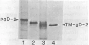
FIG. 5. Endo H digestion of gD-2 translated and processed in vitro. Fluorogram of a 9 to12% gradient SDS-polyacrylamide gel. Lane 1, pgD-2 prepared as in Fig. 1, lane 6. Lanes 2 and 3, Cytoplasmic mRNA from HSV-2-infected cells translated in the presence of microsomal vesicles. After translation, thelysate was sedimented through a sucrose cushion, solubilized in 1 x buffered detergent, and immunoprecipitated with anti-gD-1 serum.Theimmunoprecipitate was disrupt-ed by boiling for5 min in 1% SDS-mercaptoethanol andeither mock digested (lane 2) or digested (lane 3) with Endo H. Lane 4, TM-gD-2. All polypeptides were labeled with
[35S]methionine.
Lanes 1 and 4of this gel were exposed to X-ray film for 7 days, and lanes 2 and 3 were exposed for 21 days.ure 8B shows a
comparison of the fragments
derived from modified gD-2 with trypsin (Fig. 8B, lane 1), chymotrypsin (Fig. 8B, lane 2), or a
combination
oftrypsin
andchymotrypsin (Fig.
8B,
lane3).
In asimilar
experiment
withprotein-ase K, only modified gD-2 was found to be
sensitive
(data
notshown).
Theseproteolysis
experiments
indicated thatalthough
asimilarly
sized
portion
of thepolypeptide
chain of invitro-modified
gD-1
andgD-2
was located outside themicrosomal
vesicles,
it wasstructurally
distinct in the twoglycoproteins.
Endo Hsensitivity ofinvitro-modifiedgD. The
N-asparagine-linked oligosaccharide chains
add-ed toglycoproteins
during
invitro
processing
are located on that
portion of
themolecule
which is resistant
toproteolysis, suggesting
thatthey
arelocated
within the lumina of the
micro-somal vesicles
(28,
29,
39).
Totestwhether
this was the case forgD,
the molecule was treated withtrypsin
andimmunoprecipitated (Fig.
9, lane2).
Thetrypsinized molecule
wasthen treat-edwith
Endo H(Fig.
9,
lane3).
Aseries of four
bands were
generated,
thelargest of which
co-migrated with
thetrypsin-resistant portion
of modified gD-2 (Fig. 9, lane 2) and the smallest of which had a molecular size that was ca. 3,000 daltons less than that ofTM-gD-2
(Fig. 9,
lane 4). This pattern was similar to that observed when modified gD-2 was treated with Endo H (seeFig.
5, lane 3), except that inFig.
9each of the Endo Hdigestion products
was ca.3,000
daltons smaller. The results are
consistent
with theidea that all threeN-asparagine-linked
oligo-saccharides ofin vitro-modified
gD
are located within thelumina of
themicrosomal
vesicles.48,
on November 10, 2019 by guest
http://jvi.asm.org/
[image:7.491.274.421.82.165.2] [image:7.491.48.241.367.540.2]1 2 3 4 5
6
A
1 2 3 4 5: 1 2 3 45
FIG. 6. Cotranslational insertion of gD into micro-somal vesicles. Fluorogramofa9to12%gradient gel. HSV-1 mRNA (A) and HSV-2 mRNA (B)were
trans-lated invitro, and microsomeswereaddedatdifferent times. After 2 h, the sampleswereimmunoprecipitated with anti-gD-1 serum. Lanes 1, Translationwas
car-riedoutin theabsence of microsomes; lanes 2,
micro-somes were addedat the same time as mRNA(zero
time) and incubated for 2 h; lanes 3, microsomeswere
added atzerotime, and RNase wasadded 1 hlater; lanes 4,microsomes and RNasewereadded 1 h after mRNAwasadded, and sampleswereincubated foran
additional 1 h; lanes 5, microsomesand RNasewere
added 1 h after mRNA was added, and the mixture
was incubatedfor 1 h. Trypsin was then added, and the mixturewasincubated for 2 hat0°C.
Orientation of modified gD within the micro-somal vesicles. The previous experiments
em-ployed polyclonal anti-gD-1 serum to immuno-precipitate gD molecules. We screened apanel
of monoclonal antibodies (11) and found that
groupVandgroupVIIantibodiesrecognized in vitro-synthesized gD-1 polypeptides. GroupVII also recognized in vitro-synthesized gD-2 poly-peptides.
Experiments were performed to determine
Pg
l 2 3 4 5 6
FIG. 7. SDS-PAGE analysis of gD-1 and gD-2 translated and processed in vitro and treated with trypsin. Fluorogramofa9to12%gradientgel.HSV-1 and HSV-2 mRNAweretranslated in thepresenceof microsomes (modified gD). Trypsin (50 ,ug/ml) was addedtothesampleseither before orafter Triton X-100wasadded. Thesampleswereincubated for 2 hat
0°C and then immunoprecipitated with anti-gD-1 se-rum. Lane1,In vitro-modifiedgD-1 (no trypsin);lane 2, modified gD-2 (no trypsin); lane 3, modified gD-2 treated first with 0.5% Triton X-100 and then with trypsin;lane4,modifiedgD-1treated withtrypsinand then with Triton X-100;lane5, modifiedgD-2treated with trypsin and then with Triton X-100; lane 6, modifiedgD-1treated with Triton X-100 and then with trypsin. Lanes1through5wereexposedtoX-rayfilm for 7days. Lane 6was exposedfor 12days.
pgD_..-- .*
B
-opgD
12 3
FIG. 8. SDS-PAGE analysis of in vitro-modified gD polypeptides treated with trypsin and chymotryp-sin.Fluorogram ofa9to12%gradient gel. HSV-1 and HSV-2 mRNA were translated in the presence of
microsomes (modified gD). Chymotrypsin (100,ug/ml)
ortrypsin (50 p.g/ml)wasaddedtothe samples either beforeorafter Triton X-100was added. The samples
were immunoprecipitated with anti-gD-1 serum. (A)
Lane 1, modified gD-2 (no chymotrypsin); lane 2, modifiedgD-1 (nochymotrypsin); lane 3, modified gD-1treatedfirst withchymotrypsin and then with Triton X-100; lane 4, modified gD-2 treated first with chymo-trypsin and then with Triton X-100; lane 5, modified gD-2 treated first with Triton X-100 and then with chymotrypsin; lane6, modified gD-1 treated firstwith Triton X-100 and then with chymotrypsin. Lanes 1 through 4 were exposed to X-ray film for 7 days. Lanes5 and 6wereexposed for14days. (B) Lane 1, modified gD-2 treated with trypsin and then with Triton X-100; lane 2, modified gD-2 treated with chymotrypsin and then with Triton X-100; lane 3, modifiedgD-2 treatedwithchymotrypsin, trypsin,and thenTriton X-100.
whether these antibodies recognizedthe protect-ed and unprotected portions of the modified in vitro translationproducts(Fig. 10). After trans-lation and processing, microsomes were solubi-lized, and gD-1 was immunoprecipitated with
anti-gD-1 serum (Fig. 10A, lane 1), group VII monoclonal antibody 170 (11, 23) (Fig. 10A, lane 2), or group V monoclonal antibody 57S
(11, 32) (Fig. 10A, lane 3). When modifiedgD-1
wastreated withtrypsin before
immunoprecipi-tation, theprotectedportion wasrecognized by
anti-gD-1 serum (Fig. 10A, lane 4) and by 170
antibody (Fig. 10A, lane 5) but not by 57S antibody (Fig. 10A, lane6). These results
sug-gest that the 57S(group V)determinant ofgD-1 is located within the unprotected 3,000-dalton fragment and that the 170 (group VII) determi-nant is located within the fully protected
frag-ment. In thecaseof modifiedgD-2, the
untreat-A
B
J. VIROL.
on November 10, 2019 by guest
http://jvi.asm.org/
[image:8.491.279.427.72.260.2] [image:8.491.67.234.76.155.2] [image:8.491.91.209.443.518.2]IN VITRO SYNTHESIS AND PROCESSING OF HSV gD 529
ed
molecule
wasrecognized by anti-gD-1
serum(Fig. lOB,
lane1)
andby
170antibody
(Fig. lOB, lane 2), but notby
57Santibody (Fig.
lOB, lane 3). Since group V antibodies are gD-1 specific, this last resultwasexpected. The tryp-sin-protected fragment of modified gD-2 wasimmunoprecipitated by anti-gD-1
serum (Fig. 10B, lane5)
andby
170 antibody
(Fig. 10B, lane
6). These
experiments indicate
thatgD-1
and gD-2 have asimilar
orientation
withinmicro-somal vesicles.
Orientation ofgD in infected cellplasma mem-branes.
Immunofluorescence
wasused
todeter-mine
theorientation of
group V and groupVII
monoclonal
antibody determinants
ongD-1
when the
protein
waslocated
inthe
plasma
membrane of infected
KBcells. The
patternof
immunofluorescence observed
whenfixed
HSV-1-infected
KBcells
were reactedwith
different
antibodies is
shownin
Fig.
11.Normal
mouse serum(Fig. lla)
wasunreactive,
andthe
infect-ed cells
werevisualized
only with
theaid of the
rhodamine
counterstain.
Polyclonal
mouseanti-gD-1 serum
(Fig.
lib),
aswell
asgroup VII(Fig.
lic)
and
group V(Fig.
lid)
monoclonal
antibod-ies, reacted with
thefixed infected
KBcells,
yielding
a patternof
moderately intense
cyto-plasmic fluorescence. None of these antibodies
reacted with fixed
uninfected
KBcells
(data
notshown).
Unfixed infected
KBcells
were reactedwith
the
samepanel of antibodies
(Fig. 12).
Again,
normal
mouse serum(Fig.
12a)
wasnegative.
Apattern of intense focal membrane fluorescence
was
observed with either polyclonal
anti-gD-1
serum
(Fig. 12b)
orgroup VII(170) monoclonal
pgD-2--*4-TM-gD-2
1 2 3 4
FIG. 9. Endo H sensitivity of the trypsin-resistant fragmentofmodified gD-2. Fluorogram ofa9 to12% gradient gel. HSV-2 mRNA was translated in the presence of microsomes (modified gD). Trypsin (50
Rg/ml)
wasadded toone sample after 1 h, and anothersample was mockdigested. Both samples were incu-bated for 2 h at0°C, centrifuged through a sucrose cushion, diluted with buffered detergent, and immuno-precipitated with anti-gD-1 serum. The samples were suspended in buffercontaining1%SDS, and a portion wastreatedwith Endo H. Lane 1, Modified gD-2 mock digested with trypsin (no Endo H); lane 2, modified gD-2treated withtrypsin (no Endo H); lane 3, modi-fiedgD-2treatedwithtrypsinandEndoH; lane 4, TM-gD-2 prepared as described in the legend to Fig. 1, lane 4, and runas acontrol.
A
--pre-gD
_~~~ ~ 0
...0
1 2 3 4 5 6
B
.- -4-pre-gD
[image:9.491.251.444.72.292.2]1
2
345
FIG. 10. Orientation of gD-1 and gD-2 in micro-somal vesicles. Fluorogram (A) or autoradiogram (B) ofa9to12% gradient gel. HSV-1mRNA(A)orHSV-2 mRNA(B) was translated in the presence of micro-somes (modified gD). After translation, one sample was mockdigested, and the other was digested with 50 j±g of trypsin per ml for 2 h at 0°C. The samples were solubilized with detergent and immunoprecipitated. pre-gD, Invitro-synthesized form. (A) Lane 1, modi-fied gD-1 mock digested with trypsin and immunopre-cipitated with anti-gD-1 serum; lane 2, modified gD-1 mock digested with trypsin and immunoprecipitated with group VII (170) monoclonal antibody; lane 3, modified gD-1 mock digested with trypsin and immu-noprecipitated with group V (57S) monoclonal anti-body; lane 4, modified gD-1 digested with trypsin and immunoprecipitated with anti-gD-1 serum; lane 5, modified gD-1 digested with trypsin and immunopre-cipitated with 170 antibody; lane 6, modified gD-1 digested with trypsin and immunoprecipitated with 57Santibody. Lanes1 through 3wereexposed to X-rayfilm for7 days. Lanes4through 6were exposed for14days. (B) Lane 1, modified gD-2 mockdigested with trypsin and immunoprecipitated with anti-gD-1 serum; lane 2, modified gD-2 mock digested with trypsin and immunoprecipitated with 170 monoclonal antibody; lane 3, modified gD-2 mock digested with trypsin and immunoprecipitated with 57S monoclonal antibody; lane 4, modified gD-2 digested with trypsin andimmunoprecipitated withanti-gD-1 serum; lane 5, modified gD-2 digested withtrypsinand immunopre-cipitated with 170monoclonalantibody.
antibody
(Fig. 12c).
No fluorescence was ob-served withgorup V(57S)
monoclonalantibody
(Fig. 12d). None of the antibodies reacted with unfixed uninfected KB cells
(data
notshown).
Theresults
indicate
that thegroup VII determi-nantis
located on theoutside of infected
cells(corresponding
totheluminaof microsomal
ves-VOL.48,1983
on November 10, 2019 by guest
http://jvi.asm.org/
[image:9.491.72.216.458.530.2]....
FIG. 11. Immunofluorescence studies of fixed HSV-1-infected KB cells. Monolayers were infected withHSV-1,fixed with 3.7% formaldehyde, and dehy-drated with acetone at 10 h postinfection. The cells
were incubated with normal mouse serum (diluted
1:20) (a); mouse anti-gD-1 serum (diluted 1:80) (b);
170 monoclonal antibody (diluted 1:640) (c); or57S
monoclonal antibody(diluted1:40)(d). Each
monolay-er was then reacted with fluorescein isothiocyanate-conjugatedgoatanti-mouseimmunoglobulinG. A
rep-resentative field observed with each of these antibodiesis shown.
icdes),whereas thegroupVdeterminant is locat-ed on the inside of the cells (corresponding to the outside of the microsomal vesicles). Thus, gD has the orientation in infected cell plasma membranes that would be predicted from in vitro processing experiments.
DISCUSSION
Inpreviousstudiesof the synthesisand
proc-essingof HSV glycoprotein D, we documented
someof the structural changesinthe proteinas
itwasfound in HSV-infected cells(5, 6, 10, 12).
In those studies, the first precursor that was
detected was already glycosylated, even when
weusedradioactivepulse-labelingtimesasshort as2 min (6). To detect the unglycosylated
pre-cursortogD-1 in infected cells. TM was added
toinhibitthe firststepofglycosylation (24). The experiments inthe present studyhave provided
detailsconcerning the earliest events in synthe-sis, as well as the processing and membrane orientation ofgD. All ofourfindings agree with predictions made about the orientation and structureofgD-1 as atransmembrane glycopro-tein, based on its primary amino acid sequence (41). Moreover, we have shown that gD-2 hasa structure and an orientation similar to those of gD-1.
With polyclonal anti-gD-1 serum, we identi-fied the primary unglycosylated translation products of gD by using an in vitro translation system and HSV-1 mRNA or HSV-2 mRNA. Each of the primary products had an apparent molecular size, measured by
SDS-PAGE,
of 49K. Our results have confirmed and extended the work ofInglis and Newton (15), as well as that of Lee et al. (19), who showed that the primary translation product of gD-1 had a molec-ular weight of ca. 50K. We found that the [35S]methionine-labeled tryptic peptide profiles ofthe in vitro precursors are indistinguishable from theprofiles of thecorresponding pgD mole-cules isolated from HSV-1- and HSV-2-infected cells. Each of the invitro-synthesized molecules was alsofound
to beslightly larger
than thecorresponding
polypeptides produced
in the presence of TM. This difference in size, as estimatedbySDS-PAGE,
isconsistent with the presenceoftransientsignalpeptide sequences in gD-1 and gD-2 which aremissing
in the TM-treated gD molecules. The fact that thein
vitro-synthesized forms of gD-1 and gD-2 did not contain any additional methionine tryptic pep-tides indicates that these signal
peptides
proba-blydo not contain methionine.A difference in signal
peptide
sizewould
ac-countfor thesimilarityin the molecularweights of in vitro-synthesized gD-1 and gD-2 as op-posed to thedifference in molecularweights
of each of theprocessedforms ofthe two glycopro-teins.Thus,
it ispossible
thatthesignal
peptide ofgD-2 has amolecularweighthigher
thanthat of gD-1. However, all of the estimations of molecular weight based on migration in SDS-polyacrylamide gels appearto be somewhat in-accurate, since the molecularweight
of gD-1 predicted from the deduced amino acid se-quence is 43,291 and the size estimated from SDS-PAGE was found to be 49K. A similardiscrepancy
betweenpredicted
and actual mo-lecularweightswasnoted forgD(41),
aswellas forgC (14), and in both cases it was suggested that thedifference
could be due to thehigh
proline
contentof
theprotein (14, 41).
Inthe present
studies,
wefound that each of the invitro-synthesized
forms ofgD
was proc-essedto ahigher-molecular-weight
form.
These in vitro-modifiedpolypeptides
comigrated
on SDS-PAGE withpgD-1
andpgD-2.
ExperimentsJ.VIROL.
on November 10, 2019 by guest
http://jvi.asm.org/
IN VITRO AND gD 531
with Endo H
showed
thateach
of the in
vitro-processed
molecules contained
threeN-aspara-gine-linked oligosaccharides, suggesting
that in vitro processing resembles the process which occurs in vivo. Thus, we would predict that allof
thepotential N-asparagine glycosylation
sites are used for in vitro and in vivoprocessing
of gD-1. Thepossibility
that gD-2contains
addi-tional potential glycosylation
sites willbest
be assessed from its sequence. For certainglyco-proteins,
someglycosylation
sitespredicted
from primary amino acid sequence data are not used
(37).
Because
non-glycosylated
forms of
gD are notnormally isolated from infected cells, it is
rea-sonable
to supposethat gD-1
and gD-2,like
many
other cell and
viral glycoproteins
(17, 28, 39), arecotranslationally
processed. For otherglycoproteins, it is
thought that this earlyproc-essing
stepprobably involves simultaneous
translocation
into the lumen of the roughendo-plasmic reticulum
(28, 30).Our experiments
areconsistent
withthe
idea that insertion
andproc-essing of
gD occurcotranslationally.
Thus, wefound that
invitro
processing did
not occur when microsomal vesicles were added 1 h after mRNA wasadded
to thein
vitro
translation system.Moreover, under these
conditions,
new-ly
synthesized
gD was notinserted into
themicrosomal
vesicles, since
itremained totally
sensitive
todegradation by trypsin.
When
membranes
were presentduring
trans-lation,
themodified
forms of gD-1 and gD-2 wereonly partially trypsin sensitive,
and afragment
of
ca.3,000
daltons was removedfrom
eachprotein.
Noother
fragments
weregenerated by
this
treatment. Weinterpret
these results to mean thatmodified
gD-1 and gD-2 are eachasymmetrically oriented
in thevesicles
astrans-membrane
proteins, with
ca. 3,000daltons
ex-posed
totrypsin. The
amountof gD
onthe
outside of
thevesicles could actually
be greaterdepending
onthe number of
trypsin sites
ex-posed. However, for gD-1,
this size estimate agreesquite well with predictions
based on thededuced
sequence (41).The last
30residues
ofgD-1
include
6arginines
and 2 lysines. Onewould
predict
thattrypsin
would probably re-move29 of the last 30
amino
acids of
gD-1
or ca. 3,600daltons
of the carboxy terminus.The exposed
portions of
modifiedgD-4
and gD-2 appeared todiffer
in proteasesusceptibil-ity, implying
that they also differed in structure.Thus, only
modified gD-2 appeared to be sensi-tive tochymotrypsin
orproteinase
K, whereasboth
gD-1 and gD-2 were sensitive to trypsin.According
to the deduced aminoacid sequenceof gD-1
(41),only
thelast
two residues arepotentially chymotrypsin
sensitive beyond the membraneinsertion sequence. Removal of theseFIG. 12. Immunofluorescence studies of unfixed HSV-1-infected KB cells. Suspension cultures were infected with HSV-1 for 10h and washed with PBS. The cells werethen incubated with 100
,u1
ofnormal mouse serum(diluted1:10inPBS)(a); mouse anti-gD-1 serum (diluted 1:20) (b); 170 monoclonalantibody (diluted1:80) (c); or57Smonoclonalantibody (diluted 1:10) (d). The cells were centrifuged, washed with PBS, and incubated with fluorescein isothiocyanate-conjugatedgoatanti-mouseimmunoglobulinG.residues would
probably
notbe
detectable in our system. It issomewhat
puzzling
thatmodified
gD-1
wasinsusceptible
toproteinase
K, since
this
enzyme has abroad
specificity.
Itmight
be that thesecondary
structureof
thecarboxy
terminus of gD-1
accountsfor
this result.Our data
suggest that the threeN-asparagine-linked
oligosaccharides
addedduring
the invitrotranslation
andprocessing of gD
arelocatedon aportion
of
theglycoprotein
which
is within thelumina of
themicrosomes.
According
to thededuced
amino acid sequence ofgD-1
(41), themembrane
insertion sequence is located closetothe
carboxy
terminus. Thepredicted
orientation of gD is thus similar to thatreported
for a number of viralglycoproteins (27, 28, 38).
This orientation alsoimplies
that the group V(57S)
determinant is located within the last 30 residues
of gD-1.
Thefact
that this determinant is also gD-1specific emphasizes
thedifference in struc-tureof
gD-1 andgD-2
in thisregion
that wasimplied
from theproteaseexperiments.
The dataon November 10, 2019 by guest
http://jvi.asm.org/
[image:11.491.252.445.64.353.2]in
Fig.
11and
12show that gD-1
is
oriented in
the
plasma membrane
of HSV-1-infected
cellsin
the
mannerpredicted from in vitro
processing.
Thus,
the57S
(groupV) determinant
(presum-ably
atthe
carboxy
end) faces
thecytoplasm,
and the
170
(groupVII)
determinant
(presum-ably
atthe
amino
terminus) faces outside.
Thesedeterminants would be expected
tohave
asimi-lar
orientation in the virion envelope.
Itis
note-worthy that
group VIIantibodies
arecapable of
virus
neutralization and
thatgroup
Vantibodies
are not.
Experiments
are nowin
progress tofurther delineate the
precise locations of these
two
determinants.
ACKNOWLEDGMENTS
Thisinvestigation was supported by Public Health Service grants DE-02623 from the National Institute of Dental Re-searchand AI-18289from the NationalInstituteofAllergy and Infectious Diseases. J.T.M. was a predoctoral trainee support-ed by Public Health Service grants NS-07180 from the Nation-alInstitute of Neurological and Communicative Disorders and Stroke.
Wethank Manuel Ponce de Leon for help in preparation of HSV-2 mRNA and Madeline Cohen and Deborah Long for excellent technical assistance. We are indebted to Lenore Pereira, Berge Hampar, and Martin Zweig for supplying monoclonal antibodies used in this study. We also acknowl-edge the help of William Wunner in preparation of this manuscript.
LITERATURECITED
1. Blobel,G.,and B. Dobberstein.1975.Transfer ofproteins
across membranes. I. Presence ofproteolytically proc-essed and unprocessed nascent immunoglobulin light chainsonmembrane-bound ribosomesof murine
myelo-ma.J.Cell Biol. 67:835-851.
2. Bonner,W.M.,and R. A.Laskey. 1974.Afilm detection methodfor tritium labeledproteinsand nucleic acids in polyacrylamidegels.Eur.J. Biochem. 46:83-88. 3. Braell, W., and H. Lodish. 1981. Biosynthesis of the
erythrocyte anion transport protein. J. Biol. Chem. 256:11337-11344.
4. Cohen, G.H.,M. N.Factor,andM. Ponce de Leon.1974. Inhibition ofherpes simplexvirus type 2replication by thymidine. J. Virol. 14:20-25.
5. Cohen, G. H., M. Katze, C. Hydrean-Stern, and R. J. Eisenberg. 1978. Type-common CP-1 antigen ofherpes simplex virus is associated with a 59,000-molecular-weightenvelopeglycoprotein. J. Virol. 27:172-181. 6. Cohen, G. H., D. Long, and R. J. Eisenberg. 1980.
Synthesisandprocessingofglycoproteins gDandgCof herpessimplex virus type1.J.Virol.36:429-439. 7. Cohen,G. H.,D.Long, J.T.Matthews,M.May,and R.
Eisenberg. 1983.Glycopeptides ofthetype-common gly-coprotein gDofherpessimplex virus types 1 and 2. J. Virol. 46:679-689.
8. Cohen, G. H., M. Ponce deLeon,and C. Nichols. 1972. Isolation of a herpes simplex virus-specific antigenic fraction which stimulates the production ofneutralizing antibody. J. Virol. 10:1021-1030.
9. Dobberstein, B.,H.Garoff, G.Warren,and P.J. Robin-son.1979.Cell freesynthesis and membrane insertion of mouseH-2Dhistocompatibility antigenand
,B2-microglo-bulin.Cell17:759-769.
10. Eisenberg, R. J., C. Hydrean-Stern, and G. H. Cohen. 1979.Structuralanalysis of precursorandproduct forms oftype-commonenvelopeglycoprotein D(CP-1 antigen) ofherpes simplexvirustype1.J.Virol. 31:608-620. 11. Eisenberg, R. J., D. Long, L. Pereira, B. Hampar, M.
Zweig, and G. H. Cohen. 1982. Effect of monoclonal antibodies onlimited proteolysis of nativeglycoprotein gD ofherpes simplex virustype 1.J.Virol. 41:478-488. 12. Eisenberg, R. J., M. Ponce de Leon, and G. H. Cohen.
1980. Comparative structural analysisof glycoproteingD ofherpes simplexvirus types1 and 2. J. Virol. 35:428-435.
13. Eisenberg,R.J.,M. Ponce de Leon, L.Pereira,D.Long, andG.H. Cohen.1982.Purification of glycoprotein gD of herpes simplex virustypes 1and 2byuseof monoclonal antibody. J. Virol. 41:1099-1104.
14. Frink, R. J., R.Eisenberg, G. Cohen,andE. K. Wagner. 1983. Detailed analysis of the portion of the herpes simplex virustype1genomeencoding glycoprotein C. J. Virol. 45:634-647.
15. Inglis,M. M., and A. A. Newton. 1982.Identification of the polypeptide precursors to HSV-1 glycoproteins by cell-free translation. J. Gen. Virol. 58:217-222. 16. Irving, R.,F.Toneguzzo, S. Rhee, T. Hofmann, and H.
Ghosh. 1979.Synthesis and assembly of membrane glyco-proteins: presence of leader peptide in nonglycosylated precursorsof membraneglycoprotein of vesicular stoma-titis virus. Proc. Natl. Acad.Sci. U.S.A. 76:570-574. 17. Katz, F, J. Rothman, D. Knipe, and H. Lodish. 1977.
Membrane assembly: synthesis and intracellular process-ing of the vesicularstomatitis viralglycoprotein.J. Supra-mol.Struct. 7:353-370.
18. Kreil, G. 1981. Transfer of proteinsacrossmembranes. Annu.Rev. Biochem. 50:317-348.
19. Lee, G. T.-Y., M. F. Para, and P. G. Spear. 1982. Location of the structuralgenesforglycoproteins gD and gE and for other polypeptides in the S component of herpes simplex virustype1 DNA. J. Virol.43:41-49. 20. Palfreyman,J. W., L. Haarr, A.Cross,R.G. Hope,and
H. S. Marsden. 1983.Processing of herpes simplex virus type1glycoproteins: two-dimensional gel analysis using monoclonalantibodies. J. Gen. Virol. 64:873-886. 21. Palmiter,R. D., S. N.Thibodeau, G.Rogers,and I.Boime.
1980.Co-translational sequestration of egg white proteins andplacental lactogen inside membrane vesicles. Ann. N.Y.Acad.Sci.343:192-209.
22. Pelham, H., and R.Jackson. 1976. Anefficient mRNA dependent translation system from reticulocyte lysates. Eur. J. Biochem. 67:247-256.
23. Pereira,L.,D. V.Dondero,D.Gallo,V.Devlin, and J. D. Woodie. 1982.Serological analysis ofherpes simplexvirus types1and 2 with monoclonalantibodies. Infect.Immun. 35:363-367.
24. Pizer,L.I., G.H.Cohen, and R. J. Eisenberg. 1980. Effect oftunicamycinonherpessimplex virusglycoproteinsand infectious virusproduction. J. Virol. 34:142-153. 25. Preston, C. M. 1977. The cell-free synthesis of herpes
simplex virus-inducedpolypeptides. Virology 78:349-353. 26. Reed, C.L., G.H.Cohen, andF.Rapp.1975.Detection of avirus-specificantigenonthe surfaceofherpes simplex virus-transformed cells. J. Virol. 15:668-670.
27. Rose, J., W. Welch, B. Sefton,F.Esch,and N.Ling.1980. Vesicular stomatitis virusglycoprotein isanchoredinthe viral membrane by a hydrophobic domain near the COOH terminus.Proc.Natl. Acad. Sci. U.S.A.77:3884-3888. 28. Rothman, J., and H. Lodish. 1977. Synchronized
trans-membrane insertion andglycosylation ofanascent mem-braneprotein.Nature(London)269:775-780.
29. Sabbatini, D. D., G. Kreibich, T. Morimoto, and M. Adesnik. 1982.Mechanisms for theincorporationof pro-teins intomembranes andorganelles.J.Cell Biol.92:1-22. 30. Scheele, G.,R.Jacoby, andT.Carne.1980.Mechanisms ofcompartmentation ofsecretory proteins: transportof exocrine proteinsacross the microsomal membrane. J. Cell Biol. 87:611-628.
31. Shields, D., andG. Blobel. 1978. Efficientcleavage and segregation of nascent presecretoryproteins ina reticulo-cytelysatesupplementedwithmicrosomalmembranes.J. Biol.Chem. 253:3753-3756.
32. Showalter,S.D.,M.Zweig,and B.Hampar.1981.
on November 10, 2019 by guest
http://jvi.asm.org/
IN gD
clonal antibodiestoherpes simplex virustype1proteins, including the immediate-earlyprotein ICP 4. Infect.
Im-mun.34:684-692.
33. Spear, P. G. 1975. Glycoproteins specified by herpes simplex virus type 1: their synthesis, processing and antigenic relatednesstoHSV-2glycoproteins,p.49-61. In
G. De The, M. A. Epstein, and H. zur Hausen (ed.),
Oncogenesis andherpesviruses. II. Part 2. International Agencyfor ResearchonCancer, Lyons,France. 34. Spear, P. G. 1976.Membraneproteins specified by herpes
simplex viruses. I. Identification of four glycoprotein
precursorsandtheirproductsintype1-infected cells. J. Virol. 17:991-1008.
35. Spear, P. G. 1980.Herpesviruses, p.709-750. In H. A. Blough and J. M. Tiffany (ed.), Cell membranes and viral envelopes, vol. 2. Academic Press, Inc., New York. 36. Spear, P. G., M.Sarmiento,and R.Manservigi. 1978. The
structuralproteins andglycoproteinsofherpesviruses:a
review,p.157-167. In G. de The, W. Henle, and F. Rapp (ed.), Oncogenesis andherpesviruses.III.Part 1.
Interna-tionalAgencyfor ResearchonCancer,Lyons,France. 37. Struck,D.,andW. Lennarz. 1980. The function of
saccha-ride-lipids in synthesis of glycoproteins. In W. Lennarz (ed.), The biochemistry of glycoproteins and
proteogly-cans.PlenumPublishing Corp., New York.
38. Sveda, M., L. Markoff, and G. Lai. 1982. Cell-surface expression of the influenza virus hemagglutinin requires the hydrophobic carboxy-terminal sequences. Cell 30:649-656.
39. Toneguzzo, F., and H. Ghosh. 1977. Synthesis and glyco-sylation in vitro of glycoprotein of vesicular stomatitis virus. Proc.Natl. Acad. Sci. U.S.A. 74:1516-1520. 40. Villa-Komaroff, L., M. McDowell, D. Baltimore, and H.
Lodish.1974. Translation of reovirus mRNA, poliovirus RNA andbacteriophage QP RNA in cell freeextractsof mammaliancells. Methods Enzymol. 30:709-723. 41. Watson, R. J., J. H. Weis, J. S. Salstrom, and L. W.
Enquist. 1982.Herpes simplex virus type-1 glycoprotein D
gene: nucleotidesequenceand expression in Escherichia coli.Science 218:381-384.

![FIG. NUMBERlabeledpreparedaatthethionine-labeledsynthesizedFig. Chromabeads 6 vitro precipitate h 1, postinfection lane [35S]methionine- polypeptides as (A) 1)](https://thumb-us.123doks.com/thumbv2/123dok_us/1441597.96561/5.491.107.401.69.476/numberlabeledpreparedaatthethionine-labeledsynthesizedfig-chromabeads-vitro-precipitate-postinfection-methionine-polypeptides.webp)