SHORT REPORT
Direct molecular detection of a
broad range of bacterial and viral organisms
and
Streptococcus pneumoniae
vaccine
serotypes in children with otitis media
with effusion
Robert Slinger
1*, Melanie Duval
1, Jonathan Langill
1, Matthew Bromwich
2, Johnna MacCormick
2, Francis Chan
1and Jean‑Philippe Vaccani
2Abstract
Background: Otitis media with effusion (OME) causes significant morbidity in children, but the causes of OME and methods for prevention are unclear. To look for potential infectious etiologies, we performed a pilot study using multiple‑target real‑time polymerase chain reaction (qPCR) for 27 infectious agents, including nine bacterial organ‑ isms and 18 respiratory viruses in middle ear fluids (MEFs) from children with OME. QPCR was also performed for the 13 Streptococcus pneumoniae serotypes contained in the current vaccine.
Results: Forty‑eight MEF samples were obtained and qPCR detected bacterial nucleic acid (NA) in 39/48 (81 %) and viral NA in 7/48 (15 %). Alloiococcus otitidis and S. pneumoniae were both detected in 15/48 (31 %) MEFs, followed by M. catarrhalis in 14/48 (29 %), H. influenzae in 5/48 (10 %) and M. pneumoniae in 4/48 (8 %). Rhinoviruses were most common virus type detected, found in 4/48 (8 %) MEFs. Serotypes included in the current 13‑serotype vaccine were detected in only 3/15 (20 %) S. pneumoniae qPCR‑positive MEFs.
Conclusions: Bacteria may play an important role in OME, since over 80 % of MEFs contained bacterial NA. Further research into the role of A. otitidis in OME will be helpful. Serotypes of S. pneumoniae not included in the current 13‑serotype vaccine may be involved in OME. Larger studies of OME S. pneumoniae serotypes are needed to help determine which additional serotypes should be included in future vaccine formulations in order to try to prevent OME.
Keywords: Otitis media with effusion, PCR, Bacteria, Viruses, Streptococcus pneumoniae
© 2016 Slinger et al. This article is distributed under the terms of the Creative Commons Attribution 4.0 International License (http://creativecommons.org/licenses/by/4.0/), which permits unrestricted use, distribution, and reproduction in any medium, provided you give appropriate credit to the original author(s) and the source, provide a link to the Creative Commons license, and indicate if changes were made. The Creative Commons Public Domain Dedication waiver (http://creativecommons.org/ publicdomain/zero/1.0/) applies to the data made available in this article, unless otherwise stated.
Background
Otitis media with effusion (OME) is a common child-hood condition in which fluid persists in the middle ear cavity for 3 months or more. It has been estimated that up to 80 % of children have been affected by OME at some time by the age of four [1]. Many children suffering
from this condition will have a mild to moderate hearing loss, averaging 27 decibels, which is sufficient to lead to language development delay [2].
While the exact pathogenesis of OME remains unknown, multiple hypotheses have been put forward. The position and length of the Eustachian tube in chil-dren and a persistent inflammatory process in the middle ear cavity due to frequent upper respiratory tract infec-tions are both thought to contribute to the frequency of OME in childhood [1].
Open Access
*Correspondence: slinger@cheo.on.ca
1 Department of Laboratory Medicine and Pathology, Children’s Hospital of Eastern Ontario, University of Ottawa, 401 Smyth Rd, Ottawa, ON K1H 8L1, Canada
Since bacterial cultures of middle ear fluid (MEF) are negative in the majority of children with OME, OME was once thought to be an inflammatory but non-infectious condition. However, with the use of more sensitive molec-ular detection methods such as polymerase-chain reaction (PCR), studies have shown that many bacterial culture-negative OME specimens contain bacterial DNA [3–6].
Based on studies by Hall-Stoodley and others [7], it is now thought that bacteria persist chronically in the mid-dle ear in the form of biofilms in many or most children with OME. Biofilms are known to be difficult to eradicate with antimicrobial therapy, which could account for the persistence of effusions in children treated with antibiotics for OME in clinical trials [8]. The biofilm hypothesis would also fit with the observation that bacterial cultures of MEF are often negative, as bacteria in biofilms are more difficult to culture than free-floating (planktonic) bacteria [8, 9].
Bacteria detected by molecular methods from OME middle ear fluid include those known to cause acute oti-tis media (AOM), namely, Streptococcus pneumoniae, Haemophilus influenzae and Moraxella catarrhalis [3–
6]. As well as accepted otitis pathogens, other bacteria whose pathogenicity is the middle ear is uncertain, such as Alloiococcus otitidis and Helicobacter pylori have also been detected by molecular studies in OME samples [10–13].
Viruses have also been detected in some MEF from OME. Viral RNA was detected in approximately 1/3 of MEF samples from children undergoing tympanostomy tube insertion [14, 15]. Viruses detected by PCR have included rhinoviruses, enteroviruses, respiratory syncyt-ial virus (RSV), and human coronaviruses [14–16].
In order to determine the prevalence of bacterial and viral agents in children with OME in our region, we per-formed a pilot study using real-time PCR (qPCR) for twenty-seven bacterial and viral pathogens in MEF sam-ples. Also, for the first time to our knowledge, MEFs were also tested directly for the 13 serotypes contained in the conjugate pneumococcal vaccine (Prevnar 13, Pfizer) to assess the potential impact of the 13-serotype S. pneumo-niae on OME associated with this bacterium [17]. Our overall objectives were to better understand the microbi-ology of OME in children, and to determine if the current conjugated pneumococcal vaccine would likely lead to prevention of OME.
Methods
Study outcome measures
1. Presence or absence of the bacteria and viruses listed in Table 1 as determined by real-time PCR from OME fluids.
2. Comparison of PCR and culture results for bacterial detection from OME fluids.
3. For S. pneumoniae, qPCR-positive OME fluids, the results of PCR serotyping for the serotypes contained in the 13-serotype S. pneumoniae vaccine.
Study design
This was a prospective observational study performed from Oct. 2011 to Oct 2012 at the Children’s Hospital of Eastern Ontario, Ottawa, Ontario, Canada, a 165 bed ter-tiary care hospital serving a catchment area 1.5 million. In the province of Ontario, a 7- serotype S. pneumoniae
vaccine was introduced for in 2002 and a 13-serotype vaccine in Dec. 2010. Both were part of the routine pub-licly funded immunization schedule for infants beginning at 2 months of age. Children also received infant immu-nization against H. influenzae type B.
Ethics and subject recruitment
Ethics approval was obtained for the study from the hos-pital Research Ethics Board. Parents of children seen at the pediatric otolaryngology clinic at the CHEO for treat-ment of OME who were to undergo tympanostomy tube insertion were identified and invited to participate in the study by the treating otolaryngologist. Informed con-sent was sought from parents or guardians who wished to participate to allow the MEF to be collected and sent for bacterial culture and PCR studies. Immunization sta-tus, age and sex of the child were recorded from hospital records in a study form.
The inclusion criteria were patients under the age of 18 years of age requiring ventilation tube insertion for OME for whom informed consent was obtained. Patients not meeting these age, diagnosis, and consent criteria were excluded. OME was defined as the presence of fluid in the middle ear without signs or symptoms of acute ear infection that was associated with a conductive hearing loss of at least 30 decibels in two consecutive frequencies.
Collection of MEFs
In patients who consented to participate in the study, the external auditory canal was cleaned by bathing the canal in 70 % isopropyl alcohol for 60 s. A myringotomy was then performed and middle ear fluid from each ear was suctioned into a Juhn Tym-Tap suction container. For each ear, the fluid obtained was then divided into two portions, with one portion sent for standard microbiol-ogy analysis (culture) and the second portion stored at
−80 °C until for PCR testing was performed.
Laboratory methods Culture methods
conditions at 37 °C and examined daily for five days. An aliquot was inoculated into thioglycollate broth and ali-quots were plated on anaerobic agar media and kept for 5 days under strict anaerobic conditions; these were also
checked daily after being left initially for 48 h. Bacteria were identified used standard laboratory methods [18]. We did not perform viral or Mycoplasma pneumoniae
cultures.
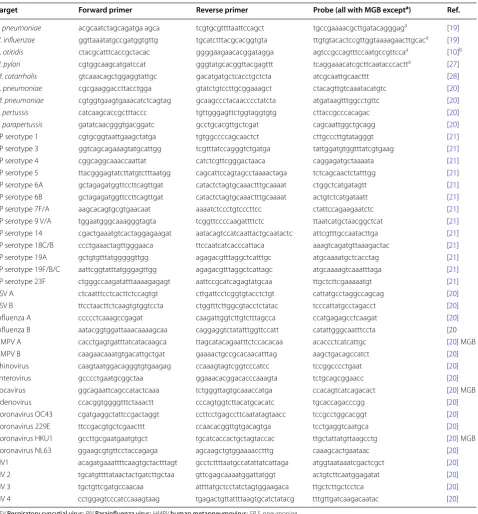
Table 1 Real-time PCR assays for bacteria, viruses and Streptococcus pneumoniae serotypes
RSV Respiratory syncytial virus; PIV Parainfluenza virus; HMPV human metapneumovirus; SPS. pneumoniae
a Probes contained an internal quencher and no minor groove binder. All other probes contained a minor groove binder and no internal quencher b Primers from published reference, probe this study
Target Forward primer Reverse primer Probe (all with MGB excepta) Ref.
S. pneumoniae acgcaatctagcagatga agca tcgtgcgttttaattccagct tgccgaaaacgcttgatacagggaga [19]
H. influenzae ggttaaatatgccgatggtgttg tgcatctttacgcacggtgta ttgtgtacactccgttggtaaaagaacttgcaca [19]
A. otitidis ctacgcatttcaccgctacac ggggaagaacacggatagga agtccgccagtttccaatgccgttccaa [10]b
H. pylori cgtggcaagcatgatccat gggtatgcacggttacgagttt tcaggaaacatcgcttcaatacccactta [27]
M. catarrhalis gtcaaacagctggaggtattgc gacatgatgctcacctgctcta atcgcaattgcaacttt [28]
C. pneumoniae cgcgaaggaccttacctgga gtatctgtccttgcggaaagct ctacagttgtcaaatacatgtc [20]
M. pneumoniae cgtggtgaagtgaaacatctcagtag gcaagccctacaacccctatcta atgataagtttggcctgttc [20]
B. pertussis catcaagcaccgctttaccc tgttgggagttctggtaggtgtg cttaccgcccacagac [20]
[image:3.595.61.540.99.614.2]QPCR methods
Nucleic acids (NA) were extracted from MEF using an automated extraction device (iPrep, Life Technologies, Carlsbad, California). The qPCR primers and probes used for bacteria viruses, and S. pneumoniae serotype detec-tion are shown in Table 1. All assays used 5′ exonuclease probes labeled at the 5′end with fluorescein amidite and with a quencher at the 3′ end.
For A. otitidis, we used previously published PCR prim-ers for this bacterium [5, 10] but designed a 5′ exonucle-ase probe for qPCR rather than using intercalating dye detection [10]. A commercial qPCR program (Allele ID, Premier Biosoft) was used to design the probe and speci-ficity was checked using BLAST searches and by testing the assay against multiple reference strains of bacteria.
QPCR methods were performed as described previ-ously [19, 20]. All probes were labeled with fluorescein amidite (FAM). Probes containing minor groove binders (MGB) were obtained from Life Technologies (Carlsbad, CA). All other probes were obtained from Integrated DNA Technologies (Coralville, IA) and contained a pro-prietary internal quencher (ZEN quencher, IDT). Briefly, singleplex real-time 5′exonuclease qPCR assays were prepared in 20 µL volumes in 96-well qPCR plate. Posi-tive and negaPosi-tive control (no template) was performed with each qPCR plate run. QPCR plates were covered with MicroAmp® Optical Adhesive Film (Life Tech-nologies Carlsbad, CA) to prevent cross-contamination. QPCR was performed with a 96 well fast cycling block on a ViiA7 thermocyler (Life Technologies Inc.) using 40 cycles of 2-temperature thermocyling (95 °C × 3 s and
60 °C × 30 s).
Only samples which were positive with the S. pneu-moniae qPCR assay were further tested using published serotype-specific qPCR assays for the 13 serotypes con-tained in the current conjugated vaccine (serotypes 1, 3, 4, 5, 6A, 6B, 7F, 9 V, 14, 18C, 19A, 19F, and 23F) [21]. QPCR serotyping assays were performed as above, with the exception that longer cycling times were used (40 cycles of 2-temperature thermocyling were used, with 95 °C × 15 s and 60 °C × 60 s).
Results
Thirty-one children were enrolled in the study. One child was excluded as tympanostomy was performed for AOM rather than OME, leaving 30 that were evaluated. Forty-eight MEF samples were obtained from these 30 chil-dren. Sixteen children were female and 14 were male. The median age was 34 months with range 11–120 months. Twenty-seven of the 30 (90 %) children had received the original 7-serotype S. pneumoniae vaccine as part of routine infant immunization program, while three older children born prior to introduction of this vaccine had
not. None had received the 13-serotype S. pneumoniae
vaccine.
The number of MEF specimens in which bacterial and viral organisms NA were detected by qPCR, both alone and with other agents, is shown in Table 2. Overall, qPCR detected bacterial DNA in 39/48 (81 %) MEFs from 26/30 (87 %) children and detected viral NA in 7/48 (15 %) MEF from 5/30 (17 %) children. A.otitidis and S. pneumoniae
DNA were both detected by qPCR in 15/48 (31 %) MEF, with A.otitidis in 11/30 (37 %) children and S. pneumo-niae in 10/33 (33 %) children. These were followed by M. catarrhalis DNA in 14/48 (29 %) MEF from 10/30 (33 %) children, H. influenzae DNA in 5/48 MEF (10 %) from 4/30 (13 %) children, and M. pneumoniae DNA in 4/48 (8 %) MEF from 2/30 (7 %) children. Among the viruses tested for, rhinovirus RNA was present in 4/48 (8 %) MEF, coronavirus OC43 RNA in 2/48 (4 %) MEF, and influenza B RNA in 1/48 (2 %) MEF.
Bacteria were grown in culture from 11/48 (23 %) MEF from 10/30 (33 %) children. M. catarrhalis was isolated from five MEF, H. influenzae from 3 MEF, and S. pneu-moniae from 2 MEF. All culture positive samples for these three bacteria were also PCR-positive. One MEF grew Streptococcus pyogenes (group A streptococcus), a bacterium not included in the PCR testing. A. otitidis
was not grown in culture, and no anaerobic bacteria were grown in culture.
With regard to detection of multiple organisms (detec-tion of ≥ bacterial or viral NA in the same MEF), there were 28/48 (58 %) MEF with NA from a single organism detected, 6/48 (13 %) MEF with NA from 2 organisms detected and 6/48 (13 %) MEF with NA from 3 organisms detected, and 8/48 with no NA detected. Of note, 6/7
Table 2 Bacterial and viral nucleic acid detected by qPCR from middle ear fluid (MEF) samples from children under-going tympanostomy tube insertion for otitis media with effusion
Organism
name Number of PCR-positive MEF (n = 48)
Number
detected alone Number detected with other bacteria and/or viruses
S. pneumoniae 15 5 10
A.otitidis 15 6 9
M. catarrhalis 14 9 5
H. influenzae 5 4 1
M. pneumoniae 4 2 2
Rhinoviruses 4 0 4
Coronavirus
OC43 virus 2 0 2
[image:4.595.305.539.562.725.2](86 %) viral- NA positive samples also had bacterial DNA detected by qPCR.
Streptococcus pneumoniae serotyping by qPCR detected serotype 19A DNA in MEF in 3/15 (20 %) S. pneumoniae qPCR-positive specimens. All three samples with serotype 19A DNA detected from children who had received the 7 serotype vaccine. Tests for the other 12 vaccine serotypes were negative.
Discussion
The finding that over 80 % of MEF samples from chil-dren with OME contained bacterial DNA supports the idea that OME may be an infectious process. A. otiti-dis DNA was frequently detected but the pathogenicity of this organism in OME is uncertain. Several studies have reported detecting A. otitidis by PCR in MEF [10,
11]. One report suggests this bacterium may be part of the normal flora of the external ear, and not a middle-ear pathogen [22]. However, other studies show A. otitidis
can stimulate an inflammatory response, suggesting it may cause disease in the middle ear. For example, A. oti-tidis elicited higher levels of inflammatory mediators than S. pneumoniae in a cell line model [23].
The fact that we did not isolate A.otitidis in culture is consistent with findings that this organism typically does not grow in culture using the methods standardly used in clinical microbiology laboratories. For example, in one study, A. otitidis was not grown from any MEFs but 35 % were PCR-positive [11]. Improved methods to culture this organism from MEFs are being sought [24].
As seen in other studies, S. pneumoniae, M. catarrhalis, and H. influenzae DNA were detected in a much higher proportion of MEF by PCR than culture [4–6]. This may be due to the greater sensitivity of PCR over culture, or could reflect that PCR detects both live and dead bacteria, while culture requires live bacteria. Similar to our find-ings, Hendolin reported 84 % of MEF were PCR-positive for bacteria while 32 % were culture positive [5], while Park reported 36.7 % MEF were PCR-positive for bacte-ria and 14 % of samples were positive by culture [6]. This common trend may reflect the greater sensitivity of PCR for detecting bacteria in biofilm-related infections [8].
Among the accepted bacterial pathogens, S. pneu-moniae was detected more frequently by qPCR than H. influenzae or M. catarrhalis. Since S. pneumoniae could be cultured from only 2/15 (13 %) qPCR positive MEFs, culture-based detection of S. pneumoniae in OME may grossly underestimate the importance of this bacterium, and serotyping of positive culture samples may similarly provide very limited information. Molecular serotyping methods using qPCR or other techniques that are cul-ture-independent thus may have be a better approach to determining which S. pneumoniae serotypes need to be
included in future vaccines to help prevent OME. To our knowledge, this is the first study in which molecular sero-typing for S. pneumoniae has been performed directly on MEFs from children with OME.
Serotype 19A was the only serotype of the 13 included in the latest S. pneumoniae vaccine that was detected in the MEF in this study. Serotype 19A was not included in the original 7-serotype vaccine, but is included in the 13 serotype vaccine, so this vaccine may decrease otitis caused by this serotype. However, our finding that only a minority of samples contained a serotype included in the new vaccine is concerning, as it suggests that non-vaccine serotypes are important in OME. In this pilot study, we were unable to perform qPCR serotyping for all of the approximately 90 S. pneumoniae serotypes, but we plan to test for a greater number of serotypes in subse-quent studies to help determine which serotypes should be included in future vaccines.
The detection of M. pneumoniae PCR in several sam-ples is also of interest. M. pneumoniae has been detected in OME before, but it appears to be infrequent cause. For example, Strogard reported that only 1/150 MEF was PCR positive for M. pneumoniae [25].
Helicobacter pylori, known as a cause of peptic ulcer disease, has also recently been reported into be present in MEFS of children with OME in several studies, In a study from Iran, 43 % MEFs and 25 % of adenoid samples from children undergoing myringotomy were positive for H. pylori [12]. Similarly, a study from Turkey reported detection of H. pylori in 47 % of MEFs from children with OME [13]. The absence of this organism in our samples may reflect geographic variability in H. pylori exposure in children.
In terms of viral agents, despite testing for a large num-ber of respiratory viral agents, RNA from rhinoviruses, which are the major cause of the common cold, were the most frequently detected viruses in this study, and have also been detected in some other OME studies [14, 16]. Virus NA was detected in a much smaller proportion of MEF than bacteria and was often detected with bacterial DNA. This suggests that viruses may not be a common direct cause of OME. However, it is possible that viral infections of the upper respiratory tract could lead to Eustachian tube dysfunction, which could contribute to OME. Studies examining both nasopharyngeal and MEF samples for bacteria and viruses by qPCR may help clar-ify the role of viruses as possible co-factors in OME.
large number of PCR assays (27 microorganism assays and 13 serotype assays for S. pneumoniae) to guide us as to priorities for future OME research. Similarly, given the small number of S. pneumoniae qPCR-positive samples available for molecular serotyping, it is not possible to make definite conclusions regarding the distribution of serotypes. Nevertheless, the findings suggest that molec-ular serotyping will provide much more information than traditional culture-based serotyping, and also that sero-types other than those contained in the 13-valent vaccine will need to be tested for in future studies.
Second, as noted above, PCR-positive results can occur from live or dead bacterial or viral micro-organisms. However, for bacteria, Post and others have shown in an animal model that purified DNA and DNA from intact but non-viable bacteria do not persist in the middle ear. In contrast, DNA from live bacteria could be detected for 3 weeks post-middle ear inoculation even when antibiotic treatment caused bacterial culture to become negative [9].
Additional work that supports the concept that bac-terial DNA detected by PCR represents live organisms comes from the study by Rayner and others [26]. This group used reverse transcriptase-polymerase chain reac-tion (RT-PCR) to determine if bacterial messenger RNA (mRNA) was present in pediatric OME samples that con-tained bacterial DNA but were sterile by standard cul-tural methods. Since bacterial mRNAs have a very short half-life of seconds to minutes, detection of bacteria-specific mRNAs suggests metabolically active organisms are present. This group found that 29/29 H. influenzae
PCR- positive samples were also positive for H. influen-zae mRNA by RT-PCR, which suggests that viable, meta-bolically active organisms were present in these samples.
With respect to the correlation between the detection of viral NA detection by PCR and the presence of live virus in patient samples, one study has shown that this appears to depend on the type of virus. The duration of viral shedding for influenza viruses was not significantly different when measured by culture and PCR (13 vs. 14 days, respectively). However, for RSV and parainflu-enza viruses shedding lasted significantly longer by PCR than by culture. Unfortunately, rhinoviruses, the most common viruses found in our study, were not examined in this report [29].
Finally, although we tested for a wide range of organ-isms, there have been additional agents found in OME for which we did not perform PCR. For example, the anaerobic organism Fusobacterium nucleatum was found in 6/20 (30 %) OME samples using PCR in one study [30]. Although anaerobic bacterial cultures in our study were negative, it is possible that F. nucleatum or other anaero-bic bacteria may have been detected had we used PCR for these organisms.
Conclusion
Bacterial infections may play a role in OME since over 80 % of MEFS were qPCR-positive for bacterial organ-isms, while viruses were found in a much smaller propor-tion. Further studies to determine the pathogenicity of A. otitidis are needed. Additional S. pneumoniae serotypes not included in the 13-serotype vaccine may be important in OME, but this needs to be confirmed by larger studies.
Abbreviations
OME: otitis media with effusion; MEF: middle ear fluid; AOM: acute otitis media; qPCR: real‑time PCR.
Authors’ contributions
RS contributed to study design and drafted the primary manuscript. MD, FC, and J‑PV contributed to study design and execution and manuscript review. JL performed laboratory work and manuscript review. MB and JM contributed to study execution and manuscript review. All authors read and approved the final manuscript.
Author details
1 Department of Laboratory Medicine and Pathology, Children’s Hospital of Eastern Ontario, University of Ottawa, 401 Smyth Rd, Ottawa, ON K1H 8L1, Canada. 2 Department of Surgery, Children’s Hospital of Eastern Ontario, University of Ottawa, Ottawa, ON, Canada.
Acknowledgements None.
Availability of data and material
The data sets supporting the results of this article are included within this article.
The dataset supporting the conclusions of this arti‑ cle is available in the LabArchives repository at the follow‑ ing URL: https://mynotebook.labarchives.com/share/Slinger1/
MjIuMXw4NTg5OC8xNy01L1RyZWVOb2RlLzM1ODc0Mjk4MDh8NTYuMQ==.
Competing interests
The authors declare that they have no competing interests.
Ethics approval and consent to participate
Ethics approval was obtained for the study from the Children’s Hospital of Eastern Ontario hospital Research Ethics Board.
Funding
Funding for this study was provided by the Department of Surgery, Children’s Hospital of Eastern Ontario.
Received: 26 January 2016 Accepted: 14 April 2016
References
1. Williamson I. Otitis media with effusion in children. BMJ Clin Evid. 2011;2011:0502.
2. Bennett KE, Haggard MP. Behaviour and cognitive outcomes from middle ear disease. Arch Dis Child. 1999;80:28–35.
3. Holder RC, Kirse DJ, Evans AK, Peters TR, Poehling KA, Swords WE, et al. One third of middle ear effusions from children undergoing tympanos‑ tomy tube placement had multiple bacterial pathogens. BMC Pediatr. 2012;28(12):87.
• We accept pre-submission inquiries
• Our selector tool helps you to find the most relevant journal
• We provide round the clock customer support
• Convenient online submission
• Thorough peer review
• Inclusion in PubMed and all major indexing services
• Maximum visibility for your research
Submit your manuscript at www.biomedcentral.com/submit
Submit your next manuscript to BioMed Central
and we will help you at every step:
5. Hendolin PH, Markkanen A, Ylikoski J, Wahlfors JJ. Use of multiplex PCR for simultaneous detection of four bacterial species in middle ear effusions. J Clin Microbiol. 1997;35:2854–8.
6. Park CW, Han JH, Jeong JH, Cho SH, Kang MJ, Tae K, et al. Detection rates of bacteria in chronic otitis media with effusion in children. J Korean Med Sci. 2004;19:735–8.
7. Hall‑Stoodley L, Hu FZ, Gieseke A, Nistico L, Nguyen D, Haves J, et al. Direct detection of bacterial biofilms on the middle‑ear mucosa of children with chronic otitis media. JAMA. 2006;296:202–11.
8. Coticchia JM, Chen M, Sachdeva L, Mutchnick S. New paradigms in the pathogenesis of otitis media in children. Front Pediatr. 2013;1:52. 9. Post JC, Aul JJ, White GJ, Wadowsky RM, Zavoral T, Tabari R, et al. PCR‑
based detection of bacterial DNA after antimicrobial treatment is indica‑ tive of persistent, viable bacteria in the chinchilla model of otitis media. Am J Otolaryngol. 1996;17:106–11.
10. Marsh RL, Binks MJ, Beissbarth J, Christensen P, Morris PS, Leach AJ, et al. Quantitative PCR of ear discharge from Indigenous Australian children with acute otitis media with perforation supports a role for Alloiococ-cus otitidis as a secondary pathogen. BMC Ear Nose Throat Disord. 2012;3(12):11.
11. Aydın E, Taştan E, Yücel M, Aydoğan F, Karakoç E, Arsian N, et al. Concur‑ rent assay for four bacterial species including alloiococcus otitidis in middle ear, nasopharynx and tonsils of children with otitis media with effusion: a preliminary report. Clin Exp Otorhinolaryngol. 2012;5:81–5. 12. Saki N, Samarbaf Zadeh AR, Sheikhpour Jonaky R, Noori SM, Kayedani GA, Nikakhlagh S. The prevalence rate of helicobacter pylori infection in chronic otitis media with effusion patients. Jundishapur J Microbiol. 2014;7:e15694.
13. Yilmaz MD, Aktepe O, Cetinkol Y, Altuntaş A. Does Helicobacter pylori have a role in development of otitis media with effusion? Int J Pediatr Otorhinolaryngol. 2005;69:745–9.
14. Pitkäranta A, Jero J, Arruda E, Virolainen A, Hayden FG. Polymerase chain reaction‑based detection of rhinovirus, respiratory syncytial virus, and coronavirus in otitis media with effusion. J Pediatr. 1998;133:390–4. 15. Rezes S, Söderlund‑Venermo M, Roivainen M, Kemppainen K, Szabó Z,
Sziklai I, et al. Human bocavirus and rhino‑enteroviruses in childhood otitis media with effusion. J Clin Virol. 2009;46:234–7.
16. Chantzi FM, Papadopoulos NG, Bairamis T, Tsiakou M, Bournousouzis N, Constantopoulos AG, et al. Human rhinoviruses in otitis media with effu‑ sion. Pediatr Allergy Immunol. 2006;7:514–8.
17. Zhao AS, Boyle S, Butrymowicz A, Engle RD, Roberts JM, Mouzakes J. Impact of 13‑valent pneumococcal conjugate vaccine on otitis media bacteriology. Int J Pediatr Otorhinolaryngol. 2014;78:499–503.
18. L. Garcia (ed.), Clinical Microbiology Procedures Handbook. 3rd ed. Ameri‑ can Society for Microbiology. Washington, DC: ASM Press; 2010. pp 1–3. 19. Pernica JM, Moldovan I, Chan F, Slinger R. Real‑time polymerase chain
reaction for microbiological diagnosis of parapneumonic effusions in Canadian children. Can J Infect Dis Med Microbiol. 2014;25:151–4. 20. Ellis C, Misir A, Hui C, Jabbour M, Barrowman N, Langill J, et al. Detection
of respiratory viruses and bacteria in children using a twenty‑two target reverse‑transcription real‑time PCR (RT‑qPCR) panel. World J Pediatr. 2016;12:183–9.
21. Slinger R, Hyde L, Moldovan I, Chan F, Pernica JM. Direct Streptococcus pneumoniae real‑time PCR serotyping from pediatric parapneumonic effusions. BMC Pediatr. 2014;14:189.
22. Tano K, von Essen R, Eriksson PO, Sjöstedt A. Alloiococcus otitidis—otitis media pathogen or normal bacterial flora? APMIS. 2008;116:785–90. 23. Ashhurst‑Smith C, Hall ST, Burns CJ, Stuart J, Blackwell CC. In vitro inflam‑
matory responses elicited by isolates of Alloiococcus otitidis obtained from children with otitis media with effusion. Innate Immun. 2014;20:320–6. 24. Matsuo J, Harimaya A, Fukumoto T, Nakamura S, Yoshida M, Takahashi
K, et al. Impact of anaerobic and oligotrophic conditions on survival of
Alloiococcus otitidis, implicated as a cause of otitis media. J Infect Chem‑ other. 2011;17:478–82.
25. Storgaard M, Tarp B, Ovesen T, Vinther B, Andersen PL, Obel N, et al. The occurrence of Chlamydia pneumoniae, Mycoplasma pneumoniae, and herpesviruses in otitis media with effusion. Diagn Microbiol Infect Dis. 2004;48:97–9.
26. Rayner MG, Zhang Y, Gorry MC, Chen Y, Post JC, Ehrlich GD. Evidence of bacterial metabolic activity in culture‑negative otitis media with effusion. JAMA. 1998;279:296–9.
27. Schabereiter‑Gurtner C, Hirschl AM, Dragosics B, Hufnagl P, Puz S, Kovách Z, et al. Novel real‑time PCR assay for detection of helicobacter pylori infection and simultaneous clarithromycin susceptibility testing of stool and biopsy specimens. J Clin Microbiol. 2004;42:4512–8.
28. Heiniger N, Troller R, Meier PS, Aebi C. Cold Shock response of the uspa1 outer membrane adhesin of Moraxella catarrhalis. Infect Immun. 2005;73:8247–55.
29. Richardson L, Brite J, Del Castillo M, Childers T, Sheahan A, Huang YT, Dougherty E, Babady NE, Sepkowitz K, Kamboj M. Comparison of respira‑ tory virus shedding by conventional and molecular testing methods in patients with haematological malignancy. Clin Microbiol Infect. 2016;22:380.e1–7.