RESEARCH ARTICLE
Insight to changing morphologic
patterns of glomerulopathy in adult Pakistani
patients: an institutional perspective
Atif Ali Hashmi
1, ZubaidaFida Hussain
1, Muhammad Muzzammil Edhi
2, Shazia Mumtaz
1, Naveen Faridi
1and Mehmood Khan
3*Abstract
Background: Idiopathic nephrotic syndrome encompasses diverse histogenetic patterns and depicts socioeco-nomic and demographic differences attributable to genetic profile, environmental factors and prevalence of infec-tious diseases. A lack of renal registry in our country necessitates a need to document changing histologic patterns of nephrotic syndrome as noted in different parts of the world.
Methods: We retrospectively analyzed 140 patients who underwent renal biopsy at Liaquat National Hospital from January 2009 to December 2013 over a period of 3 years. On the basis of clinical profile cases were segregated into nephritic and nephrotic syndrome and histologic and immunoflourescence findings were analyzed.
Results: Among 140 cases of glomerulonephritis diagnosed in the study period, 98 cases (70 %) were those of primary glomerulonephritis and 42 were of secondary glomerulopathy (30 %). Membranous glomerulonephritis was the most common primary glomerulonephritis (33.6 %) followed by focal segmental glomerulosclerosis FSGS (20.4 %), whereas lupus nephritis is the most common secondary glomerulopathy (47.6 %) followed by amyloidosis and dia-betic glomerulosclerosis (16.6 % each).
Conclusion: We found a considerable high incidence of membranous glomerulonephritis and FSGS in our popula-tion that entails a need to investigate prevalence of associated factors like Hepatitis B and HIV infecpopula-tions in populapopula-tion at risk. Moreover, renal biopsy registry would be instrumental in this regard to record changing disease pattern in this part of the world.
Keywords: Nephrotic syndrome, Renal biopsy, Membranous glomerulonephritis, Focal segmental glomerulosclerosis
© 2016 Hashmi et al. This article is distributed under the terms of the Creative Commons Attribution 4.0 International License (http://creativecommons.org/licenses/by/4.0/), which permits unrestricted use, distribution, and reproduction in any medium, provided you give appropriate credit to the original author(s) and the source, provide a link to the Creative Commons license, and indicate if changes were made. The Creative Commons Public Domain Dedication waiver (http://creativecommons.org/ publicdomain/zero/1.0/) applies to the data made available in this article, unless otherwise stated.
Background
Renal diseases accredit major health problem in devel-oping countries [1, 2]. While chronic diseases like dia-betes and hypertension still accounts for the major bulk of chronic renal failure patients, idiopathic glomerulone-phritis (GN) poses a major diagnostic challenge to both nephrologists and pathologists. Degree of proteinuria often helps clinicians to narrow down their differentials to two major clinical patterns: nephritic and nephrotic. While the epidemiology of nephritic syndrome changed
quite a bit over the years due to changing public health protocols, prevailing infections and vaccinations, etiology of nephrotic syndrome showed more dramatic fluctua-tions in etiology and depicts a racial and age preference [3, 4]. Whether this represents a change in the disease pattern or better understanding of glomerular disorders is a matter of debate. However it has been suggested that apart from genetic profile, environmental factors and prevalence of infectious diseases attribute to socio-economic and demographic differences seen in patterns of glomerulopathy. Acute nephritic syndrome is usually related to underlying immunologic injury, the culprit of which is attributed mostly to infectious agents [5, 6]. However, nephritic syndrome is usually self limited and
Open Access
*Correspondence: [email protected]
3 Dhaka Medical College, Dhaka, Bangladesh
renal biopsy is not always indicated. On the other hand nephrotic syndrome encompasses diverse histogenetic patterns ranging from minimal change disease (MCD) to collapsing form of focal segmental glomerulosclerosis (FSGS).
Renal biopsy remained the gold standard for the diag-nosis of primary glomerulonephritis with ancillary immunoflorescent and electron microscopic examina-tion. Most common indication for renal biopsy is unex-plained elevation of renal parameters and proteinuria. Renal biopsy is often performed in systemic diseases like systemic lupus erythematosus when active renal involvement is suspected due to presence of hematuria or proteinuria and elevated serologic markers of lupus nephritis. In diabetes and hypertension, renal biopsy is not indicated as progressive renal involvement is usually present in uncontrolled cases of long duration disease; however it is usually done in atypical presentations to rule out other causes of glomerulopathy or de novo pri-mary glomerulonephritis.
In addition to being different histologically, disease pat-tern of GN also differs significantly in clinical parameters. GN typically behaves in a self limited fashion in children, while it poses a major risk when occurring in adults. Data of adult western population show FSGS to be the most frequent pattern in African Americans while membra-nous GN dominates the clinical picture of nephrotic syndrome in whites. It is of immense importance to rec-ognize common pathologic patterns of idiopathic GN in a population to devise better therapeutic protocols. Data pertaining to this clinical entity is limited in our setup, as renal biopsy registries are not currently available and changing patterns of glomerulopathy have not been well demonstrated in our setup as noted in other populations [7, 8]. Therefore we aimed to determine histopathologic patterns of both secondary and idiopathic (primary) GN in adult patients undergoing renal biopsy.
Methods
We retrospectively analyzed 140 patients who underwent renal biopsy at Liaquat National Hospital from Janu-ary 2009 to December 2013 over a period of 3 years. An approval from research and ethical review committee were taken antecedent to conducting the study. Data was retrieved by reviewing pathology request forms of renal biopsy specimens received at Histopathology laboratory during the study period. This included patient major signs and symptoms, indication of renal biopsy, labora-tory findings like degree of hematuria, proteinuria, uri-nalysis results and positive serum markers like ANA, anti-dsDNA and c-ANCA. Cases with insufficient clini-cal information were excluded from the study. A total of 30 biopsies were excluded by this criterion. Moreover
five cases were also excluded due to inconclusive findings on renal biopsy and two due to inconclusive immuno-flourescence results. Subsequently cases were segregated according to mode of presentation and lab findings into nephrotic and nephritic syndrome. Cases with >3.5 g proteinuria with edema were defined as nephrotic syn-drome and cases with <3.5 g proteinuria along with hematuria and RBC casts were classified as nephritic syndrome. General indications of renal biopsy at our institution were nephrotic or acute nephritic syndrome, unexplained rise in urea and creatinine, evaluation of extent of renal involvement in secondary disorders like SLE, DM etc. In SLE, renal biopsy was performed when there is clinical or laboratory evidence of lupus nephritis (e.g. elevated anti dsDNA levels). In diabetes and chronic hypertension, renal biopsy was only performed when some other superimposed glomerular pathology was sus-pected. Percutaneous renal biopsy was performed under aseptic precautions with a 16–18 gauge needle. Informed written consent was taken prior to conducting the pro-cedure. Two separate cores were taken for light micros-copy and immunoflourscence studies. Nine to 12 thin levels (2 µm) were examined on 3 to 4 slides stained with hematoxylin& eosin for light microscopy along with peri-odic acid shift, trichome and silver stains on each biopsy. Stains for amyloid were performed when needed. Cases with atleat six glomeruli and one artery were considered adequate. All biopsies were evaluated by two senior his-topathologists trained in renal pathology. Immunofluo-rescence studies were performed using antibodies against IgG, IgM, IgA, C3c and C1q. Cases with provisional diagnosis of MCD or inconclusive light and immuno-flourence findings were referred for electron microscopy at another institution as it was not routinely performed at our institution. Statistical analysis was performed using Chi square on SPSS version 20.
Results
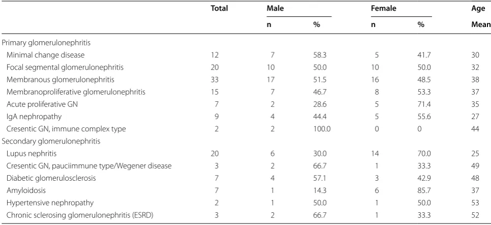
mean age for lupus nephritis, IgA nephropathy and MCD were slightly lower than other forms of GN, however the difference was not statistically significant. Frequency of primary and secondary GN alongwith age and sex distri-bution is shown in Tables 1 and 2.
Among 98 cases of primary GN, 84 cases (85 %) pre-sented with nephrotic syndrome. Table 3 shows the different histologic patterns of primary GN. Among nephrotic syndrome, most common pattern was mem-branous GN (38 %), followed by FSGS (21 %) and MPGN (16 %). On the other hand acute post streptococcal GN was the most common pattern found in nephritic syn-drome patents (42 %) followed IgA nephropathy (28 %). Table 4 shows the IMF pattern of primary and secondary GN. All cases of MPGN were of type I. There was no case of goodpasture syndrome or myeloma related disorder except for amyloidosis. There were five cases of cresentic
GN; out of which, two cases showed immune complex deposits on IF in the absence of significant primary GN pattern therefore they were categorized as primary GN. The rest of 3 cases were pauci-immune and they were c-ANCA positive with systemic evidence of Wegener’s syndrome, therefore they were classified as secondary GN. There were 7 cases of amyloidosis, out of which 3 were known cases of multiple myeloma, however no fur-ther stains were performed to sub classify cases of amy-loidosis on renal biopsy.
Discussion
Our institution represents one of the largest tertiary care centers in country with well developed nephrology unit. Renal biopsy is routinely performed as part of workup in patients with unexplained deterioration of renal func-tions or to determine extent of renal dysfunction in
Table 1 Gender distribution and mean age of primary and secondary glomerulonephritis
Total Male Female Age
n % n % Mean
Primary glomerulonephritis
Minimal change disease 12 7 58.3 5 41.7 30
Focal segmental glomerulonephritis 20 10 50.0 10 50.0 32
Membranous glomerulonephritis 33 17 51.5 16 48.5 38
Membranoproliferative glomerulonephritis 15 7 46.7 8 53.3 37
Acute proliferative GN 7 2 28.6 5 71.4 35
IgA nephropathy 9 4 44.4 5 55.6 27
Cresentic GN, immune complex type 2 2 100.0 0 0 44
Secondary glomerulonephritis
Lupus nephritis 20 6 30.0 14 70.0 25
Cresentic GN, pauciimmune type/Wegener disease 3 2 66.7 1 33.3 49
Diabetic glomerulosclerosis 7 4 57.1 3 42.9 48
Amyloidosis 7 1 14.3 6 85.7 37
Hypertensive nephropathy 2 1 50.0 1 50.0 53
Chronic sclerosing glomerulonephritis (ESRD) 3 2 66.7 1 33.3 52
Table 2 Age distribution of primary glomerulonephritis
Less than 18 years 19–30 years 31–50 years More than 50 years
n % n % n % n %
Minimal change disease 5 41.7 2 16.7 3 25.0 2 16.7
Focal segmental glomerulonephritis 3 15.0 9 45.0 5 25.0 3 15.0
Membranous glomerulonephritis 3 9.1 10 30.3 12 36.4 8 24.2
Membranoproliferative glomerulonephritis 2 13.3 4 26.7 5 33.3 4 26.7
Acute proliferative GN 2 28.6 1 14.3 2 28.6 2 28.6
IgA nephropathy 2 22.2 3 33.3 4 44.4 0 0
[image:3.595.55.538.320.541.2] [image:3.595.58.542.589.724.2]some secondary renal diseases like systemic lupus ery-thematosus, Diabetes and hypertension. Nephrotic syn-drome is the most common clinical picture in patients undergoing renal biopsies as demonstrated in our study. A survey of renal biopsies performed in United States from 1995 to 1997 showed that FSGS is the most com-mon cause of nephrotic syndrome accounting for 35 % of all cases and more than 50 % cases among blacks [9]. An 11-fold rise in the incidence of FSGS as a cause of end stage renal disease was identified in recent year in US [10]. These changing histologic trends were also demonstrated in India. FSGS was found to be the
dominant morphologic pattern in different studies. Rathi et al. found a frequency of 30 %, while Chandrika found 18 % frequency of FSGS [11, 12]. This shows a rising fre-quency of FSGS in some parts of the world. The statistics differ in other regions of the world. Renal biopsy registry in Spain shows membranous GN as the most common pattern (24 %), followed by MCD (6 %) and lupus nephri-tis (14 %) [13]. On the other hand, renal biopsy registry of china uncovered different trends. In that population 34.5 % of cases were diagnosed as IgA nephropathy fol-lowed by MCD (12.4 %) in recent years, according to Czech registry of renal biopsies [14]. Another study from Chinese population showed an even higher frequency of IgA nephropathy (40 %) [15]. In Romania, membrano-proliferative GN was found to be the changing dominant histologic pattern comprising of 29 % cases of primary GN [16].
In our population definitive trends cannot be reliably determined as there is lack of renal biopsy registry in our country. A few studies conducted so far showed differing patterns. A study involving 316 patients with nephrotic syndrome showed FSGS as the most common histo-logic pattern (39 %) followed by membranous nephropa-thy (26 %) [17]. Same trend was found in another study conducted in Karachi involving 60 cases of renal dys-function with FSGS as the most common histologic pat-tern [18]. On the other hand study, Rabbani et al. found membrano-proliferative GN to be at the top of the list (28 %) followed by membranous GN (19 %) [19]. A study
Table 3 Clinical presentation of different types of primary glomerulonephritis
Nephrotic
syndrome Nephritic syndrome
N % N %
Primary glomerulonephritis
Minimal change disease 12 14.2 0 0 Focal segmental glomerulonephritis 18 21.4 2 14.2 Membranous glomerulonephritis 32 38.0 1 7.1 Membranoproliferative glomerulonephritis 14 13.0 1 7.1 Acute proliferative GN 1 1.1 6 42.8 IgA nephropathy 5 5.9 4 28.5 Cresentic GN, immune complex type 2 2.3 0 0
Total 84 14
Table 4 Immunoflourescence patterns of primary and secondary glomerulonephritis
Immunoglobulins
IgG IgM IgA C3c C1q
N % N % N % N % N %
Primary glomerulonephritis
Minimal change disease 2 16.6 7 58.3 4 33.3 5 41.6 2 16.6
Focal segmental glomerulonephritis 2 10.0 17 85.0 5 25.0 12 60.0 7 35.0 Membranous glomerulonephritis 28 84.8 27 81.8 18 54.5 29 87.8 23 69.6 Membranoproliferative glomerulonephritis 10 66.7 13 86.7 13 86.7 15 100 10 66.7
Acute proliferative GN 6 85.7 4 57.1 4 57.1 7 100 4 57.1
IgA nephropathy 1 11.1 9 100 9 100 8 88.9 4 44.4
Cresentic GN, immune complex type 1 50.0 2 100 2 100 2 100 2 100 Secondary glomerulonephritis
Lupus nephritis 20 100 20 100 19 95.0 20 100 20 100
Cresentic GN, pauciimmune type/Wegener disease 1 33.3 2 66.7 0 0 2 66.7 1 33.3 Diabetic glomerulosclerosis 4 57.1 6 85.7 4 57.1 4 57.1 1 14.2
Amyloidosis 4 57.1 3 42.8 2 28.6 4 57.1 2 28.6
Hypertensive nephropathy 2 100 2 100 1 50.0 2 100 1 50.0
[image:4.595.58.290.113.273.2] [image:4.595.56.543.482.720.2]involving population from another part of the coun-try found that MCD was the most common diagnosis on histology comprised of 40 % of cases [20]. We found a higher frequency of membranous GN. These conflict-ing patterns may represent different set of population characteristics in these studies. The studies with FSGS as the dominant pattern mostly represent rural popula-tion. On the other hand our study involved patients from urban parts of the country. Another explanation could be changing histologic patterns over a period of time as seen in other parts of the world.
Membranous GN is the term applied when renal biopsy shows diffuse thickening of glomerular basement membrane in the absence of significant hypercellularity [21, 22]. On Immunoflourescence studies IgG and C3c are usually demonstrated. In our study apart from IgG and C3c, significant number of cases also showed deposi-tion of IgM and C1q. While most cases of membranous GN are idiopathic, in endemic areas like our country symptomatic Hepatitis B carrier state may be considered as a significant associated factor for the occurrence of membranous GN and its prevalence should be sought in populations at risk. Antibodies against M-type phospho-lipase A2 receptor (PLA2R) are serological markers of disease activity in patients with idiopathic membranous nephropathy. Some authors proposed that assessment of PLA2R antigen in biopsy specimens is a sensitive marker for the diagnosis of PLA2R-related MN [23]. We did not perform PLA2R antigen testing in our biopsy specimens.
In our study FSGS was the second most common pat-tern accounting for 18 % of cases of nephrotic syndrome. This represents a significant bulk of renal pathology in our population. An association of HIV infection with FSGS has been well established in western population, therefore HIV status should be determined in these cases.
One of the major limitations of our study was that all cases were of a single institution which could be a major source of bias, however as this is one of the largest ter-tiary care center in the province, therefore the patients which were referred to the nephrologist represent a major part of population including urban and rural parts of the province.
Conclusion
In conclusion membranous GN is the most common his-tologic pattern seen in nephrotic syndrome followed by FSGS in our population, therefore we suggest that under-lying risk factors like HBV and HIV infection should be determined in patients presenting with idiopathic nephrotic syndrome. More over there is a need for renal biopsy registry to determine evolving trends of nephrotic syndrome in our population.
Consent
Written informed consent was obtained from the patients for publication of the data. Ethics committee of Liaquat National hospital approved the study.
Authors’ contributions
AAA, the main author of manuscript, has made substantial contributions to conception and design of study. ZFH and SM have been involved in requisi-tion of data. MME has been involved in analysis of the data. NF gave final approval and revision of the manuscript. MK has been involved in drafting the manuscript. All authors read and approved the final manuscript.
Author details
1 Department of Histopathology, Liaquat National Hospital and Medical
College, Karachi, Pakistan. 2 Liaquat National Hospital and Medical College,
Karachi, Pakistan. 3 Dhaka Medical College, Dhaka, Bangladesh.
Acknowledgements
We gratefully acknowledge all staff members of Pathology, Liaquat National Hospital, Karachi, Pakistan for their help and cooperation.
Competing interests
The authors declare that they have no competing interests. Received: 12 March 2015 Accepted: 19 January 2016
References
1. Chang JH, Kim DK, Kim HW. Changing prevalence of glomerular diseases in Korean adults: a review of 20 years of experience. Nephrol Dial Trans-plant. 2009;24:2406–10.
2. Naumovic R, Pavlovic S, Stojkovic D, et al. Renal biopsy registry from a single centre in Serbia: 20 years of experience. Nephrol Dial Transplant. 2009;24:877–85.
3. Braden GL, Mulhern JG, O’Shea MH, et al. Changing incidence of glo-merular diseases in adults. Am J Kidney Dis. 2000;35:878.
4. Korbet SM, Genchi RM, Borok RZ, Schwartz MM. The racial prevalence of glomerular lesions in nephrotic adults. Am J Kidney Dis. 1996;27:647. 5. Kong D, Wu D, Wang T, Li T, Xu S, Chen F, Jin X, Lou G. Detection of viral
antigens in renal tissue of glomerulonephritis patients without serologi-cal evidence of hepatitis B virus and hepatitis C virus infection. Int J Infect Dis. 2013;17(7):e535–8.
6. Nasr SH, Radhakrishnan J, D’Agati VD. Bacterial infection-related glomeru-lonephritis in adults. Kidney Int. 2013;83(5):792–803.
7. Volovăt C, Cãruntu I, Costin C, Stefan A, Popa R, Volovăt S, Siriopol D, Voro-neanu L, Nistor I, Segall L, Covic A. Changes in the histological spectrum of glomerular diseases in the past 16 years in the North-Eastern region of Romania. BMC Nephrol. 2013;15(14):148. doi:10.1186/1471-2369-14-148. 8. Sugiyama H, Yokoyama H, Sato H, Saito T, Kohda Y, Nishi S, Tsuruya K,
Kiyo-moto H, Iida H, Sasaki T, Higuchi M, Hattori M, Oka K, Kagami S, Kawamura T, Takeda T, Hataya H, Fukasawa Y, Fukatsu A, Morozumi K, Yoshikawa N, Shimizu A, Kitamura H, Yuzawa Y, Matsuo S, Kiyohara Y, Joh K, Nagata M, Taguchi T, Makino H. Japan Renal Biopsy Registry and Japan Kidney Disease Registry: committee Report for 2009 and 2010. Clin Exp Nephrol. 2013;17(2):155–73. 9. Haas M, Meehan SM, Karrison TG, Spargo BH. Changing etiologies of
unexplained adult nephrotic syndrome: a comparison of renal biopsy findings from 1976–1979 and 1995–1997. Am J Kidney Dis. 1997;30:621. 10. Kitiyakara C, Eggers P, Kopp JB. Twenty-one-year trend in ESRD due to
focal segmental glomerulosclerosis in the United States. Am J Kidney Dis. 2004;44:815.
11. Rathi M, Bhagat RL, Mukhopadhyay P, Kohli HS, Jha V, Gupta KL, Sakhuja V, Joshi K. Changing histologic spectrum of adult nephrotic syndrome over five decades in north India: a single center experience. Indian J Nephrol. 2014;24(2):86–91.
• We accept pre-submission inquiries
• Our selector tool helps you to find the most relevant journal
• We provide round the clock customer support
• Convenient online submission
• Thorough peer review
• Inclusion in PubMed and all major indexing services
• Maximum visibility for your research
Submit your manuscript at www.biomedcentral.com/submit
Submit your next manuscript to BioMed Central
and we will help you at every step:
13. Rivera F, Lopez-Gomez JM, Perez-Garcia R, Spanish Registry of Glomeru-lonephritis. Clinicopathologic correlations of renal pathology in Spain. Kidney international. 2004;66(3):898–904.
14. Rychlík I, Jančová E, Tesař V, Kolský A, Lácha J, Stejskal J, Stejskalová A, Dušek J, Herout V. The Czech registry of renal biopsies. Occurrence of renal diseases in the years 1994–2000. Nephrol Dial Transplant. 2004;19(12):3040–9.
15. Chen H, Tang Z, Zeng C, Hu W, Wang Q, Yu Y, Yao X, Wang J, Zhu M, Zhou H, Liu H. Pathological demography of native patients in a nephrology center in China. Chin Med J. 2003;116(9):1377–81.
16. Volovăt C, Cãruntu I, Costin C, Stefan A, Popa R, Volovăt S, Siriopol D, Voro-neanu L, Nistor I, Segall L, Covic A. Changes in the histological spectrum of glomerular diseases in the past 16 years in the North-Eastern region of Romania. BMC Nephrol. 2013;15(14):148.
17. Kazi JI, Mubarak M, Ahmed E, Akhter F, Naqvi SA, Rizvi SA. Spectrum of glomerulonephritides in adults with nephrotic syndrome in Pakistan. Clin Exp Nephrol. 2009;13(1):38–43.
18. Sabir S, Mubarak M, Ul-Haq I, Bibi A. Pattern of biopsy proven renal diseases at PNS SHIFA, Karachi: a cross-sectional survey. J Ren Inj Prev. 2013;2(4):133.
19. Rabbani MA, Memon GM, Ahmad B, Memon S, Tahir SA, Tahir S. Percu-taneous renal biopsy results: a retrospective analysis of 511 consecutive cases. Saudi J Kidney Dis Transplant. 2012;23(3):614–8.
20. Akhtar SZ, Ali A. Histological patteren of nephrotic syndrome in elderly patients. J Ayub Med Coll Abbottabad. 2008;20(4):97–9.
21. Austin HA 3rd, Antonovych TT, MacKay K, et al. NIH Conference. Membra-nous nephropathy. Ann Intern Med. 1992;116:672.
22. Wasserstein AG. Membranous glomerulonephritis. J Am Soc Nephrol. 1997;8:664.