White Rose Research Online URL for this paper:
http://eprints.whiterose.ac.uk/110318/
Version: Accepted Version
Article:
Levdikov, Vladimir M, Blagova, Elena V, Young, Vicki L et al. (4 more authors) (2016)
Structure of the Branched Chain Amino Acid and GTP Sensing Global Regulator, CodY,
from Bacillus subtilis. The Journal of biological chemistry. pp. 1-36. ISSN 1083-351X
https://doi.org/10.1074/jbc.M116.754309
[email protected] https://eprints.whiterose.ac.uk/ Reuse
Items deposited in White Rose Research Online are protected by copyright, with all rights reserved unless indicated otherwise. They may be downloaded and/or printed for private study, or other acts as permitted by national copyright laws. The publisher or other rights holders may allow further reproduction and re-use of the full text version. This is indicated by the licence information on the White Rose Research Online record for the item.
Takedown
If you consider content in White Rose Research Online to be in breach of UK law, please notify us by
1
Structure of the Branched Chain Amino Acid and GTP Sensing Global Regulator, CodY, from Bacillus subtilis
Vladimir M. Levdikov1, Elena Blagova1, Vicki L. Young1, Boris R. Belitsky2, Andrey Lebedev3, Abraham L. Sonenshein2 and Anthony J. Wilkinson1
From the 1Structural Biology Laboratory, Department of Chemistry, University of York, York YO10 5DD, UK and 2Tufts University School of Medicine, Department of Molecular Biology and
Microbiology, Boston, Massachusetts 02111, USA and 3STFC Rutherford Appleton Laboratory, Harwell Oxford, Didcot, Oxfordshire, OX11 0QX, UK.
Running Title: Structure of Global Regulator CodY
To whom correspondence should be addressed: Anthony J. Wilkinson, Structural Biology Laboratory, Department of Chemistry, University of York, York YO10 5YW, UK, Telephone: +44-1904-328261; Fax. +44-1904-328366; E-mail [email protected]
Keywords: CodY/ /transcription regulation/GAF domain/wHTH domain
_____________________________________________________________________________________
ABSTRACT
CodY is a branched-chain amino acid (BCAA) and GTP sensor, and a global regulator of transcription in low G + C Gram-positive bacteria. It controls the expression of over 100 genes and operons, principally by repressing during growth genes whose products are required for adaptations to nutrient limitation. However, the mechanism by which BCAA binding regulates transcriptional changes is not clear. It is known that CodY consists of a GAF domain that binds BCAAs and a winged helix-turn-helix (wHTH) domain that binds to DNA, but the way in which these domains interact and the structural basis of the BCAA-dependence of this interaction are unknown. To gain new insights, we determined the crystal structure of unliganded CodY from Bacillus subtilis revealing a 10-turn -helix linking otherwise discrete GAF and wHTH domains. The structure of CodY in complex with isoleucine revealed a reorganised GAF domain. In both complexes CodY was tetrameric. Size exclusion chromatography with multiangle laser light scattering (SEC-MALLS) experiments showed that CodY is a dimer at concentrations found in bacterial cells. Comparison of structures of dimers of unliganded CodY and CodY-Ile derived from the tetramers showed a splaying of the wHTH domains when Ile was bound; splaying is likely to account for the increased affinity of Ile-bound CodY for DNA. Electrophoretic mobility shift and SEC-MALLS analyses of CodY binding to 19-36 base-pair operator fragments are consistent with isoleucine-dependent binding of two CodY dimers per duplex. The implications of
these observations for effector control of CodY activity are discussed.
________________________________________
In low G+C Gram-positive bacteria, CodY is a global regulatory protein that controls the transcription of numerous genes. In Bacillus subtilis, the species in which it was first identified, CodY is a nutrient sensor that represses during growth genes that are turned on under conditions of nutrient limitation (1). The CodY regulon in this organism, encompassing more than 100 genes and operons, encodes degradative enzymes, transporter proteins, catabolic enzymes, antibiotic synthesis pathways, and factors involved in the development of genetic competence, and sporulation (2,3). In human pathogens, such as Staphylococcus aureus (4,5), Streptococcus pneumoniae (6), S. pyogenes (7), Listeria monocytogenes (8,9), B. anthracis (10), and Clostridium difficile (11), CodY also regulates virulence gene expression and provides a regulatory link between metabolism and pathogenesis (12).
2
others (2). As the cells begin to experience nutrient limitation, the concentrations of BCAAs and GTP drop, leading to dissociation of CodY from the DNA and derepression of genes that are required for adaptation to nutrient-poor conditions. In the majority of cases, CodY is a repressor competing for DNA binding with RNA polymerase or with other positive regulators or causing premature termination of transcription by a roadblock mechanism (15). At some target genes, CodY acts as an activator either directly or indirectly (2,16,17). CodY binds to a 15-nucleotide canonical consensus sequence AATTTTCWGAAAATT that was first identified for the protein from Lactococcus lactis (18,19), and later shown to play an important role in B. subtilis (20).
The scope and complexity of CodY regulation of gene expression in B. subtilis have been revealed by genome-wide studies (2,3,21,22). These identified the substantial complement of CodY binding sites on the chromosome of B. subtilis and the broad range of affinities CodY has for these sites (22), consistent with the notion that the threshold concentration of CodY activity needed to trigger a transcriptional response varies from gene to gene. This gives rise to hierarchical control of gene expression in the CodY regulon allowing differential transcriptional responses according to the extent of nutrient limitation (3).
CodY from B. subtilis consists of 259 amino acid residues. The crystal structures of two fragments of CodY from B. subtilis that constitute its effector (residues 1-155) and DNA binding (residues 168-259) domains have been solved (23). CodY(1-155) is a dimer of GAF (cGMP-stimulated phosphodiesterases, adenylate cyclases, FhlA (24)) domains. It consists of three tiers, a basal three-helix bundle which forms a dimerisation surface, a central five-stranded -sheet, and a distal region formed by two extended loops which connect adjacent strands on the sheet. In complexes with isoleucine or valine, these loops embrace the ligand; comparison with the uncomplexed GAF domain reveals up to 15 Å displacements of backbone atoms in these loops upon effector binding (25). The C-terminal fragment CodY(168-259) has a winged helix-turn-helix (wHTH) domain fold.
These structures provided insights into the mode of BCAA binding and a basis for interpreting sequence conservation in CodY orthologues, but they gave no insights into the juxtaposition of the domains in the intact molecule
and thus it was not possible to infer how effector binding alters the structure of the protein so as to regulate DNA binding. Here, we describe the crystal structure of full length CodY in the unliganded state, together with that of a point mutant, CodY(L3S) in complex with isoleucine. These data, together with light scattering data on CodY and its complexes with DNA in solution, allow a mechanism for isoleucine control of DNA binding by CodY to be proposed.
RESULTS
Domain Organisation in CodY - The crystallisation and preliminary X-ray analysis of the full length CodY protein were reported previously (26). However, the crystals were not easy to reproduce and we were unable to solve their structure either by ab initio phasing or by molecular replacement using the coordinates for the isolated GAF domain in complex with isoleucine together with the coordinates for the wHTH domain (23). The structure of the unliganded form of the GAF domain (25) provided a search model that allowed successful structure solution by molecular replacement methods.
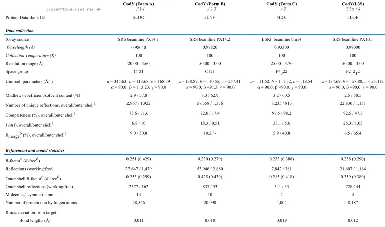
We have solved the structure of unliganded CodY in three different crystal forms containing 14 (Form A), 10 (Form B) and 2 (Form C) molecules in the asymmetric unit from data sets extending to 4.6 Å, 3.0 Å and 3.7 Å spacing, respectively (Table 1). These crystals were grown in the presence of different buffers, precipitants and additives as described in Materials and Methods. For two of the crystallisations, GTP or a GTP analogue was present though neither was observed bound to the protein. The following descriptions relate to the structure of the Form B crystals, since these crystals diffracted to the highest resolution (3.0 Å). In addition, we have solved the structure of a leucine3 to serine mutant, CodY(L3S), in complex with isoleucine at 3.0 Å resolution in a crystal form containing four molecules per asymmetric unit (Table 1). We have been unable to obtain crystals of either liganded wild-type CodY or unliganded CodY(L3S) suitable for X-ray analysis.
3
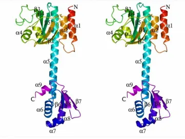
thereby creating a single -helix spanning residues 137 – 177. For the full-length CodY, the secondary structure elements are renumbered accordingly in Fig. 1C. The inter-domain linker portion of this helix has an unusual sequence with 9 of the 12 residues in the 156-167 segment possessing ionisable side-chains including five glutamates in a string of six residues. Within the CodY chain, there are no non-covalent interactions between the domains that exist as discrete entities. As a result, the isoleucine-binding site in the GAF domain and the helix-turn-helix, which constitutes the DNA binding site in the wHTH domain, are remote from one another (Fig. 1A and B).
The Branched-Chain Amino Acid Binding Pocket - Isoleucine was bound to the GAF domains of all four subunits in the CodY(L3S) tetramer. The ligand was almost completely enclosed in a pocket formed by the and
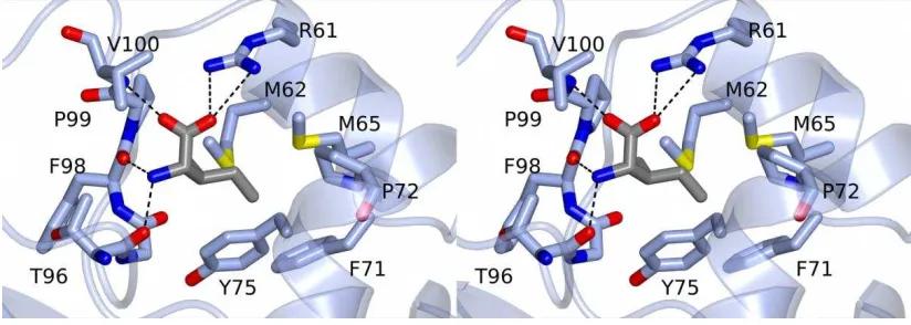
segments of the polypeptide (Fig. 1B and C). This pocket is distal to the site of the Leucine-3 to Serine substitution which resides in helix 1 at the N-terminus. The ligand carboxylate forms an ion-pair with the side chain of Arg61 and a further polar contact with the main chain amide of Val100 while its amino group forms polar contacts to the main chain carbonyl groups of Thr96 and Phe98 (Fig. 1D). The isobutyl side chain of the isoleucine ligand projects into the protein parallel to the -sheet and is surrounded by the side chains of Met62, Met65, Phe71, Pro72, Tyr75, and Pro99 (Fig. 1B and D). Various amino acid substitutions at positions 61, 71 and 98 have been shown to have a range of effects on CodY activity (3,27-29). This mode of isoleucine binding to CodY(L3S) exactly recapitulates that seen previously in the structure of isolated GAF domain of the native protein (23). When comparing chains from the CodY(1-155) and full length CodY(L3S) crystal structures, the average pairwise root mean squared deviation (rms) of equivalent C atoms is 0.7 Å. We conclude that ligand binding is essentially unchanged in CodY(L3S) and that the effects of the leucine3 to serine mutation on the tertiary structure of the GAF domain are barely discernible.
As may be seen in Fig. 1E, in the absence of ligand, the GAF domain structure in CodY is significantly altered in the vicinity of the ligand binding site. The positional rms following least squares superposition of 154 equivalent GAF domain C atoms is 3.4 Å. However, the structural changes are unevenly distributed (133
Csuperpose with an rms = 1.2 Å) because very large deviations are seen in residues 94-108. Here, >10 Å atomic positional displacements are observed as the segment undergoes dramatic rearrangement accompanying the binding and release of the ligand, so much so that the ligand-binding pocket is not formed in the absence of the ligand. Again, the unliganded GAF domain conformation seen in the full length protein resembles that seen in the crystal structure of unliganded CodY(1-155) with a positional rms of 0.4 Å for equivalent C atoms. As a result, the structural changes accompanying isoleucine binding to the full-length protein closely match those reported previously for ligand binding to the isolated GAF domain (25).
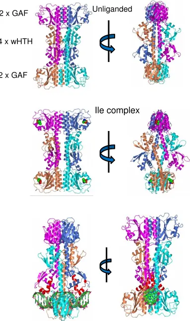
Quaternary structure in the crystal - Examination of the molecular packing in the crystals of unliganded CodY reveals that the basic building block of all three crystals is a tetramer. The tetramer in the Form C crystals and one of the tetramers in both the Form A and the Form B crystals are generated by crystallographic 2-fold symmetry. Superposition of the eight tetramers from the three crystal forms suggests that they are essentially identical within the limits of the data. As before, the following description relates to a tetramer from the Form B crystals since these crystals diffracted to the highest resolution.
4
four wHTH domains was observed in the crystal structure of CodY(168-259) determined previously (1b18) (23); these quaternary interactions were considered to be without physiological significance, in part because these domains possessed an artificial N-terminus (23).
The domain topology of the isoleucine-bound CodY(L3S) tetramer is superficially similar to that of the unliganded wild-type form, however the packing of the chains is significantly different (Fig. 2B). As shown in the right hand panels of Fig. 2, the lateral protrusion of the wHTH domains is more striking in the liganded form. This results from changes in the relative orientations of the long interdomain helices, as discussed later. For both the liganded and unliganded CodY tetramers, while the wing elements of the wHTH are on the outside of the assembly, the so-called recognition helices of the HTH (8) are nearer the centre of the tetramer and residues from the turn connecting the two helices of the HTH are in close proximity to the GAF domains of the distal subunits. As a result, in both the unliganded CodY and isoleucine-bound CodY(L3S) tetramers, DNA is sterically hindered from binding to the wHTH domains (Fig. 2C).
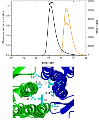
SEC-MALLS Reveals a CodY Dimer - Since CodY is thought to be a dimer (23,25), the observation of tetramers in the crystals was unexpected even though earlier gel-filtration data had shown evidence for a possible dimer-tetramer equilibrium (26). We therefore re-investigated the quaternary structure of CodY by performing a size exclusion chromatography and multi-angle laser light scattering (SEC-MALLS) analysis. In these experiments, CodY (1 mg.ml-1) samples were incubated in the presence and absence of isoleucine (10 mM), and passed down a Superdex S200 column. As the material eluted from the column, the refractive index was recorded to give a measure of protein concentration and the light scattering was recorded to allow calculation of the molar mass. As shown in Fig. 3A, CodY elutes as a single symmetric peak in these experiments and the molecular weight calculations reveal a stable species of mass 58 kDa. CodY has 259 amino acid residues and a calculated mass of 29 kDa and we conclude that the protein is dimeric under these solution conditions. The inclusion of isoleucine in the column chromatography buffer had no effect on the trace. These experiments show that CodY is a dimer at a concentration of 1 mg.ml-1 (~ 35 M) in solution. In contrast, at the 10 to 20-fold higher CodY concentrations used for crystallisation, CodY is evidently able to form tetramers. The
intracellular concentration of CodY would be ~3
M if we make a generous estimate of 10,000 CodY protomers per B. subtilis cell (30). We conclude that CodY is a dimer under physiological conditions.
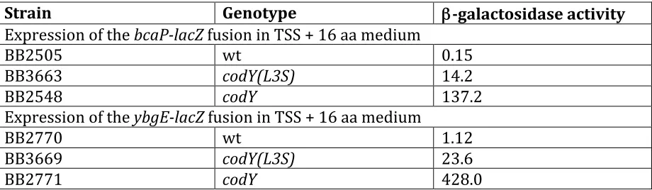
CodY(L3S) is predominantly monomeric -These SEC-MALLS experiments were repeated for the CodY(L3S) mutant. The profiles revealed a lower molecular weight species of ~ 34 kDa indicating that this protein is predominantly monomeric (Fig. 3A). Leu3 is situated in the interface between the GAF domains in the tetrameric assembly where it forms interactions with the side chains of Leu3 and Ile141 in the partner subunit that contribute to a local intermolecular hydrophobic core (Fig. 3B). Substitution of the Leu3 residues with smaller and polar serines would be expected to destabilise this interface. However, at higher protein concentrations, it is evident that CodY(L3S) assembles to form dimers as well as the tetramers that are observed in the crystals.
5
The CodY Dimer is Most Likely Formed through GAF-GAF domain Interactions - The observation of dimers of wild type CodY in the SEC-MALLS experiment prompted us to consider which of the pairs of chains in the tetramer observed in the crystal constitute the dimer observed in solution. There are three possibilities as illustrated in Fig. 3C - AB (equivalent to CD), AC (equivalent to BD) and AD (equivalent to BC). The interfacial areas in the three possible dimers are 830 Å2 (AB), 720 Å2 (AC) and 1300 Å2 (AD) in the unliganded protein and 830 Å2 (AB), 130 Å2 (AC) and 1,410 Å2 (AD) in the CodY(L3S) isoleucine complex.
We favour the AB dimer for three reasons. Firstly, the AB dimer preserves the interface observed in the structure of the isolated GAF domain dimer and the residues of this interface are well-conserved among CodY orthologues (23). Moreover, much of the surface of the protein that contributes to this interface is hydrophobic. Thus the exposure of this surface in the AC and AD dimers would be expected to be energetically unfavourable. Secondly, the L3S mutation, which disrupts CodY dimer formation, maps to the GAF-GAF interface which is unique to the AB dimer. Finally, simple modelling shows that the wHTH elements in the AB dimer would be able to bind to DNA in a conventional manner. In contrast, the AC and AD dimers would be severely sterically hindered from doing so by the presence of the distal GAF domains. We conclude that the AB dimer is most likely to be the form observed in solution by SEC-MALS.
Structural Changes Accompanying Ligand Binding - As previously stated, we were unable to obtain crystals of liganded wild-type CodY or unliganded CodY(L3S). Therefore, to explore the basis of branched-chain amino acid regulation of DNA binding, we compared the three-dimensional structures of the unliganded wild-type CodY and liganded CodY(L3S) forms, mindful of the caveats associated with the L3S mutation.
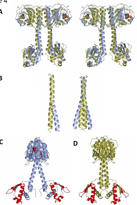
Thus the unliganded CodY and liganded CodY(L3S) dimers were superposed by least squares minimisation procedures applied to the positions of the backbone C atoms of the GAF domains from the AB dimers (Fig. 4). This gives a positional rms of 3.5 Å for 304 equivalent C atoms. However, 270 of these C atom pairs can be superposed with an rms of 1.3 Å, emphasising that very large structural changes in the GAF domains are localised to the site of isoleucine
binding (Fig. 4A). Following this superposition, it is apparent that isoleucine binding to CodY(L3S) is associated with displacements in the long helix and a significant separation of the two wHTH domains in the dimer (Fig. 4). This results from changes in the directions of the interdomain helices as they emerge from the GAF domains (Fig. 4B). The structural changes can be described as a combination of bending and twisting motions (Fig. 4A). In the dimer, the structural changes in each subunit lead to a significant splaying of the two helices and a noticeable separation of the wHTH domains. wHTH domains typically bind to DNA by aligning their recognition helices with the major groove of the DNA, with their wing elements forming contacts with the ribose-phosphate backbone and in some instances extending into the adjacent minor groove. Dimeric wHTH domain-containing DNA binding proteins often bind to recognition sequences with elements of two-fold symmetry, often referred to as palindromic, that match the two-fold symmetry of the protein itself. The distance of separation of the wHTH domains is an important determinant of DNA binding, so that the alteration in the juxtaposition of the DNA binding domains of CodY upon ligand binding would be expected to change the ability of CodY to bind to DNA. At the resolution of the structures determined here, however, it is difficult to discern how changes in the GAF domain upon ligand binding lead to changes in helix 5 and the relative arrangement of the wHTH domains.
6
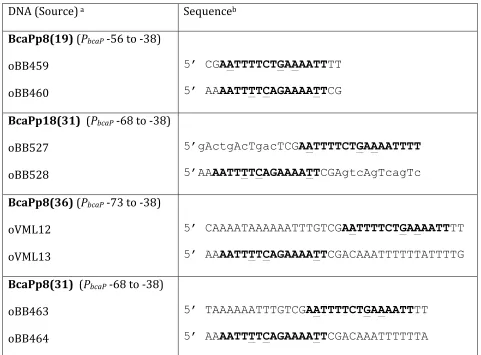
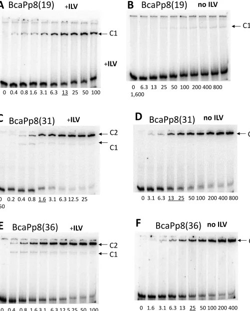
In electrophoretic mobility shift assays, wild-type CodY in the presence of a mixture of isoleucine, leucine and valine (ILV) was able to bind the BcaPp8(19) duplex, indicating that this short sequence is sufficient for protein-DNA interaction (Fig. 5A). Binding of wild-type CodY to the BcaPp8(31) or BcaPp8(36) duplexes was even more efficient, with an apparent Kd of 1.6 nM (Fig. 5C and E). The affinity of CodY for the longer duplexes is almost identical to its affinity for the extended 235-bp bcaPp8 fragment (1 nM) (31), indicating that the 31-bp duplex contains all the determinants important for binding. Interestingly, CodY formed only one type of complex (C1) with the BcaPp8(19) duplex; however, an additional, lower mobility complex (C2) was the predominant form in the presence of the BcaPp8(31) or BcaPp8(36) duplexes (Fig. 5A, C and E). The two complexes are likely to represent species with different CodY binding stoichiometries, BcaPp8(19) is presumably bound by one CodY dimer whereas BcaPp8(31) and BcaPp8(36) are each bound by two CodY dimers. In the absence of ILV, CodY formed complexes with DNA with considerably lower affinity (Fig. 5B, D and F). The loss of affinity was very dramatic in the case of the BcaPp8(19) duplex. No higher-mobility complex was observed with the longer duplexes, suggesting impaired stability of this complex in the absence of ILV (Fig. 5D and F).
Binding of CodY(L3S) to the BcaPp8(19) and BcaPp8(36) duplexes was less efficient than that of the wild-type CodY, consistent with the lower activity of the mutant protein in vivo (Fig. 6). CodY(L3S) bound very weakly to the BcaPp8(19) duplex even in the presence of ILV (Fig. 6A and B). Moreover, it did not form the higher mobility complex with the BcaPp8(36) duplex (Fig. 6C and D). The weak capacity of CodY(L3S) to form the higher mobility complex is consistent with its defect in dimer formation in solution. However, CodY(L3S) dimers are apparently stabilized if two of them are allowed to interact together, as revealed by the relatively efficient formation of the lower mobility complex with the BcaPp8(36) duplex.
The interaction of CodY with the BcaPp8(36) duplex DNA was further examined using SEC-MALLS. As shown in Fig. 7, the DNA duplex eluted as a single symmetrical peak from the Superdex S200 column with an Mr value calculated from the light scattering data of 23 kDa. This is consistent with its molar mass of 22 kDa. Similarly, in the absence of DNA, the
experimentally derived Mw of CodY is 58 kDa, consistent with a dimer. In the two subsequent experiments, CodY was incubated with BcaPp8(36) in the presence and absence of 10 mM isoleucine. In the absence of isoleucine, we observed a somewhat asymmetric peak in the differential refractive index profile centred at 28 mins (Fig. 7B), a lower retention time than that of CodY alone. The molar mass associated with this species is ~70 kDa. In contrast, the presence of isoleucine caused a significant reduction in the retention time of the principal species present and a significant rise in its molecular mass to ~130 kDa. We interpret the 130 kDa species to be a complex formed between four molecules of CodY and one BcaPp8(36) duplex (138 kDa). We observed a similar ~130 kDa species when analysing a mixture of CodY and a 32 base pair duplex derived from the CodY-regulated yurP (frlB) promoter (20) in the presence of isoleucine but not in its absence (data not shown).
The 70 kDa species observed in the absence of isoleucine has a higher mass than that expected for a CodY dimer (58 kDa) but lower than that expected for a dimer of CodY bound to one BcaPp8(36) duplex (81 kDa). It can be seen that the peak on the chromatogram for this complex overlaps with the leading edge of the peak for the free DNA trace. The presence of the uncomplexed DNA would lower the weight average molecular weight of the eluting species calculated by MALLS, consistent with the presence of a higher molecular weight protein-DNA complex such as CodY2-BcaPp8(36). The EMSA experiments suggest however that this species would not be observed and it is more likely that the profile reflects the extensive dissociation of CodY4-BcaPp8(36) complexes accompanying chromatography in the absence of Ile.
These results confirm that the C1 species observed in the EMSA experiments represent CodY2 complexes with DNA and that the lower mobility C2 species are complexes of CodY4 and BcaPp8(31) or BcaPp8(36), implying the presence of two CodY2 binding subsites on these longer duplexes. Therefore, one might expect that inactivation of one of the subsites within such a DNA duplex would reduce binding of one of the CodY dimers resulting in the formation of a DNA complex containing only one CodY dimer. Indeed, when
we introduced multiple mutations in the 5’ part
7
reduction of C2 complex formation and the emergence of C1 as the predominant complex with CodY (Fig. 5G).
DISCUSSION
CodY is a Dimer which Binds Cooperatively to bcaP DNA - The structures of full length CodY from B. subtilis determined here reveal the effector binding domain and the DNA binding domain as discrete entities separated by a helical linker. In the crystals, four CodY protomers are assembled into an unusual tetramer in which four wHTH domains are sandwiched between two GAF domain dimers. For the unliganded form in particular, the tetramer is compact with extensive subunit interfaces. From within this tetramer structure, there is no possibility that the wHTH domains of CodY could bind to DNA in a conventional manner, because the GAF domains of partner subunits in the tetramer occupy a substantial volume that would be taken up by the DNA duplex. (Fig. 2C). We considered therefore that this tetrameric form might represent an inactive state of CodY that prevails in the absence of nutritional signals. However, we found no evidence for CodY tetramers in SEC-MALLS experiments, which conclusively showed that CodY is a dimer even at protein concentrations higher than those found in vivo (Fig. 3A). Furthermore, unliganded CodY can bind to DNA, albeit with lower affinity than the BCAA-liganded protein (Fig. 5). Thus, there is no evidence to suggest that the CodY tetramers observed here by crystallography are physiologically relevant. There are many cases where plausible, stable interfaces between molecules in crystals prove to have no functional significance, including examples from studies of proteins involved in gene regulation in Bacillus (32) (33).
An interesting and important finding from this work is the establishment that CodY forms alternate complexes with short DNA fragments derived from the bcaP promoter. With the shorter 19 bp duplex, CodY forms a single complex. With the 31 bp and 36 bp duplexes, it forms two complexes as shown schematically in Fig. 8A. The first, C1, has a mobility similar to that of the complex formed with the 19-mer, while the second, predominant complex, C2, has lower electrophoretic mobility consistent with a larger size (Fig. 5). SEC-MALLS analysis of CodY mixed with BcaPp8(36) in the presence of isoleucine showed the formation of a complex
consistent with four CodY protomers bound to a single duplex (Fig. 7). Collectively, our data suggest that C1 is CodY2-BcaPp8 with one CodY dimer bound per duplex and that C2 is CodY4-BcaPp8 with two dimers per duplex. Interestingly, in the absence of branched chain amino acids, the C1 complex appears to be much less stable for BcaPp8(19) and is not discernible at all for BcaPp8(31) or BcaPp8(36) (Fig. 5D). These data are consistent with cooperative binding of CodY to the longer duplexes with the affinity of the second dimer for the DNA augmented by the binding of the first dimer. Cooperativity, exhibited in the presence and the absence of ILV, may arise because the binding of the first CodY dimer to the DNA induces a conformational change in the duplex such that the second dimer can bind more readily. In this regard, the bcaP promoter fragments used here have a high bending propensity (34). Alternatively, the binding of the first dimer may help in the recruitment of the second dimer by contributing protein-protein interactions to supplement the protein-DNA interactions (Fig. 8A). In the case of CodY, these interdimer interactions may be contributed either by the GAF domains or, as shown schematically in Fig. 8A, by the wHTH domains.
The exact role of protein-protein interactions in binding of two dimers and the precise contribution of each of the two subsites (Fig. 8A) to CodY binding remain unknown. It is also not known whether all CodY-binding sites are composed of two (or more) subsites; the exact stoichiometry of CodY binding to all CodY-binding sites may be not identical.
Structural Implications for DNA Binding - Comparisons of the structures of unliganded CodY and effector-bound CodY(L3S) reveal striking differences in the relative positions of the wHTH domains (Fig. 4). These differences could reflect the effect of Ile binding on CodY conformation which would lead us to conclude that the splayed conformation of CodY is more conducive to DNA binding, since Ile-bound CodY has a higher affinity for all bona fide binding sites tested to date. This conclusion relies, however, on the assumption that the mode of DNA binding is same for CodY(L3S) and wild type CodY bound to Ile.
8
transcription factors (TFs) whose structures have been solved in complex with DNA. The most closely similar such structure is of the quinone sensing response regulator QsrR from S. aureus whose subunits can be overlaid on the wHTH domain of CodY with an rms of 2.9 Å for 85 aligned residues giving a Z-score of 4.1 (35). In the complex of QsrR with a 17 bp duplex, the respective recognition helices bind in consecutive major grooves of the DNA. These
-helices are related by a 28 Å translation and a 70° rotation (Fig. 9A). Binding to the operator DNA in this system is reversed upon covalent attachment of menadione to Cys5 of QsrR, which leads to a 10° rotation and 9 Å separation of the subunits (35). Another structurally well-characterised example is FadR of E. coli which regulates genes encoding enzymes of fatty acid metabolism. In the structure of its complex with a 19 bp fadB operator DNA, the two recognition helices of the wHTH domains are closely abutting (their helix axes can be superposed following a 9 Å translation and a 30° rotation) in the major groove of the DNA giving rise to a compact protein DNA complex (Fig. 9B). Binding of the effector, myristoyl-CoA, to the N-terminal domain leads to separation of the recognition helices by a further 7 Å, loss of DNA binding, and relief of repression (36). In IscR, which regulates genes involved in iron-sulphur cluster biogenesis, longer DNA recognition sequences spanning 26 to 29 bp were used in crystallisation (37) (38) (Fig. 9C). In these complexes, the wHTH domains are separated by long C-terminal helices which mediate dimer formation such that the recognition helices of the wHTH in the respective subunits are related by a displacement of 36 Å and a 71° rotation. In this system, the iron-sulphur cluster effector binds within the wHTH itself so as to change the promoter specificity of the repressor.
To facilitate comparison, we superimposed the wHTH domains of one of the chains (chain A) of unliganded CodY and Ile-bound CodY(L3S) onto one of the corresponding domains of QsrR, FadR and IscR in their DNA complexes and observed the juxtaposition of the recognition helix in the partner wHTH domains and the DNA (Fig. 9D). This simple modelling suggests that the unliganded CodY dimer, in which the recognition helices from each chain are related by a 25 Å translation and a 40° rotation, could bind to a DNA duplex of ~20 bp. For the
Ile-bound CodY(L3S) structure, where splaying increases the translational and rotational separation of the recognition helices to 34 Å and 45°, a longer DNA recognition sequence of ~30 base pairs would be needed.
This analysis indicates that 19 bp is just sufficient to bind a CodY dimer in the more compact conformation we observe in the unliganded CodY crystals. Meanwhile, a pair of CodY dimers in the splayed conformation could bind to the 31-mer (BcaPp8(31)) and 36-mer (BcaPp8(36)) DNA duplexes only if the binding sites for each dimer are significantly overlapping as depicted in Fig. 8A and modelled in Fig. 8C. Such a mode of binding has been proposed previously (39). However, the modelling predicts that the splayed CodY dimer would be unable to bind to the 19-mer, BcaPp8(19). This is consistent with the poor ability of CodY(L3S) to bind the BcaPp8(19) duplex, but contrary to the experimental data obtained with the wild-type CodY. One explanation for this is that the splayed conformation of CodY obtained with CodY(L3S) is specific to the mutant form of the protein. Alternatively, the CodY dimer may be able to engage the 19-mer duplex using just a single wHTH domain.
Concluding remarks - Crystals of CodY grew irreproducibly perhaps due to inherent flexibility in the CodY dimer, arising from relative motions of the wHTH domains relative to each other and the GAF domains. At low frequency, tetramers are evidently assembled in
which these domain motions are ‘frozen out’ by
dimer-dimer interactions and this overcomes the barrier to lattice formation. A precedent for this is calmodulin, where early structures revealed independent calcium binding domains connected by a long -helix in a dumb-bell arrangement (40). Later structures of calmodulin-target protein complexes showed altered conformations of the linker region and a spectrum of more compact relative domain arrangements (41) (42).
9
electrostatically the compact helical conformation in this region of the subunit, possibly giving rise to a more extended conformation in solution. Interestingly, it has been shown for some promoters that the extent of CodY binding to DNA and CodY activation by isoleucine is pH-dependent (39). This behaviour may be associated with the titration of one or more of these glutamates. Regardless, this flexibility could allow the wHTH domains to be brought into contact with the GAF domains within the dimer so that they are able to sense directly the conformational changes brought about by BCAA binding as shown schematically in Fig. 8B. This would provide a basis for explaining the puzzling absence in the crystal structures of an obvious route for transducing the substantial structural changes upon ligand binding to the GAF domain, into structural alterations of the wHTH domains. A rigorous elucidation of the structural basis of (i) effector control of CodY activity and (ii) CodY-recognition of the 15-bp consensus DNA binding motif, with its considerable degeneracy but clear two-fold symmetry, awaits the determination of structures of CodY-DNA complexes.
While this manuscript was under review, crystal structures of CodY from B. cereus (BcCodY) and S. aureus (SaCodY) were reported (43). The domain arrangement in these orthologous proteins is similar to that observed in CodY from B. subtilis and tetramers similar to those reported here were observed for BcCodY. Curiously, the BCAA binding site in unliganded BcCodY resembles that of the liganded CodY from B. subtilis. Most interestingly, the SaCodY structure contains GTP which is bound at the junction of the long helix and the GAF domain in a dimeric form of the protein. The authors combine their structures to propose a model in which tetramers represent a physiologically inactive state of CodY. This model is yet to be rigorously tested and is unlikely to apply to CodY from B. subtilis which is clearly dimeric under conditions found in the cell.
EXPERIMENTAL PROCEDURES
Bacterial strains and culture media -The B. subtilis strains used in this study are described in the text and were all derivatives of strain SMY (44) and grown at 37oC in TSS - 0.5% glucose - 0.2% NH4Cl minimal medium supplemented with a mixture of 16 amino acids (45,46). The
mixture contained all of the amino acids commonly found in proteins except for glutamine, asparagine, histidine, and tyrosine; the concentrations of isoleucine, leucine, and valine were 200 µg/ml each. DS nutrient broth medium with addition of agar was used for growth of bacteria on plates(46). Escherichia coli strain JM107 (47) was used for isolation of plasmids and was grown in L broth or on L agar plates (48). The following antibiotics were used when appropriate: tetracycline, 15 µg/ml; spectinomycin, 50 µg/ml; or the combination of erythromycin, 0.5 µg/ml, and lincomycin, 12.5 µg/ml, for B. subtilis strains; ampicillin, 50 µg/ml, for E. coli strains.
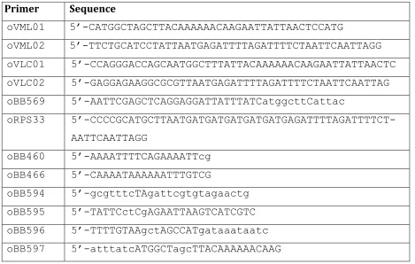
DNA manipulations - Methods for general DNA manipulation, transformation, DNA sequencing, gel shift assays, and sequence analysis were as previously described (20,49). Oligonucleotides used in this work are described in Table 2. Chromosomal DNA of B. subtilis strain SMY or plasmid DNA was used as template for PCR as appropriate. All cloned PCR-generated fragments were verified by sequencing.
CodY expression and purification - CodY was initially produced using the pBAD30 (50) derivative plasmid, pKT1(26), encoding residues Ala2-His259 of CodY with a C-terminal pentahistidine tag, with the protein purified as described previously(26). This protein was also used for the EMSA studies together with CodY(L3S)-His5 produced using the plasmid pBB1737, which was created by cloning the PCR fragment, synthesized using oligonucleotides oBB569 and oRPS33, into pBAD30 as described previously(27).
10
for 24 hours at 30°C before harvesting by centrifugation, resuspension in 20 mM sodium phosphate buffer pH 7.5, 0.5 M NaCl, 30 mM imidazole (Buffer A) supplemented with 1 mM 4-(2-aminoethyl) benzene sulphonyl fluoride (AEBSF) and lysis by sonication. Following clarification by centrifugation, the supernatant was loaded onto a 1 ml HisTrap HP column that had been pre-charged with Ni2+ ions and equilibrated in Buffer A. The column was washed extensively with Buffer A and developed with a 30 mM – 0.5 M linear imidazole gradient in Buffer A. After the nickel chelation chromatography step, the protein was exchanged into 20 mM Tris-HCl buffer pH 7.5 with 50 mM NaCl and treated overnight with 1 unit/mg thrombin (BD Biosciences) to remove the polyhistidine tag and generate CodY protein with a residual tripeptide tag (GlySerHis) at the amino terminus and a Leucine 3 to Serine substitution. This CodY(L3S) protein was further purified by gel filtration chromatography on a Superdex S200 16/60 column.
A third construct was generated to direct the production of CodY, without the Leucine-3 substitution and with an N-terminal human rhinovirus 3C protease cleavable polyhisitdine tag. Following polymerase chain reaction using the primers oVLC01 and oVLC02 (Table 4) and the pBAD30-CodY template, the amplified DNA was inserted into the vector pET-YSBLIC3C using ligation independent cloning methods (51), and E. coli NovaBlue cells harbouring a suitable recombinant plasmid were identified among kanamycin-resistant transformants. Following sequencing to confirm its authenticity, pET-YSBLIC-BsCodY was introduced in E. coli BL21(DE3) for protein production. The recombinant protein was purified as described above except that the polyhistidine tag was removed by overnight cleavage with human rhinovirus 3C protease. A second nickel column was run to separate the cleaved protein from the uncleaved protein and the cleaved tag. The flow-through fraction was collected, buffer exchanged into 20 mM Tris-HCl, 100 mM NaCl, and loaded onto a heparin column which was developed in this buffer over a 100-500 mM NaCl.
The protein was then fractionated by gel filtration on a Superdex 200 column and concentrated by centrifugal membrane filtration using a 10 kDa molecular weight cutoff membrane filter (AminconR), to yield CodY with an N-terminal Gly-Pro-Ala tag in 20 mM Tris-HCl, 100 mM NaCl.
Crystallisation - Form A crystals of unliganded CodY were grown in hanging drops formed by mixing 1 l of 10-20 mg.ml-1 CodY with 1 l of well solution containing 1 M sodium citrate pH 6.0 and 5 % glycerol. Form B crystals were similarly prepared by mixing 1 l of 10-20 mg.ml-1 CodY containing 10 mM GTP with 1 l of well solution containing 0.1 M sodium citrate pH 5.6, 1.7 M lithium sulphate and 3 % dioxane. Finally, the Form C crystals were prepared by mixing 1 l of 10-20 mg.ml-1 CodY containing 5
mM GTP--S and 2 mM
Tris(2-carboxyethyl)phosphine (TCEP) with 1 l of well solution containing 0.1 M sodium citrate pH 5.6, 20 mM Tris-HCl pH 7.5, 35 % ammonium sulphate.
Crystals of CodY(L3S) in complex with isoleucine were grown in hanging drops prepared by mixing equal volumes of 18 mg.ml-1 protein containing 20 mM isoleucine with a well solution which contained 15 % polyethylene glycol 5000, 5 % tacsimate in 0.1 M HEPES buffer pH7.5.
Data Collection, Structure Solution and Refinement - After transfer to solutions of mother liquor containing a suitable cryoprotectant, a preliminary X-ray analysis of crystals of CodY was carried out in-house using K radiation from a copper rotating anode. Diffraction was initially very weak, but the quality of the diffraction data was significantly improved following cycles of crystal reannealing, that is, by repeated thawing in cryoprotectant and cryo-cooling of the crystals. Full three-dimensional data were collected from the strongest diffracting crystals on beamlines at the Diamond Light Source (Harwell, UK) and the European Synchrotron Radiation Source (Grenoble, France) as detailed on Table 1. The data were processed and scaled in HKL2000 (52). As shown in Table 1, three crystal forms (A, B and C) of unliganded CodY were obtained with 14, 10 and 2 molecules in the asymmetric unit respectively.
11
the basis of whether they gave rise to GAF domain dimers reminiscent of that contained in the crystal structure of the search model. Suitable solutions were identified and fixed and a subsequent search was carried out using the wHTH domain (coordinate set; 1B0L).
The resulting structure was refined using REFMAC (55) with non-crystallographic symmetry restraints applied. This revealed electron density for the ‘missing’ residues which form connecting helices. These were built manually in Coot (56). From the refined coordinates of this Form B crystal structure, a dimer was used in molecular replacement calculations to solve the Form A and Form C crystal structures.
Later the structure of CodY(L3S)-Ile was solved similarly but using the coordinate set 2B18 representing the GAF domain with bound isoleucine followed by refinement using REFMAC (55). Figures of the structures of CodY were made using CCP4MG (57). The coordinates and structure factors for the models reported in this work have been deposited in the protein data bank with PDBIDs 5LOO, 5LNH and 5LOJ for the unliganded CodY crystal forms with 16, 10 and 2 molecules per asymmetric unit respectively and 5LOE for CodY(L3S) in complex with isoleucine.
Annealing DNA oligonucleotides – Oligonucleotides, purified by HPLC, were purchased from Eurofins MWG Operon. Complementary oligonucleotides were resuspended in deionised water and incubated at a 1:1 molar ratio at a concentration of 50 M in a buffer composed of 25 mM Tris pH 8.5, 200 mM NaCl, 1 mM DTT and 0.5 mM EDTA. The
oligonucleotides were heated to 90˚C for 5
minutes to ensure full denaturation, followed by slow cooling to room temperature.
Labelling of DNA fragments - The BcaPp8(19), BcaPp8(31) and BcaPp18(31) DNA duplexes were generated by annealing corresponding pairs of oligonucleotides (Table 3); the oligonucleotide mixtures in 10 mM Tris-Cl (7.5) - 50 mM KCl were heated for 5 min in a boiling water bath and allowed to cool to room temperature. The BcaPp8(36) DNA duplex was synthesized by PCR using oligonucleotides oBB466 and oBB460 as primers and pBB1622 as template. In each case, one of the oligonucleotides was pre-labeled using T4 polynucleotide kinase and [-32P]-ATP.
Gel shift experiments - Incubation of CodY with the 32P-labelled DNA duplexes was performed in 20 mM Tris-Cl (pH 8.0), 50 mM KCl, 2 mM MgCl2, 5% glycerol, 0.5 mM EDTA, 1 mM DTT, 0.05% Nonidet P, 40-25 µg/ml salmon sperm DNA as a binding buffer. Samples (10 µl) containing varying amounts of CodY and less than 1 fmole of DNA were incubated for 16 min at room temperature and separated on 8% non-denaturing 50 mM Tris, 384 mM glycine, 1 mM EDTA polyacrylamide gels in 35 mM HEPES, 43 mM imidazole buffer. In some experiments, 10 mM ILV was added to the incubation mixture, gel, and electrophoresis buffer. The gels were dried, and the radioactive bands were detected and quantified using storage screens, PhosphorImager, and the ImageQuant software (GE Healthcare).
SEC-MALLS - A Wyatt Dawn HELEOS-II 18-angle light scattering detector and Wyatt Optilab rEX refractive index monitor linked to a Shimadzu HPLC system and SPD20A UV/Vis detector was used for SEC-MALLS. A Superdex S200 HR 10/30 size exclusion column was attached to the HPLC and equilibrated in a running buffer consisting of 50 mM TrisHCl pH 7.5, 150 mM NaCl in the presence or absence of 10 mM isoleucine. An SIL-20A Autosampler was
used to inject 100 l samples of 1 mg.ml-1 CodY samples, 19 µM DNA or mixtures of the two. Data were analysed with Astra software using a dn/dc value of 0.186 for protein and protein-DNA complexes and 0.168 for DNA (58,59)
Introduction of the codY(L3S) mutation in the B. subtilis chromosome -A PCR fragment containing the full-length codY (L3S) gene and the flanking sequences was synthesized by two-step overlapping PCR. In the first two-step, the PCR
product containing the 5’-part of the codY gene was synthesized by using oligonucleotide oBB594 as the forward primer and mutagenic oligonucleotide oBB596 as the reverse primer. In a similar manner, the PCR product containing the
12
The spliced PCR product was digested with XbaI and XhoI and cloned in an integrative plasmid pBB1579 (bgaB neo)(60), creating pBB1756 [codY(L3S) bgaB neo]. The latter plasmid was introduced by a single-crossover, homologous recombination event into the bcaP chromosomal locus of strain SMY. White Neos colonies indicating excision of pBB1756 from the chromosome were searched for on plates containing XGal (5bromo4chloro3indolyl
-D-galactopyranoside), the coloured substrate of bgaB-encoded -galactosidase. Multiple colonies were tested for acquisition of the codY(L3S) mutation by sequencing PCR products from the chromosomal codY allele.
Enzyme assays - -Galactosidase activity was determined as described previously (61).
Acknowledgements: This work was supported by Grant GM042219 from the US National Institute of
General Medical Sciences and by Grants from the BBSRC (BBS/B1213X) and Wellcome Trust (082829/Z/07/Z). We are grateful to Dr Johan Turkenburg and staff at the ESRF, Grenoble, France and DIAMOND, Harwell, UK for help with data collection.
Conflict of interest: The authors declare that they have no conflicts of interest.
Author Contributions: ALS and AJW conceived the study and coordinated the work. VML
determined the crystal structures with expert input from AL. EVB and VML made constructs for expression of purified proteins with EVB then growing the crystals and VLY performing the biophysical experiments. BRB performed the EMSA experiments and constructed and analysed strains for in vivo analyses. The paper was written by AJW with input from all authors.
REFERENCES
1.
Slack, F., Serror, P., Joyce, E., and Sonenshein, A. L. (1995) A gene required for
nutritional repression of the Bacillus subtilis dipeptide permease operon Mol.
Microbiol. 15 689-702
2.
Molle, V., Nakaura, Y., Shivers, R. P., Yamaguchi, H., Losick, R., Fujita, Y., and
Sonenshein, A. L. (2003) Additional targets of the Bacillus subtilis global regulator
CodY identified by chromatin immunoprecipitation and genomw-wide transcript
analysis. J. Bacteriol. 185, 1911-1922
3.
Brinsmade, S. R., Alexander, E. L., Livny, J., Stettner, A. I., Segre, D., Rhee, K. Y.,
and Sonenshein, A. L. (2014) Hierarchical expression of genes controlled by the
Bacillus subtilis global regulatory protein CodY. Proc. Natl. Acad. Sci. U. S. A. 111,
8227-8232
4.
Pohl, K., Francois, P., Stenz, L., Schlink, F., Geiger, T., Herbert, S., Goerke, C.,
Schrenzel, J., and Wolz, C. (2009) CodY in Staphylococcus aureus: a regulatory link
between metabolism and virulence gene expression. J. Bacteriol. 191, 2953-2963
5.
Majerczyk, C. D., Sadykov, M. R., Luong, T. T., Lee, C., Somerville, G. A., and
Sonenshein, A. L. (2008) Staphylococcus aureus CodY negatively regulates virulence
gene expression. J. Bacteriol. 190, 2257-2265
6.
Hendriksen, W. T., Bootsma, H. J., Estevao, S., Hoogenboezem, T., de Jong, A., de
Groot, R., Kuipers, O. P., and Hermans, P. W. (2008) CodY of Streptococcus
pneumoniae: link between nutritional gene regulation and colonization. J. Bacteriol.
190, 590-601
13
8.
Lobel, L., Sigal, N., Borovok, I., Ruppin, E., and Herskovits, A. A. (2012) Integrative
genomic analysis identifies isoleucine and CodY as regulators of Listeria
monocytogenes virulence. PLoS Genet. 8, e1002887
9.
Bennett, H. J., Pearce, D. M., Gelenn, S., Taylor, C. M., Kuhn, M., Sonenshein, A. L.,
Andrew, P. W., and Roberts, I. S. (2007) Characterisation of relA and codY mutants
of Listeria monocytogenes: identification of the CodY regulon and its role in
virulence. Mol. Microbiol. 63, 1453-1467
10.
van Schaik, W., Chateau, A., Dillies, M. A., Coppee, J. Y., Sonenshein, A. L., and
Fouet, A. (2009) The global regulator CodY regulates toxin gene expression in
Bacillus anthracis and is required for full virulence. Infect. Immun. 77, 4437-4445
11.
Dineen SS, McBride, S., and Sonenshein, A. (2010) Integration of metabolism and
virulence by Clostridium difficile CodY. J. Bacteriol. 192, 5350-5362
12.
Sonenshein, A. L. (2005) CodY, a global regulator of stationary phase and virulence
in Gram-positive bacteria Curr. Opin. Microbiol. 8 203-207
13.
Shivers, R. P., and Sonenshein, A. L. (2004) Activation of the Bacillus subtilis global
regulator CodY by direct interaction with branched-chain amino acids. Mol.
Microbiol. 53 599-611
14.
Ratnayake-Lecamwasam, M., Seror, P., Wong, K. W., and Sonenshein, A. L. (2001)
Bacillus subtilis CodY represses early stationary-phase genes by sensing GTP levels.
Genes Dev. 15, 1093-1103
15.
Belitsky, B. R., and Sonenshein, A. L. (2011) Roadblock repression of transcription
by Bacillus subtilis CodY. J. Mol. Biol. 411, 729-743
16.
Shivers, R. P., Dineen, S. S., and Sonenshein, A. L. (2006) Positive regulation of
Bacillus subtilis ackA by CodY and CcpA: establishing a potential hierarchy in
carbon flow. Mol. Microbiol. 62, 811-822
17.
Preis, H., Eckart, R. A., Gudipati, R. K., Heidrich, N., and Brantl, S. (2009) CodY
activates transcription of a small RNA in Bacillus subtilis. J. Bacteriol. 191,
5446-5457
18.
den Hengst, C. D., van Hijum, S. A., Geurts, J. M., Nauta, A., Kok, J., and Kuipers,
O. P. (2005) The Lactococcus lactis CodY regulon: identification of a conserved
cis-regulatory element. J. Biol. Chem. 280, 34332-34342
19.
Guedon, E., Sperandio, B., Pons, N., Ehrlich, S. D., and Renault, P. (2005) Overall
control of nitrogen metabolism in Lactococcus lactis by CodY, and possible models
for CodY regulation in Firmicutes. Microbiology 151, 3895-3909
20.
Belitsky, B. R., and Sonenshein, A. L. (2008) Genetic and biochemical analysis of
CodY-binding sites in Bacillus subtilis. J. Bacteriol. 190, 1224-1236
21.
Nicolas, P., Mader, U., Dervyn, E., Rochat, T., Leduc, A., Pigeonneau, N., Bidnenko,
E., Marchadier, E., Hoebeke, M., Aymerich, S., Becher, D., Bisicchia, P., Botella, E.,
Delumeau, O., Doherty, G., Denham, E. L., Fogg, M. J., Fromion, V., Goelzer, A.,
Hansen, A., Hartig, E., Harwood, C. R., Homuth, G., Jarmer, H., Jules, M., Klipp, E.,
Le Chat, L., Lecointe, F., Lewis, P., Liebermeister, W., March, A., Mars, R. A.,
Nannapaneni, P., Noone, D., Pohl, S., Rinn, B., Rugheimer, F., Sappa, P. K., Samson,
F., Schaffer, M., Schwikowski, B., Steil, L., Stulke, J., Wiegert, T., Devine, K. M.,
Wilkinson, A. J., van Dijl, J. M., Hecker, M., Volker, U., Bessieres, P., and Noirot, P.
(2012) Condition-dependent transcriptome reveals high-level regulatory architecture
in Bacillus subtilis. Science 335, 1103-1106
14
23.
Levdikov, V. M., Blagova, E., Joseph, P., Sonenshein, A. L. and Wilkinson, A. J.
(2006) The structure of CodY, a GTP- and isoleucine-responsive regulator of
stationary phase and virulence in Gram-positive bacteria. J. Biol. Chem. 281,
11366-11373
24.
Aravind, L., and Ponting, C. P. (1997) The GAF domain: an evolutionary link
between diverse phototransducing proteins. Trends in biochemical sciences 22,
458-459
25.
Levdikov, V. M., Blagova, E., Colledge, V. L., Lebedev, A. A., Williamson, D. C.,
Sonenshein, A. L., and Wilkinson, A. J. (2009) Structural rearrangement
accompanying ligand binding in the GAF domain of CodY from Bacillus subtilis. J.
Mol. Biol. 390, 1007-1018
26.
Blagova, E. V., Levdikov, V. M., Tachikawa, K., Sonenshein, A. L., and Wilkinson,
A. J. (2003) Crystallization of the GTP-dependent transcriptional regulator CodY
from Bacillus subtilis. Acta Crystallogr. D Biol. Crystallogr. 59, 155-157
27.
Villapakkam, A. C., Handke, L. D., Belitsky, B. R., Levdikov, V. M., Wilkinson, A.
J., and Sonenshein, A. L. (2009) Genetic and biochemical analysis of the interaction
of Bacillus subtilis CodY with branched-chain amino acids. J. Bacteriol. 191,
6865-6876
28.
Belitsky, B. R., Brinsmade, S. R., and Sonenshein, A. L. (2015) Intermediate Levels
of Bacillus subtilis CodY Activity Are Required for Derepression of the
Branched-Chain Amino Acid Permease, BraB. PLoS Genet. 11, e1005600
29.
Belitsky, B. R. (2015) Role of branched-chain amino acid transport in Bacillus
subtilis CodY activity. J. Bacteriol. 197, 1330-1338
30.
Maass, S., Wachlin, G., Bernhardt, J., Eymann, C., Fromion, V., Riedel, K., Becher,
D., and Hecker, M. (2014) Highly precise quantification of protein molecules per cell
during stress and starvation responses in Bacillus subtilis. Mol. Cell. Proteomics 13,
2260-2276
31.
Belitsky, B. R., and Sonenshein, A. L. (2011) Contributions of multiple binding sites
and effector-independent binding to CodY-mediated regulation in Bacillus subtilis. J.
Bacteriol. 193, 473-484
32.
Lewis, R. J., Muchova, K., Brannigan, J. A., Barak, I., Leonard, G., and Wilkinson,
A. J. (2000) Domain swapping in the sporulation response regulator Spo0A. J. Mol.
Biol. 297, 757-770
33.
Levdikov, V. M., Blagova, E. V., Rawlings, A. E., Jameson, K., Tunaley, J., Hart, D.
J., Barak, I., and Wilkinson, A. J. (2012) Structure of the phosphatase domain of the
cell fate determinant SpoIIE from Bacillus subtilis. J. Mol. Biol. 415, 343-358
34.
Travers, A. A., and Klug, A. (1987) The bending of DNA in nucleosomes and its
wider implications. Philos. Trans. R. Soc. Lond. B Biol. Sci. 317, 537-561
35.
Ji, Q., Zhang, L., Jones, M. B., Sun, F., Deng, X., Liang, H., Cho, H., Brugarolas, P.,
Gao, Y. N., Peterson, S. N., Lan, L., Bae, T., and He, C. (2013) Molecular mechanism
of quinone signaling mediated through S-quinonization of a YodB family repressor
QsrR. Proc. Natl. Acad. Sci. U. S. A. 110, 5010-5015
36.
van Aalten, D. M., DiRusso, C. C., and Knudsen, J. (2001) The structural basis of
acyl coenzyme A-dependent regulation of the transcription factor FadR. EMBO J. 20,
2041-2050
37.
Rajagopalan, S., Teter, S. J., Zwart, P. H., Brennan, R. G., Phillips, K. J., and Kiley,
P. J. (2013) Studies of IscR reveal a unique mechanism for metal-dependent
15
38.
Santos, J. A., Alonso-Garcia, N., Macedo-Ribeiro, S., and Pereira, P. J. (2014) The
unique regulation of iron-sulfur cluster biogenesis in a Gram-positive bacterium.
Proc. Natl. Acad. Sci. U. S. A. 111, E2251-2260
39.
Wray, L. V., Jr., and Fisher, S. H. (2011) Bacillus subtilis CodY operators contain
overlapping CodY binding sites. J. Bacteriol. 193, 4841-4848
40.
Babu, Y. S., Bugg, C. E., and Cook, W. J. (1988) Structure of calmodulin refined at
2.2 A resolution. J. Mol. Biol. 204, 191-204
41.
Drum, C. L., Yan, S. Z., Bard, J., Shen, Y. Q., Lu, D., Soelaiman, S., Grabarek, Z.,
Bohm, A., and Tang, W. J. (2002) Structural basis for the activation of anthrax
adenylyl cyclase exotoxin by calmodulin. Nature 415, 396-402
42.
Wilkinson, A. J. (1996) Accommodating structurally diverse peptides in proteins.
Chem. Biol. 3, 519-524
43.
Han, A. R., Kang, H. R., Son, J., Kwon, D. H., Kim, S., Lee, W. C., Song, H. K.,
Song, M. J., and Hwang, K. Y. (2016) The structure of the pleiotropic transcription
regulator CodY provides insight into its GTP-sensing mechanism. Nucleic acids
research 44, 9483-9493
44.
Zeigler, D. R., Pragai, Z., Rodriguez, S., Chevreux, B., Muffler, A., Albert, T., Bai,
R., Wyss, M., and Perkins, J. B. (2008) The origins of 168, W23, and other Bacillus
subtilis legacy strains. J. Bacteriol. 190, 6983-6995
45.
Atkinson, M. R., Wray, L. V., Jr., and Fisher, S. H. (1990) Regulation of histidine and
proline degradation enzymes by amino acid availability in Bacillus subtilis. J.
Bacteriol. 172, 4758-4765
46.
Fouet, A., and Sonenshein, A. L. (1990) A target for carbon source-dependent
negative regulation of the citB promoter of Bacillus subtilis. J. Bacteriol. 172,
835-844
47.
Yanisch-Perron, C., Vieira, J., and Messing, J. (1985) Improved M13 phage cloning
vectors and host strains: nucleotide sequences of the M13mp18 and pUC19 vectors.
Gene 33, 103-119
48.
Miller, J. H. (1972). Experiments in Molecular Genetics, Cold Spring Harbor
Laboratory, Cold Spring Harbor, N.Y
49.
Sambrook, J., Fritsch, E. F., and Maniatis, T. J. (1989). Molecular Cloning: a
Laboratory Manual, Cold Spring Harbor Laboratory, Cold Spring Harbor, N.Y
50.
Guzman, L. M., Belin, D., Carson, M. J., and Beckwith, J. (1995) Tight regulation,
modulation, and high-level expression by vectors containing the arabinose PBAD
promoter. J. Bacteriol. 177, 4121-4130
51.
Fogg, M. J., and Wilkinson, A. J. (2008) Higher-throughput approaches to
crystallization and crystal structure determination. Biochem. Soc. Trans. 36, 771-775
52.
Otwinowski, Z., and Minor, W. (1997) Processing of X-ray diffraction data collected
in oscillation mode. Method Enzymol 276, 307-326
53.
Lebedev, A. A., and Isupov, M. N. (2014) Space-group and origin ambiguity in
macromolecular structures with pseudo-symmetry and its treatment with the program
Zanuda. Acta Crystallogr. D Biol. Crystallogr. 70, 2430-2443
54.
Vagin, A., and Teplyakov, A. (2010) Molecular replacement with MOLREP. Acta
Crystallogr. D Biol. Crystallogr. 66, 22-25
55.
Murshudov, G. N., Vagin, A. A., and Dodson, E. J. (1997) Refinement of
macromolecular structures by the maximum-likelihood method. Acta Crystallogr. D
Biol. Crystallogr. 53, 240-255
16
57.
McNicholas, S., Potterton, E., Wilson, K. S., and Noble, M. E. (2011) Presenting your
structures: the CCP4mg molecular-graphics software. Acta Crystallogr. D Biol.
Crystallogr. 67, 386-394
58.
Wen, J., and Arakawa, T. (2000) Refractive index of proteins in aqueous sodium
chloride. Anal. Biochem. 280, 327-329
59.
Nicolai, T., van Dijk, T. N., van Dijk, J. A., and Smit, J. A. (1987) Molar mass
characterization of DNA fragments by gel permeation chromatography using a
low-angle laser light-scattering detector. J. Chromatogr. 389, 286-292
60.
Belitsky, B. R. (2011) Indirect repression by Bacillus subtilis CodY via displacement
of the activator of the proline utilization operon. J. Mol. Biol. 413, 321-336
17
FIGURE LEGENDS
FIGURE 1. The domain structure of CodY and the ligand binding site. Ribbon renderings of single
chains from the crystal structures of unliganded CodY (A) and the CodY(L3S)-isoleucine complex (B). The chains are colour ramped from the N-terminus (red) to the C-terminus (magenta) with the helix-turn-helix motif of the wHTH domain also coloured in red. The isoleucine ligand is shown as van der Waals spheres coloured by atom type, carbon, grey; oxygen, red; nitrogen blue. The GAF and wHTH domains are labelled. The sequence of the glutamate-rich central domain is also highlighted. (C) Stereo representation of the unliganded CodY structure with the secondary structure elements labelled. (D) and (E), Stereo views of the ligand binding pocket. In (D), residues surrounding the isoleucine effector are shown with polar protein-ligand interactions represented by dashed lines. The carbon atoms of the protein are shown in light blue and sulphur atoms are yellow. In (E), the GAF domains of unliganded CodY and the isoleucine liganded protein are superimposed and shown as white ribbons with the loops connecting
2 and 3, and 3 and 4 coloured in gold and blue respectively. The side chains of T96, F98 and R61 are labelled in normal font in the unliganded domain and italics for the isoleucine complex so as to illustrate the extent of the conformational changes that accompany ligand binding.
FIGURE 2. The tetrameric structures of CodY in the crystal. The four chains that form an obvious
tetramer are shown in ribbon representation and with chains A, B, C and D coloured magenta, light blue, cyan and coral respectively. (A). The unliganded CodY is shown in two approximately orthogonal orientations following rotation about a vertical axis. The two pairs of GAF domains flanking the four central wHTH domains are evident. (B). The isoleucine liganded CodY(L3S) represented as above. The amino acid effector bound to each of the four GAF domains is shown as van der Waals spheres coloured by atom type, carbon, green; oxygen, red; nitrogen blue. (C) Modelled DNA complex of the unliganded CodY tetramer observed in the crystal, illustrating the steric clash between the DNA duplex and the distal pair of GAF domains. The duplex DNA is shown as a ribbon backbone (green) and the nucleic acid atoms are shown in ball-and-stick and coloured by atom type. The DNA molecule has been juxtaposed with the CodY protomers using rigid body superposition methods so that the HTH (coloured red) and wing elements of the wHTH domains in the proposed AB dimer fit into the major and minor groove of DNA as observed in the structure of the FadR-DNA complex (PDB code 1h9t). In these orthogonal views it is evident that the GAF domains from chains C and D clash sterically with the DNA.
FIGURE 3. The quaternary structure of CodY. (A). Molecular mass measured from SEC-MALLS
analysis. 100 l of 1 mg.ml-1 protein samples were loaded onto a Superdex S200 10/300 gel filtration column. The continuous thin lines trace the refractive index of the eluate from this column as a function of time. The short thicker lines represent the weight average molecular weight of the species in the eluate calculated using the light-scattering measurements. The black lines show the data from the experiment with wild type CodY while the orange lines show the data for CodY(L3S). (B). The GAF domain interface of chains A and B in the dimer. The contributions of the pair of Leu3 residues (side chains shown as cylinders) to the local hydrophobic core, through interactions with Leu4, Ile141 and Leu142 residues (side chains shown as ball-and-stick), are evident. (C). Possible dimers arising from the CodY tetramer. The unliganded CodY tetramer, coloured by chain, is shown on the left with the three possible dimers AB, AC and AD shown moving from left to right.
FIGURE 4. Structural changes in the CodY dimer upon ligand binding. (A) Stereo view of the
unliganded CodY dimer (gold) and the isoleucine liganded CodY(L3S) dimer (blue) following superposition of the C atoms from their GAF domains. The isoleucine ligand is shown as van der Waals spheres coloured by element. (B) The long interdomain helices following superposition of the unliganded CodY dimer (gold) and the isoleucine liganded CodY(L3S) (blue) as in (A). These are shown for a single subunit (left) where the changing course of this -helix is observed, and for the dimer (right) where the splaying of the pair of helices can be seen. (C) and (D) The overall structures of the liganded CodY(L3S) with chains coloured blue in (C) and unliganded CodY with chains coloured gold in (D). The orientation of the dimers in (C) and (D) is approximately orthogonal to that in (A). The splaying of the long helices and the increased separation of the wHTH domains accompanying isoleucine binding can be seen. Residues 203-240 spanning the helix-turn helix and the associated wing, consisting of a loop and flanking