Targets TANK Binding Kinase 1 To Negatively Regulate the Host
Type I Interferon Response
Hye-Ri Kang,aWoo-Chang Cheong,aJi-Eun Park,aSeungbo Ryu,aHye-Jeong Cho,aHyunyee Youn,a,bJin-Hyun Ahn,c Moon Jung Songa
Virus-Host Interactions Laboratory, Department of Biosystems and Biotechnology, Division of Biotechnology, College of Life Sciences and Biotechnology, South Korea University, Seoul, Republic of Koreaa; Laboratory of Protein Immunology, Biomedical Research Institutes, Seoul National University Hospital, Seoul, Republic of Koreab; Department of Molecular Cell Biology, School of Medicine, Sungkyunkwan University, Suwon, Gueonggido, Republic of Koreac
ABSTRACT
Upon viral infection, type I interferons, such as alpha and beta interferon (IFN-
␣
and IFN-

, respectively), are rapidly induced
and activate multiple antiviral genes, thereby serving as the first line of host defense. Many DNA and RNA viruses counteract the
host interferon system by modulating the production of IFNs. In this study, we report that murine gammaherpesvirus 68
(MHV-68), a double-stranded DNA virus, encodes open reading frame 11 (ORF11), a novel immune modulator, to block IFN-

produc-tion. ORF11-deficient recombinant viruses induced more IFN-

production in fibroblast and macrophage cells than the
MHV-68 wild type or a marker rescue virus. MHV-68 ORF11 decreased IFN-

promoter activation by various factors, the
signal-ing of which converges on TBK1-IRF3 activation. MHV-68 ORF11 directly interacted with both overexpressed and endogenous
TBK1 but not with IRF3. Physical interactions between ORF11 and endogenous TBK1 were further confirmed during virus
repli-cation in fibroblasts using a recombinant virus expressing FLAG-ORF11. ORF11 efficiently reduced interaction between TBK1
and IRF3 and subsequently inhibited activation of IRF3, thereby negatively regulating IFN-

production. Our domain-mapping
study showed that the central domain of ORF11 was responsible for both TBK1 binding and inhibition of IFN-

induction,
while the kinase domain of TBK1 was sufficient for ORF11 binding. Taken together, these results suggest a mechanism
underly-ing inhibition of IFN-

production by a gammaherpesvirus and highlight the importance of TBK1 in DNA virus replication.
IMPORTANCE
Gammaherpesviruses are important human pathogens, as they are associated with various kinds of tumors. Upon virus
infec-tion, the type I interferon pathway is activated by a series of signaling molecules and stimulates antiviral gene expression. To
subvert such interferon antiviral responses, viruses are equipped with multiple factors that can inhibit its critical steps. In this
study, we took an unbiased genomic approach using a mutant library of murine gammaherpesvirus 68 to screen a novel viral
immune modulator that negatively regulates the type I interferon pathway and identified ORF11 as a strong candidate.
ORF11-deficient virus infection produced more interferon than the wild type in both fibroblasts and macrophages. During virus
replica-tion, ORF11 directly bound to TBK1, a key regulatory protein in the interferon pathway, and inhibited TBK1-mediated
inter-feron production. Our results highlight a crucial role of TBK1 in controlling DNA virus infection and a viral strategy to curtail
host surveillance.
V
irus infection induces various immune responses in the host
which control virus replication and limit its spread. One of the
earliest and most potent innate immune responses to virus
infec-tion is the transcripinfec-tional activainfec-tion of type I interferons (IFNs),
such as IFN-

and multiple IFN-
␣
species. Upon secretion, all
type I IFNs bind to a common IFN-
␣
/

receptor and activate
signaling through the classical Janus kinase (JAK) signal
trans-ducer and activator of transcription (STAT) pathway, which
sub-sequently induces transcription of hundreds of IFN-stimulated
genes (ISGs) with diverse antiviral responses. ISGs directly inhibit
protein translation, degrade viral mRNAs, and induce apoptosis
in infected cells (
1–4
). Indirectly, IFNs activate immune cells, such
as natural killer cells and macrophages, and increase antigen
pre-sentation on the cell surface, which further limits virus
propaga-tion
in vivo
(
5–8
).
Type I IFN production is orchestrated by amplification of an
initial wave of IFN-

that promotes expression of IFN-
␣
.
Inter-feron regulatory factor 3 (IRF3) and IRF7 are critical
transcrip-tional activators for IFN production (
9
). In response to viral
in-fection, cytoplasmic IRF3 becomes phosphorylated, forms
dimers, and translocates into the nucleus, where it binds to
CREB-binding protein (CBP)/P300 and initiates the transcription of type
I IFN genes and IFN stimulatory response elements (ISREs), a
consensus promoter sequence found in interferon-stimulated
genes (
10
). IRF3 is mainly activated by two noncanonical I
B
kinases, the TANK-binding kinase (TBK1; NAK or T2K) and
in-ducible IKK (IKKi or IKK
ε
) (
9
,
11–14
). TBK1 and IKK
ε
can be
activated by engagement of PAMPs by the PRRs, including
Toll-Received23 November 2014Accepted27 March 2014 Published ahead of print2 April 2014
Editor:K. Frueh
Address correspondence to Moon Jung Song, [email protected].
Copyright © 2014, American Society for Microbiology. All Rights Reserved.
doi:10.1128/JVI.03460-13
on November 7, 2019 by guest
http://jvi.asm.org/
like receptors (TLRs), cytoplasmic RIG-1-like receptors (RLRs),
or cytosolic DNA sensors (
15–22
). Recently, the adaptor protein
STING was found to play an essential role in the signaling
re-sponse to cytoplasmic double-stranded DNA (dsDNA),
promot-ing TBK1-specific activation of IRF3 (
23
,
24
). Ubiquitously
ex-pressed TBK1 plays a critical role in type I IFN induction,
particularly upon DNA virus infection, as evidenced in
impair-ment of IFN production against DNA virus infection in TBK1
⫺/⫺mice (
25
,
26
). Both murine embryonic fibroblasts (MEFs) and
bone marrow-derived macrophages (BMDMs) of TBK1
⫺/⫺mice
failed to produce type I IFNs against DNA virus infection, while
they were able to produce normal levels of IFNs against RNA virus
infection (
25
,
26
). In contrast, BMDMs of IKK
ε
⫺/⫺mice did not
show any defects in production of type I IFNs against DNA virus
infection, suggesting functional differences between TBK1 and
IKK
ε
in DNA virus-mediated IFN responses (
25
,
26
).
Herpesviruses are large, double-stranded DNA viruses with the
ability to persist in the host by establishing latency and by evading
host immune surveillance. Two human gammaherpesviruses,
Ep-stein-Barr virus (EBV) and Kaposi’s sarcoma-associated
herpes-virus (KSHV), are known as causative agents for various kinds of
tumors (
27
,
28
). Murine gammaherpesvirus 68 (MHV-68 or
␥
HV-68) is genetically and biologically related to EBV and KSHV
and is considered an important experimental system to study
vi-rus-host interactions and viral pathogenesis (
29–32
). MHV-68
productively replicates in epithelial and fibroblast cells and
estab-lishes latency mainly in B cells and macrophages (
33–37
). A
whole-genome-wide library of MHV-68 mutants tagged with
dis-tinct sequences has been generated (
38
), which allows us to
con-duct a forward genetic screening for the phenotype of interest.
To successfully infect and persist in the host, herpesviruses are
equipped with multiple strategies to subvert host immune
re-sponses (
39
). Understanding the mechanisms of how
gammaher-pesviruses modulate host immune responses leading to persistent
infection is essential to control virus infection and their associated
diseases. In this study, we sought to screen a novel viral factor that
modulates the host type I IFN response using the genome-wide
mutant library of MHV-68 and identified that open reading frame
11 (ORF11), the tegument protein of previously unknown
func-tion, inhibited the transactivation of the IFN-

promoter induced
by various stimuli, the signaling of which converges into TBK1
and IRF3 activation. MHV-68 ORF11 directly binds to
endoge-nous TBK1, but not to IKK
ε
, and subsequently interferes with
TBK1-IRF3 interactions, leading to efficient inhibition of IFN-

production in virus-susceptible macrophages as well as
fibro-blasts. Our results highlight the importance of TBK1 as a central
factor in regulating the type I IFN response against DNA virus
infection and a DNA viral strategy to curtail host IFN responses.
MATERIALS AND METHODS
Cells, viruses, and plaque assays.HEK293T, Raw264.7, HeLa, and Vero cells were cultured in complete Dulbecco’s modified Eagle’s medium con-taining 10% fetal bovine serum (FBS; HyClone) and supplemented with
100 U/ml penicillin and 100g/ml streptomycin (HyClone), while NIH
3T3 cells were cultured in 10% bovine calf serum (HyClone). MEF (mu-rine embryonic fibroblast) cells were obtained from BALB/c and C57BL/6 mouse embryos (13.5 days) and cultured with 10% fetal bovine serum (HyClone). Bone marrow-derived macrophages (BMDMs) were cultured in RPMI 1640 medium supplemented with 10% FBS, 10 mM HEPES, 100
U/ml penicillin, 100g/ml streptomycin (HyClone), 50M
beta-mer-captoethanol (Sigma), and 30% macrophage colony-stimulating factor
(M-CSF) from the L929 cell line for 7 days. NIH 3T3 cells stably
contain-ing 5⫻ISRE-Luc (5⫻ISRE/NIH 3T3) were made by stably transfecting
5⫻ISRE-Luc plasmid and pBABE-puro (a puromycin resistance gene
plasmid) at a ratio of 10:1.
MHV-68 was originally obtained from the American Type Culture Collection (ATCC VR1465). The titers of amplified or reconstituted vi-ruses were determined by plaque assay using Vero cells overlaid with 1% methylcellulose (Sigma) in normal growth media. After 5 days of infec-tion, the cells were fixed and stained with 2% crystal violet in 20% ethanol.
Plaques were then counted to determine the titers. MHV-68 ORF11null
and ORF36nullviruses were generated byin vitroMu transposition with an
infectious bacterial artificial chromosome (BAC) clone of MHV-68 (pMHV-68) and purified STM transposons as described previously (38). Sendai virus (SeV) was originally obtained from Peter Palese (Icahn School of Medicine at Mount Sinai, USA) and propagated in 10-day-old embryonated eggs at 37°C for 48 h. After chilling at 4°C overnight, the titer of amplified SeV was determined using hemagglutinin assays. The viruses
were stored in aliquots at⫺70°C until use.
Plasmids and molecular cloning.MHV-68 ORF11 wild-type (WT) and domain mutant constructs were prepared in pENTR vector of the
Gate-way system (Invitrogen) using the following primers: ORF11 WT (F, 5=-TC
GACTGGATCCATGGCGGAGAGTCACCC-3=; R, 5=-AGATATCTCGAG
TTTGAAACAGTTGGGGA-3=), ORF11⌬⌬C1(1–195) (F, 5=-CCGGAATT
CATGGCGGAGAGTCACCCATGG-3=; R, 5=-ATAAGAATGCGGCCGCT
CAGACGGCTGTGAAGACGC-3=), ORF11⌬C2(1–288) (F, 5=-CCGGAAT
TCATGGCGGAGAGTCACCCATGG-3=; R, 5=-ATAAGAATGCGGCCGC
TCAAATACTGATGGTTTCCAGCGG-3=), ORF11⌬N1(196 –388) (F, 5=-C
CGGAATTCATGTTTCCCGGGCTCCACAGG ⫺3=; R, 5=-ATAAGAAT
GCCGGCCGCTCATTTGAAACAGTTGGGGAGGG-3=), ORF11⌬N2(85–
388) (F, 5=-CCGGAATTCATGGGCCTGCTCCTGTTTGG-3=; R, 5=-ATAA
GAATGCGGCCGCTCATTTGAAACAGTTGGGGAGGG-3=), and ORF11
CD(85–288) (F, 5=-CCGGAATTCATGTTTCCCGGGCTCCACAGG-3=;
R, 5=-ATAAGAATGCGGCCGCTCAAATACTGATGGTTTCCAGCGG-3=).
The entry clones were further transferred to FLAG-tagging (pTAG-attR-C1) and hemagglutinin (HA)-tagging (pSG5-HA) destination vectors ac-cording to the manufacturer’s instructions. In particular, Myc-tagged (pCS3-MT-6-Myc) ORF11 was generated using a modified version of the
pCS-MT plasmid as a destination vector containing the 6⫻Myc tag with
additional sequences. For construction of ORF11-expressed lentiviral vector, an amplified ORF11 PCR product was cloned into pCMV2-FLAG
using primers 5=-GGATAAGCTTGCAGCTGCAATGGCGGAGAGTCA
CCCATGG-3=(F) and 5=-GTGTAGGATCCTTATTTGAAACAGTTGG
GGAG-3=(R) and then subcloned into modified lentiviral
pCDH-MCS-T2A-copGFP-MSCV vector (CD523A-1; System Biosciences, Mountain
View, CA) (40) using primers 5=-GCCAATATAGCTAGCACCATGGAC
TACAAAGAC-3=(F) and 5=-GCTCTATGCGGCCGCTTTGAAACAGT
TGGGGAGGG-3=(R). IFI16 (from Andrew Bowie, Trinity College
Dub-lin, Ireland), STING, TBK1, TBK1 K38A, IKKε(from Kate Fitzgerald,
University of Massachusetts Medical School, USA), RIG-I, TRIF (from Joo-Young Lee, Gwang-Ju Institute of Science and Technology, Republic
of Korea), IRF3, IRF3-5D, IFN--Luc, 5⫻ISRE-Luc, and MAVS (from
Ren Sun and Genhong Cheng, University of California, Los Angeles, CA) plasmids were prepared by following a standard protocol (Qiagen). In-serts for TBK1 domain mutants were amplified with the following primers
and cloned into pCDNA-FLAG: TBK1 KD/ULD (F, 5=-GTGTAGGATCCG
CCACCATGGACTACAAG-3=; R, 5=-GCCTCTAGACTAGCTTACTACAA
ATATAGG-3=) and TBK1 KD (F, 5=-GTGTAGGATCCGCCACCATGGAC
TACAAG-3=; R, 5=-GCCTCTAGACTATTCTGCAAAAAACTGG-3=).
Generation of 11ST, 11ST/MR, and FLAG-ORF11/MHV-68 viruses.
To generate an ORF11-deficient recombinant MHV-68 clone, a shuttle plasmid based on pGS284 (kindly provided by Greg Smith, Northwestern University, USA) was constructed. The translational stop codons at the ORF11 locus were introduced by a two-step PCR approach. The se-quences upstream (nucleotides [nt] 23228 to 23688) of the stop codons
were amplified by primers 11AF (F, 5=-tatagatctttcaccaagtctgtggctac-3=)
on November 7, 2019 by guest
http://jvi.asm.org/
and 11AR (F, 5=
-cagatcatactggaagacgcTAACTGAGTaagcttgcggccgcgggc-3=), and the downstream sequences (nt 23681 to 24121) were amplified by
primers 11BF (F, 5=-acgcTAACTGAGTaagcttgcggccgcgggcaccgtggggtccgc
cc-3=) and 11BR (F, 5=-attgctagccaaatcccataaaatttagg-3=). The uppercase
letters indicate the stop codon sequences, and italic letters indicate the restriction enzyme sites containing BglII, HindIII, NotI, and NheI sites. In a subsequent PCR, two PCR products were mixed as templates and am-plified with primers of AF and BR. The final PCR products were cloned into pGS285 using BglII and NheI sites (pGS284-11ST). The WT se-quences of ORF11 were amplified with primers of AF and BR using bac-terial artificial chromosome (BAC) DNA of MHV-68 as a template and cloned into pGS284 to generate a marker rescue virus (11ST/MR). The re-combinant MHV-68 BAC plasmids of 11ST and 11ST/MR were generated by
the two-step allelic exchange method (41,42). Insertion or removal of stop
codons was screened by PCR and restriction enzyme digestion and confirmed by sequencing. The genome integrity of positive clones was further examined by restriction enzyme digestion and Southern blot analysis. A BAC plasmid of 11ST or 11ST/MR was reconstituted in BHK21 cells by cotransfecting a Cre recombinase-expressing plasmid using Lipofectamine Plus (Invitrogen) to excise the BAC sequences. The genome integrity of the produced viruses was determined by restriction enzyme digestion and Southern analysis, and their titers were measured by plaque assays. To generate a FLAG-ORF11/MHV-68
virus, we produced PCR constructs containing 3⫻FLAG coding sequence on
an inducible I-SceI and kanamycin-resistant gene cassette. 3⫻FLAG coding
se-quences were inserted between nt 24651 and nt 24652 (tagggataacagggtaat tgatagggataacagggtaataccgccATGGACTACAAAGACCATGACGGT GATTATAAAGATCATGACATCGATTACAAGGATGACGATGA CAAGtga). The uppercase letters indicate the FLAG coding sequence, the italic letters indicate the Kozak sequence, and the lowercase letters indi-cate the viral genome sequence. The recombinant MHV-68 BAC plasmids of FLAG-ORF11/MHV-68 were generated by a two-step red-mediated recombination method (43). The genome integrity of positive clones was examined by restriction enzyme digestion.
Southern blot analysis.BAC plasmids of MHV-68 WT, 11ST, and 11ST/MR were incubated with EcoRI and NotI. Digested DNA was loaded into 0.7% agarose gel and transferred to a nylon membrane (Amersham Biosciences). The probe was a PCR product (nt 23228 to 24121) amplified with primers ORF11 AF and ORF11 BR and generated by a random
prim-ing method with [␣-32P]dCTP. Radioactivity was detected and analyzed
by using a multiplex bioimaging system (FLA-7000; Fujifilm).
Multiple growth curves of viruses.The replication kinetics of the WT, 11ST, and 11ST/MR viruses were assayed in NIH 3T3 and MEF cells. The cells were incubated with viral inocula for 1 h at an MOI of 0.05. After 1 h of incubation, the inocula were removed, and the cells were washed three times with phosphate-buffered saline (PBS) and added to fresh medium. The cells and the supernatants were harvested together at various time points and subjected to three rounds of freezing and thawing. The virus titers of the supernatants were analyzed by plaque assays.
Transfection and transduction.Polyethylenimine (1 mg/ml) (Sigma) was used for 293T cell transfection, while Lipofectamine Plus (Invitrogen) and Lipofectamine 2000 (Invitrogen) were used according to the manu-facturer’s instructions for Vero cells and NIH 3T3 cell transfection, re-spectively. To produce the lentivirus, a modified lentiviral vector (from pCDH-MCS-T2A-copGFPMSCV; System Biosciences) was used to clone FLAG-ORF11 (40). The ORF11-expressing lentiviral vector (pCDH-MCS-T2A-copGFPMSCV-ORF11) and packaging vectors (pMD2.G and pspA-X2) were cotransfected into HEK293T cells. Supernatants were har-vested every 12 h after the transfection and changed to complete media for 3 days. The supernatants were incubated with Raw264.7 cells and changed every 24 h for 3 days for transduction. The lentivirus-transduced cells expressing green fluorescent protein (GFP) were sorted by a FACSAria (BD Bioscience).
Luciferase reporter assays.The luciferase reporter assay system (Pro-mega) was used to measure promoter activity. The cell lysates were
washed with 1⫻PBS and incubated with 100l of passive lysis buffer
(10% glycerol, 1% Triton X-100, 2 mM EDTA, 2 mM dithiothreitol [DTT], and 24 mM Tris-HCl [pH 7.8]). Lysates were frozen, thawed once, and centrifuged at top speed in a centrifuge for 5 min. In all assays, firefly
luciferase activity from the reporters was normalized with-galactosidase
(-gal), GFP, orrenilla.
ELISA.Cells were infected with MHV-68 or SeV at the indicated MOI, and the supernatants were harvested at the indicated time points. Released
amounts of IFN-were measured by IFN-enzyme-linked
immunosor-bent assay (ELISA) kits (PBL) according to the manufacturer’s instruc-tions.
Quantitative real-time PCR (RT-qPCR).For quantification of cellu-lar transcripts, total RNA was extracted using Tri Reagent (MRC) and chloroform extraction methods. The cDNAs were synthesized using a RevertAid first-strand cDNA synthesis (Fermentas, South Korea) with
random hexamers. Transcripts were quantified by using IFN-primers
(F, 5=-AAACTCATGACCAGTCTGCA-3=; R, 5=-AGGAGATCTTCAGT
TTCGGAGC-3=) and normalized by actin primers (F, 5=-GTATCCTGA
CCCTGAAGTACC-3=; R, 5=-TGAAGGTCTCAAACATGATCT-3=). The
experiment was performed on an iCycler iQ multicolor real-time PCR detection system and analyzed on Optical system software (Bio-Rad). Reverse-transcribed cDNAs were mixed with a homemade mix. SYBR green PCR was run at 95°C for 15 min and 45 cycles of 95°C for 30 s, 55°C for 30 s, and 72°C for 30 s, followed by melting curve analysis. All quan-titative PCRs were performed and analyzed in an iCycler iQ (Bio-Rad).
Coimmunoprecipitation and antibodies. HEK293T cells were seeded, transfected with the indicated plasmids, and incubated for 48 h. Cells were scraped and resuspended in the immunoprecipitation (IP) buf-fer (20 mM HEPES, pH 7.4, 100 mM NaCl, 0.5% Nonidet P-40, and 1% Triton X-100) supplemented with a 1/100 volume of protease inhibitor cocktail (Sigma). Cells were rotated at 4°C for 1 h, and cell debris was removed by centrifugation (12,000 rpm, 4°C, 10 min). Appropriate anti-bodies were added, and lysates were incubated at 4°C with rotating. After
that, 30l protein A/G agarose beads (Pierce) was added and further
incubated at 4°C. The beads were washed extensively by IP buffer, and proteins were analyzed by Western blot analysis. The samples were probed with primary antibodies to FLAG-M2 (1:2,000; Sigma), GFP (1:500; Santa Cruz), HA (1:300; Santa Cruz), Myc-c-horseradish peroxidase (HRP) (1: 5,000; Roche), phospho-IRF3 (Ser396) (1:200; Cell Signaling), TBK1 (1: 100; Cell Signaling), and tubulin (1:2,000; Sigma). Goat anti-rabbit or goat anti-mouse immunoglobulin G conjugated with horseradish perox-ide secondary antibody (Santa Cruz) was detected by ECL plus Western blot detection reagents (ELPIS), and the signals were detected and ana-lyzed using LAS-4000, a chemiluminescent image analyzer (Fujifilm).
Immunofluorescence assay (IFA) and confocal microscopy.HEK293T cells were seeded on the cover glass of a 24-well plate. On the following day, 293T cells were transfected with the indicated plasmids. After 24 h, cells were fixed with 4% paraformaldehyde and 0.15% picric acid in PBS. A blocking step was performed with 10% normal goat serum and 0.3%
Triton X-100 in 0.1% bovine serum albumin (BSA) containing 1⫻PBS.
For staining, anti-FLAG-M2 (Sigma), anti-HA probe (Santa Cruz), and anti-TBK1 (Abcam) were used as a first antibody for 12 h at 4°C, and rab-bit-IgG was used as a control antibody. Mouse-Cy3, rabbit-Cy3, and mouse-FITC (Jackson ImmunoResearch) were used as secondary anti-bodies for 45 min at room temperature. DAPI stain (1:1,000) was used for nuclear staining for 3 min at room temperature. Thereafter, cover glass was mounted onto slide glass using a Shandon Immu-Mount (Thermo
Scientific). The stained cells were visualized at⫻1,000 magnification
un-der a confocal laser scanning microscope (LSM 5 Exciter; Zeiss).
RESULTS
Identification of a viral factor that regulates the host type I IFN
response.
To screen a viral factor that counteracts the host
anti-viral IFN response during MHV-68 replication, we generated an
IFN-

reporter cell line harboring the 5
⫻
ISRE-Luc reporter
plas-mid in NIH 3T3 cells (5
⫻
ISRE/3T3), which were activated by
on November 7, 2019 by guest
http://jvi.asm.org/
IFN-

treatment in a dose-dependent manner (data not shown).
The 5
⫻
ISRE/3T3 reporter cells were infected with MHV-68 at an
MOI of 1 for 30 to 32 h, followed by IFN-

treatment (500 U/ml)
for 6 to 8 h. MHV-68 infection decreased transactivation of
5
⫻
ISRE-Luc induced by IFN-

treatment compared to mock
in-fection, suggesting that MHV-68 expresses viral factors that block
the IFN-

signaling and/or production upon virus infection (
Fig.
1A
). To systematically identify such a viral factor antagonizing the
host IFN response, we took advantage of our genome-wide
repli-cation-competent MHV-68 mutant library (
38
) and screened for
a mutant that reversed the WT phenotype in 5
⫻
ISRE/3T3 cells
following IFN-

treatment (500 U/ml). We found that
ORF36-deficient virus (ORF36
null) infection did not lower 5
⫻
ISRE-Luc
activation (
Fig. 1B
). ORF36, a conserved herpesviral kinase, was
previously reported to inhibit IRF3-mediated type I IFN-

pro-duction (
44
), validating our screening system. We also found that
ORF11-deficient virus (ORF11
null) infection failed to decrease the
IFN-

-induced 5
⫻
ISRE activity (
Fig. 1B
).
MHV-68 ORF11 is classified as an early-late gene (
45–47
) and is
associated with virions (
48
). Although ORF11 was dispensable for
viral growth
in vitro
(
38
,
48
), ORF11 deficiency led to attenuation in
lytic replication
in vivo
and delayed seeding to the spleen for latency
(
48
). However, the function of MHV-68 ORF11 is unknown. To
further examine whether ORF11 can interfere with type I IFN-

pro-duction similarly to ORF36, we infected the WT or ORF11
nullvirus
into murine embryonic fibroblasts (MEFs) and Raw264.7
macro-phage cells at an MOI of 2 for 24 h and measured the relative activity
of released IFN-

by transferring the supernatants from the infected
cells into 5
⫻
ISRE/3T3 cells (
Fig. 1C
and
D
). ORF11
nullvirus infection
in macrophages and MEFs induced significantly higher levels of
IFN-

than the WT virus, as shown in higher 5
⫻
ISRE activity of
transferred supernatants. Consistent with this, a higher level of
IFN-

protein was detected in ORF11
nullinfection than in WT
infection according to ELISA (
Fig. 1E
). In summary, our
screening system newly identified ORF11 with a previously
un-known function as a viral immune modulator that may inhibit
IFN-

production in both fibroblasts and macrophages during
virus infection.
2 4 8
0 6
Mock
no IFN-β IFN-β
WT
relative 5X ISRE activity by
IFN-β
(%)
20 60
0 100
Mock WT 120
40 80
ORF1
1
null
IFN-β (pg/ml)
Mock WT
ORF1
1
null
1 3
0 5 6
2 4
D
C
***Mock WT
ORF1
1
null
1 3
0 4
2
E
Mock WT
ORF1
1
null
5X ISRE activity
( x 10,000)
relative 5X ISRE activity
relative 5X ISRE activity
ORF36
null
*
100
0 2000
1000
50 *
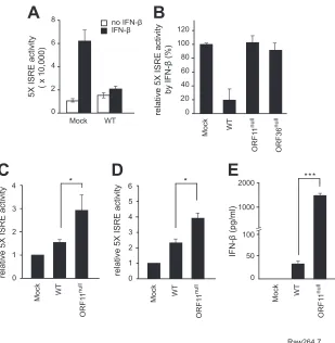
FIG 1Identification of ORF11 as a viral immune modulator that downregulates the IFN-signaling pathway. (A) MHV-68 infection downregulated ISRE
activation induced by IFN-treatment. NIH 3T3 cells harboring the 5⫻ISRE-Luc reporter plasmid (5⫻ISRE/3T3) were mock infected or infected with MHV-68
for 30 to 32 h (MOI, 1) and treated with mouse IFN-(500 U/ml) for 6 to 8 h. The harvested cell lysates were analyzed for luciferase activities. The results were
from three independent experiments, and standard deviations are shown. (B) Screening of replication-competent MHV-68 mutants in 5⫻ISRE/3T3 cells.
Replication-competent MHV-68 mutants were individually infected in 5⫻ISRE/3T3 cells for 30 to 36 h (MOI, 1), and then mouse IFN-(500 U/ml) was
supplied for 6 to 8 h. At least three independent experiments were performed for individual viruses, and relative activities of 5⫻ISRE-Luc were calculated with
the 5⫻ISRE activity of mock infection set as 100% for each experiment. Results with WT, ORF11null, and ORF36nullviruses are shown as representative results
from our screenings. (C and D) ORF11nullvirus infection elevated the levels of IFN-produced in fibroblasts and macrophages upon infection. MEFs (C) and
Raw264.7 cells (D) were infected with WT or ORF11nullvirus at an MOI of 2 for 6 h. The supernatants were transferred to 5⫻ISRE/3T3 cells and incubated for
an additional 12 h, and relative 5⫻ISRE activity was measured and compared to that of mock infection (n⫽3). (E) Raw264.7 cells were infected with WT or
ORF11nullvirus at an MOI of 2 for 12 h. IFN-amounts in the supernatants were measured by ELISA. Each error bar shows the means⫾standard deviations.
*,P⬍0.05 (Student’sttest).
on November 7, 2019 by guest
http://jvi.asm.org/
[image:4.585.139.448.62.378.2]ORF11 deficiency affected the host type I IFN response in
fibroblasts and macrophages.
ORF11
nullharbors a 1.2-kb-long
transposon which may affect neighboring gene functions. To
con-firm that the phenotype of ORF11
nullwas due to disrupted ORF11
expression, another ORF11-deficient virus (11ST) containing
tri-ple stop codons at the ORF11 locus (nt 23685) and its marker
rescue revertant (11ST/MR) were generated using the allelic
ex-change method (
Fig. 2A
). The viral genome integrities of 11ST
and 11ST/MR were confirmed by restriction enzyme mapping
and Southern blotting (
Fig. 2B
). Consistent with previous reports
(
38
,
48
), multiple-step growth curves of the 11ST virus showed no
replication defect in NIH 3T3 and MEF cells (
Fig. 2C
and
D
).
However, 11ST infection induced higher ISRE activation in
5
⫻
ISRE/3T3 cells and elevated levels of IFN-

protein in bone
marrow-derived macrophages (BMDMs) than WT or 11ST/MR
infection (
Fig. 2E
and
F
). These results confirm our previous
re-sults that ORF11 deficiency increased the host type I IFN response
in fibroblasts and macrophages.
ORF11 inhibits IFN-

promoter activity.
Infection of
ORF11-deficient viruses into fibroblasts and macrophages led to elevated
levels of IFN-

(
Fig. 1C
to
E
and
2E
and
F
). To confirm the
func-tion and understand the mechanisms of ORF11 in negatively
reg-ulating IFN-

production, we examined the effects of ORF11 on
transactivation of the IFN-

promoter by various stimuli. Sendai
virus (SeV) is known to elicit strong IFN-

responses in a
RIG-I-dependent manner (
49
). When HEK293T cells were infected with
0.25 0.5 0.75 1 1.5 2 2.5 3 4 6 10 23
(Kb)
TR TR
BAC7
nt 20000 nt 30000
ORF10 pMHV-68
ORF11ST
nt 23228 nt 24121
ORF11
ORF11ST/MR
B
EcoR I
ST/BAC ST/MR/BAC ST/BAC ST/MR/BAC
Not I
Lamda
Hind
III
1kb Ladder
Log
10
virus titers
(PFU / ml)
Hours post-infection
Log
10
virus titers
(PFU / ml)
3
0 6
1 2 4 5
WT 11ST 11ST/MR
0 24 48 72 96
Hours post-infection MEFs
(h)
D
E
F
20 60
0 40
IFN-β (pg/ml)
100
80
1 3
0 4
2
relative 5X ISRE activity
3
0 8
1 2 4 6
WT 11ST 11ST/MR
0 24 48 72 96
NIH3T3
(h) 7
5
5X ISRE/3T3 BMDM BAC7 BAC7
EcoR I
ST/BAC ST/MR/BAC ST/BAC ST/MR/BAC
Not I
BAC7 BAC7
Mock WT 11ST 11ST /MR
WT 11ST 11ST /MR
nt 23685
ttcaccaagtctgtggctac TAACTGAGT aagcttgcggccgc gggcaccgtggggtccgccc
Triple stop codonHindIII NotI
ttcaccaagtctgtggctac gggcaccgtggggtccgccc
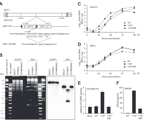
FIG 2Infection of MHV-68 lacking ORF11 induces the IFN-signaling pathway. (A) Schematic diagram of the ORF11 locus in the MHV-68 genome. (B)
Construction of the 11ST and 11ST marker rescue (11ST/MR) viruses. Stop codons were introduced into the ORF11 locus (nt 23685) using two-step allelic exchange in the MHV-68 BAC clone. BAC DNAs for the wild type, 11ST, and 11ST/MR viruses were digested by EcoRI and NotI, resolved in 0.7% agarose gel,
and subjected to Southern blot analysis using a32P-labeled ORF11-specific probe. (C and D) Virus growth of the WT, 11ST, and 11ST/MR viruses in fibroblasts.
NIH 3T3 (C) and MEFs (D) were infected with WT, 11ST, or 11ST/MR virus at an MOI of 0.05 and harvested at the indicated time points. The virus titers in the
cells and the supernatants were analyzed by plaque assays. (E and F) 11ST virus infection resulted in increased production of IFN-in fibroblasts and
macrophages upon infection. (E) The 5⫻ISRE-Luc reporter cells were infected with WT, 11S, and 11S/MR viruses (MOI, 2), and relative 5⫻ISRE activity was
measured at 24 h postinfection and compared to that of mock infection. (F) BMDMs were infected with WT, 11S, and 11S/MR viruses (MOI, 2) for 8 h, and the
amounts of IFN-in the supernatants were measured by ELISA. Error bars show the means⫾standard deviations.
on November 7, 2019 by guest
http://jvi.asm.org/
[image:5.585.46.540.61.471.2]SeV following transient transfection with FLAG-ORF11, the
en-dogenous
Ifnb
mRNA was significantly decreased compared to
that of the vector control (
Fig. 3A
). To test whether ORF11
func-tion can be recapitulated in macrophages, we established a
Raw264.7 cell line stably expressing ORF11 by transducing a
FLAG-ORF11 lentivirus (
Fig. 3B
, right) and found that the
endog-enous
Ifnb
mRNA was significantly decreased in
ORF11-express-ing macrophages upon SeV infection (
Fig. 3B
). ORF11 expression
reduced transactivation of the IFN promoter (IFN-

-Luc)
in-duced by RIG-I in HEK293T cells (
Fig. 3C
). Similarly, ORF11
decreased IFN-

-Luc activated by MAVS, a downstream adaptor
of RIG-I (
Fig. 3D
). ORF11 also reduced the IFN promoter activity
induced by IFI16, a cytoplasmic DNA sensor (
Fig. 3E
). STING, a
downstream adaptor of cytosolic DNA sensors, was also tested,
and the results indicated that ORF11 downregulated
STING-me-diated activity (
Fig. 3F
). When TRIF, an adaptor protein inducing
IFN-

from TLRs, stimulated the IFN-

-Luc transactivation,
ORF11 overexpression reduced its activation to a lesser extent
(
Fig. 3G
). The expression levels of these stimulators remained
similar even in the presence of ORF11, suggesting that ORF11
does not directly affect the stability of these molecules (
Fig. 3H
).
Although some stimulators, such as RIG-I and TRIF, can also
activate the interferon promoter in a TBK1-independent manner,
these stimuli are known to commonly activate TBK1-IRF3 for
type I interferon production. Therefore, our results led us to
hy-pothesize that ORF11 targets TBK1-IRF3 activation during virus
10001 100000
-
-
++
FLAG-ORF11
-
+
+
SeV (10 HA) 10000
relative IFN-β mRNA
levels
(IFN-β /
Actin)
-
-
++
FLAG-ORF11
-
+
+
FLAG-RIG-I
relative
IFN-β-luc
activity
80 120
0 160
-
-
++
FLAG-ORF11
-
+
+
FLAG-TRIF
relative
IFN-β-luc
activity
*
40
D
1.0 2.0
0 3.5
0.5
relative
IFN-β-luc
activity
-
-
++
FLAG-ORF11
-
+
+
HA-IFI16 1.5 2.5 3.0
* 1000
1 10000000
100000
relative IFN-β mRNA
levels
(IFN-β /
Actin)
Lenti-Cont.
Lenti-ORF11 Mock
SeV
E
80 120
0 200
-
-
++
FLAG-ORF11
-
+
+
FLAG-MAVS
relative 5X ISRE activity
***
40
G
160
2 4
0 7
1 3 5 6
**
Raw264.7
20
0
-
-
++
FLAG-ORF11
-
+
+
FLAG-STING
relative 5X ISRE activity
**
10
F
30
IB : α-FLAG
IB : α-Tubulin Lenti-Cont. Lenti-ORF1
1
FLAG-ORF11 Control
FLAG-RIG-I
FLAG-MAVS
HA-IFI16
FLAG-STING
-
+
FLAG-TRIF
-
+
-
+
-
+
-
+
-
+
Stimulators
ORF11
Tubulin
H
*** ***
FIG 3MHV-68 ORF11 inhibits the IFN-production and promoter activity. (A) MHV-68 ORF11 decreased the level of the IFN-transcripts upon Sendai virus
(SeV) infection in fibroblasts. HEK293T cells were transiently transfected with a vector alone or FLAG-ORF11 for 24 h and infected with SeV (10 HA units). After
18 h postinfection, the total RNA was extracted and relativeIfnbmRNA levels were measured and normalized to actin mRNA levels by RT-qPCR. (B) ORF11
ablated SeV-induced IFN-mRNA levels in macrophages. Raw264.7 cells stably expressing a control vector (Lenti-Cont.) or FLAG-ORF11 (Lenti-ORF11) were
generated by lentiviral transduction and infected with SeV (10 HA units) for 18 h. RelativeIfnbmRNA levels were analyzed as mentioned for panel A. The
expression of ORF11 was shown in the transduced cells using Western blot analysis with anti-FLAG (right). IB, immunoblot. (C and G) FLAG-ORF11 inhibited
IFN-promoter activity. Vector alone or FLAG-OF11 (400 ng) was transiently cotransfected into HEK293T cells with RIG-I (500 ng; C), MAVS (20 ng; D), IFI16
(400 ng; E), STING (50 ng; F), or TRIF (50 ng; G). The cells were harvested at 24 h posttransfection and subjected to luciferase reporter assays. The-gal (C, E,
and G) orrenillaluciferase activities (D and F) were used as a control. (H) HEK293T cells were cotransfected with various expression vectors in the presence
or absence of ORF11, harvested at 24 h posttransfection, and analyzed by Western blotting. The data are shown as means⫾standard deviations. *,P⬍
0.05; **,P⬍0.01; ***,P⬍0.001 (Student’sttest).
on November 7, 2019 by guest
http://jvi.asm.org/
replication to negatively modulate the host type I interferon
re-sponse.
ORF11 inhibits IFN-

promoter activated by IRF3, not by
IRF3-5D.
To further investigate the effect of ORF11 on
TBK1-IRF3 activation, the reporter plasmid IFN-

-Luc or 5
⫻
ISRE-Luc
was cotransfected into HEK293T cells with or without ORF11 in
the presence of IRF3. ORF11 efficiently inhibited the activation of
IFN-

-Luc and 5
⫻
ISRE-Luc by IRF3 (
Fig. 4A
and
B
). However,
ORF11 had no effect on the promoter activity of IFN-

-Luc and
5
⫻
ISRE-Luc activated by IRF3-5D, a constitutively active mutant
form of IRF3 (
Fig. 4C
and
D
). Consistent with these results,
ORF11 transfection failed to decrease the endogenous
Ifnb
mRNA
induced by IRF3-5D (
Fig. 4E
). Our repeated attempts to show
physical interaction between ORF11 and IRF3 were not successful,
as shown in the coimmunoprecipitation assay (
Fig. 4F
). These
results suggest that ORF11 targets sequence upstream of IRF3 to
block IFN production rather than directly targeting IRF3.
ORF11 inhibits the IFN-

promoter activated by TBK1 via
direct binding to TBK1.
We next examined whether ORF11
tar-gets an upstream kinase of IRF3, TBK1, that is ubiquitously
ex-pressed in most cell types. When transfected in HEK293T cells,
ORF11 efficiently inhibited the activation of IFN-

-Luc and
5
⫻
ISRE-Luc induced by TBK1 in a dose-dependent manner (
Fig.
5A
and
B
). Accordingly, ORF11 also decreased the endogenous
Ifnb
mRNA induced by TBK1 in HEK293T cells (
Fig. 5C
). We
further examined direct interaction of ORF11 with TBK1 (
Fig. 5D
to
F
). When overexpressed in HEK293T cells, ORF11 was
coim-munoprecipitated with TBK1 and colocalized with TBK1 in the
cytoplasm of HEK293T cells (
Fig. 5D
and
E
). To further confirm
the physiological interaction between ORF11 and TBK1, we
im-C
E
2 4 8
0 10
6
relative 5X ISRE activity
- - + ++
FLAG-ORF11
- + + +
GFP-IRF3
***
10 20 30
0 40
relative
IFN-β
activity
- - + ++
FLAG-ORF11
- + + +
GFP-IRF3
* *
20 40 60
0 100
80
relative 5X ISRE activity
- - + ++
FLAG-ORF11
- + + +
GFP-IRF3-5D
N.S.
N.S.
20 40 60
0 80
relative
IFN-β
activity
++
FLAG-ORF11
- + + +
GFP-IRF3-5D
- - +
N.S.
N.S.
10 100
1 1000
relative IFN-β mRNA
levels
(IFN-β /
Actin)
- - +
FLAG-ORF11
F
D
GFP-IRF3 FLAG-ORF11
-
-
+
-
+
-IB : α-GFP IB : α-FLAG IB : α-GFP IB : α-FLAG
+
+
Cell lysates IP : α-FLAG
N.S.
FIG 4MHV-68 ORF11 inhibits IFN-promoter activation by IRF3, not by IRF3-5D. (A and B) ORF11 inhibited IRF3-induced IFN-promoter and 5⫻ISRE
activities. GFP-IRF3 (300 ng) was cotransfected into HEK293T cells with either vector alone or FLAG-ORF11 (250 ng and 500 ng) in the presence of IFN--Luc
(A) or 5⫻ISRE-Luc (B). After 24 h, 5⫻ISRE-Luc and IFN--Luc reporter gene activities were measured and normalized by-gal activities. (C and D) ORF11 did
not inhibit transactivation of the IFN-promoter and 5⫻ISRE induced by IRF3-5D, a constitutive active mutant of IRF3. GFP-IRF3-5D (300 ng) was
cotransfected into HEK293T cells with FLAG-ORF11 (250 ng and 500 ng) in the presence of IFN--Luc (C) or 5⫻ISRE-Luc (D), and the cell lysates were analyzed
as described above. (E) ORF11 did not decrease theIfnbmRNA levels induced by IRF3-5D. HEK293T cells were transfected with GFP-IRF3-5D (300 ng) and
FLAG-ORF11 (500 ng) for 24 h. RelativeIfnbmRNA levels were measured and normalized to actin mRNA levels by RT-qPCR. (F) ORF11 did not directly interact
with IRF3. GFP-IRF3 (9g) and FLAG-ORF11 (9g) were cotransfected into HEK293T cells. After 48 h, the cells were lysed and immunoprecipitated (IP) with
␣-FLAG. The arrow represents the expected size of GFP-IRF3. The data are shown as means⫾standard deviations. *,P⬍0.05; **,P⬍0.01 (Student’sttest).
on November 7, 2019 by guest
http://jvi.asm.org/
[image:7.585.136.457.64.444.2]munoprecipitated ORF11 in Raw264.7 cells stably expressing
FLAG-ORF11 (
Fig. 3B
, right) and found that ORF11 interacted
with endogenous TBK1, suggesting that ORF11 targets TBK1 to
inhibit the host type I interferon response during virus infection
(
Fig. 5F
). The interaction of TBK1 with ORF11 was independent of its
kinase activity, as shown in strong binding of ORF11 with TBK1
K38A, a kinase null mutant of TBK1 (
Fig. 5G
). IKK
ε
is another
up-stream kinase that can activate IRF3 mainly in immune cells. ORF11
20 40 60
0 100
80
relative 5X ISRE activity
20 40 80
0 140
100
60 120
-
-
+
++
FLAG-ORF11
-
+
+
+
FLAG-TBK1
relative
IFN-β
activity
-
-
+
++
FLAG-ORF11
-
+
+
+
FLAG-TBK1
A
F
HA-ORF11 FLAG-TBK1
-
-
+
-
+
-IB : α-HA
IB : α-FLAG
IB : α-HA
IB : α-FLAG
+
+
Cell lysates IP : α-FLAG
MYC-ORF11 FLAG-TBK1
FLAG-TBK1 K38A
-
+
-+
+
+
-
-
+
IB : α-MYC
IB : α-FLAG
IB : α-MYC
IB : α-FLAG
-Cell lysates IP : α-FLAG
***** ***
***
100
10 1000
relative IFN-β mRNA
levels
(IFN-β /
Actin)
-
-
+
FLAG-ORF11
-
+
+
FLAG-TBK1
C
ORF11
Merged DAPI
TBK1
B
IB : α-TBK1
IB : α-FLAG
IB : α-TBK1
IB : α-FLAG Cell lysates
IP : α-FLAG
Raw264.7
G
FLAG-ORF1
1
Cont.V
ector
D
E
HA-ORF11 FLAG-TBK1
FLAG-IKKε
-
-
+
-
+
+
-
-
-IB : α-HA
IB : α-FLAG
IB : α-HA
IB : α-FLAG
-+
+
Cell lysates IP : α-FLAG
H
I
40 80 120
0 160
-
-
++
FLAG-ORF11
-
+
+
FLAG-IKKε
relative
IFN-β
activity
**
***
FIG 5MHV-68 ORF11 inhibits TBK1-induced IFN-promoter activation via direct binding. (A and B) ORF11 inhibited TBK1-induced IFN-promoter and 5⫻ISRE
activities. FLAG-TBK1 (100 ng) was cotransfected into HEK293T cells with FLAG-ORF11 (200 ng and 400 ng) in the presence of IFN--Luc (A) or 5⫻ISRE-Luc (B).
After 24 h, 5⫻ISRE-Luc and IFN--Luc reporter gene activities were measured and normalized to-gal activities. (C) ORF11 decreased theIfnbmRNA levels induced
by TBK1. HEK293T cells were transfected with FLAG-TBK1 (100 ng) and FLAG-ORF11 (500 ng) for 24 h. RelativeIfnbmRNA levels were measured and normalized to
the actin mRNA levels by RT-qPCR. (D) Direct interaction of ORF11 with TBK1. FLAG-TBK1 (9g) and HA-ORF11 (9g) were cotransfected into HEK293T cells.
After 48 h, the cells were lysed and immunoprecipitated with␣-FLAG. (E) Colocalization of ORF11 and TBK1 in the cytoplasm. HA-ORF11 and FLAG-TBK1 were
transfected into HEK293T cells, and the cells were fixed at 24 h. HA-ORF11 and FLAG-TBK1 were stained for ORF11 (FITC; green) and TBK1 (Cy3; red), respectively,
while the nuclei were stained with the DNA-intercalating dye DAPI (blue). All of the panels were under a magnification of⫻1,000. (F) ORF11 interacted with
endogenous TBK1. Raw264.7 cells stably expressing a control vector or FLAG-ORF11 were lysed and immunoprecipitated with␣-FLAG and immune blotted with
␣-TBK1 to detect the endogenous TBK1. (G) ORF11 interacted with a TBK1 kinase null mutant. FLAG-TBK1 K38A (9g) or FLAG-TBK1 (9g) was cotransfected
with MYC-ORF11 (9g) into HEK293T cells. After 48 h, the cells were lysed and immunoprecipitated with␣-FLAG antibody. (H) ORF11 inhibited IKKε-induced
IFN-promoter activities. FLAG-IKKε(100 ng) was cotransfected into HEK293T cells with FLAG-ORF11 (200 ng, 400 ng) in the presence of IFN--Luc. After 28 h,
5⫻ISRE-Luc and IFN--Luc reporter gene activities were measured and normalized as described for panels A and B. (I) ORF11 did not directly interact with IKKε.
FLAG-IKKε(9g) and HA-ORF11 (9g) were cotransfected into HEK293T cells. After 48 h, the cells were lysed and immunoprecipitated with␣-FLAG. Each error bar
shows means⫾standard deviations. **,P⬍0.01; ***,P⬍0.001 (Student’sttest).
on November 7, 2019 by guest
http://jvi.asm.org/
inhibited IKK
ε
-induced IFN-

promoter activation, albeit to a lesser
degree than its inhibition of TBK1-activated transactivation (
Fig. 5H
).
There was no direct association of ORF11 and IKK
ε
in transfected
HEK293T cells (
Fig. 5I
). Our results suggest that ORF11 specifically
targets TBK1 to inhibit the host type I interferon response.
To further investigate whether ORF11 interacts with TBK1
during virus replication, we constructed a recombinant virus
ex-pressing 3
⫻
FLAG-tagged ORF11 (FLAG-ORF11/MHV-68) (
Fig.
6A
). The viral genome integrity of FLAG-ORF11/MHV-68 was
confirmed by restriction enzyme mapping (
Fig. 6B
), and its
repli-cation was similar to that of the WT (data not shown). When Vero
cells were infected, the FLAG-ORF11/MHV-68 virus expressed
the FLAG-ORF11 protein as detected with the FLAG
anti-body (
Fig. 6C
). FLAG-ORF11 was expressed in both the
cyto-plasm and the nucleus of infected HeLa and MEF cells, while it was
localized more in the cytoplasm than in the nucleus of infected
HEK293T cells (
Fig. 6D
). Upon infection of FLAG-ORF11/MHV-68
into MEF cells (MOI, 2) for 24 h, FLAG-ORF11 physically interacted
with endogenous TBK1 and was colocalized with TBK1, as shown in
coimmunoprecipitation and IFA, respectively (
Fig. 6E
and
F
). Taken
together, these results demonstrate that MHV-68 ORF11 physically
interacts with TBK1 in the context of virus replication to inhibit IFN
production.
ORF11 blocks IRF3 activation by interfering with the
inter-action between TBK1 and IRF3.
Upon RNA and DNA virus
in-fections, TBK1 plays a critical role in activation of IRF3, such as its
phosphorylation, dimerization, and nuclear translocation,
lead-ing to transactivation of the IFN-

promoter. We next examined
the effect of ORF11-TBK1 interactions on IRF3 activation (
Fig. 7
).
When IRF3 and TBK1 were cotransfected into HEK293T cells
with ORF11, ORF11 decreased IRF3 phosphorylation induced by
TBK1 in a dose-dependent manner (
Fig. 7A
). The level of
HA-TR TR
BAC7
nt 20000 nt 30000
pMHV-68
FLAG-ORF11/ MHV-68
nt 23488 nt 24651
ORF11
accgcc ATGGACTACAAAGACCATGACGGTGATTATAAAG
ATCATGACATCGATTACAAGGATGACGATGACAAG tga ttcttg
F
3X FLAG tag
ClaI nt 24652
nt 24727 STOP codon
HeLa
B
C
(Kb)
EcoRI
FLAG-ORF1
1
Lamda
Hind
III
1kb Ladder BAC7 FLAG-ORF1
1
BAC7 FLAG-ORF1
1
BAC7 Lamda
Hind
III
1kb Ladder
2.5 3 4 6 10 23
1.5 2
1
HindIII ClaI
*
HEK293T
MEF
D
ORF1
1-FLAG
/MHV
-68
IB : α-FLAG
IB : α-Tubulin
Mock
F
IB : α-TBK1
IB : α-FLAG IP : α-FLAG
FLAG-ORF1
1
/MHV
-68
Mock
IB : α-TBK1
IB : α-Tubulin Cell lysates
MEF
*
ORF11
ORF11
ORF11 TBK1
IgG Merged
Merged DAPI
DAPI
TBK1 Merged DAPI
IgG Merged DAPI
Mock
FLAG-ORF11/ MHV-68
ORF11 Merged DAPI
ORF11 Merged DAPI
ORF11 Merged DAPI
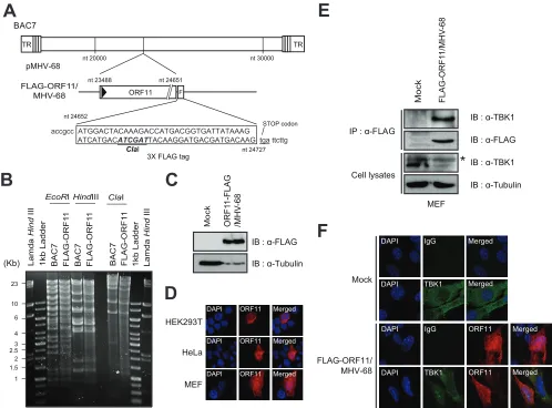
FIG 6ORF11 interacts with endogenous TBK1 during MHV-68 infection. (A) Schematic diagram of the FLAG-tagged ORF11 recombinant virus
(FLAG-ORF11/MHV-68) in the MHV-68 genome. (B) Construction of the FLAG-ORF11/MHV-68 virus. The 3⫻FLAG sequences were introduced into the ORF11
locus (nt 24652) of the MHV-68 BAC clone (pMHV-68). BAC DNAs for the wild type and FLAG-ORF11 were digested by EcoRI and ClaI and resolved in 0.7% agarose gel. (C) Expression of FLAG-ORF11 during MHV-68 replication. Vero cells were infected with the FLAG-ORF11/MHV-68 virus at an MOI of 0.05 for 72 h cells. The expression of ORF11 was analyzed by Western blotting using anti-FLAG. (D) HEK293T, HeLa, and MEF cells were infected with FLAG-ORF11/ MHV-68 at an MOI of 1 for 24 h. (E) Interaction of ORF11 with endogenous TBK1 during virus infection. MEF cells were infected with FLAG-ORF11/MHV-68
(MOI, 1) for 24 h, subjected to immunoprecipitation with␣-FLAG, and monitored by immunoblotting with␣-TBK1. The asterisk indicates a nonspecific band.
(F) Colocalization of ORF11 and TBK1 during viral infection. MEF cells were infected with FLAG-ORF11/MHV-68 at an MOI of 1, and the cells were fixed at 24 h postinfection. ORF11 was stained with Cy3 (red), and TBK1 was stained with FITC (green), while the nuclei were stained with DAPI (blue). All of the panels
were under a magnification of⫻1,000.
on November 7, 2019 by guest
http://jvi.asm.org/
[image:9.585.43.541.65.433.2]IRF3 immunoprecipitated with GFP-IRF3 in the presence of
TBK1 was decreased with increasing doses of ORF11, indicating
that ORF11 inhibited dimer formation of IRF3 induced by TBK1
(
Fig. 7B
). Furthermore, nuclear translocation of IRF3 following
SeV infection (10 HA units) for 6 h was efficiently blocked in MEF
cells when ORF11 was overexpressed (
Fig. 7C
). Since IRF3 is a
substrate of TBK1 and ORF11 binds to TBK1, we tested whether
ORF11 can interfere with TBK1 and IRF3 interactions. In
coim-munoprecipitation assays, the interactions between IRF3 and
TBK1 were inhibited by ORF11 (
Fig. 7D
). These results suggest
that ORF11 outcompetes IRF3 for TBK1 binding and
subse-quently inhibits TBK1-induced IRF3 activation.
The central domain of ORF11 is essential for TBK1 binding
and IFN inhibition, while the kinase domain of TBK1 is
impor-tant for ORF11 binding.
The ORF11 protein of MHV-68 consists
of 388 amino acids. To map the domain of ORF11 that is required
for TBK1 binding and IFN inhibition, we generated ORF11
do-main mutants. Since computational structure analysis of the
ORF11 protein revealed no currently known domain or motif
except weak homology for the 2=-deoxyuridine 5=-triphosphate
pyrophosphatase (dUTPase)-like motif (
50
,
51
), we made a series
of N terminus and C terminus truncation mutants (
Fig. 8A
).
Co-immunoprecipitation results of ORF11
⌬
N1 and
⌬
C1 mutants
with TBK1 showed that both the N terminus (domain I) and C
terminus (domain IV) of ORF11 were dispensable for TBK1
bind-ing. However, as shown in the results for ORF11
⌬
N2 and
⌬
C2
mutants, deletion of the central domains (domains II and III)
abolished ORF11 and TBK1 interactions. Similarly, ORF11 lost its
inhibition of TBK1-mediated IFN-

-Luc transactivation when
the central domain of ORF11 was disrupted in
⌬
N2 and
⌬
C2
mutants (
Fig. 8D
), suggesting that the central domain of ORF11
(domains II and III) is required for both TBK1 binding and IFN
inhibition of ORF11. To test whether the central domain of
ORF11 is sufficient to confer TBK1 binding and IFN inhibition,
we constructed ORF11 CD, expressing the central domain
(do-mains II and III) alone, and found that the central domain of
ORF11 alone was sufficient for TBK1 binding (
Fig. 8B
).
Further-more, ORF11 CD was also sufficient to inhibit IRF3
phosphory-lation and the IFN-

promoter activity induced by TBK1 (
Fig. 8C
and
D
). Taken together, these results suggest that the central
do-main of ORF11was sufficient to bind and inhibit TBK1, leading to
inhibition of IRF3 activation and IFN-

production.
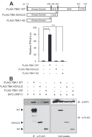
To further map the minimal domain of TBK1 to interact with
ORF11, we constructed TBK1 domain mutants, TBK1-KD/ULD
and TBK1-KD, according to the previous report (
52
) (
Fig. 9A
).
TBK1-KD (amino acids [aa]1 to 301) deletes both ULD and SDD/
CTD, leaving the kinase domain alone, while TBK1-KD/ULD (aa
1 to 383) contains the kinase domain and ubiquitin-like domain
FLAG-ORF11 HA-IRF3 FLAG-TBK1
+ + +
- - +
- + +
IB : α-P-IRF3
IB : α-HA
IB : α-FLAG P-IRF3
IB : α-FLAG ORF11
TBK1 IRF3
+
++ +
FLAG-ORF11 GFP-IRF3
FLAG-TBK1
+ + +
- - +
- + +
+
++ +
HA-IRF3 + + + +
IP : α-GFP IB : α-HA
IB : α-P-IRF3 IB : α-GFP
HA-ORF11 FLAG-TBK1
IB : α-GFP
IB : α-FLAG
IB : α-GFP
IB : α-HA Cell lysates
IP : α-FLAG
A
B
D
GFP-IRF3IB : α-FLAG
C
DAPI
ORF11 IRF3
Merged
Mock
SeV
- - +
FLAG-ORF11
+ + +
GFP-IRF3
Merged Merged
DAPI IRF3
DAPI IRF3
60 80
0 100
40
20
Percentage (%)
Cytosol Nucleus
Cell lysates
IB : α-HA
IB : α-P-IRF3 IB : α-GFP
IB : α-FLAG
+ + +
- - ++
- + +
+
+ +
+ + + +
+ + +
- - +
- + +
+
++ +
FIG 7MHV68 ORF11 interferes with TBK1-induced IRF3 activation via abolishing the interactions between TBK1 and IRF3. (A) ORF11 inhibited the phosphorylation
of IRF3. HA-IRF3 (300 ng), FLAG-TBK1 (100 ng), and FLAG-ORF11 (500 ng) were cotransfected into HEK293T cells. After 24 h, the transfected cells were harvested
and analyzed by Western blotting using␣-phospho-IRF3 (Ser396). (B) ORF11 inhibited the dimerization of IRF3. IRF3 with two different tags (HA-IRF3 and
GFP-IRF3) (3g), FLAG-TBK1 (3g), and FLAG-ORF11 (5g) were transfected into HEK293T cells. After 24 h, the cells were lysed and immunoprecipitated with
␣-GFP and analyzed by Western blotting. (C) MEF cells were transfected with GFP-IRF3 (4g) and FLAG-ORF11 (6g) and were infected with SeV (50 HA units). After
6 h, we examined the location of IRF3 (GFP; green) by immunofluorescence. ORF11 was stained by Cy3 (red), and the nuclei were stained by DAPI (blue). Every sample
had more than 100 cells, and the percentage of the cells is shown for cytoplasmic or nuclear localization. All of the panels were under a magnification of⫻1,000. (D)
ORF11 disrupted the interaction between TBK1 and IRF3. GFP-IRF3 (4g), FLAG-TBK1 (5g), and HA-ORF11 (4g and 9g) were cotransfected into HEK293T
cells. At 24 h, the cells were lysed and immunoprecipitated with␣-FLAG and analyzed by Western blotting.
on November 7, 2019 by guest
http://jvi.asm.org/
[image:10.585.63.519.68.355.2]and lacks two coiled-coil domains at the C terminus, which serve
as a scaffold/dimerization domain (SDD) (
52–54
). Consistent
with the previous report (
52
), these two mutants failed to activate
IFN-

production in reporter assays (
Fig. 9A
). In our
coimmuno-precipitation assays, TBK1-KD bound to ORF11 at a level similar
to that of full-length TBK1, while TBK1-KD/ULD showed no or
little binding to TBK1 (
Fig. 9B
), suggesting that the TBK1 kinase
domain is a minimal domain required for ORF11 interaction.
DISCUSSION
While the production of type I interferon is a fundamental host
response to combat viral invasion, viral pathogens develop
multi-ple strategies to subvert such host IFN responses. Here, we
con-firmed that lytic MHV-68 infection blocks type I interferon
sig-naling and identified MHV-68 ORF11 as a negative regulator of
interferon-

production via an unbiased genomic approach using
a transposon mutant library of MHV-68. ORF11 expression
in-hibited activation of the IFN-

promoter by various factors.
ORF11 interacted directly with both overexpressed and
endoge-nous TBK1, which was further confirmed in the context of virus
replication using a recombinant virus expressing FLAG-ORF11.
Interactions between ORF11 and TBK1 disrupted the interaction
between TBK1 and IRF3, thereby blocking IRF3 phosphorylation,
FLAG-TBK1 ORF11 Δ C1
aa 1 38885
ORF11 WT
ORF11 Δ N2 ORF11 Δ N1 ORF11 Δ C2
195 288
- + +
+ -
-- -
-- -
-- -
-- -
-MYC-ORF11 Δ C2 MYC-ORF11 Δ N1 MYC-ORF11 Δ N2 MYC-ORF11 Δ C1 MYC-ORF11 WT
- -
-- -
-- - +
+ +
-+ -
-- -
-+ - +
- -
-- +
-- -
-+ - +
- + +
Cell lysates IP : α-FLAG
IB : α-MYC IB : α-FLAG
IB : α-MYC
Relative IFN-β-Luc (%) 20 40 80
0 100
60 120
ORF11 CD
FLAG-TBK1 MYC-ORF11 WT MYC-ORF11 CD
+ -
-+ - +
- + +
IB : α-MYC IB : α-FLAG IB : α-MYC Cell lysates
IP : α-FLAG
D
FLAG-TBK1
- - +
- +
-- -
-- -
-FLAG-ORF11 Δ N1 FLAG-ORF11 Δ C2 FLAG-ORF11 Δ N2 FLAG-ORF11 Δ C1 FLAG-ORF11 WT
- -
-- -
-- +
-+ -
-+ + +
- -
-+ + +
- - +
+
-TBK1 binding
++
-+
-TBK1 binding
+ ++
FLAG-ORF11 WT FLAG-TBK1 GFP-IRF3
- + +
- - +
+ + +
IB : α-P-IRF3 IB : α-GFP IB : α-FLAG (WT) IB : α-FLAG (CD)
+
-+
-FLAG-ORF11 CD - - - +
-C
I II III IV
aa 1 38885
ORF11 WT
195 288
I II III IV
+
*
***
***
**
N.S.
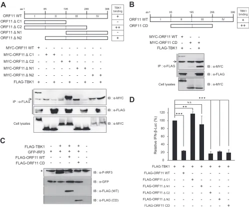
FIG 8Functional domain mapping of MHV-68 ORF11 required for TBK1 interaction and inhibition. (A) The central domain of ORF11 (CD; aa 85 to 288) was
necessary to bind to TBK1. Full-length MYC-ORF11 (WT),⌬C1,⌬C2,⌬N1, or⌬N2 (9g) was transfected with FLAG-TBK1 (9g) into HEK293T cells. After
48 h, the cells were lysed and immunoprecipitated with␣-FLAG and analyzed by Western blotting. An arrow indicates IgG bands. (B) The central domain of
ORF11 (CD; aa 85 to 288) was sufficient for TBK1 binding. Full-length MYC-ORF11 (WT) (9g) and the ORF11 CD (9g) were transfected with FLAG-TBK1
(9g) into HEK293T cells. After 48 h, the cells were lysed and immunoprecipitated with␣-FLAG and analyzed by Western blotting. An arrow indicates IgG
bands, while the asterisk indicates a nonspecific band, serving as a loading control. (C) ORF11 CD was sufficient to inhibit the phosphorylation of IRF3 by TBK1. FLAG-TBK1 (100 ng) and GFP-IRF3 (300 ng) were cotransfected with WT FLAG-ORF11 or FLAG-ORF11 CD (500 ng) into HEK293T cells. After 24 h, the
transfected cells were harvested and analyzed by Western blotting using␣-phosphoIRF3 (Ser396). N.S. indicates nonspecific bands. (D) ORF11 CD was sufficient
to inhibit TBK1-induced IFN-promoter activity. Full-length FLAG-ORF11 (WT),⌬C1,⌬C2,⌬N1,⌬N2, or CD (400 ng) was transfected with FLAG TBK1 (100
ng) into HEK293T cells. IFN--Luc reporter gene activities were measured and normalized to-gal activities. Each error bar shows means⫾standard deviations.
**,P⬍0.01; ***,P⬍0.001 (Student’sttest).
on November 7, 2019 by guest
http://jvi.asm.org/
[image:11.585.43.541.65.480.2]dimerization, and nuclear translocation. The central domain of
ORF11 was necessary and sufficient for both TBK1 binding and
IFN-

promoter inhibition, while the kinase domain of TBK1 was
the minimal domain required for interaction with ORF11. To our
knowledge, MHV-68 ORF11 is the first viral factor that is
identi-fied to block TBK1 function among the genes of
gammaherpesvi-ruses.
TBK1 functions as a key node protein in several cell signaling
pathways, including antiviral innate immune response,
au-tophagy related to bacterial invasion, cell growth, and
prolifera-tion (
55
). Among them, antiviral innate immune activity of TBK1
has been the most extensively studied. TBK1 activities are tightly
regulated in various ways, such as phosphorylation,
ubiquitina-tion, kinase activity modulaubiquitina-tion, and prevention of functional
TBK1-containing complexes (
55
,
56
). Several viral factors have
been reported to modulate the TBK1 activity to circumvent IFN
responses (
56
). The
␥
134.5 protein of herpes simplex virus type 1
(HSV-1) and the NS3 protein of hepatitis C virus (HCV) were
shown to directly interact with TBK1, which disrupts the
interac-tion of TBK1 and IRF3 (
57–59
). ORF11 has a mechanism similar
to those of HSV
␥
134.5 and HCV NS3 proteins in that it directly
binds to TBK1 and blocks IRF3 activation by inhibiting the
inter-actions of TBK1 and IRF3, but there is no sequence homology
among these proteins (data not shown). However, the fact that
MYC-ORF11 FLAG-TBK KD/ULD FLAG-TBK1 KD FLAG-TBK1 WT
-
-
+
+
+
+
-
-
-+
+
-
+
-
--
-
+
+
+
+
-
-
-+
+
-
+
-
-IB : α-MYC
IB : α-FLAG FLAG-TBK KD/ULD
aa 1 730299
FLAG-TBK1 WT
FLAG-TBK1 KD
383 Kinase Domain
Kinase Domain
Kinase Domain
ULD
ULD
SDD
KD/ULD
KD WT
9 305
383
301
B
20 40 80
0 60 100
Relative
IFN-β-Luc
FLAG-TBK KD/ULD FLAG-TBK1 KD FLAG-TBK1 WT
-
-
+
-
-
-+
-
+
-
-CTD 657
*** N.S.
FIG 9Kinase domain of TBK1 is sufficient to bind to MHV-68 ORF11. (A) Activity of TBK1 domain mutant constructs. The upper panel illustrates schematic
diagrams of WT TBK1 and mutants containing the kinase domain (KD), ubiquitin-like domain (ULD), scaffold/dimerization domain (SDD), and/or C-terminal
domain (CTD). FLAG-TBK1 WT, KD/ULD, or KD (100 ng) was cotransfected with full-length ORF11 into HEK293T cells. IFN--Luc promoter activities were
measured and normalized by-gal activities. (B) Kinase domain of TBK1 is sufficient to bind to ORF11. FLAG-TBK1 WT, KD/ULD, or KD (9g) was
cotransfected with MYC-ORF11 (9g) into HEK293T cells. After 48 h, the cells were lysed, immune precipitated with␣-FLAG, and analyzed by Western
blotting. Each error bar shows means⫾standard deviations. ***,P⬍0.001 (Student’sttest).
on November 7, 2019 by guest
http://jvi.asm.org/
[image:12.585.148.444.63.504.2]viruses encode viral proteins targeting TBK1-mediated signaling
pathways highlights a critical role of TBK1 in antiviral immunity.
Although TBK1 and IKK
ε
have similar biochemical properties
in
vitro
, they have very distinct functions
in vivo
(
60
,
61
). TBK1 is
activated by pattern recognition receptors, such as Toll-like
recep-tors, and intracellular receprecep-tors, such as RIG-I, MDA5, and DAI,
and it phosphorylates IRF3 and IRF7 and other target proteins
(
62
). TBK1 is essential for the activation of type I IFN
in vivo
, while
IKK
ε
is not. In fact, IKK
ε
is required for the activation of
IFN-stimulated genes
in vivo
but is not required for IFN expression.
IKK
ε
functions by phosphorylating a specific serine residue in the
transcription factor STAT1, thereby controlling the assembly of
IFN-inducible transcription factor complexes (
60
,
61
). Although
TBK1 is highly homologous to IKK
ε
, TBK1, not IKK
ε
, was
re-quired for controlling DNA virus infection (
25
,
26
). It is
interest-ing that ORF11 bound to TBK1, not to IKK
ε
, a closely related
kinase, although it inhibited the IFN-

promoter activity induced
by both TBK1 and IKK
ε
, suggesting that ORF11 indirectly inhibits
the IKK
ε
signaling or targets the sequence downstream of IKK
ε
in
a TBK1-independent manner. Although HSV
␥
134.5 and HCV
NS3 proteins inhibited the IKK
ε
signaling, interaction of HSV
␥
134.5 or HCV NS3 proteins with IKK
ε
has not been shown.
The ORF11 protein of MHV-68 consists of 388 amino acids
and is conserved among gammaherpesviruses. The maximum
identities of homologues between MHV-68 and KSHV or
MHV-68 and EBV are 22% or 24%, respectively, while those
be-tween KSHV ORF11 and EBV LF2 are up to 43%. All of these
ORF11 homologues are predicted to contain a limited motif
re-lated to herpesviral dUTPase (
50
), but their actual dUTPase
activ-ities have never been shown. While little has been studied about
the function of KSHV ORF11, multiple functions of EBV LF2 were
reported (
63
). Interestingly, EBV LF2 was also shown to inhibit
type I IFN production (
63
). EBV LF2 was identified from a
screen-ing of EBV ORFs for its ability to block SeV-induced IFN-
␣
pro-moter activity. EBV LF2 directly binds to IRF7 and blocks IRF7
dimerization rather than affecting phosphorylation and nuclear
translocation of IRF7. However, the mechanism of EBV LF2
seems to differ from that of MHV-68 ORF11 in that EBV LF2 had
no effect on IRF3-induced ISRE activation (
63
). Therefore,
MHV-68 ORF11 and EBV LF2 may have a similar function via
independent mechanisms during virus replication. Recently, a
conserved viral dUTPase (ORF54) of MHV-68 was shown to
downregulate type I IFN signaling by inducing the degradation of
the type I interferon receptor protein independent of its dUTPase
enzymatic activity (
64
). KSHV ORF10, with weak homology to
viral dUTPase, was also shown to block IFN-mediated signal
transduction by forming inhibitory complexes with the type I IFN
receptor subunit (
65
). Moreover, the KSHV viral dUTPase ORF54
was reported to downregulate a ligand for the NK-activating
re-ceptor NKp44 without requiring viral dUTPase (
66
). Although it
is not clear whether viral dUTPase motifs of MHV-68 ORF11
would contribute to its inhibition of type I IFN production, it is
interesting that all ORFs of gammaherpesviruses with predicted
homology to viral dUTPase confer the functions to inhibit host
innate immunity, especially the type I IFN system, via various
mechanisms.
It has been shown that type I IFNs have roles not only in
con-trolling acute replication of MHV-68 but also in modulating
tent gene expression and inhibiting viral reactivation during
la-tency (
67
). Given that MHV-68 ORF11 is a virion-associated
tegument protein (
48
), the role of ORF11 in inhibiting type I IFNs
is 2-fold: it can modulate virus replication in permissive
fibro-blasts and the host immune milieu in immune cells, such as
mac-rophages or dendritic cells. Our results indicate that ORF11
effi-ciently blocked type I IFN production from both fibroblasts and
macrophages, supporting this notion (
Fig. 2
and
3
). Since
macro-phages or dendritic cells are
in vivo
latent reservoirs of MHV-68 in
addition to B cells, ORF11 may affect latent infection in addition
to acute lytic replication, possibly by regulating reactivation
fre-quency. Consistent with these results, ORF11 deficiency led to
significantly reduced acute replication in the lung and a delay in
seeding to the spleen for latency following intranasal infection (
38
,
48
). However, we and others found little difference between
ORF11-deficient viruses and their revertants in viral growth in
cultured cells (
Fig. 2
), suggesting that the type I IFN response is
more critical in controlling virus replication
in vivo
than
in vitro
.
Our screening results suggest that multiple viral proteins act
together to curtail the host type I IFN response during
gammaher-pesvirus