COMPARISON OF DIAGNOSTIC ACCURACY OF CONVENTIONAL
INTRAORAL PERIAPICAL RADIOGRAPH, DIGITAL
RADIOVISIOGRAPH AND DIGITAL ORTHOPANTOMOGRAPH IN
DETECTING INTERDENTAL BONE LOSS
Dissertation submitted to
THE TAMILNADU DR. M.G.R. MEDICAL UNIVERSITY
In partial fulfilment for the degree of
MASTER OF DENTAL SURGERY
BRANCH-IX
ORAL MEDICINE AND RADIOLOGY
THE TAMILNADU DR. M.G.R. MEDICAL UNIVERSITY
CHENNAI-600032
ACKNOWLEDGEMENT
Praises and thanks to the God, the Almighty, for His showers of blessings throughout
my research work to complete the research successfully
First and foremost, I would like to record my gratitude to my guide, Dr. S. Elangovan.
M.D.S., Professor and Head of Department of Oral Medicine and Radiology for the vision
and foresight which inspired me to conceive this project. I sincerely thank Dr. S. Elangovan sir
for his concern, motivation and support throughout this project. His unending belief in me was
the key element which helped me to bring this work to a successful conclusion. His immense
knowledge and perseverance has always been the guiding force in realizing my goals in time.
I would like to express my heartfelt gratitude to Professor Dr. Suman Jaishankar.
M.D.S., Dept of Oral Medicine, for her constant support, inspiration, for giving me ideas to
proceed with this study and for being the guiding force throughout the course of this study.
I would like to express my sincere gratitude to my advisors Dr. Senthil Kumar. M.D.S.,
Dr. Gomathi M.D.S., and Dr.Sakthi Saranya Devi M.D.S for their continuous support,
motivation and enthusiasm in my study and research.
I would like to express my deepest thanks toDr.G.S.Kumar M.D.S, Principal, K.S.R.
I am thankful to my friend Dr. SriRam, for aiding me in Biostatistics work.
I would like to express my heartiest thanks to my dear colleagues, Dr. Sri Jaya
Ranjitha.J and Dr. Silpa Ramachandran and my seniors, my juniors for their unyielding
support during the period of study.
I am extremely grateful to my dear Parents Mr. K. Chellan and Mrs. M.G. Indira, my
Wife Mrs. Sindhuja and my little charm Anvitha for their love, prayers, caring and sacrifices
Dedicated to my father
Mr. K. Chellan
, my mother
Mrs. Indira
, my wife
Mrs.
T. Sindhuja
for their care, love, support and prayers to overcome all my
CONTENTS
S.NO TITLE PAGE NO.
1 INTRODUCTION 1
2 AIMS AND OBJECTIVES 5
3 REVIEW OF LITERATURE 6
4 MATERIALS AND METHODS 13
5 STATISTICAL ANALYSIS 25
6 RESULTS 26
7 DISCUSSION 33
8 SUMMARY AND CONCLUSION 38
9 BIBLIOGRAPHY 40
LIST OF TABLES
S.NO TITLE PAGE NO
1 Dimensions of Interdental Bone loss in Conventional Intraoral periapical radiographs, Digital Radiovisiographs and Digital
Orthopantomographs
28
2 Dimensions of Interdental Bone loss in Conventional Intraoral periapical radiographs, Digital Radiovisiographs and Digital
Orthopantomographs, among the Gender.
30
LIST OF FIGURES
S.NO TITLE PAGE NO
1 PATIENT POSITIONED FOR CONVENTIONAL INTRAORAL PERIAPICAL RADIOGRAPH
19
2 MEASUREMENT OF ALVEOLAR BONE LOSS USING CONVENTIONAL INTRAORAL PERIAPICAL RADIOGRAPH
20
3 PATIENT POSITIONED FOR DIGITAL RADIOVISIOGRAPHY 21
4 MEASUREMENT OF ALVEOLAR BONE LOSS USING DIGITAL RADIOVISIOGRAPHY
22
5 PATIENT POSITIONED FOR DIGITAL ORTHOPANTOMOGRAPHY
23
6 MEASUREMENT OF ALVEOLAR BONE LOSS USING DIGITAL ORTHOPANTOMOGRAPHY
LIST OF GRAPHS
S.NO GRAPHS PAGE. NO
1 Dimensions of Interdental Bone loss in Conventional Intraoral periapical radiographs, Digital Radiovisiographs and Digital
Orthopantomographs
29
2 Dimensions of Interdental Bone loss in Conventional Intraoral periapical radiographs, Digital Radiovisiographs and Digital
Orthopantomographs, among the Gender
INTRODUCTION
[Type text] Page 1
INTRODUCTION
The position and shape of the alveolar margins are indices of health and disease to
both the general dentist and the periodontist. In examining these, the use of radiographs
plays an important diagnostic role. Bjorn, Hailing and Thyberg (1969), who mounted
intra-oral radiographs in frames and projected the images onto a back-projection table on
which a scale had been drawn. They established bone heights relative to tooth lengths by
means of this method. (1)
The development of direct digital radiographic methods has made it possible to
reduce the radiation dose and to enhance the image quality after image acquisition. The
diagnostic accuracy of direct digital radiography has been shown to be comparable to that
of conventional film radiography for the detection of experimental bone tissue lesions. (2)
The shortcomings of film-based radiography, which have been dealt with in
previous studies, include processing errors, increased radiation dose in comparison with
direct digital images, poor imaging geometry, and lack of post-imaging enhancement
facilities. (3)
Along with marginal inflammation, periodontal pocket formation, and attachment
loss, alveolar bone loss is a primary feature of periodontitis. The height of the alveolar
INTRODUCTION
[Type text] Page 2
examination; however, radiographic assessment tends to underestimate the amount of
bone loss. (5)
Alveolar bone loss is the main feature of destructive inflammatory periodontal
disease. The height of the alveolar bone may be evaluated by radiographic examination.
However, conventional radiographic assessment tends to underestimate the amount of
bone loss. Digital measurement with RVG may improve diagnostic interpretation of
radiographs in terms of accuracy (7).
Alveolar bone loss is a main characteristic of destructive inflammatory
periodontal disease. The height of the alveolar bone may be evaluated by radiographic
examination. However, radiographic assessment tends to underestimate the amount of
bone loss (8).
Digital radiology was invented because of the need for improvement in diagnostic
imaging. The digital system presents features that provide greater dynamism to images,
facilitates interpretation and diagnosis of proximal changes. In addition, the accuracy of
diagnosis can be enhanced by programs that filter the images. These programs can adjust
the brightness and contrast, determine the gray level, inverte the shades of gray, and
INTRODUCTION
[Type text] Page 3
The correct diagnosis of periapical lesions by radiographs should be carefully
done and the diagnosis will define the treatment choice and prognosis. In addition, the
radiographic examination is fundamental to assess the repair or the persistence of
post-treatment periapical lesions. (10)
Radiographs have been used in medicine since 1895 when Wilhelm Conrad
Roentgen discovered the roentgen rays. One year later, the radiographic technique was
used by Morton in the diagnosis of periodontal disease. With the Introduction of the
concept of focal infection, radiographs became commonly accepted in dentistry. In
periodontics, radiographs have mainly
been used to assess the loss and destruction of alveolar hone and to confirm a clinical
diagnosis of trauma from occlusion. Intraoral radiographs are generally preferred due to
their sharpness and ability to demonstrate structural details (Barr 1966). (12)
Bone loss has been expressed as a percent of total root length or of total tooth
length, and more recently in terms of absolute measurements in millimeters. Low
sensitivity for subtle changes is considered to be the major limitation of these
conventional interpretations of radiographie images of periodontal bone support. (13)
Periodontitis is an inflammatory disease of the supporting tissues of the teeth
INTRODUCTION
[Type text] Page 4
ligament and alveolar bone. Progressive loss of alveolar bone is an important feature of
periodontal disease. Accurate detection of periodontal disease with the use of radiographs
helps in diagnosis, treatment plan and prognosis. Bone loss at the crest of the alveolar
bone and interdental osseous defects are the frequent sequelae of periodontal disease.
Diagnosing their presence and establishing their morphology before surgical
access requires a careful clinical examination combined with diagnostic quality
AIMS AND OBJECTIVES
[Type text] Page 5
AIMS OF THE STUDY
To compare the diagnostic accuracy of conventional intraoral periapical
radiograph, digital radiovisiograph and digital orthopantamograph in detecting interdental
alveolar bone loss using digital radiovisiographic measurements as the gold standard.
OBJECTIVE OF THE STUDY
The main objective of this study is to estimate the diagnostic accuracy of
conventional intraoral periapical radiographs, digital radiovisiography and digital
orthopantomography in detecting interdental alveolar bone loss using digital
radiovisiography measurements as the gold standard and to suggest the most accurate
REVIEW OF LITERATURE
[Type text] Page 6
REVIEW OF LITERATURE
Volchansky et al. (1976)1 selected radiographic landmarks for measurement
purposes; and secondly the determination of the accuracy and reproducibility of intra-oral
radiographs taken under standardised conditions. They have concluded that long cone
parallel technique ensures reasonably distortion-free intra-oral radiographs; the use of a
metal sphere on the film or tooth, indicates whether there is distortion or false
magnification of the object or not; and the measuring table onto which the radiographs
are projected at predetermined magnification makes it possible to see the landmarks more
clearly.
Salonen et al. (1991)2 conducted a cross-sectional epidemiologic study to assess
the interproximal alveolar bone level within the dentition of randomly selected adult
individuals, stratified according to gender and age and reduction in mean alveolar
bone/root ratio with age. They concluded that women had a significantly more favorable
mean alveolar bone height/root length ratio than men in the ages above 40 years.
Maurizio S. Torniti et al. (1993)3 compared the probing attachment level and
radiographic bone linear measurements to a gold standard obtained as intrasurgical
clinical measurements at baseline. They have concluded that probing attachment level
gain showed better diagnostic accuracy than radiographic bone gain to correctly
REVIEW OF LITERATURE
[Type text] Page 7
Peter Eickholz et al. (1998)4 compared radiographic assessment of interproximal
bone loss using a Ioupe with a 0.1 mm calibrated grid and a computer-assisted analysis
system (LMSRT). They have concluded that the computer-assisted analysis of linear
distances on radiographs underestimated the amount of interproximal bone loss
significantly less than conventional measurements using a calibrated loupe and, vertical
and particularly horizontal angulation differences between the central beam and the
orthoradial projection increased the risk of underestimating interproximal bone loss by
radiographic examination.
Madhu K. Nair et al. (1998)5 evaluated the accuracy of alveolar crestal bone
detection in a comparison of unenhanced and enhanced Sidexis (Siemens Medical
Systems, Inc., Bensheim, Germany) digital images with Ektaspeed Plus (Eastman Kodak,
Rochester, N.Y.) films by means of receiver operating characteristic analysis. They
concluded that Sidexis digital imaging system was not significantly different from
Ektaspeed Plus film for crestal bone evaluation in their in vitro study.
Eickholz et al. (2000)6 examined the accuracy of linear measurements on
radiographs of interproximal bone loss in intrabony defects utilizing the gold standard of
surgical measurements. They have concluded that computer-assisted analysis of linear
distances on radiographs underestimated the amount of interproximal bone loss as
REVIEW OF LITERATURE
[Type text] Page 8
Britta Wolf et al. (2001)7 assessed the reproducibility and validity of linear
measurements of interproximal bone loss in intrabony defects on digitized radiographic
images after application of different filters and magnifications. They have concluded that
the chosen digital manipulations (filters: spreading, structure) of radiographic images
failed to result in significantly more reproducible or valid measurements of interproximal
bone loss within intrabony defects when compared to the digitized but unchanged
images. All radiographic assessments on the digitized images except for use of
enhancement of grey level differences (structure) came close to the intrasurgical gold
standard.
A.R. Talaiepour et al. (2005)8 evaluated the accuracy of RadioVisioGraphy
(RVG) in the linear measurement of interproximal bone loss in intrabony defects. They
have concluded that radiographic assessment by either the CEJ or occlusal references
overestimated bone loss as compared to the intrasurgical gold standard.
Parissis et al. (2005)9 compared the image quality characteristics of conventional
radiographs and their digital counterparts. They have concluded that digitized
radiographs appeared to be of higher density than the conventional ones. Moreover, they
demonstrated a narrower density range. Resolution was similar for both types of images.
Gang LI et al. (2007)10 compared the accuracy and precision of marginal bone
REVIEW OF LITERATURE
[Type text] Page 9
that color-coding digital radiographs with such a color scale that brightness, hue and
saturation, as well as the response of the human visual system to light intensities, were
taken into account did not affect the accuracy and precision of measurements of marginal
alveolar bone levels, neither positively nor negatively, compared to measurements in
black-and-white radiographs.
Benedicta K.J. Wong et al. (2007)11 determined the anatomical variations in the
radiographic distance between the cemento-enamel junction and the alveolar crest with
respect to ethnic heritage and gender in New Zealand dental students. They demonstrated
that within this group of dental students, those of Asian descent had a significantly
greater CEJ-AC distance than their non-Asian counterparts by 20 years of age. This
finding suggested that these Asians might have had an increased history of periodontal
disease.
Rui Vicente Oppermann et al. (2007)12 conducted a review to identify the
presence of periodontal diseases and the relative importance of known risk factors in
Latin American countries. The retrieved data were sparse and inconsistent, lacking
information for the majority of the countries. They have concluded that periodontal
diseases are highly prevalent in Latin American populations.
David L. Cochran et al. (2008)13suggested that Interleukin-1 and tumor necrosis
REVIEW OF LITERATURE
[Type text] Page 10
and subsequent bone loss via the receptor activator of nuclear factor-kappa B (RANK)–
RANK ligand (RANKL)–osteoprotegerin (OPG) axis. Increase in receptor activator of
nuclear factor-kappa B ligand mRNA expression and protein production increased the
receptor activator of nuclear factor-kappa B ligand/osteoprotegerin ratio and stimulated
the differentiation of macrophage precursor cells into osteoclasts. They also stimulated
the maturation and survival of the osteoclasts, leading to bone loss.
Ajay Parihar et al. (2010)14 investigated the accuracy of diagnosing periapical
lesions through conventional radiography and direct digital radiography technique. Both
the conventional and digital images were taken with the same exposure parameters
keeping the film without lead foil and sensor simultaneously. They have concluded that
digital images had the highest diagnostic value according to sensitivity.
Farzad Esmaeli et al. (2012)15 assessed the correlation between indirect digital
radiographic measurements and clinical measurements in determining the topography of
interproximal bony defects. They have concluded that indirect digital radiographic
technique could be used to diagnose intra-osseous defects, providing a better opportunity
to treat bony defects and digitized parallel periapical radiographs have a high level of
correlation with clinical measurements..
Wilton Mitsunari Takeshita et al (2013)17 compared the diagnostic accuracy of
REVIEW OF LITERATURE
[Type text] Page 11
proximal defects. They have concluded that digital subtraction radiography was the best
method of diagnosis.
Karanprakash Singh et al. (2015)18 assessed the depth of alveolar bone loss by
using conventional radiography (IOPA) and digital radiography (RVG) techniques in
periodontitis as it affects the connective tissue attachment and supporting bone around the
teeth. They concluded that the digital radiographs had an upper hand when compared to
conventional radiographs in terms of alveolar bone loss. Although RVG had superior
image recording capabilities compared to conventional radiographs, its cost factor was an
important point of consideration, which could limit its use.
Hans R Preus et al. (2015)19 tested a rapid and indirect, semi-automated
radiographic method for periodontal bone level assessments that reduces the distortion of
radiographic bone level (RBL) changes caused by varying angles when obtaining the
radiographic image. They have concluded that when examining radiographic images for
longitudinal changes of the periodontal bone level, the direct technique were markedly
biased when the angle between the X-ray beam and the sensor was 30°. This was not
observed with the indirect, length-adjusted technique proposed in the present study. Thus
the presented, indirect technique seemed to be an appropriate radiographic method for
REVIEW OF LITERATURE
[Type text] Page 12
Ashwinirani et al. (2015)21 compared the efficacy of conventional intraoral
periapical (IOPA) and direct digital radiographs (RVG) in detecting interdental alveolar
bone loss using intrasurgical (IS) measurements as the gold standard. They concluded
that both the radiographic techniques IOPA and RVG underestimated bone loss by 1.5–
2.5 mm. RVG was superior to IOPA for the detection of interdental bone loss due to
reduced time and radiation exposure to obtain the same diagnostic information.
Mojdeh Mehdizadeh et al. (2016)22 compared the accuracy of determining the
distance between alveolar crest and cementoenamel junction in digital radiography with
two image processing software programs. They have concluded measurements made to
determine the distance from the cementoenamel junction to the alveolar crest with Dental
Eye and Scanora, relative to each other, and relative to the standard mode, were accurate
only on distal surfaces of teeth.
Daniele Lucca Longo et al. (2017)23 has evaluated the correlation between
conventional and digital radiographic methods in the measurement of periapical lesions
in primary molars and compared the time used to obtain the radiographic images between
both methods. They have concluded that the digital method had a shorter time amount to
obtain the images and strong correlation between the lesion measurements than the
conventional method, and therefore, the digital radiograph method was preferable for
MATERIALS AND METHODS
[Type text] Page 13
MATERIALS AND METHODS:
Study type: Observational study
Study design: Cross-sectional study
Study duration: October 2017 –September 2018
Source of Data collection: The size of the study sample consisted of 50 patients who were
randomly selected from the OPD of Department of Oral Medicine and Radiology. in K.S.R
dental college, Tamil Nadu, India, between October 2017 to September 2018, after obtaining
MATERIALS AND METHODS
[Type text] Page 14
INCLUSION CRITERIA:
Patients who were diagnosed with moderate to severe chronic periodontitis.
EXCLUSION CRITERIA:
Patients with drifted teeth, supraerupted teeth.
Those who were contraindicated for any radiographic procedure.
Supernumerary teeth adjacent to mandibular first molars.
Three rooted mandibular first molars.
Patients undergoing orthodontic treatment.
MATERIALS AND METHODS
[Type text] Page 15
METHODOLOGY:
50 subjects have been enrolled who voluntarily signed an informed consent after obtaining
institutional ethical committee clearance. Patients were informed about the objectives of the
study and explained about the benefits and risks involved.
Fifty inter-dental sites were considered for the study. The sites included distal surface of the
mandibular first molar.
Patients having generalized mild to severe chronic periodontitis as assessed by measuring
attachment loss and categorized as mild: 1-2 mm, moderate: 3-4 mm, severe: ≥ 5 mm.
CONVENTIONAL INTRAORAL PERIAPICAL RADIOGRAPH:
A series of conventional intraoral periapical radiographs were taken for each of fifty patients
having chronic periodontitis using Confident dental X-ray unit operated at 70 Kvp and 8mA with
a radiation exposure of 0.8 seconds using paralleling angle technique. Constant source to object
and object to film distance was maintained for all the radiographs.
The film used was Kodak E - speed, number 2 of size 41 x 31 mm (Ekta speed, Eastman Kodak,
Rochester, USA) and processing of the film was done manually using visual method.
MEASUREMENT OF BONE LOSS:
Radiographs were mounted on x-ray viewer and alveolar bone loss was measured in the
interproximal region between first and second mandibular molars by keeping divider on the
Cemento Enamel junction to the most apical level of alveolar bone. Later transparent ruler was
MATERIALS AND METHODS
[Type text] Page 16
periapical radiographs were measured after blocking the light around the periphery of the film
with an opaque paper.
DIGITAL RADIOVISIOGRAPHS:
Similarly a series of digital radiovisiographs were taken for each of fifty patients having chronic
periodontitis using Satelec dental X-ray unit operated at 70 Kvp and 8mA with a radiation
exposure of 0.2 seconds and SOPIX2 sensor.
To ensure maximum hygiene, the sensor was covered with plastic sleeves and for each patient a
new plastic cover was used.
MEASUREMENT OF BONE LOSS:
The measurement in digital radiovisiographs was done using SOPRA Imaging software –
Version 1.71 A. Alveolar bone loss was measured from the Cemento Enamel junction to the
most apical level of marginal bone.
DIGITAL ORTHOPANTAMOGRAPH:
Similarly a series of digital orthopantamographs were taken for each of fifty patients having
chronic periodontitis using SIRONA dental X-ray unit operated at 64 Kvp and 8mA with a
radiation exposure of 14.1 seconds.
To ensure maximum hygiene, the bite block was covered with plastic sleeves and for each
MATERIALS AND METHODS
[Type text] Page 17
MEASUREMENT OF BONE LOSS:
The measurement in digital orthopantamographs was done using SIDEXIS Imaging software.
Alveolar bone loss was measured from the Cemento Enamel junction to the most apical level of
MATERIALS AND METHODS
[Type text] Page 18
ARMAMENTARIUM:
Intraoral Periapical radiographic film (Film Size 30.5 x 40.5 mm )
Film Holder
Disposable gloves and mouth masks
Divider
Transparent ruler
Disposable plastic sleeves
Position indicating device
MATERIALS AND METHODS
[Type text] Page 19
PATIENT POSITIONED FOR CONVENTIONAL INTRAORAL PERIAPICAL
MATERIALS AND METHODS
[Type text] Page 20
MEASUREMENT OF ALVEOLAR BONE LOSS USING CONVENTIONAL
MATERIALS AND METHODS
[Type text] Page 21
MATERIALS AND METHODS
[Type text] Page 22
MEASUREMENT OF ALVEOLAR BONE LOSS USING DIGITAL
MATERIALS AND METHODS
[Type text] Page 23
MATERIALS AND METHODS
[Type text] Page 24
MEASUREMENT OF ALVEOLAR BONE LOSS USING DIGITAL
STATISTICAL ANALYSIS
[Type text] Page 25
STATISTICAL ANALYSIS
Data obtained was analyzed using Statistical package for Social Sciences (SPSS)
software version 12.0. Intra and intergroup analysis was done using ANOVA and t-test.
In the present study, the level of significance (α) was fixed at 5%. (p≤ 0.05). Comparison
of radiographic dimensions between Conventional Intraoral periapical radiographs,
Digital Radiovisiographs and Digital Orthopantomographs were performed using
RESULTS
[Type text] Page 26
RESULTS
In our study, 50 Conventional Intraoral periapical radiographs, 50 Digital
Radiovisiographs and 50 Digital Orthopantomographs were taken in patients with
periodontal disease. Comparison of the radiographic measurements of Conventional
Intraoral periapical radiographs, Digital Radiovisiographs and Digital
Orthopantomographs were performed using t-test, ANOVA and chi-square test. The
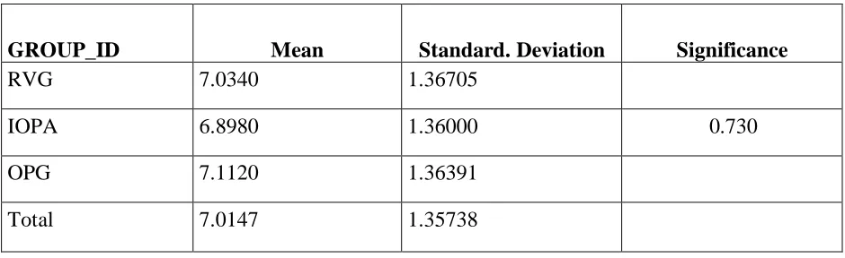
mean radiographic measurements in Conventional Intraoral periapical radiographs were
6.8980 mm and the mean Digital Radiovisiograph measurements were 7.0340 mm and
the mean Digital Orthopantomographic measurements were 7.1120 mm. (Table 1).
It was observed that Conventional Intraoral periapical radiographs evaluated
about 0.136 mm lesser bone loss on an average than Digital Radiovisiograph. Digital
Orthopantomographs evaluated about 0.078 mm lesser bone loss on an average than
Digital Radiovisiograph.
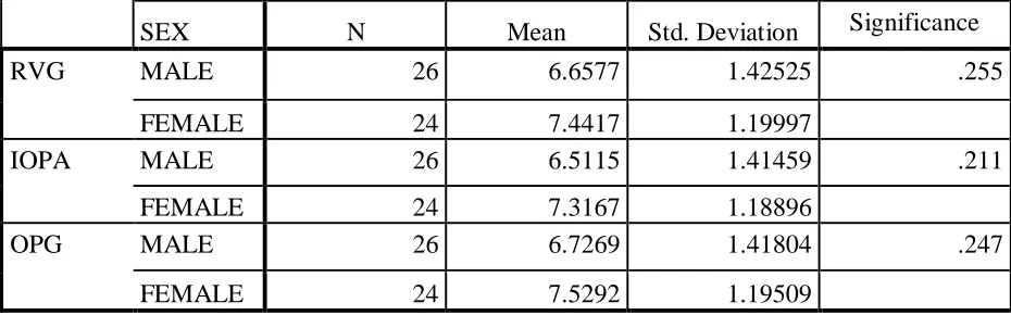
On comparing the radiographic dimensions between the genders, regarding the
Conventional Intraoral periapical radiographs, the mean value was 6.5115 in males and
7.3167 in females. In Digital Radiovisiograph, the mean value was 6.6577 in males and
7.4417 in females. In Digital Orthopantomographs, the mean value was 6.7269 in males
RESULTS
[Type text] Page 27
The comparison between them by Non-parametric one-way analysis of variance
(ANOVA) did not show statistically significant difference between Conventional
Intraoral periapical radiographs, Digital Radiovisiographs and Digital
Orthopantomographs.
We also compared the radiographic dimensions between Conventional Intraoral
periapical radiographs, Digital Radiovisiographs and Digital Orthopantomographs using
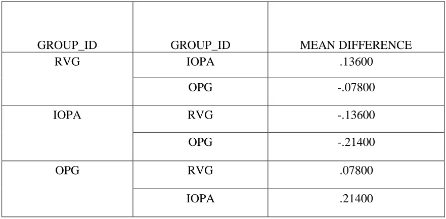
Post Hoc Tests. The mean difference between Digital Radiovisiographs and Conventional
Intraoral periapical radiographs was 0.13600 and the mean difference between Digital
Radiovisiographs and Digital Orthopantomographs was -0.07800. The mean difference
between Conventional Intraoral periapical radiographs and Digital Orthopantomographs
was -0.21400. The overall results showed the mean statistical difference between Digital
Radiovisiographs and Digital Orthopantomographs as 0.005, which is statistically
significant. The overall results showed the mean statistical difference between
Conventional Intraoral periapical radiographs and Digital Radiovisiographs as 0.005,
RESULTS
[Type text] Page 28
Table 1 : Dimensions of Interdental Bone loss in Conventional Intraoral periapical
radiographs, Digital Radiovisiographs and Digital Orthopantomographs.
GROUP_ID Mean Standard. Deviation Significance
RVG 7.0340 1.36705
IOPA 6.8980 1.36000 0.730
OPG 7.1120 1.36391
RESULTS
[Type text] Page 29
GRAPH 1 : Dimensions of Interdental Bone loss in Conventional Intraoral periapical
radiographs, Digital Radiovisiographs and Digital Orthopantomographs.
6.75 6.8 6.85 6.9 6.95 7 7.05 7.1 7.15
RVG IOPA OPG
MEAN
RESULTS
[Type text] Page 30
Table 2 : Dimensions of Interdental Bone loss in Conventional Intraoral periapical
radiographs, Digital Radiovisiographs and Digital Orthopantomographs, among the
Gender.
SEX N Mean Std. Deviation Significance RVG MALE 26 6.6577 1.42525 .255
FEMALE 24 7.4417 1.19997
IOPA MALE 26 6.5115 1.41459 .211
FEMALE 24 7.3167 1.18896
OPG MALE 26 6.7269 1.41804 .247
RESULTS
[Type text] Page 31
GRAPH 2 : Dimensions of Interdental Bone loss in Conventional Intraoral
periapical radiographs, Digital Radiovisiographs and Digital Orthopantomographs,
among the Gender.
6 6.2 6.4 6.6 6.8 7 7.2 7.4 7.6 7.8
RVG IOPA OPG
MALE
RESULTS
[Type text] Page 32
Table 3: Post Hoc Tests:
GROUP_ID GROUP_ID MEAN DIFFERENCE
RVG IOPA .13600
OPG -.07800
IOPA RVG -.13600
OPG -.21400
OPG RVG .07800
DISCUSSION
[Type text] Page 33
DISCUSSION
Periodontitis is an inflammatory disease of the supporting tissues of the teeth
caused by specific microorganisms, resulting in destruction of the periodontal ligament
and alveolar bone. Progressive loss of alveolar bone is the salient feature of periodontal
disease. Periodontitis involves progressive loss of alveolar bone around the teeth, if left
untreated leads to subsequent loss of teeth. It is characterized by periods of activity in
which the periodontal supporting structures are destroyed by the action of chemical
mediators of inflammation. Accurate detection of periodontal disease with the use of
radiographs helps in diagnosis, treatment and prognosis. The goal of dental radiology is
to make an accurate diagnosis using the most effective imaging modality with the lowest
radiation possible. (21)
Radiographs provide unique information about the status of the periodontium and
a permanent record of the condition of the bone throughout the course of the disease.
Radiographs aid the clinician in identifying the extent of destruction of alveolar bone,
local contributing factors, and features of the periodontium that influence the prognosis.
The diagnosis of periodontal disease is primarily based on clinical examination. The
clinical findings of periodontal osseous destruction can be confirmed by radiographic
DISCUSSION
[Type text] Page 34
Radiography is a well established procedure in daily dental practice and is still the
most basic and an important diagnostic tool available. Radiographs play an integral role
in the assessment of periodontal diseases. Conventional bitewing and intra oral periapical
radiographs are commonly used to detect alveolar bone loss associated with periodontal
disease. They provide unique information about the status of the periodontium and a
permanent record of the bone throughout the course of the disease. (18)
Monitoring bone changes with relatively simple radiographic procedures has
proven to be an elusive objective in clinical periodontics. Bone loss has been expressed
as a percent of total root length or of total tooth length, and more recently in terms of
absolute measurements in millimeters. Low sensitivity for subtle changes is considered to
be the major limitation of these conventional interpretations of radiographic images of
periodontal bone support.
Along with marginal inflammation, periodontal pocket formation, and attachment
loss, alveolar bone loss is a primary feature of periodontitis. The height of the alveolar
bone may be evaluated by intrasurgical inspection or, less invasively, by radiographic
examination; however, radiographic assessment tends to underestimate the amount of
bone loss. Changes of mineralized tissue like alveolar bone may be detected
DISCUSSION
[Type text] Page 35
The advent of direct digital imaging has introduced a versatile imaging tool that
can be used for a variety of tasks, including detection of alveolar crestal bone defects.
The reported prevalence of alveolar bone loss, which is a common dental disease state,
may vary depending on the epidemiologic conditions of the study. (5)
In periodontal diseases, the bone destruction pattern is divided into horizontal
(even) and oblique (vertical/angular) defects. In the vertical pattern, bone destruction
does not proceed in a symmetrical pattern. The severity of bone destruction varies in
different parts around the tooth, which explains why the alveolar crest does not
correspond to cemento-enamel junction and is not parallel to it. This bone destruction
pattern gives rise to bony defects in which the base of the defect is located more apical to
the alveolar crest.
Diagnosis and accuracy in determining the exact location, extent and
configuration of bony defects are of utmost importance to determine prognosis, to plan
treatment and to preserve the teeth in the long run. Because determination of the depth
and to some extent, the width of bony defects is an important parameter in the prognosis
of treatment, it is important to accurately measure these two parameters on radiographs to
develop a correct and appropriate treatment plan. Recently, digital radiography has
attracted a lot of attention in determining the depth, width and topography of bony
DISCUSSION
[Type text] Page 36
evaluated using an automated instrument to diagnose periodontal lesions and assess the
treatment success. (15)
The mean radiographic measurements in conventional intraoral periapical
radiographs were 6.8980 mm and the mean digital radiovisiograph measurements were
7.0340 mm and the mean digital orthopantomographic measurements were 7.1120 mm.
It was observed that conventional intraoral periapical radiographs evaluated about
0.136 mm lesser bone loss on an average than digital radiovisiograph. Digital
orthopantomographs evaluated about 0.078 mm lesser bone loss on an average than
digital radiovisiograph.
The results in our study showed that the mean difference between digital
radiovisiographs and conventional intraoral periapical radiographs was 0.13600 and the
mean difference between digital radiovisiographs and digital orthopantomographs was
-0.07800. The mean difference between conventional intraoral periapical radiographs and
digital orthopantomographs was -0.21400. The overall results showed the mean statistical
difference between digital radiovisiographs and digital orthopantomographs as 0.005,
which is statistically significant. (21)
Karanprakash Singh (2015)18 conducted a study on comparision between
conventional radiography (Intraoral periapical radiographs) and digital radiography using
DISCUSSION
[Type text] Page 37
mandible conventional images showed average bone loss of 3.3 mm, while digital images
showed 4.0 mm of average bone loss which is statistically significant, which is similar to
this study.
On comparing the radiographic dimensions between the genders, regarding the
conventional intraoral periapical radiographs, the mean value was 6.5115 in males and
7.3167 in females. In digital radiovisiograph, the mean value was 6.6577 in males and
7.4417 in females. In digital orthopantomographs, the mean value was 6.7269 in males
and 7.5292 in females, which is contradictory to the study conducted by Karanprakash
SUMMARY AND CONCLUSION
[Type text] Page 38
SUMMARY:
The present study consisted of 50 samples. Subjects were of both sexes, who were
40-70 years with chronic periodontitis and were attending K.S.R Institute of Dental
Science and Research, Tiruchengode, Tamil Nadu, India. Radiographs were taken after
obtaining their informed consent. Conventional intraoral periapical radiographs, digital
radiovisiographs and digital orthopantamographs were taken for all the patients to
measure the interdental alveolar bone loss. Digital radiovisiographs was considered as the
gold standard in measuring the interdental alveolar bone loss. Alveolar bone loss was
measured from the Cemento Enamel junction to the most apical level of marginal bone.
In this study the mean value was compared between the three radiographic techniques
and it was revealed the mean value of conventional intraoral periapical radiographs was
less when compared to the mean value of digital radiovisiographs, and the mean value of
digital orthopantamographs was more when compared to the mean value of digital
radiovisiographs. Alveolar bone loss was compared between males and females. Females
SUMMARY AND CONCLUSION
[Type text] Page 39
CONCLUSION:
Based on the results of this study we conclude that digital radiovisiograph was
superior to conventional intraoral periapical radiograph and digital orthopantamograph
for the detection of interdental bone loss, due to reduced time and radiation exposure to
obtain the same diagnostic information. Digital radiographs showed better results when
compared to conventional radiographs in terms of alveolar bone loss as digital
BIBLIOGRAPHY
[Type text] Page 40
BIBLIOGRAPHY:
1. Volchansky A. A technique for the radiographic assessment of marginal alveolar bone.
2. Salonen LW, Frithiof L, Wouters FR, Helldén LB. Marginal alveolar bone height in an
adult Swedish population: A radiographic cross‐sectional epidemiologic study. Journal of
clinical periodontology. 1991 Apr;18(4):223-32.
3. Tonetti MS, Prato GP, Williams RC, Cortellini P. Periodontal regeneration of human
infrabony defects. III. Diagnostic strategies to detect bone gain. Journal of
periodontology. 1993 Apr;64(4):269-77.
4. Eickholz P, Kim TS, Benn DK, Staehle HJ. Validity of radiographic measurement of
interproximal bone loss. Oral Surgery, Oral Medicine, Oral Pathology, Oral Radiology
and Endodontics. 1998 Jan 1;85(1):99-106.
5. Nair MK, Ludlow JB, Tyndall DA, Platin E, Denton G. Periodontitis detection efficacy
of film and digital images. Oral Surgery, Oral Medicine, Oral Pathology, Oral Radiology
and Endodontics. 1998 May 1;85(5):608-12.
6. Eickholz P, Hausmann E. Accuracy of radiographic assessment of interproximal bone
loss in intrabony defects using linear measurements. European journal of oral sciences.
2000 Feb;108(1):70-3.
7. Wolf B, Bethlenfalvy EV, Hassfeld S, Staehle HJ, Eickholz P. Reliability of assessing
interproximal bone loss by digital radiography: intrabony defects. Journal of clinical
BIBLIOGRAPHY
[Type text] Page 41
8. Talaiepour AR, Panjnoush M, Soleimanishayeste Y, Abesi F, Sahba S. A survey on the
accuracy of radiovisiography in the assessment of interproximal intrabony defects.
Journal of Dentistry of Tehran University of Medical Sciences. 2005;2(1):29-32.
9. Parissis N, Kondylidou-Sidira A, Tsirlis A, Patias P. Conventional radiographs vs
digitized radiographs: image quality assessment. Dentomaxillofacial Radiology. 2005
Nov;34(6):353-6.
10. Li G, Engström PE, Welander U. Measurement accuracy of marginal bone level in
digital radiographs with and without color coding. Acta Odontologica Scandinavica. 2007
Jan 1;65(5):254-8.
11. Wong BK, Leichter JW, Chandler NP, Cullinan MP, Holborow DW. Radiographic
study of ethnic variation in alveolar bone height among New Zealand dental students.
Journal of periodontology. 2007 Jun;78(6):1070-4.
12. Oppermann RV. An overview of the epidemiology of periodontal diseases in Latin
America. Brazilian Oral Research. 2007;21(SPE):8-15.
13. Cochran DL. Inflammation and bone loss in periodontal disease. Journal of
periodontology. 2008 Aug;79(8S):1569-76.
14. Cochran DL. Inflammation and bone loss in periodontal disease. Journal of
periodontology. 2008 Aug;79(8S):1569-76.
15. Esmaeli F, Shirmohammadi A, Faramarzie M, Abolfazli N, Rasouli H, Fallahi S.
BIBLIOGRAPHY
[Type text] Page 42
indirect digital radiographic measurement and clinical measurement. Iranian Journal of
Radiology. 2012 Jun;9(2):83.
16. Vijay G, Raghavan V. Radiology in periodontics. Journal of Indian Academy of Oral
Medicine and Radiology. 2013;25(1):24.
17. Takeshita WM, Iwaki LC, Da Silva MC, Iwaki Filho L, Queiroz AD, Geron LB.
Comparison of the diagnostic accuracy of direct digital radiography system, filtered
images, and subtraction radiography. Contemporary clinical dentistry. 2013 Jul;4(3):338.
18. Singh S. Comparision between Conventional Radiography (IOPA) and Digital
Radiography using Bitewing Technique in Detecting the Depth of Alveolar Bone Loss.
Global Journal of Medical Research. 2015 May 28.
19. Preus HR, Torgersen GR, Koldsland OC, Hansen BF, Aass AM, Larheim TA,
Sandvik L. A new digital tool for radiographic bone level measurements in longitudinal
studies. BMC oral health. 2015 Dec;15(1):107.
20. Zaki HA, Hoffmann KR, Hausmann E, Scannapieco FA. Is radiologic assessment of
alveolar crest height useful to monitor periodontal disease activity?. Dental Clinics. 2015
Oct 1;59(4):859-72.
21. Ashwinirani SR, Suragimath G, Jaishankar HP, Kulkarni P, Bijjaragi SC, Sangle VA.
Comparison of diagnostic accuracy of conventional intraoral periapical and direct digital
radiographs in detecting interdental bone loss. Journal of clinical and diagnostic research:
BIBLIOGRAPHY
[Type text] Page 43
22. Mehdizadeh M, Maarefat N, Bagherieh S. Comparison of Accuracy of determining
the Distance between Alveolar Crest and Cementoenamel Junction in Digital
Radiography with Scanora and DentalEye Software Programs. The journal of
contemporary dental practice. 2016 Oct;17(10):815-9.
23. Longo DL, Fumes AC, de Oliveira DS, de Oliveira KH, Romualdo PC, Kuchler EC,
da Silva LA. Comparison of digital and conventional radiographic techniques. RSBO.
ANNEXURE
[Type text] Page 44
ANNEXURE-I
INFORMED CONSENT FORM
I ………. hereby declare that I clearly understood the
procedures of the study. Also, I declare that I give permission for the above mentioned
individual/organization/hospital to do the procedure to the individual/organization listed
above.
Signature ………. Date ………..
I have explained the above and answered all questions asked by the participant.
ANNEXURE
[Type text] Page 45
ANNEXURE
[Type text] Page 46
COMPARISON OF DIAGNOSTIC ACCURACY OF CONVENTIONAL
INTRAORAL PERIAPICAL RADIOGRAPH, DIGITAL
RADIOVISIOGRAPH AND DIGITAL ORTHOPANTOMOGRAPH IN
DETECTING INTERDENTAL BONE LOSS
ABSTRACT:
AIM: To compare the diagnostic accuracy of conventional intraoral periapical radiograph,
digital radiovisiograph and digital orthopantamograph in detecting interdental alveolar bone loss
using digital radiovisiographic measurements as the gold standard.
OBJECTIVES: The main objective of this study is to estimate the diagnostic accuracy of
conventional intraoral periapical radiographs, digital radiovisiography and digital
orthopantomography in detecting interdental alveolar bone loss using digital radiovisiography
measurements as the gold standard and to suggest the most accurate technique to be used in the
clinical departments.
MATERIALS AND METHODS: The size of the study sample consisted of 50 patients who
were randomly selected from the OPD of Department of Oral Medicine and Radiology. in K.S.R
dental college, Tamil Nadu, India, between October 2017 to September 2018, after obtaining
their informed consent. Fifty inter-dental sites were considered for the study. The sites included
distal surface of the mandibular first molar. Patients having generalized mild to severe chronic
periodontitis as assessed by measuring attachment loss and categorized as mild: 1-2 mm,
moderate: 3-4 mm, severe: ≥ 5 mm. A series of conventional intraoral periapical radiographs,
digital radiovisiographs and digital orthopantamographs were taken. Alveolar bone loss was
RESULTS:It was observed that Conventional Intraoral periapical radiographs evaluated lesser
bone loss on an average than Digital Radiovisiograph. Digital Orthopantomographs evaluated
lesser bone loss on an average than Digital Radiovisiograph.
CONCLUSION: Based on the results of this study we conclude that digital radiovisiograph was
superior to conventional intraoral periapical radiograph and digital orthopantamograph for the
detection of interdental bone loss, due to reduced time and radiation exposure to obtain the same
diagnostic information. Digital radiographs showed better results when compared to
conventional radiographs in terms of alveolar bone loss as digital radiographs has superior image