COMPARISON OF PRIMING MECHANISM OF ACTIVE PASSIVE
BILATERAL THERAPY AND MIRROR THERAPY ON HAND
FUNCTION IN POST – STROKE PATIENTS
A RANDOMIZED CONTROL STUDY
Dissertation submitted in
the Partial fulfilment
for the degree of
MASTER OF PHYSIOTHERAPY
(Neurology)
The Tamil Nadu DR. M.G.R. Medical University
Chennai
May 2019
PSG COLLEGE OF PHYSIOTHERAPY
PSG COLLEGE OF PHYSIOTHERAPY
Coimbatore
CERTIFICATE
This is to certify that the research work entitled
“COMPARISON OF
PRIMING MECHANISM OF ACTIVE PASSIVE BILATERAL THERAPY
AND MIRROR THERAPY ON HAND FUNCTION IN POST STROKE
PATIENTS – A RANDOMIZED CONTROL STUDY”
was carried out by
Reg.
No. 271720241,
of P.S.G. College of Physiotherapy, towards the partial fulfilment
for the degree of
MASTER OF PHYSIOTHERAPY (Physiotherapy in
Neurology),
affiliated to The Tamil Nadu Dr. M.G.R. Medical University,
Chennai.
Internal Examiner
External Examiner
PSG COLLEGE OF PHYSIOTHERAPY
Coimbatore
CERTIFICATE
This is to certify that the dissertation work entitled
“COMPARISON OF
PRIMING MECHANISM OF ACTIVE PASSIVE BILATERAL THERAPY
AND MIRROR THERAPY ON HAND FUNCTION IN POST STROKE
PATIENTS – A RANDOMIZED CONTROL STUDY”
was carried out by
PERIYASAMY.A, Reg. No. 271720241
of P.S.G. College of Physiotherapy,
towards
the
partial
fulfilment
for
the
degree
of
MASTER
OF
PHYSIOTHERAPY (Physiotherapy in Neurology),
affiliated to The Tamil
Nadu Dr. M.G.R. Medical University, Chennai.
PRINCIPAL
Prof. R.MAHESH, MPT,
Principal
P.S.G. College of Physiotherapy
Coimbatore - 641 004.
Place: Coimbatore
PSG COLLEGE OF PHYSIOTHERAPY
Coimbatore
CERTIFICATE
This is to certify that the research work entitled
“COMPARISON OF
PRIMING MECHANISM OF ACTIVE PASSIVE BILATERAL THERAPY
AND MIRROR THERAPY ON HAND FUNCTION IN POST STROKE
PATIENTS – A RANDOMIZED CONTROL STUDY”
was carried out by
PERIYASAMY.A, Reg. No. 271720241
of P.S.G. College of Physiotherapy,
towards the partial fulfilment for the degree of
MASTER OF PHYSIOTHERAPY
(Physiotherapy in Neurology),
affiliated to The Tamil Nadu Dr. M.G.R. Medical
University, Chennai, under my guidance.
Dr. R.BALAKRISHNAN MD, DM (Neuro), DNB (Neuro),
Professor & HOD,
Department of Neurology,
P.S.G Hospitals,
Coimbatore – 641 004.
Place: Coimbatore
PSG COLLEGE OF PHYSIOTHERAPY
Coimbatore
CERTIFICATE
This is to certify that the dissertation work entitled
“COMPARISON OF
PRIMING MECHANISM OF ACTIVE PASSIVE BILATERAL THERAPY
AND MIRROR THERAPY ON HAND FUNCTION IN POST STROKE
PATIENTS – A RANDOMIZED CONTROL STUDY”
was carried out by
PERIYASAMY.A, Reg. No. 271720241
of P.S.G. College of Physiotherapy,
towards
the
partial
fulfilment
for
the
degree
of
MASTER
OF
PHYSIOTHERAPY (Physiotherapy in Neurology),
Coimbatore, affiliated to
The Tamil Nadu Dr. M.G.R. Medical University Chennai, under our guidance.
Guide Co-Guide
Prof. R.MAHESH, MPT, Mr. J. Raja Regan, MPT,
Principal Associate Professor
P.S.G. College of Physiotherapy P.S.G. College of Physiotherapy
Coimbatore - 641 004. Coimbatore - 641 004.
Place: Coimbatore
ACKNOWLEDGEMENT
It is my privilege to express my deep sense of gratitude to the
GOD
for
showering his blessings, who has always been my source of strength and who
guides me throughout.
For the ancestors who paved the path before me upon whose shoulders I
stand. I dedicate this study to
my Dad (ANGAMUTHU), Mom (SELVI)
for
providing their moral support and love in each and every step of my life.
With due respect, I would like to express my immense gratitude to
Professor R. Mahesh, MPT, Principal,
PSG College of Physiotherapy,
Coimbatore, for his encouragement and inspiration during the course of my study.
I am thankful to my project guide
Mr. J. Raja Regan, MPT
for his
encouragement, inspiration and untiring efforts given throughout the study.
I feel it my duty to thank Professor
Dr. R. Balakrishnan MD, DM (Neuro), DNB
(Neuro),
Department of Neurology, PSG IMS&R Hospitals for his constant and
unwavering encouragement, who rendered his invaluable experience as guidance
to this project.
I also thank Professor
Dr. V. Ramamoorthy, MD, HOD,
Department of
PMR, PSG IMS& R Hospitals for his encouragement, who rendered his invaluable
experience as guidance to this project.
I am grateful to
Mr. A. Parthiban
,
MPT,
for his expert guidance and
constant support throughout the study.
My special thanks to
Dr. ANIL MATHEW Ph.D., Professor,
Department
of Biostatistics, PSG Institute of Medical Science and Research who gave me a
helping hand in statistical method of data analysis.
I thank all the members of
Institutional Review Committee of Research,
College of Physiotherapy and Human Ethics Committee of PSG Institute of
Medical Science and Research for their kind suggestions to complete the
dissertation.
Of all I would like thank
My Friends (SRIMATHI and NIVETHA),
seniors and juniors
for sharing their knowledge, love, support and exclusive
cooperation.
I also thank all the staff members of the PSG College of Physiotherapy and
Department of Physiotherapy for helping me to complete this project successfully.
Finally, I thank all the patients for their kind co-operation. Without their
involvement this project would have not been possible.
ABBREVIATIONS
APBT
-
Active Passive Bilateral Therapy
MT
-
Mirror Therapy
AFMUE
-
Abbreviated Fugl Meyer Upper Extremity
CONTENTS
CHAPTER TITLE PAGE NO
I INTRODUCTION 1
1.1 Need for the Study 3
1.2 Objective 3
1.3 Hypothesis 4
1.4 Operational Definitions 4
II LITERATURE REVIEW 5
III MATERIALS AND METHODS 9
3.1 Materials 9
3.2 Study Design 9
3.3 Study Setting 10
3.4 Human Participation Protection 10
3.5 Population/Participants 10
3.6 Sampling 10
3.7 Intervention 10
3.8 Criteria for Sample Selection 10
3.8.1 Inclusion Criteria 10
3.8.2 Exclusion Criteria 11
3.9 Study Duration 11
3.10 Instrument and Tools for Data collection 11
3.11 Technique of Data Collection 11
3.12 Technique of Data Analysis and Interpretation 13
IV DATA ANALYSIS AND INTERPRETATION 15
V RESULTS AND DISCUSSION 26
VI SUMMARY AND CONCLUSION 29
BIBLIOGRAPHY 30
LIST OF ANNEXURES
Annexure
Content
I.
Ethical Committee Clearance Letter
II.
Neurological Assessment Form for Stroke
III.
Record form
IV.
Informed Consent (English and Tamil)
V.
Assessment Tools
1
CHAPTER I
INTRODUCTION
Stroke is a disease of the central nervous system caused by impaired blood supply to brain, and it
disturb the normal function of motor system, perception, sensory, language and especially in
activities of daily living. We need our upper limb for most of the activities but in case of stroke,
upper limb movements are impaired. So our rehabilitation should be focused on upper limb
recovery [1].
Most of our activities of daily living are accomplished by upper limb especially we need our upper
limb to perform reaching in multi direction, grasping the different size and shape of the objects,
releasing the objects and manipulative functions [2]. Special fine motor function like prehension
and precision handling are done by our distal part of the upper limb. After stroke, all upper limb
functional activities are impaired, and immediately paralysis or weakness occurs in the arm and
hand, it leads to activity limitations or restriction in upper limb and it causes major impairment in
performing all our upper limb hand functions [3].
Priming is defined as a change in behavior based on previous stimuli. Priming, which may occur
after a single learning episode, is a type of implicit learning. The role of implicit learning in
physical therapy has been the subject of recent investigation. The learning of this mechanism was
different from other types of implicit learning because skill-learning requires repetition. It was
originated in psychology, but still investigated in neuroscience, neurorehabilitation, and cognitive
neuroscience using behavioral and brain mapping techniques. Both translational and clinical
studies have been examining motor priming as a tool for inducing neuroplasticity and it also
enhance the effects of rehabilitation. Priming can be categorized as a restorative intervention that
reduces impairment. Priming stimuli can be from the same modality as the Modal-specific
(accompanying task) or Cross-modal (from a different modality). Example for Modal-specific
priming is bilateral mirror symmetrical movement (it is a movement-based priming) and it also
increase the rate of motor learning in neurological condition. Cross-modal priming can also be
used to enhance motor learning. Examples are semantic priming such as reading relevant words
2
cross modal priming. Many psychology literature results shows that same modality priming is
effective than cross modal priming [4].
Active-passive bilateral priming is a pattern of coordinated movement it assist the paretic limb by
disinhibits the motor cortex [5]. Using Active Passive Bilateral Therapy (APBT) for motor priming
is noninvasive and it has no side effect. It relies upon a device which mechanically couples the two
hands. In APBT the non-paretic wrist actively perform flexion and extension and that produces
mirror symmetric movement of paretic wrist, that was caused due to linkage mechanism and it is
the specific advantage of this device. There by it reduces short latency intracortical inhibition
(SICI) within the passive motor cortex [6].
Mirror therapy is a type of intervention which uses the movement of sound side of the body, and
this movements are reflected in a mirror, as visual feedback. These feedback promotes bilateral
motor training and it also stimulates function of the brain [7]. Mirror therapy makes a visual
imagination of patient and felt like two hands are moving simultaneously. This will activate the
cerebral hemisphere and it forms the basis of neurological mechanism to brain plasticity. It
promote the recovery of the upper limbs and enhance the motor patterns, agility, and manipulation
skills of these limbs [8].
Action Research Arm Test (ARAT) is commonly used by physiotherapists in stroke Patients. It is
used to measures the upper extremity of hand function [9]. It has four subscales such as grasp, grip,
pinch and gross movements. It contains large or small object to strength upper extremity of the
hand function. Grasp activity have 6 task, Grip activity have 4 task, Pinch activity have 6 task and
Gross movements have 3 task. ARAT using 4 points; 0 = no movements, 1= perform the task
partially, 2 = complete the task but take 5 to 60 sec, 3 = preform the task within 5 sec. Total score
is 57. The patient position was seated in a firm chair and no arm rest. The head should be straight
position and body should maintain upright posture and with the trunk contacting the back of the
chair. During task period to prevent the patient from standing up, shifting laterally and leaning
forward. The ARAT has proven to have strong validity when compared with other upper extremity
functions scale. In a study comparing scores on the ARAT in stroke population with scores on
3
ARAT has high responsiveness and the ability to detect clinically significant changes in the motor
functioning of an individual’s upper extremity, particularly in stroke recovery population [10].
The Fugl-Meyer (FM) assessment is a stroke - specific, performance - based impairment index. It
is used to assess motor functioning, balance, sensation and joint functioning in patient [11].
Fugl-Meyer assessment is quantitative measures of motor impairment following stroke, in this
assessment consist of Abbreviated Fugl Meyer Upper Extremity scale (AFMUE) [12]. This scale is
used to check the reflex activities of biceps, triceps and finger flexors, and to check the movements
in wrist and hand of coordination and speed activity. This scale was newly designed to measure
the hand function ability of stroke patient. It is a 3 point scale; 0 = cannot perform, 1 = performs
partially, 2 = performs fully [13].
1.1 NEED OF THE STUDY:
Based on the available evidence the studies suggest that priming mechanism of
active passive bilateral therapy were effective in improving the hand function of post stroke
patient. Mirror therapy were also effective in improving the hand function of post stroke patient.
There are only a few studies that have investigated the effects of active passive bilateral therapy
and mirror therapy combined with exercise on improving the hand function and activities of daily
living in stroke patients. But there are no studies to compare the effect of these two therapies as
priming techniques to improve hand function in post stroke patients.
1.2 OBJECTIVES:
To determine the effectiveness of Active Passive Bilateral Therapy as priming technique
on hand function in post stroke patients.
To determine the effectiveness of Mirror Therapy as priming technique on hand function
in post stroke patients.
To compare the effect of priming mechanism of Active Passive Bilateral Therapy and
4
1.3 HYPOTHESIS:
Null Hypothesis (Hₒ): There will be no significant difference between the effects of priming mechanism of Active Passive Bilateral Therapy and Mirror Therapy on hand function in
post stroke patients.
Alternative Hypothesis (Ha): There will be significant difference between the effects of priming mechanism of Active Passive Bilateral Therapy and Mirror Therapy on hand function in
post stroke patients.
1.4 OPERATIONAL DEFINITION:
ABBREVIATED FUGL MEYER UPPER EXTREMITY (AFMUE)
Fugl Meyer Assessment scale is an index to assess the sensorimotor motor impairments in
individuals who have had stroke. Abbreviated Fugl Meyer Upper Extremity has been tested
extensively, and it was found to have excellent psychometric properties. It is used to measure the
impairments from proximal to distal and synergistic to isolated voluntary movement. It is
considered to assess the body function according to international classification of functioning,
5
CHAPTER II
LITERATURE REVIEW
Preeti Raghavan., (2015), conducted a study on upper limb motor impairment post stroke,
understanding upper limb impairment after stroke is essential to planning therapeutic efforts to
restore function. However determining which upper limb impairment to treat and how is complex
for two reasons: 1) the impairments are not static, i.e. as motor recovery proceeds, the type and
nature of the impairments may change; therefore the treatment needs to evolve to target the
impairment contributing to dysfunction at a given point in time. 2) multiple impairments may be
present simultaneously, i.e., a patient may present with weakness of the arm and hand immediately
after a stroke, which may not have resolved when spasticity sets in a few weeks or months later;
hence there may be a layering of impairments over time making it difficult to decide what to treat
first. The most useful way to understand how impairments contribute to upper limb dysfunction
may be to examine them from the perspective of their functional consequences. There are three
main functional consequences of impairments on upper limb function are: (1) learned nonuse, (2)
learned bad-use, and (3) forgetting as determined by behavioral analysis of tasks.
Mary Ellen Stoykov, et al., (2015) conducted a study on motor priming in neurorehabilitation study suggest thatThe challenge will be to determine which methods are most effective for various
rehabilitation diagnoses and how those with various levels of impairment and disability
differentially respond to the various method available for priming.
Mary Ellen Stoykov, et al., (2010) conducted an experimental study on active-passive bilateral therapy as a priming mechanism for individuals in the sub-acute phase of post-stroke Recovery: A
feasibility study with 32 stroke patients to assess the feasibility of treating inpatient stroke
survivors with active-passive bilateral therapy as a motor priming technique before occupational
therapy. Both fugl-meyer upper extremity scores and action research arm test scores improved in
this small group of test and control patients. The magnitude of improvement was greater in test
patients who received active-passive bilateral therapy plus unilateral training and this study
6
Cathy M. Stinear, et al., (2008) conducted a study on priming the motor system enhances the effects of upper limb therapy in chronic stroke with 3 groups (Control (n =16) APBT without
cross-over (n=16) APBT with cross-over (n=21)) to improve hand function and result shows that
APBT produced sustained improvements in upper limb motor function in chronic stroke patients
and induced specific and sustained changes in motor cortex inhibitory function. We speculate that
APBT may have facilitated plastic reorganization in the brain in response to motor therapy.
Jin-Young Park, et al., (2015) conducted a study on the effect of mirror therapy on upper-extremity function and activities of daily living in stroke Patients with two groups (Mirror group (n = 15) Control group (n = 15)) was conducted to compare abilities to perform activities of daily living. Results shows that paretic upper-extremity function and hand coordination abilities were
significantly different between the mirror therapy and sham therapy groups. Intervention in the
mirror therapy group was more effective than in the sham therapy group for improving the ability
to perform activities of daily living. Self-care showed statistically significant differences between
the two groups. This study concluded that mirror therapy is effective in improving paretic upper
extremity function and activities of daily living in chronic stroke patients.
Kyunghoon Kim, et al., (2016) conducted a study on effects of mirror therapy combined with motor tasks on upper extremity function and activities daily living of stroke patients a mirror
therapy group (n=12) and a conventional therapy group (n=13) measured by using action research
arm test & fugl meyer assessment scale. Results shows that both groups showed significant
differences between measurements taken before and after four weeks of therapy. In theintergroup
comparison, the mirror therapy group showed significant improvements compared with the
conventional therapy group, both in upper limb function and activities of daily living. This study
concluded that mirror therapy is more effective than conventional therapy for the training of stroke
7
Thomas Platz, et al., (2005) conducted a study on reliability and validity of arm function assessment with standardized guidelines for the fugl-meyer test, action research arm test and box
and block test: a multicenter study. The result shows that all three motor tests showed very high
inter-rater and test-retest reliability (ICC and rho for main variables > 0.95). Correlation between
the motor scales was very high (rho > 0.92). Motor scales correlated moderately highly with the
hemispheric stroke scale, a measure of impairment (rho = 0.660-0.689), but not with the modified
barthel index, a measure of the ability to cope with basic activities of daily living (rho =
0.044-0.086). The study conclude that standardized guidelines assured comparability of test
administration and scoring across clinical facilities. The arm motor scales provided information
that was not identical to information from the hemispheric stroke scale or the modified barthel
index.
Polykarpos Angelos Nomikos, et al., (2018) conducted a study on test-retest reliability of physiotherapists using the action research arm test in chronic stroke and to asses upper limb
function activityconvenience-snowball sampleof 20 international physiotherapist (mean age and
experience=32 ± 6.8 and 7.55 ± 7.4 years) used ARAT to score chronicstroke patient’s upper limb
function, observing a video at baseline and again ≈ 2 weeks later. The results shows that Spearman’s rho was found ≈ 0.78 at a significance level of 0.00. ARAT was scored with a mean
difference of 16.6 days and a mean change of 0.6 points was observed. Limits of agreement and
coefficient of reproducibility were ± 2.3 and ± 2.6 respectively. The patient’sarm impairment was
categorized as moderate and floor or ceiling effects were not detected. The study concluded that
ARAT is consistent, valid and should be used by physiotherapist in chronic stroke.
Johan Anton Franck, et al., (2017) conducted a study onchanges in hand function and arm-hand skill performance in patients after stroke during and after rehabilitation and to assess arm-hand
function by using fugl meyer test, action research arm test and grip strength and ABHILHAND.
The Results shows that eighty-nine stroke patients (M/F: 63/23; mean age: 57.6yr (+/-10.6);
post-stroke time: 29.8 days (+/-20.1) participated. All patients improved on arm hand function and
arm-hand capacity during and after rehabilitation, except on grip strength in the severely affected
subgroup. The study concluded that a majority of stroke patients across the whole arm-hand
8
and self-perceived arm hand skill performance. These were maintained up to one year
post-rehabilitation.
Elizabeth J Woytowicz, et al., (2017) conducted a study on determining levels of upper extremity movement impairment by applying cluster analysis to upper extremity fugl-meyer assessment in
chronic stroke and to measure the quantitatively define levels of upper extremity movement
impairment using cluster analysis of Fugl-Meyer upper extremity (FM-UE) with and without
reflex items. The results shows that FM-UE scores ranged from 2–63 (mean=26.9±15.7) with
reflex items and 0–57 (mean=22.1 ±15.3) without reflex items. Three clusters were identified. The
distributions of the FM-UE scores revealed considerable overlap between the clusters, therefore
four distinct stroke impairment levels were also derived. The study conclude that reflex items make
no difference to the overall scores of the test, supporting previous recommendations for the
exclusion of these items.
David J. Gladstone, et al., (2002) conducted a study on the Fugl-Meyer Assessment of motor recovery after stroke: A critical review of its measurement Properties. The study shows that there
is good evidence from several validation studies that the Fugl-Meyer scale is indeed measuring
what it is intended to measure. Significant correlations were found between the degree of motor
impairment measured on the Fugl-Meyer motor scale and activities of daily living total score
(0.75), hygiene (0.89), locomotion (0.76), feeding (0.72), and dressing (0.76). Total Fugl-Meyer
9
CHAPTER III
MATERIALS AND METHODS
3.1 MATERIALS:
Block wood 10cm cube.
Block wood 7.5 cm cube.
Block wood 5 cm cube.
Block wood 2.5 cm cube.
Cricket ball 7.5 cm.
Stone 10×2.5×1 cm.
Glass.
Tube 2.25cm.
Tube 1×16 cm.
Washer 3.5 cm diameter.
Over bolt.
Ball bearing 6mm.
Marble 1.5 cm.
Knee hammer.
Mirror Box.
3.2 STUDY DESIGN:
10
3.3 STUDY SETTING:
Department of Neurology and Stroke Rehabilitation Centre, PSG Hospitals, Coimbatore.
3.4 HUMAN PARTICIPATION PROTECTION:
The study was reviewed and approved by Institutional Human Ethics Committee, PSG
IMS&R.
3.5 POPULATION/PARTICIPANTS:
9 hemiparetic stroke patients.3.6 SAMPLING:
Convenience Sampling Method
3.7 INTERVENTION:
GROUP A (n = 5) – Active Passive Bilateral Therapy and Training Activities. 15 repetition / 5 sets/ 60 minutes per day for 2 weeks.
GROUP B (n = 4) – Mirror Therapy and Training Activities.
15 repetition / 5 sets/ 60 minutes per day for 2 weeks.
3.8 CRITERIA FOR SAMPLE SELECTION
3.8.1 Inclusion Criteria:
Age group of 40 to 65 yrs.
Both male & female.
First episode of ischemic stroke.
Unilateral stroke with right or left hemiparesis.
Medically stable patient.
11
Abbreviated Fugl Meyer for upper extremity (wrist, hand, co-ordination section) score between 3 to 25.
3.8.2 Exclusion Criteria:
Musculoskeletal problem that affect the intervention.
Severe upper extremity spasticity with modified ashworth scale of >2
Severe sensory loss (score 2) as assessed with the National Institute of Health Stroke Scale (NIHSS).
Visual impairments.
3.9 STUDY DURATION:-
Total duration of this study was 8 months.
3.10 INSTRUMENT& TOOL FOR DATA COLLECTION:
Action Research Arm Test.
Abbreviated Fugl Meyer Upper Extremity Scale.
3.11 TECHNIQUE OF DATA COLLECTION:
Patient was assessed for eligibility based on inclusion & exclusion criteria. After obtaining
the informed consent form, they were randomly allocated to Group A or B. Initial assessment
(pre-test) was taken by using outcome measures. Then the intervention was given to each group
separately for 2 weeks. Final assessment (post-test) was taken by using same outcome measures.
Comparison of pre-test and post-test values within the group and between the groups was done
12
SCHEMATIC REPRESENTATION OF FLOW OF PARTICIPANTS
Screened for eligibility (n=35)
No. of eligible patients (n=11)
No. of patients Included (n=11)
Group A (n=7) Pre test
AFMUE and ARAT
Group B (n=4) Pre test
AFMUE and ARAT
Intervention
APBT and Training Activities
Intervention
MT and Training Activities
Group A (n=5) Post test
AFMUE and ARAT
Group B (n=4) Post test
AFMUE and ARAT
Dropped (n=2)
1-Death,
1-Developed Visual
Impairments
Data Analysis
13
3.12 TECHNIQUE OF DATA ANALYSIS & INTERPRETATION:
Data collected from subjects were analyzed using paired‘t’ test to measure changes between pretest and posttest values of outcome measures within the group. Independent‘t’
test was used to measure the changes between the groups.
Paired‘t’ test
= Calculated Mean Difference of stroke and age matched healthy subject values
SD = Standard Deviation
n = Number of samples
d = Difference between stroke and age matched healthy subject values.
d
1
)
(
2
n
d
d
SD
SD
n
d
14 Independent‘t’ test
X1 = Mean difference in Group A
X2 = Mean difference in Group B
SD = Combined standard deviation of Group A and Group B
n1 = Number of patients in Group A
n2 = Number of patients in Group B
SD1 = Standard Deviation of Group A
SD2 = Standard Deviation of Group B
15
CHAPTER - IV
STATISTICAL ANALAYSIS AND INTERPRETATION
Data analysis is the systemic organization and synthesis of research data and testing of
research hypothesis using these data. Interpretation is the process of making sense of the results of
a study and examining the implication (Polit & Beck, 2004). The Pre-test and Post-test values for
Groups A and Group B were obtained before and after intervention. The hand function activity
was measured using Abbreviated Fugl Meyer Upper Extremity Scale [AFMUE] and Action
Research Aram Test [ARAT]. The Mean, Standard deviation and Paired “t” test values were used
to find out whether there was any significant difference between pre-test and post-test values
within the groups. Statistical analysis for the present study was done using SPSS (version 21)
Independent “t” test is used to find the significant differences between the groups after
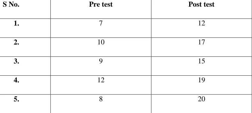
16 TABLE: 1
Pre and Post-test values of Abbreviated Fugl Meyer Upper Extremity (AFMUE) in Group A (n=5)
S No. Pre test Post test
1. 7 12
2. 10 17
3. 9 15
4. 12 19
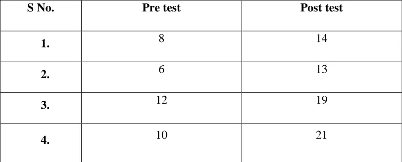
17 TABLE: 2
Pre and Post-test values of abbreviated Fugl Meyer Upper Extremity (AFMUE) in Group B (n=4)
S No. Pre test Post test
1. 8 14
2. 6 13
3. 12 19
4. 10 21
18 TABLE: 3
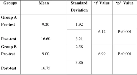
Mean, Standard Deviation and Paired‘t’ Test Values of AFMUE for Groups A & Group B
Groups
Mean
Standard
Deviation
‘t’ Value
‘p’ Value
Group A
Pre-test
Post-test
9.20
16.60
1.92
6.12
P<0.001
3.21
Group B
Pre-test
Post-test
9.00
16.75
2.58
6.99
P<0.001
3.86
Based on Table 3, the mean value of Group A 9.20 in pre-test and 16.60 in post-test,
Standard deviation was 1.92 in pre-test and 3.21 in post-test for Group A, the ‘t’ value using the
paired ‘t’ test was 6.12 which was greater than the table value of 2.77 at P<0.001. In Group B the
mean value was 9.0 in pre-test and 16.75 in post-test , standard deviation was 2.58 in pre-test and
3.86 in post-test for Group B, the ‘t’ value using the paired test was 6.99 which was greater than
the table value of 3.182 at p<0.001. This shows there is a significant improvement in Abbreviated
Fugl Meyer Upper Extremity in both groups. The result shows that pre-test and post-test mean
19 GRAPH: 1
Pre and Post-test Mean values of AFMUE for Group A & Group B
9.2 9
16.6 16.75
0 2 4 6 8 10 12 14 16 18
Group A (Active Passive Bilateral Therapy)
Group B (Mirror Therapy)
20 TABLE: 4
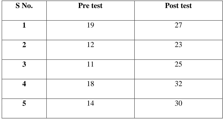
Pre and Post-test values of Action Research Arm Test (ARAT) in Group A (n=5)
S No. Pre test Post test
1 19 27
2 12 23
3 11 25
4 18 32
21 TABLE: 5
Pre and Post-test values of Action Research Arm Test (ARAT) in Group B (n=4)
S No. Pre test Post test
1. 16 34
2. 14 26
3. 11 23
22 TABLE: 6
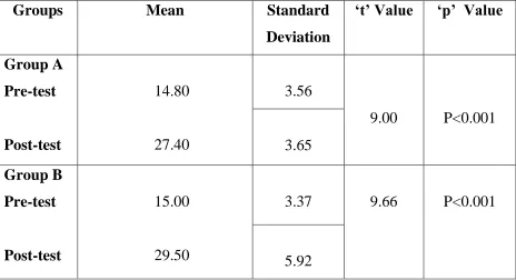
Mean, Standard Deviation and Paired‘t’ test values of ARAT for Group A & Group B
Groups
Mean
Standard
Deviation
‘t’ Value
‘p’ Value
Group A
Pre-test
Post-test
14.80
27.40
3.56
9.00
P<0.001
3.65
Group B
Pre-test
Post-test
15.00
29.50
3.37
9.66
P<0.001
5.92
Based on Table 6, the mean value of Group A was 14.80 in pre-test and 27.40 in post-test,
Standard deviation was 3.56 in pre-test and 3.65 in post-test for Group A, the ‘t’ value using the
paired ‘t’ test was 9.0 which was greater than the table value of 2.77 at P<0.001. In Group B the
mean value was 15.00 in pre-test and 29.50 in post-test, standard deviation was 3.37 in pre-test
and 5.92 in post-test for Group B, the ‘t’ value using the paired test was 9.66 which was greater
than the table value of 3.18 at P<0.001. This shows there is a significant improvement in Action
Research Arm Test in both groups. The result shows that pre-test and post-test mean difference of
23 GRAPH: 2
Pre and Post-test Mean values of ARAT for Group A and Group B
14.8 15
27.4
29.5
0 5 10 15 20 25 30 35
Group A (Active Passive Bilateral Therapy)
Group B (Mirror Therapy)
24 TABLE: 7
Comparison between the Post-test values of
Group A (Active Passive Bilateral Therapy) & Group B (Mirror Therapy)
Outcome
Measures
Mean
Standard
Deviation
“t” value
“p” value
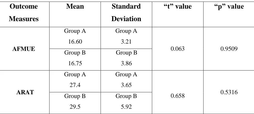
AFMUE Group A 16.60 Group A 3.21 0.063 0.9509 Group B 16.75 Group B 3.86 ARAT Group A 27.4 Group A 3.65
0.658 0.5316
Group B
29.5
Group B
5.92
The Independent‘t’ test was performed between Group A and Group B to analyse the
significance between the APBT and Mirror Therapy with Exercises on improving hand function in
post stroke patients. The Abbreviated Fugl Meyer Upper Extremity Scale between the groups
[image:34.612.74.570.165.383.2]were calculated using independent‘t’ test & the ‘t’ value was 0.063 which was lesser than the
table value of 2.36 at p>0.001.
The Action Research Arm Test between the group were calculated using independent‘t’
test & the obtained ‘t’ value is 0.658 which was lesser than that of table value of 2.36 at p>0.001.
Therefore the results of these statistical analyses showed that the Group A and Group B was same
25 GRAPH: 3
Comparison between the Post-test values of
Group A (Active Passive Bilateral Therapy) & Group B (Mirror Therapy)
16.6
16.75
16.5 16.55 16.6 16.65 16.7 16.75 16.8
Group A Group B
AFMUE
27.4
29.5
26 26.5 27 27.5 28 28.5 29 29.5 30
Group A Group B
26
CHAPTER V
RESULTS AND DISCUSSION
RESULT:
The data from Group A (APBT) and Group B (Mirror Therapy) for abbreviated fugl meyer
upper extremity were analysed using paired‘t’ test and independent ‘t’ test. The calculated value of paired‘t’ test for group A (APBT) is 6.12 and for group B (Mirror Therapy) is 6.99 which is
greater than the table value indicating there is a significant difference within both the group. The
value of independent‘t’ test for both groups is 0.063 which is lesser than the table value indicating
there is a no significant difference between the groups. Hence the null hypothesis is accepted,
alternate hypothesis is neglected.
The data from Group A (APBT) and Group B (Mirror Therapy) for action research arm
test were analysed using paired‘t’ test and independent‘t’ test. The calculated value of paired‘t’
test for Group A (APBT) is 9.00 and for Group B (Mirror Therapy) is 9.66 which is greater than
the table value indicating there is a significant difference within both the group. The value of
independent‘t’ test for both groups is 0.65 which is lesser than the table value indicating there is
no significant difference between the groups. Hence the null hypothesis is accepted, alternate
hypothesis is neglected.
The principle finding of the present study was that both Group A and Group B was
significantly effective for improving hand function activities. When comparing the results of
Group A and Group B there was no significant difference were noted. The study suggest that both
27
DISCUSSION:
Even though the various studies of active passive bilateral therapy and mirror therapy have been
shown to improve the hand function after a long term rehabilitation, to our knowledge evidence
on short term i.e., for two week effect of along with exercises in relation to hand function is not
clear. These leads a major route of idea in implenting in this study.
The aim of this study was to compare the effectiveness of Active Passive Bilateral Therapy and
Mirror Therapy along with exercise in post stroke patients.
Nine number of hemiparesis post stroke participants from inpatient department of neurology and
physical medicine and rehabilitation referred to stroke rehabilitation centre where recruited in this
study.
The participants who satisfied the selection criteria were randomly assigned into two groups by
convenient sampling. Baseline measurements were taken using the Abbreviated Fugl Meyer Upper
Extremity Scale (AFMUE) and Action Research Arm Test (ARAT) for both groups. One group
received active passive bilateral therapy along with exercise (APBT) and the other group received
mirror therapy (MT) along with exercise for 2 weeks. At the end of 2 weeks, participants were
again evaluated and measurements were taken using same outcome measures. Statistical analysis
for the present study was done using SPSS (version 21). And the results were mentioned above.
Mary Ellen Stoykov, et al., 2010 suggested that compare the conventional group and the experimental group, priming mechanism of active passive bilateral therapy is an adjuvant therapy
and useful to acute and sub-acute stroke patient. This device is not harmful to the patient. It also
restore of balance in the neural mechanism. Fugl Meyer upper extremity scale results shows greater
improvement in the hand function. Therefore experimental group of priming mechanism of active
passive bilateral therapy is more effective than the conventional group.
Kyunghoon Kim, et al., 2016 suggested that compare the mirror therapy along with exercise and the conventional therapy were applied to the stroke patient. In mirror therapy neural mechanism
of pre motor cortex area plays a major role motor recovery after brain damage. Functional
28
shows that significant improvement in upper limb function and activities of daily living was more
effective in the mirror therapy than the conventional therapy.
LIMITATION OF THE STUDY:
There was no control group without intervention, so it is difficult to exclude effects of the natural recovery process of hand function.
Within the study duration less number of patient meets the inclusion criteria, so we can’t able to complete the sample size.
SUGGESTION OF THE STUDY:
A Randomized Control Trails for large number of participant should be incorporate.
Long term follow up can be done to determine the effect of intervention.
29
CHAPTER VI
SUMMARY AND CONCLUSION
This study was conducted to compare the effect of priming mechanism of active passive bilateral
therapy and mirror therapy on hand function in post stroke patients. Thus the statically analysis
of data concluded that
There was significant improvement in hand function following active passive bilateral therapy in post stroke patient.
There was significant improvement in hand function following mirror therapy in post stroke patient.
Both active passive bilateral therapy group and mirror therapy group shows equally significant improvement on hand function.
30
BIBLIOGRAPHY
1.
Preeti Raghavan,
Upper Limb Motor Impairment Post Stroke
Phys Med
Rehabil Clin N Am. 2015 November; 26(4): 599–610.
2.
A motor relearining program for stroke
, janet H. carr, Roberta b. shepherd
butterworth-heinemann oxford 43-47.
3.
Motor Control
translating research in to clinical practice,anne shumway
cook marjorie h. woollacott wolters kulwer lippincott williams and wilkins,
475-480.
4.
Mary Ellen Stoykov,
and
Sangeetha Madhavan,
Motor Priming in
Neurorehabilitation
Neurol Phys Ther. 2015 January; 39(1): 33–42.
5.
Mary Ellen Stoykov,
and
James W. Stinear,
Active-Passive Bilateral
Therapy as a Priming Mechanism for Individuals in the Subacute Phase of
Post-Stroke Recovery:
Am J Phys Med Rehabil. 2010 November; 89(11):
873–878.
6.
Cathy M. Stinear, P. Alan Barber, James P. Coxon, Melanie K. Fleming
and Winston D.
Priming the motor system enhances the effects of upper limb
therapy in chronic stroke Brain (2008), 131, 1381^1390.
7.
Jin-Young Park, Moonyoung Chang, Kyeong-Mi Kim, Hee-Jung Kim
The effect of mirror therapy on upper-extremity function and activities of
daily living in stroke patients J. Phys. Ther. Sci. 27: 1681–1683, 2015.
8.
Kyunghoon Kim, Sukmin Lee, Donghoon Kim, Kyoungbo Lee, Youlim
Kim,
Effects of mirror therapy combined with motor tasks on upper extremity
function and activities daily living of stroke patients J. Phys. Ther. Sci. 28:
483–487, 2016.
31
10.
Polykarpos Angelos Nomikos, Nicola Spence, Mansour Abdullah
Alshehri,
Test-retest reliability of physiotherapists using the action research
arm test in chronic stroke J. Phys. Ther. Sci. 30: 1271–1277, 2018.
11.
Johan Anton Franck, Rob Johannes Elise Marie Smeets, Henk
Alexander Maria Seelen,
Changes in arm-hand function and arm-hand skill
performance in patients after stroke during and after rehabilitation PLOS
ONE https://doi.org/10.1371/journal.pone.0179453 June 14, 2017.
12.
Elizabeth J Woytowicz,
Jeremy C Rietschel,
Ronald N Goodman
,
Susan
S. Conroy
,
John D Sorkin, Jill Whitall,
and
Sandy McCombe Waller,
Determining Levels of Upper Extremity Movement Impairment by Applying
Cluster Analysis to Upper Extremity Fugl-Meyer Assessment in Chronic
Stroke
Arch Phys Med Rehabil. 2017 March; 98(3): 456–462.
ANNEXURE II
Neurological Physiotherapy Evaluation Form
I. Subjective Assessment Researcher/Therapist by:
Name: Age: Gender: M/F IP/OP
Occupation: Handedness: R/L Referred by:
Address:
Chief Complaints:
Past Medical History:
Personal History:
Family History:
Socioeconomic History:
Symptoms History:
Side: Site:
Onset: Duration:
Type: Severity:
Relieving Factors:
Vital Signs:
Temperature: Heart Rate:
Blood Pressure: Respiratory Rate:
II. Objective Examination
a)ON OBSERVATION:
Attitude of limbs:
Built:
Posture:
Gait:
Pattern of Movement:
Mode of Ventilation:
Type/ Pattern of Respiration:
Oedema:
Muscle Wasting:
Pressure Sores:
Deformity:
Wounds:
External Appliances:
b) ON PALPATION
Warmth:
Tenderness:
Swelling:
c) ON EXAMINATION
HIGHER MENTAL FUNCTIONS Level of Consciousness:
Orientation:
Person:
Place:
Time: Memory:
Immediate:
Recent:
Remote:
Verbal:
Visual:
Communication:
Cognition:
Fund of Knowledge:
Calculation:
Proverb Interpretation:
Attention:
Emotional Status:
Perception:
Body Scheme/ Body Imaging:
Special Senses:
Cranial Nerves:
Nerves Comments Nerves Comments
I - Olfactory VII - Facial
II - Optic VIII - VestibuloCochlear
III - Oculomotor IX - Glossopharyngeal
IV - Trochlear X - Vagus
V - Trigeminal XI - Accessory
VI - Abducent XII - Hypoglossal
SENSORY SYSTEM:
Location
Upper Extremity
Lower Extremity
Trunk
Comments
Sensation Rt. Lt Rt. Lt. Rt. Lt.
Superficial
Pain
Temperature
Touch
Pressure
Deep
Mov. Sense
Pos. Sense
Cortical
Tactile Localization
2 pt. discrimination
Stereognosis
Barognosis
Graphesthesia
Texture Recognition
Double Simultaneous
Stimulation
MOTOR SYSTEM:
Muscle Girth:
Voluntary Control:
Side Rt. Lt.
Upper Limb
Lower Limb
Range of Motion:
Joint Side Movement Limitation Limiting factor
Area Rt.(cm.) Lt.(cm.)
Arm
Forearm
Thigh
Shoulder
Elbow
Forearm
Wrist
Hand & Fingers
Hip
Knee
Ankle & foot
Cervical Spine
Thoracic Spine
Lumbar Spine
Limb Length
Side Rt.(cm.) Lt.(cm.)
True
Trunk Side Flexors
Pathological:
Coordination:
Trunk Extensors
Equilibrium tests Grade
Standing: Normal Posture
Standing: Normal Posture with vision occluded
Standing: Feet together
Non Equilibrium Tests Rt. Lt.
Finger to nose
Finger opposition
Mass Grasp
Pronation/Supination
Tapping (Foot)
Heel to knee
Drawing a circle(Hand)
Standing on one foot
Standing: Lateral trunk flexion
Tandem walking
Rebound test
Tapping (Hand)
Walk: Sideways
Walk: Backward
Walk in a circle
Walk on heels
Involuntary Movements:
Balance:
Sitting:
Standing:
Balance Reactions:
Posture:
Lying:
Sitting:
Standing:
Gait
Step Length:
Stride Length:
Base width:
Cadence:
Biomechanical Deviations:
Hand Functions:
Reaching:
Grasping:
Assisstive Devices:
III. Systems Review:
INTEGUMENTARY SYSTEM:
Skin Status:
Pressure Sores:
RESPIRATORY SYSTEM:
RS Status:
Secretions:
Pattern of breathing:
Chest wall/Thoracic spine deformity:
CARDIOVASCULAR SYSTEM
CVS Status:
Deep Vein Thrombosis:
MUSCULOSKELETAL SYSTEM
Contractures:
Subluxations:
Joint mobility:
Other pathology:
BLADDER & BOWEL FUNCTIONS
GASTROINTESTINAL SYSTEM
Status:
AUTONOMIC SYSTEM
Vasomotor:
Pseudomotor:
Trophic Changes:
Postural Hypotension:
Reflex Sympathetic Dystrophy:
IV. Functional Assessment: (The Functional Independence Measure)
Evaluation 1: Selfcare
Item 1. Food
Item 2. Care of appearance
Item 3. Hygiene
Item 4. Dressing upper body
Item 5. Dressing lower body
Evaluation 2: Sphincter control
Item 6. Control of bladder
Item 7. Control of bowel movements
Evaluation 3: Mobility
Item 8. Bed, chair, wheel chair
Item 9. To go to the toilets
Item 10. Bath-tub, shower
Item 11. Go, wheel chair
Item 12. Staircases
Evaluation 5: Communication
Item 13. Auditive comprehension
Item 14. Verbal expression
Evaluation 6: Social adjustment/cooperation
Item 15. Capacity to interact and to socially communicate
Item 16. Resolution of the problems
Item 17. Memory
Investigation Findings:
Problem List:
Sl. Impairment Functional Limitation
Functional Diagnosis:
V. Management
Goals:
Short term:
Long term:
Treatment:
ANNEXURE III
FOLLOW UP CHART
Name: D O A:
Age: OP NO:
Gender: IP NO:
Occupation: Address: Handedness:
Specific Complaints:
VITAL SIGNS:
Blood Pressure: Respiratory Rate: Temperature: Heart Rate:
OUTCOME MEASURE:
S.No
Outcome Measure Scores
Pre Test Post Test 1) Abbreviated FM for upper extremity (Wrist,
Hand, Co-ordination section)
2) Action Research Arm Test.
Study Volunteer ID:
ANNEXURE IV
Study Volunteer Name:PSG Institute of Medical Science and Research, Coimbatore
Institutional Human Ethics Committee
INFORMED CONSENT FORMAT FOR RESEARCH PROJECTS
I, Mr. Periyasamy. A, am carrying out a study on the topic: Comparison of Priming Mechanism of Active Passive Bilateral Therapy and Mirror Therapy on Hand Function in Post Stroke Patient as part of our research project being carried out under the aegis of the Department of:
Physiotherapy.
My research guide is: Mr. Mahesh. R MPT (Cardio Respiratory)
The justification for this study is: Bilateral priming is a neuromodulatory technique that evolved from bilateral training, which can be used to balance excitability between the cortices before training on unilateral task. The Active Passive Bilateral Therapy & Mirror Therapy can be used as a priming technique to improve hand functions. Using Active Passive Bilateral Therapy can generate crossed facilitation between the non-paretic & paretic upper limb. Mirror therapy uses the motions of the unaffected side of the body, reflected in a mirror, as visual feedback. This visual feedback enables bilateral motor training and stimulates functional improvement of brain function. There are no studies to compare the effect of these two therapies as priming techniques to improve hand function in post stroke patients. Hence there is a need for this study.
The objectives of this study are:
1. To determine the effectiveness of Active Passive Bilateral Therapy as priming technique on hand function in post stroke patient.
2. To determine the effectiveness of Mirror Therapy as priming technique on hand function in post stroke patient.
3. To compare the effect of priming mechanism Active Passive Bilateral Therapy and Mirror Therapy on hand function in post stroke patient.
Sample size: 52.
Participants are Post Ischemic Stroke – Hemiparesis Random Allocation of 26 Participant to Group A and 26 Participant to Group B.
Age group: 40 to 65 years
Location: Department of Physical Medicine Rehabilitation and Department of Neurology, PSG Hospital, Coimbatore.
Initial interview:45 minutes.
Final interview: 45 minutes.
If photograph is taken, purpose: Yes, without revealing the identity of yours we want to publish it in the project book, conferences and journals.
Data collected will be stored for a period of 5 years. We will not use the data as part of another study.
Clinical examination: Yes
Blood sample collection: Not Applicable Specify quantity of blood being drawn: ml.
No. of times it will be collected:
Whether blood sample collection is part of routine procedure or for research (study) purpose: 1. Routine procedure 2. Research purpose
Study Volunteer ID: Study Volunteer Name:
Specify purpose, discomfort likely to be felt and side effects, if any:
Whether blood sample collected will be stored after study period: Yes / No, NA it will be destroyed
Whether blood sample collected will be sold: Yes / No , NA
Whether blood sample collected will be shared with persons from another institution: Yes / No, NA
Medication given, if any, duration, side effects, purpose, benefits:
Whether medication given is part of routine procedure: Yes / No, NA (If not, state reasons for giving this medication)
Whether alternatives are available for medication given: Yes / No, NA (If not, state reasons for giving this particular medication)
Benefits from this study:
To Imrove Hand Function Activities
To Improve Activity Daily Living Risks involved by participating in this study: No Risks Involved
How the results will be used:
Peer-reviewed scientific journals
Conference presentation
Internal report
If you are uncomfortable in answering any of our questions during the course of the interview / biological sample collection, you have the right to withdraw from the interview / study at anytime. You have the freedom to withdraw from the study at any point of time. Kindly be assured that your refusal to participate or withdrawal at any stage, if you so decide, will not result in any form of compromise or discrimination in the services offered nor would it attract any penalty. You will continue to have access to the regular services offered to a patient. You will NOT be paid any remuneration for the time you spend with us for this interview / study. The information provided by you will be kept in strict confidence. Under no circumstances shall we reveal the identity of the respondent or their families to anyone. The information that we collect shall be used for approved research purposes only. You will be informed about any significant new findings - including adverse events, if any, – whether directly related to you or to other participants of this study, developed during the course of this research which may relate to your willingness to continue participation.
Consent: The above information regarding the study, has been read by me/ read to me, and has been explained to me by the investigator/s. Having understood the same, I hereby give my consent to them to interview me. I am affixing my signature / left thumb impression to indicate my consent and willingness to participate in this study (i.e., willingly abide by the project requirements).
Signature / Left thumb impression of the Study Volunteer / Legal Representative:
Signature of the Interviewer with date: Witness:
Contact number of PI: 9788128210
Contact number of Ethics Committee Office: 0422 4345818
பூ
சா
க ா
மருத்துவக்
ல்லூரி
மற்றும்
ஆராய்ச்சி
நிறுவனம்
,
க ாவவ
மனித
நெறிமுவைக்
குழு
ஒப்புதல்
படிவம்
.
தேதி:
அ
.
நபரியசாமி
ஆகிய நான் பூ சா த ா மருத்துவக் ல்லூரியின் / மருத்துவ மனையின்இயன்முவை மருத்துவ துனையின் கீழ், முதன்வம வழிமுவையான (PRIMING MECHANISM)
நசயலில் ஈடு படும் இருதரப்பு சிகிச்வச(ACTIVE PASSIVE BILATERAL THERAPY)மற்றும் ண்ணாடி சிகிச்வச (MIRROR THERAPY) பயன்படுத்தி பக் வாத பாதிப்பினால் ஏற்படும் வ நசயல்பாட்வைஅதி ரித்தல் என்ை ேனைப்பில் ஓர் ஆய்வு தமற்க ாள்ள உள்தளன்.
எண்
ஆய்வு
வழி ாட்டியில்
மாணவர் ளுக்கு
மட்டும்
:
ஆய்வு
கமற்
ந ாள்வதன்
அடிப்பவை
:
பக் வாேத்தின் பாதிப்பிைால் ன னள அனசக் முடியாே கசயனை உருவாக்கிைது.
இேைால் அன்ைாட வாழ்வில் நனடமுனைச் சார்ந்ே கசயல் ள் குனைகிைது. இந்ே ஆய்வின்
மூைம் முேன்னம வழிமுனை கசயலில் ஈடுபடும் இருேரப்பு சிகிச்னச மற்றும் ண்ணாடி
சிகிச்னச பயன்படுத்தி ன கசயல்பாட்னட அதி ரித்ேல் மற்றும் அன்ைாட கசயல்திைனை
அதி ரிக் முயற்சிக் ப்படுகிைது.
ஆய்வின்
கொக் ம்
:
1) கசயலில் ஈடுபடும் இருேரப்பு சிகிச்னச பயன்படுத்தி பக் வாே பாதிப்பிைால் ன
கசயல்பாடு மற்றும் அன்ைாட கசயல் திைனை அதி ரித்ேல்.
2) ண்ணாடி சிகிச்னச பயன்படுத்திய பக் வாே பாதிப்பிைால் ன கசயல்பாடு மற்றும்
அன்ைாட கசயல் திைனை அதி ரித்ேல்.
3) ஒப்பீட்டுத்திைன் மூைமா முேன்னம வழிமுனை கசயலில் ஈடுபடும் இருேரப்பு சிகிச்னச
மற்றும் ண்ணாடி சிகிச்னசனய பயன்படுத்தி பக் வாே பாதிப்பிைால் ன கசயல்பாடு
மற்றும் அன்ைாட கசயல்திைனை அதி ரித்ேல்.
ஆய்வில்
பங்குநபறும்
ெபர் ளின்
எண்ணிக்வ
= 52 ணினியின் ராண்டம் எண் ளின் மூைமா26 நபர் ள் A குழுவிற்கும் மற்றும் 26 நபர் ள் B குழுவிற்கும் பயன்படுத்ேப்படும்.
ஆய்வில்
பங்குநபறுகவார்
மற்றும்
வயது
: 40 - 65 வயதுள்ளவர் ள்.இந்ே ஆய்வின் எங் ளுடன் ஒத்துனைக்குமாறு த ட்டுக் க ாள்கிதைாம். நாங் ள் சிை ே வல் னள இந்ே ஆய்விற் ா தச ரிக் உள்தளாம்.
ஆய்வு
நசய்யப்படும்
முவை
:
இந்ே ஆய்வின் கமாத்ே ாை அளவு 10 மாேங் ள், இந்ே ஆய்வில் பக் வாேத்திைால்
பாதிக் ப்பட்டு ன னள அனசக் முடியாே நினையில் உள்ளார் 26 நபர் னள க ாண்டு
இருக்குழுவா பிரித்துக் க ாள்தவாம்.
முேல் மற்றும் இரண்டாம் உனரயின்தபாது பக் வாேத்திைால் ன னள அனசக் முடியாே
கசயல்திைனை அளவிடும் Abbrevated Fugl Meyer Upper Extremity (FMUE) மற்றும் Action
Research Arm Test (ARAT) என்ை படிவத்னே க ாண்டு அளவீடு ள் குறிக் ப்படும்.
ஆய்வில் ஈடுபடும் முேல் குழுவிற்கு கசயலில் ஈடுபடும் இருேரப்பு சிகிச்னச (ACTIVE PASSIVE
BILATERAL THERAPY) மற்றும் பயிற்சி ளும் இரண்டாம் குழுவிற்கு ண்ணாடி சிகிச்னச
(MIRROR THERAPY) மற்றும் பயிற்சி ள் இனணந்து க ாடுக் ப்படும்.
இச்சிகிச்னச கோடர்ந்து ஒரு வாரத்திற்கு 5 முனை வீேமா இரண்டு வாரங் ளுக்கு அளிக் ப்படும்.
இரண்டாவது வார இறுதியில் மீண்டும் (FUME மற்றும்ARAT) அளவீடு ள் அளிக் ப்படும். பின்பு
அேனை முேலில் எடுத்ே அளவீடு ளுடன் ஒப்பிட்டு ன கசய்யபாடு ஆராயப்படும்.
முதன்வம
கெர் ாணல்
:
60 நிமிடங் ள்.இந்ே ஆய்வில் கினடக்கும் ே வல் னள 5 வருடங் ள் பாது ாக் ப்படும். இந்ே ே வல் ள் தவறு
ஆய்வுக்குப் பயன்படுத்ேப் பட மாட்டாது.
சு ாதாரக்
ல்வி
: அமர்வு ள்:
2 முனை ஒரு அமர்வுக் ாை தநரம்: 45 நிமிடங் ள்.மருத்துவ
பரிகசாதவன ள்
:
உண்டு
இரத்த
மாதிரி
கச ரிப்பு
:
இல்வை
இரத்த
மாதிரி
எடுப்பது
வழக் ம�