Transcriptome Profiling of the Virus-Induced Innate Immune
Response in
Pteropus vampyrus
and Its Attenuation by Nipah Virus
Interferon Antagonist Functions
Nicole B. Glennon,a,bOmar Jabado,c*Michael K. Lo,dMegan L. Shawa
Department of Microbiology,a
Graduate School of Biomedical Sciences,b
and Institute for Genomics and Multiscale Biology, Department of Genetics and Genomic Sciences,c
Icahn School of Medicine at Mount Sinai, New York, New York, USA; Centers for Disease Control and Prevention, Viral Special Pathogens Branch, Atlanta, Georgia, USAd
ABSTRACT
Bats are important reservoirs for several viruses, many of which cause lethal infections in humans but have reduced pathogenic-ity in bats. As the innate immune response is critical for controlling viruses, the nature of this response in bats and how it may differ from that in other mammals are of great interest. Using next-generation transcriptome sequencing (mRNA-seq), we pro-filed the transcriptional response ofPteropus vampyrusbat kidney (PVK) cells to Newcastle disease virus (NDV), an avian paramyxovirus known to elicit a strong innate immune response in mammalian cells. ThePteropusgenus is a known reservoir of Nipah virus (NiV) and Hendra virus (HeV). Analysis of the 200 to 300 regulated genes showed that genes for interferon (IFN) and antiviral pathways are highly upregulated in NDV-infected PVK cells, including genes for beta IFN, RIG-I, MDA5, ISG15, and IRF1. NDV-infected cells also upregulated several genes not previously characterized to be antiviral, such as RND1,
SERTAD1, CHAC1, and MORC3. In fact, we show that MORC3 is induced by both IFN and NDV infection in PVK cells but is not induced by either stimulus in human A549 cells. In contrast to NDV infection, HeV and NiV infection of PVK cells failed to in-duce these innate immune response genes. Likewise, an attenuated response was observed in PVK cells infected with recombi-nant NDVs expressing the NiV IFN antagonist proteins V and W. This study provides the first global profile of a robust virus-induced innate immune response in bats and indicates that henipavirus IFN antagonist mechanisms are likely active in bat cells.
IMPORTANCE
Bats are the reservoir host for many highly pathogenic human viruses, including henipaviruses, lyssaviruses, severe acute respi-ratory syndrome coronavirus, and filoviruses, and many other viruses have also been isolated from bats. Viral infections are portedly asymptomatic or heavily attenuated in bat populations. Despite their ecological importance to viral maintenance, re-search into their immune system and mechanisms for viral control has only recently begun. Nipah virus and Hendra virus are two paramyxoviruses associated with high mortality rates in humans and whose reservoir is thePteropusgenus of bats. Greater knowledge of the innate immune response ofP. vampyrusbats to viral infection may elucidate how bats serve as a reservoir for so many viruses.
I
n recent years, interest in bats has steadily increased because of the discovery that they ecologically maintain viruses pathogenic to humans. To date, over 100 viruses have been isolated from bats (1,2), and they are believed to be a reservoir host for lyssaviruses (including rabies virus) (1,2), henipaviruses (3,4), filoviruses (5, 6), and severe acute respiratory syndrome coronavirus (7). Inter-estingly, current data suggest that both natural and experimental viral infections are predominantly clinically asymptomatic in bats (3,8–14). Clinical pathogenicity has been seen only with lyssavirus infections (though the severity of the infection is attenuated com-pared with that of lyssavirus infections in other mammalian spe-cies) (15–19) and Tacaribe virus infections (20), and the filovirus Lloviu virus was associated with bat die-offs in caves in Europe (21). Bats possess many characteristics that make them adept at spreading pathogens, including viruses. They are the only mam-mals that fly, enabling them to travel large distances (22,23); they have life spans of up to 35 years (24); some hibernate, allowing overwintering of pathogens (25); and they can live in crowded, large population roosts, facilitating pathogen spread (26). How-ever, none of these physical characteristics can fully explain the ability of bats to harbor so many human pathogens while rarely showing any sign of disease. Precisely what accounts for thisbal-ance between the ability of bats to support virus replication and control viral disease remains an open question.
Insight into the immune response of bats could shed light on how they function as reservoir hosts. Current research does not yield a complete picture of the immune system for any one species
Received4 February 2015Accepted2 May 2015
Accepted manuscript posted online13 May 2015
CitationGlennon NB, Jabado O, Lo MK, Shaw ML. 2015. Transcriptome profiling of the virus-induced innate immune response inPteropus vampyrusand its attenuation by Nipah virus interferon antagonist functions. J Virol 89:7550 –7566. doi:10.1128/JVI.00302-15.
Editor:D. S. Lyles
Address correspondence to Megan L. Shaw, [email protected].
*Present address: Omar Jabado, Bristol-Myers Squib, Lawrenceville, New Jersey, USA.
Supplemental material for this article may be found athttp://dx.doi.org/10.1128 /JVI.00302-15.
Copyright © 2015, American Society for Microbiology. All Rights Reserved.
doi:10.1128/JVI.00302-15
on November 7, 2019 by guest
http://jvi.asm.org/
of bats. Several studies that have examined various aspects of the immune system of a variety of bat species have been done; these studies can be summarized, with the caveat that bats are a diverse order and these findings may not hold true across all species of bats. Examination of the adaptive immune system shows that bats should have all of the cell types required for mounting an effective adaptive immune response, and sequence analysis shows that an-tibodies produced by bats should undergo class switching, VDJ recombination, and somatic hypermutation (27–31). When look-ing at the innate immune system, specifically, the production of and signaling through interferon (IFN), bats possess the necessary signaling molecules, both RIG-I-like receptors (RLRs) and Toll-like receptors (TLRs) (32,33), type I and II IFNs, and type I and II IFN receptors (34–37). Cells and tissues from bats also have the ability to respond to a variety of stimuli [poly(I·C), virus infection, IFN treatment], producing type I and III IFNs and interferon-stimulated genes (ISGs) (34,36–41). It appears as though an an-tiviral state can be established in bat cells because both IFN and supernatant from infected bat cells (which should contain se-creted IFNs and other cytokines) protect against further virus challenge (37,39,41–43).
While some differences in immunity between bats and other mammals have been discovered, the most notable differences are seen in adaptive immunity. Both B and T cells in bats display delayed and reduced responses to antigen (28,29), and there ap-pears to be less somatic hypermutation and potentially weaker antibody binding to antigens (30,31). Differences in the innate immune response could elucidate how bats can control so many different virus infections, but to date, bats do not appear to have any differences greater than the natural variation expected within an order of mammals. More insight into the innate immune sys-tem of bats is necessary to see if they possess something unique or lack a standard component or pathway that distinguishes them from other mammals.
Nipah virus (NiV) and Hendra virus (HeV) are two closely related paramyxoviruses whose reservoir hosts are thePteropus genus of bats. The first outbreak of NiV occurred in 1998 in Ma-laysia, where pigs were an intermediate and amplifying host that then spread the virus to humans (44). Since then, yearly outbreaks have occurred in Bangladesh, with the virus appearing to spread directly from bats to humans (45,46). The first outbreak of HeV occurred in Australia in 1994. This virus mainly affects horses, but spillover to humans has occurred (47). Like almost all pathogenic viruses, NiV and HeV have evolved mechanisms to block the type I IFN pathway, and these mechanisms have been characterized in human cells. The IFN antagonists for NiV and HeV are the four proteins produced from the P gene: P, V, W, and C (48). NiV and HeV are believed to share the same mechanism of IFN inhibition. The V and W proteins each block IFN production, with V inhib-iting MDA5 signaling in a mechanism conserved among all paramyxoviruses (49–51) and W blocking IFN production from both TLRs and RLRs in a step downstream of IFN regulatory fac-tor 3 (IRF3) activation (52). The P, V, and W proteins all inhibit IFN signaling by binding to STAT1 and preventing phosphoryla-tion in response to IFN (53–56). However, IFN production and signaling during NiV infectionin vitro are highly variable, de-pending on the cell type used. In the 293T cell line and human endothelial cells, IFN production is limited, but IFN production is observed in infected human neuronal cells, one of the major tar-gets of NiV replication (57,58). IFN signaling is reported to be
functional in NiV-infected 293T cells (with exogenous IFN), yet there is no evidence of phosphorylated STAT1 in NiV-infected Vero cells treated with IFN, a block relieved by a mutation in the STAT1 binding site in the P gene (57,59).
For henipavirus IFN antagonist function in bats, it is reported that HeV V can block IFN production in Tb1 Lu cells, lung cells fromTadarida brasiliensis, a bat species that is not a reservoir host for henipaviruses (60). This supports the findings of Virtue et al. (61), who report that immortalized cells from aPteropus alectobat do not produce IFN when infected with NiV or HeV, as measured by quantitative PCR. These cells also do not signal through IFN when exogenous IFN is added following infection. However, when theseP. alectocells were infected with HeV and analyzed using next-generation transcriptome sequencing (mRNA-seq), a low-level upregulation of immune genes was observed (62).
To obtain a more detailed picture of the antiviral gene profile in bats, we infected immortalized kidney cells from aPteropus vampyrusbat with Newcastle disease virus (NDV), which allowed characterization of a robust innate immune response. Next-gen-eration mRNAseq analysis identified many well-characterized IFN-related genes as well as several genes with no reported antivi-ral role that may represent previously undiscovered innate im-mune response genes in bats. When these cells were infected with NiV and HeV, no such response was observed, and through the use of recombinant NDV (rNDV) expressing the NiV V and W proteins, we show that the IFN antagonist functions of these pro-teins are active inP. vampyruscells.
MATERIALS AND METHODS
Cell culture.PrimaryP. vampyruskidney (PPVK) cells were cultured
from aP. vampyruskidney received from the Lubee Bat Conservancy in Gainesville, FL. Upon delivery, the kidney was minced, digested in tryp-sin, and suspended in fetal bovine serum (FBS). This solution was then centrifuged and filtered through a cell strainer to remove debris. Cells were resuspended in 1⫻minimal essential medium with 10% FBS and plated in a 10-cm dish. We immortalized these cells by stably expressing human telomerase (hTERT) to create the PVK4 cell line. Approximately 7⫻105GP2-293 cells (Clontech), which stably expressgagandpolfrom Moloney murine leukemia virus, were transfected in suspension with 2.5 g each of plasmids expressing vesicular stomatitis virus G glycoprotein and pBABE-hTERT-puro (Addgene plasmid 1771; a gift from Bob Wein-berg [63]) using Lipofectamine 2000 (Invitrogen). The medium was changed at 24 h postinfection (hpi), and supernatant (2 ml) from trans-fected cells was harvested at 48 h, filtered through a 0.45-m-pore-size filter, and buffered with HEPES (Corning) at a final concentration of 25 mM. Approximately 1⫻106PPVK cells were inoculated with 200l of undiluted supernatant in the presence of 8g/ml Polybrene (Sigma) for 2 h, and then the inoculum was replaced with 2 ml Dulbecco modified Eagle medium (DMEM; Corning) containing 8g/ml Polybrene. This process was repeated 24 h later. At 48 h after dosing of the initial inoculum, cells were transferred to a new dish, and 24 h later puromycin (Gemini) was added at a concentration 2g/ml (Fig. 1a). A549, MVI, and Vero cells were purchased from ATCC. Marcel A. Müller and Christian Drosten (University of Bonn, Bonn, Germany) kindly provided the EidNi/41.2 and RoNi/7.3 cells (39) and EpoNi/22.1 cells (64), whose isolation and characterization have been described previously. All cells were cultured in DMEM with 10% FBS (HyClone) and 5% penicillin-streptomycin (Corn-ing).
Viruses.rNDV B1⌬V/NDV V (NDV) has been previously described
(65) and is a recombinant virus with mutations removing the P-gene edit site, thereby deleting expression of the V protein. Expression of the V protein is restored by insertion of the V open reading frame. This virus was used because it replicates like wild-type NDV B1 (65) and serves as the
on November 7, 2019 by guest
http://jvi.asm.org/
parental control for viruses expressing the NiV IFN antagonist proteins (see below). rNDV B1⌬V/NiV V (rNDV/NiV V) and rNDV B1⌬V/NiV W (rNDV/NiV W) have been described previously (66) and are recombi-nant viruses also with mutations removing the P-gene edit site (deleting expression of the V protein). The NiV IFN antagonists V and W were inserted as additional open reading frames. Green fluorescent protein (GFP)-tagged NDV (NDV-GFP) has been described previously (67). NDVs were grown in embryonated chicken eggs, and titers were deter-mined on Vero cells by immunofluorescence with an antinucleoprotein (anti-NP) polyclonal antibody (P. Palese lab, Icahn School of Medicine at Mount Sinai [ISMMS]). All NDV infections were performed as follows: NDV was diluted in 200l phosphate-buffered saline (PBS; Corning) with 5% penicillin-streptomycin and 0.3% bovine serum albumin (Fi-scher) and was inoculated on 1⫻106cells. After 1 h, the inoculum was replaced with 500l Opti-MEM medium (Life Technologies). For NDV-GFP, infections were performed in a 96-well format with 4⫻104cells per well.
All infections with HeV and NiV were performed under biosafety level 4 (BSL4) containment conditions at the CDC laboratory. HeV and NiV growth curve assays were performed in PVK4 cells. PVK4 cells (1⫻105) were seeded into each well of 12-well plates and grown overnight. On the following day, cells were infected with 1⫻10550% tissue culture infective doses (TCID50s) (multiplicity of infection [MOI]⫽1) of NiV (Malaysian strain, GenBank accession numberAF212302) or HeV (GenBank acces-sion numberAF017149) in 250l for 2 h, and then the inoculum was replaced with growth medium. At 12, 24, 48, and 72 hpi, supernatants were harvested for determination of the TCID50s. Cytopathic effect-based TCID50assays were performed by infecting Vero cells in a 10-fold dilution series of samples (10⫺1to 10⫺8) in a 96-well plate format, infecting 9 wells per sample and dilution step. Plates were read after 7 days to ensure a clear distinction between infected and uninfected wells at the highest dilutions.
RNA isolation.RNA from PVK4, PPVK, MVI, EidNi/41.2, RoNi/7.3,
EpoNi/22.1, and A549 cells infected with NDVs or treated with universal IFN was purified using an RNeasy miniprep (Qiagen) kit following the manufacturer’s protocols. RNA was then digested with Turbo DNA-free DNase (Ambion). RNA from PVK4 cells infected with HeV or NiV was isolated using a MagMAX sample preparation system (ABI) and DNase treated according to the manufacturer’s protocols.
mRNA-seq.PPVK cells were infected with NDV at an MOI of 0.2 for
24 h as described above. Purified RNA from NDV- or mock-infected cells was poly(A) selected, converted to cDNA, ligated to platform-specific adapters, and then amplified using an Illumina TruSeq RNA kit. Mas-sively parallel sequencing was carried out on an Illumina HiSeq 2000 sequencer at a density of one sample per lane. Reads containing rRNA or Illumina adapters were filtered. The remaining reads were mapped to the
P. vampyrusgenome build 1 using the TopHat (v2) program and the Ensembl (release 75) gene annotation set. The Cufflinks (v2) program was used to create ade novotranscriptome annotation set, using the Ensembl (release 75) gene annotation set as a reference. Mapped read files were analyzed by use of the HTSeq-Count script to generate a list of reads per exon gene model, and then the DESeq R package was used to perform differential expression analysis. We used the suggested parameters in DESeq for analysis of nonreplicate data sets.
All gene ontology studies were performed on annotated genes differ-entially regulated more than 2-fold. The list of genes was uploaded into the Interferome (v2.01) database (68). The list of genes and expression data were also uploaded for analysis into Qiagen’s Ingenuity pathway analysis database. Genes were mapped to canonical pathways in the Inge-nuity database according to the ratio of genes present in our data set to the total number of genes in the pathway, and thePvalue was calculated by Fisher’s exact test. Graphical representation of the Ingenuity data was generated by creating bubble graphs for the canonical pathways, with each
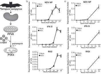
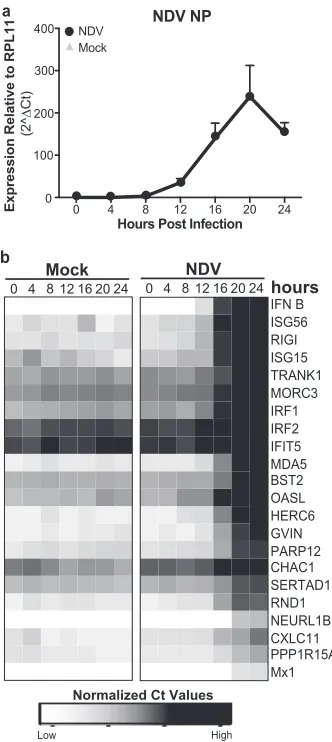
FIG 1Formation and characterization ofPteropus vampyruscells. (a) PPVK cells were cultured from a kidney and then immortalized by ectopically expressing human telomerase using a Moloney murine leukemia virus system, creating the cell line PVK4. (b) qRT-PCR expression of NDV NP, IFN-, and RIG-I in PPVK cells infected with NDV at an MOI of 0.2 or mock infected. The data shown are normalized to the level of actin expression. Error bars represent standard deviations. No data point signifies that no signal was detected for that sample. (c) qRT-PCR expression of NDV NP, IFN-, and RIG-I in PVK4 cells infected with NDV at an MOI of 2 or mock infected. The data shown are normalized to the level of actin expression. Error bars represent standard deviations. No data point signifies that no signal was detected for that sample.
Glennon et al.
on November 7, 2019 by guest
http://jvi.asm.org/
[image:3.585.120.473.67.326.2]pathway representing one bubble. The area of the bubble was calculated using the negative log of thePvalue. The expression dots of the genes present in each pathway were calculated by making a heat map of the mRNA-seq expression data in the R Studio interface.
For genes that were not identified with the Ensembl or BLAST pro-gram, we identified putative genes by evidence of expression in mRNA-seq and homology on the basis of similarity to known proteins. Putative mRNA sequences were extracted using the Cufflinks program-derived region on the genome and then stored as a FastA file of nucleotide se-quences. The sequences were compared to those in the nonredundant UniRef protein database (http://www.uniprot.org/uniref/) using a trans-lated nucleotide query against the protein sequences. The top-scoring protein in the UniRef database was used to annotate the putative bat gene.
qRT-PCR.PPVK, EidNi/41.2, RoNi/7.3, EpoNi/22.1, MVI, or A549
cells (1⫻105) were infected with NDV at an MOI of 2 as described above, and RNA was harvested at 24 hpi. PVK4 cells were infected with NDV at an MOI of 2 as described above, and RNA was harvested every 4 h for 24 h. PVK4 cells (1⫻105) were treated with 10,000 or 2,000 U/ml of univer-sal IFN (Sigma) diluted in 2 ml of DMEM for 24 h. EidNi/41.2, RoNi/7.3, EpoNi/22.1, MVI, or A549 cells (1⫻105) were treated with 2,000 U/ml of universal IFN (Sigma) diluted in 2 ml of DMEM for 24 h. PVK4 cells (1⫻ 105) were infected with HeV and NiV at an MOI of 10. RNA was isolated as described above. cDNA was synthesized using Invitrogen SuperScript III first-strand synthesis (Life Technologies). Fifteen nanograms of cDNA was used in each quantitative reverse transcription-PCR (qRT-PCR). qRT-PCR was performed on a Roche LightCycler 480 apparatus using the Roche LightCycler 480 SYBR green I master mix reagent. Validated prim-ers used for human RPS18, RND1, MORC3, SERTAD1, and PPP1R15A were purchased from Bio-Rad. All other primers were designed and syn-thesized (sequences are available upon request).
Data were analyzed using the normalized expression (2⫺⌬CT) or fold
induction (2⫺⌬⌬CT) threshold cycle (C
T) method as noted previously
(69). Values represent the means from three replicates, and error bars represent standard deviations. Heat maps were made using the⌬CT
method in the R Studio program.
NDV-GFP cytokine bioassay.PVK4 cells were infected with NDV at
an MOI of 2 or PVK4 cells were infected with HeV or NiV at an MOI of 10 for 24 h. The supernatant was UV inactivated and then incubated on 4⫻ 104fresh PVK4 cells (NDV, HeV, NiV) or Vero cells (NDV) for 24 h. Cells were then infected with NDV-GFP at an MOI of 1 for 24 h. Cells were fixed with 4% paraformaldehyde (Fisher) and permeabilized with 0.05% Triton (Fisher) in PBS. Cells were then stained with ethidium bromide for 1 h. The levels of GFP and ethidium bromide were read on a TTP Labtech Accumen HCI laser scanning microplate imaging cytometer. Values are presented as the ratio of the GFP signal to the ethidium bromide signal, and then the level of infection for the control sample treated with DMEM only was normalized to 100%. All other values are relative to that value. Values are the averages from three replicates, and error bars represent standard deviations.
Antibodies, immunofluorescence, and Western blotting.PVK4 cells
(1⫻106) were incubated with 10,000 U/ml universal IFN for 1, 3, or 6 h. Cells were fixed with 4% paraformaldehyde and permeabilized with 0.05% Triton X-100 in PBS. Cells were stained using a human monoclonal STAT1 antibody (catalog number 610185; BD Sciences). Images were taken on a Leica SP5 DM microscope with a 63⫻immersion lens objec-tive.
PVK4 cells (1⫻105) were seeded into each well of 12-well plates overnight. On the following day, cells were infected with NiV or HeV at an MOI of 1, 5, or 10 in 250l for 2 h, and then the inoculum was replaced with growth medium. At 12, 24, 48, and 72 hpi, the supernatant was discarded and cells were fixed for 20 min in 10% formalin (Sigma) solu-tion. Cells were then permeabilized with PBS with 0.1% Triton X-100 for 15 min and then incubated overnight with an anti-N antibody (which is cross-reactive against both NiV and HeV) in 5% milk in PBS containing 0.1% Tween 20 (PBS-T) at a dilution of 1:1,000. On the following day, the
cells were washed 3 times with PBS-T and incubated with anti-mouse Dylight 488 secondary antibody (Bethyl Laboratories) in PBS-T contain-ing 5% milk at dilutions of 1:500 and 1:1,000, respectively. After 3 washes, fluorescent images were taken on a Zeiss Axiovert 200 microscope at⫻20 magnification.
PVK4 cells infected with rNDV/NDV V, rNDV/NiV V, or rNDV/NiV W were infected as described above and harvested at 0, 8, and 24 h in 200 l 2⫻SDS lysis buffer with-mercaptoethanol (Sigma), freeze-thawed once, and boiled. Five percent of the lysate was separated on an SDS-polyacrylamide gel (Bio-Rad) and transferred to a polyvinylidene difluo-ride membrane (Bio-Rad). The membrane was blocked in PBS with 0.05% Tween 20 (Fisher) and 5% milk. Polyclonal antibodies against NiV V and NiV W (70) and NDV NP (a gift from the P. Palese lab, ISMMS) and a monoclonal antibody against human actin (Sigma) were diluted in the PBS–Tween 20 –milk solution, and the mixture was incubated on individ-ual membranes overnight at 4°C. Following washing, membranes were incubated with the appropriate horseradish peroxidase-conjugated sec-ondary antibody (Invivogen). Following washing, the membranes were illuminated using enhanced chemiluminescence (PerkinElmer), and the signal was detected on a ChemiDoc imager (Bio-Rad).
RESULTS
Primary and immortalizedP. vampyruscells mount an immune response to NDV infection.To generateP. vampyruscells for our studies, we obtained a kidney from a euthanizedP. vampyrusbat at the Lubee Bat Conservancy in Gainesville, FL. Primary cells were cultured from this kidney, establishing the primaryP. vampyrus kidney (PPVK) cell culture. We then immortalized these primary cells by transduction with a human telomerase-expressing retro-virus to generate the cell line PVK4 (Fig. 1a). To examine the antiviral response ofP. vampyruscells, we infected both primary and immortalized cell cultures with Newcastle disease virus (NDV), a potent activator of the mammalian antiviral response (65). Infection resulted in increased NDV NP expression both in PPVK cells (Fig. 1b) and in PVK4 cells (Fig. 1c), as assayed by quantitative RT-PCR (qRT-PCR). This signifies that cultures of both types of cells can support NDV infection and gene expression, establishing that NDV can be used to explore the response ofP. vampyruscells to virus infection. Since beta IFN (IFN-) and RIG-I are activation markers of the host innate im-mune response, we investigated their expression and found that NDV infection of PPVK and PVK4 cells elicited IFN-production and increased the levels of RIG-I expression, indicative of IFN signaling (Fig. 1bandc).
The innate immune response of PVK4 cells protects against virus challenge.To determine if the antiviral response produced in NDV-infected PVK4 cells is functional, we tested whether the cytokine-containing supernatant from infected cells would be able to protect naive cells from infection. This was achieved by overlaying UV-inactivated supernatant from either NDV- or mock-infected PVK4 cells onto both fresh PVK4 cells and Vero cells and then challenging the cells with NDV-GFP 24 h later (Fig. 2a). Supernatant from infected PVK4 cells restricted NDV-GFP infection in PVK4 cells but not in Vero cells (Fig. 2b). This indicates a species-specific protective activity of the antiviral cyto-kines produced byP. vampyruscells. In contrast, universal IFN, which is a recombinant form of IFN-␣that is functional in many mammalian species (71), protected both PVK4 and Vero cells against NDV-GFP infection (Fig. 2b). As another indicator of a functional antiviral response, we examined the nuclear transloca-tion of STAT1 as a measure of JAK/STAT signaling. In PVK4 cells, 1 h of treatment with universal IFN (10,000 U/ml) caused the
on November 7, 2019 by guest
http://jvi.asm.org/
translocation of STAT1 into the nucleus, which returned to its more diffuse cytoplasmic location after 6 h of treatment (Fig. 2c). This further exemplifies the activation of JAK/STAT signaling in response to type I IFN inP. vampyruscells.
Transcriptome analysis of NDV-infectedP. vampyruscells reveals global immune pathway activation.To obtain a global view of theP. vampyrusinnate immune response following NDV infection and to explore the identity and profile of virus-regulated genes, we performed next-generation mRNAseq of NDV-infected (MOI⫽0.2, 24 h) and mock-infected PPVK cells using the Illu-mina platform. As summarized inTable 1, this analysis yielded 7⫻107to 10⫻107total reads and 3⫻107to 5⫻107unique reads per sample, which indicates considerable depth. A draft genome of P. vampyrusis publically available, and 82% of the total reads in the mock-infected cells and 64% of the total reads in the infected cells were mapped to the genome using TopHat. Of the total reads that remained unmapped by TopHat, 8% (mock-infected cells) and 7% (infected cells) could be mapped to theP. vampyrus ge-nome using the BLAST program. We also mapped 19% of the total reads (5% unique reads) from the infected cells to the rNDV ge-nome, but reassuringly, a negligible number of reads were mapped to the virus from the mock-infected sample. This demonstrates a
relatively good infection level in the infected cells and little cross contamination in the mock-infected cells. We also analyzed the data for the presence of other microbes (viruses, bacteria, fungi, parasites) but found that sequences for those microbes comprised nothing greater than 0.0001% of total reads (data not shown), suggesting that theP. vampyruscells are not harboring any known
[image:5.585.135.452.64.406.2]FIG 2Functional characterization of the PVK4 cell innate immune response. (a) NDV-GFP cytokine bioassay. (b) NDV-GFP infection (MOI⫽1) in PVK4 and Vero cells treated with universal IFN or UV-inactivated supernatant from NDV-infected (MOI⫽2) or mock-infected PVK4 cells. The data shown are the ratio of the GFP signal to the ethidium bromide signal for staining of nuclei. The level of infection for supernatant containing DMEM only was normalized to 100%, and the results for the other samples are shown as a percentage of that value. Error bars represent standard deviations. (c) Immunofluorescence staining of STAT1 in PVK4 cells following universal IFN treatment over 6 h.
TABLE 1mRNA-seq reads from NDV-infected PPVK cells
Read group and infection
Total no. of readsa
% reads:
Mapped toP. vampyrusgenome
Mapped to
NDV Unmapped TopHat BLAST
All reads
Mock 90,272,827 82 8 0 10 NDV 74,396,949 64 7 19 10
Unique reads
Mock 54,744,789 79 6 0 15 NDV 38,574,846 74 8 5 13
aFiltered for rRNA and quality. Glennon et al.
on November 7, 2019 by guest
http://jvi.asm.org/
[image:5.585.299.544.589.715.2]microbial species and that potentially latent viruses were not acti-vated under our culture conditions. Approximately 10% of reads remained unmapped in both mock-infected and infected samples. This value is considered reasonable for this type of analysis.
We next performed quantitative analysis using DeSeq to exam-ine regulated gene expression. Assessment was limited to the reads that were mapped to theP. vampyrusgenome using TopHat, and compared with the gene expression in mock-infected cells, NDV infection upregulated 309 genes and downregulated 16 genes ( Ta-ble 2). TheP. vampyrusgenome is only partially annotated; there-fore, gene names could be assigned to only 267 of these genes. Annotation was carried out using Ensembl and homology with other mammalian species (Table 2; see also Table S1 in the sup-plemental material). We attempted to identify the 52 genes that remained unidentified after analysis with the Ensembl and BLAST programs by examining homology on the basis of similarity to known proteins. Briefly, putative mRNA sequences were extracted using the Cufflinks-derived region on the genome and were then compared to the sequences in the nonredundant UniRef protein database using a translated nucleotide search against the protein sequences (BLASTX). Portions of some of these transcripts showed homology at the protein level to known and putative genes in other species. For example, one of the transcripts showed homology to TRIM3 (the highest percent identity was toMyotis lucifugus; data not shown); however, this homology was not seen at the nucleotide level, potentially indicating a gene that is highly divergent in bats compared to its degree of divergence in other mammalian species.
Since NDV is a potent activator of the IFN response, we com-pared the genes differentially expressed during NDV infection with the genes in a public database of known IFN-regulated genes in mice and humans (Interferome, v2.0 [68]) to see how many of the genes induced during NDV infection were ISGs. We found that 156 genes differentially expressed during NDV infection of PPVK cells are known ISGs in either humans or mice (Table 2; see also Table S1 in the supplemental material). Further gene ontol-ogy analysis was performed using Ingenuity pathway analysis, which determined the cellular canonical pathways significantly enriched in the data set. Significant pathways included many im-mune pathways as well as virus detection pathways and apoptosis pathways (Fig. 3; see also Table S2 in the supplemental material). The most significantly enriched pathway within our data set was that of interferon signaling (P⫽5⫻10⫺20), followed by
activa-tion of an IRF by cytosolic pattern recogniactiva-tion receptors (P⫽1⫻ 10⫺15) and role of pattern recognition receptors in recognition of
bacteria and viruses (P⫽3.16⫻10⫺12). The top 10 pathways are illustrated inFig. 3, with the size of the gray circle corresponding to thePvalue. The regulated genes contributing to each pathway
are shown in blue, with higher levels of expression being indicated by the darker colors, and vice versa. Many genes are present in more than one pathway, showing the high degree of overlap be-tween different immune pathways.
We chose 22 of the 306 upregulated genes for validation using qRT-PCR analysis, including 6 genes (CHAC1, MORC3, PPP1R15A, RND1, SERTAD1, NEURL1B) not present in the Interferome (v2.0) database. Primers specific to these genes were designed on the basis of the sequence information from the mRNAseq data. We infected PVK4 cells with NDV over a 24-h time course and harvested RNA every 4 h for analysis by qRT-PCR. Virus infection increased the expression of all 22 genes, con-firming the mRNAseq results (Fig. 4b). Host gene induction in response to NDV infection occurred at between 12 and 20 h, reaching a maximum at 24 h. While PVK4 cells expressed some genes (e.g., IFN-, NEURL1B, Mx1) at a low basal level and others (e.g., IRF1, IRF2, BST2) at a higher basal level, NDV infection induced the expression of all genes tested, regardless of basal ex-pression levels. These included CHAC1, MORC3, PPP1R15A, RND1, SERTAD1, and NEURL1B, which are not known to be ISGs, at least not in human or murine systems. This gene expres-sion profile is likely representative of an antiviral state in infected PVK4 cells.
Gene expression profiling of NDV-infectedP. vampyruscells elucidates potential differences between the regulation of bat and human antiviral genes.Genes that are transcriptionally up-regulated by NDV infection most likely include typical ISGs that respond to activation of the IFN-␣/receptor, but they could also include genes that are activated by the virus inde-pendently of IFN. To distinguish between the two, we either treated PVK4 cells with universal IFN (10,000 U/ml, 24 h) or infected PVK4 cells with NDV (MOI⫽2, 20 h) and monitored the expression of select genes identified by the mRNAseq study, including both known ISGs and five of the genes yet to be described as ISGs. We found that IFN treatment of PVK4 cells upregulated all genes known to be ISGs (Fig. 5a). These genes included signaling molecules involved in the production of IFN-(RIG-I, MDA5), transcription factors (IRF1, IRF2), ef-fector molecules (Mx1, BST2), and some with poorly charac-terized functions (GVIN, TRANK1). While treatment with universal IFN did upregulate all of these genes, the levels of upregulation of many of these genes did not reach the same levels seen with NDV infection. This suggests that virus infec-tion has secondary signals that amplify these genes further.
We examined the data for those genes not previously described to be ISGs (CHAC1, MORC3, PPP1R15A, RND1, SERTAD1) in more detail to see if their regulation is IFN de-pendent or indede-pendent (Fig. 5b). Since stimulation with IFN induced RND1 and MORC3 more than 5-fold in PVK4 cells compared with their level of induction in mock-treated cells, we believe that these genes may be ISGs inP. vampyruscells. However, SERTAD1 did not show a statistically significant in-crease in the level of induction upon IFN stimulation, and PPP1R15A and CHAC1 showed increases of less than 5-fold com-pared with their levels of induction in mock-treated cells, so we conclude that these three genes are likely not ISGs inP. vampyrus cells (Fig. 5aandb). A lower concentration of universal IFN (2,000 U/ml) provided results that were the same as those depicted inFig. 5aandb(data not shown).
[image:6.585.40.287.79.159.2]MORC3, PPP1R15A, RND1, and SERTAD1 are all poorly
TABLE 2Differentially expressed genes in NDV-infected PPVK cells
Gene
No. of genes
Upregulated Downregulated
Total 309 16
Annotated 257a 10
Unannotated 52 6
Known IFN stimulated 155 1
a
Includes 6 gene families and 3 duplicates.
on November 7, 2019 by guest
http://jvi.asm.org/
characterized in other mammalian species. To determine if induc-tion of these genes is regulated similarly or differently in human cells, we treated cells of the human A549 lung cell line with uni-versal IFN (2,000 U/ml, 24 h) or infected them with NDV (MOI⫽ 2, 24 h) and examined gene expression levels via qRT-PCR (Fig. 5c). As a control for IFN signaling, we observed the induction of ISG56 (which is regulated through IFN-dependent and -indepen-dent mechanisms) following treatment with universal IFN or NDV infection. Only expression of SERTAD1 was significantly increased by universal IFN, but it was increased less than 2-fold compared with the level of expression in mock-infected cells, so it appears that MORC3, PPP1R15A, RND1, and SERTAD1 are probably not ISGs in human cells, at least A549 cells. In response to NDV infection, the expression levels of RND1, PPP1R15A, and SERTD1 did increase; however, MORC3 expression was not in-duced upon NDV infection (Fig. 5c). Therefore, we have identi-fied potential species-specific differences in the regulation of
cer-tain antiviral response genes. MORC3, which is upregulated in response to both IFN and virus in PVK4 cells, did not respond to either stimulus in A549 cells, while RND1 was regulated by virus infection in both species but responded only to IFN in PVK4 cells. Since bats are a very diverse order, we wanted to determine if IFN or NDV regulation of RND1 and MORC3 occurs in other bat species or if the regulation isP. vampyrusspecific. We infected and treated cell lines from four additional species of bats,Eidolon hel-vum(EidNi/41.2 cells),Epomops buettikoferi(EpoNi/22.1 cells), Rousettus aegyptiacus(RoNi/7.3 cells), andMyotis velifer incautus (MVI cells), with NDV and IFN and tested for the expression of RND1 and MORC3. These cells represent cells of kidney origin from three megabat species (EidNi/41.2, EpoNi/22.1, and RoNi/ 7.1 cells) and cells of skin tumor origin from one microbat species (MVI cells). qRT-PCR primers were designed using the sequence of theP. vampyrusgene. The increased expression of NDV NP in all infected cell types demonstrates that all four cell lines are
sus-FIG 3Canonical pathways enriched during NDV infection. Genes upregulated during NDV infection were clustered using the Ingenuity pathway analysis software. Shown here are the top 10 canonical pathways and the genes upregulated during NDV infection in each pathway. The area of the circle is inversely proportional to thePvalue of the pathway. Genes with darker colors are expressed at higher levels. See Table S2 in the supplemental material for a complete list of canonical pathways. IL-17A, interleukin-17A; PKR, RNA-dependent protein kinase.
Glennon et al.
on November 7, 2019 by guest
http://jvi.asm.org/
[image:7.585.113.473.70.471.2]ceptible to NDV infection (Fig. 6a), and the increased expression of ISG56 in infected and IFN-treated cells demonstrates that all four cell lines respond to both NDV infection and IFN treatment (Fig. 6b). However, RND1 and MORC3 did not behave similarly in PVK4 cells and cells from other bat species. NDV induced RND1 expression in EidNi/41.2, EpoNi/22.1, RoNi/7.1, and MVI cells, as it did in both PVK4 and A549 cells, while IFN treatment induced RND1 only in PVK4 cells and not EidNi/41.2, EpoNi/ 22.1, RoNi/7.1, or MVI cells (Fig. 6c). Therefore, RND1 may be a P. vampyrus-specific ISG. The regulation of MORC3, however, varied among the cells from the different bat species. As in PVK4 cells, MORC3 expression was induced in EidNi/41.2 cells upon both NDV infection and IFN treatment (Fig. 6d). In EpoNi/22.1, RoNi/7.1, and MVI cells, NDV infection but not IFN induced MORC3 expression (Fig. 6d). This differs from the regulation in A549 cells, where neither NDV nor IFN induced MORC3 expres-sion.
PVK4 cells support henipavirus replication but do not elicit an innate immune response following infection.NDV is not a natural pathogen ofP. vampyrusor any other species of bat that we are aware of, but it is a useful tool to test the innate immune response of a mammalian species to a virus infection because the NDV IFN antagonist protein is nonfunctional in mammalian cells (65). To test the ability ofP. vampyruscells to support and respond to one of its natural pathogens, we examined the growth of and antiviral response to NiV and HeV in PVK4 cells. All experiments using NiV and HeV were performed under BSL4 containment conditions at the CDC. To examine if PVK4 cells could support the growth and replication of NiV and HeV, we infected PVK4 cells with NiV and HeV (MOI⫽1) and harvested virus at 12, 24, 48, and 72 h. Both viruses grew to a titer of approximately 106 TCID50s/ml (Fig. 7a). Further demonstrating the ability of PVK4
cells to support henipavirus infection, the infected cells stained positively with an antibody that cross-reacts to the nucleocapsid proteins of both NiV and HeV (Fig. 7b). Cells were infected at MOIs of 1, 5, and 10. Though the monolayer was not completely infected at any MOI, we did see a high percentage of cells infected, with the number of cells infected increasing both with time and with MOI.
To maximize the number of infected cells for analysis of the host immune response, we infected PVK4 with HeV and NiV at an MOI of 10 and extracted RNA at 12, 24, 48, and 72 h postinfection for qRT-PCR analysis of host and viral genes. Both NiV- and HeV-infected PVK4 cells showed an increase in L expression over 72 h (Fig. 7c). We examined the expression of 11 genes confirmed to be upregulated by NDV in PVK4 cells, and in contrast to NDV, nei-ther NiV nor HeV induced any of these genes over the 72-h time course (Fig. 7d). To verify this lack of response, we examined the supernatants of HeV- and NiV-infected PVK4 cells (MOI⫽10, 24 h) for the presence of protective cytokines. UV-inactivated super-natants were overlaid onto fresh PVK4 cells, which were subse-quently challenged with NDV-GFP. In contrast to the results for the IFN-treated control, we did not see protection against NDV-GFP infection with supernatants from HeV- or NiV-infected cells (Fig. 7e), reinforcing the qRT-PCR data showing that henipavirus infection does not upregulate antiviral cytokines inP. vampyrus cells. These data confirm and expand on the inhibition of IFN production seen inP. alectocells following henipavirus infection (61).
Two reasons can explain why NiV- and HeV-infected PVK4 cells do not mount an IFN response. Either these viruses do not infect to a high enough level and do not produce enough of a signal to trigger a response, or the virus has IFN antagonist mech-anisms to block the response. The most well-characterized IFN antagonists of henipaviruses are the V and W proteins; they block both IFN production and signaling (72). To see if the IFN antag-onists from NiV are active in PVK4 cells, we infected cells with recombinant NDVs that express either the NiV V (rNDV/NiV V) or NiV W (rNDV/NiV W) protein as an additional open reading frame. These viruses do not express the NDV V protein and have been shown to block the IFN response in human dendritic cells (66). PVK4 cells infected with rNDV expressing NiV V or NiV W showed expression of NDV NP at levels equal to (rNDV/NiV V) or greater than (rNDV/NiV W) the levels of expression by rNDV, demonstrating adequate infection levels (Fig. 8a). The presence of an NiV IFN antagonist also resulted in decreased expression of IFN-and all ISGs tested (ISG56, ISG15, MDA5, RIG-I, Mx1,
FIG 4Confirmation of genes upregulated during NDV infection of PVK4 cells. (a and b) qRT-PCR for gene expression in PVK4 cells infected with NDV at an MOI of 2. (a) Level of expression of NDV NP normalized to the level of expression of the housekeeping gene RPL11. (b) Heat map of qRT-PCR expression for the indicated genes, which were selected on the basis of mRNA-seq analysis (see Table S1 in the supplemental material). Data are normalizedCTvalues (CTtarget⫺CTRPL11). Darker boxes indicate higher lev-els of expression.
on November 7, 2019 by guest
http://jvi.asm.org/
[image:8.585.79.245.64.435.2]TRANK1, RND1, MORC3) compared with the level of expression by the control NDV (Fig. 8a). rNDV/NiV W decreased expression of these genes to a greater extent than rNDV/NiV V, but this could have been due to different expression levels of the NiV IFN antag-onists. These results show that the IFN antagonists of NiV retain their activity in PVK4 cells and that blocking of the innate im-mune response observed in NiV-infectedP. vampyruscells is most likely mediated by the V and W proteins. Two genes that we had determined were regulated independently of IFN, SERTAD1 and CHAC1, showed comparable expression levels following infection with rNDV/NiV V, rNDV/NiV W, or the parental NDV (Fig. 8a). Therefore, NiV V and W do not block the mechanism by which
these genes are induced in response to NDV. As they are not in-duced by NiV infection, either induction is a specific effect of NDV or alternative NiV proteins are responsible for their inhibi-tion.
DISCUSSION
We generated primary (PPVK) and immortalized (PVK4) cells from the kidney of aP. vampyrusbat and show that these cells support infection with NDV, as well as NiV and HeV. NDV infec-tion ofP. vampyruscells produced a robust and protective antivi-ral immune response. In contrast, NiV and HeV infections as well as infections with rNDVs that express the IFN antagonists of NiV
FIG 5Regulation of gene expression in PVK4 cells (a and b) and A549 cells (c) treated with IFN or infected with NDV at an MOI of 2 for 24 h. (a) Heat map of qRT-PCR data for the indicated genes, which were selected from those inFig. 4b. Data are calculated as normalizedCTvalues (CTtarget⫺CTRPL11). Darker boxes indicate higher levels of expression. (b and c) Bar graphs of the fold induction of select genes from PVK4 cells (b) and A549 cells (c). Data are presented as the fold induction relative to that for mock-infected cells, and error bars represent standard deviations. Statistical significance was determined by Student’sttest. *,P⬍
0.05; **,P⬍0.01; ***,P⬍0.001; ****,P⬍0.0001; NS, no statistically significant difference.
Glennon et al.
on November 7, 2019 by guest
http://jvi.asm.org/
[image:9.585.96.492.65.520.2]in PVK4 cells did not elicit an innate immune response, leading to the conclusion that the IFN antagonist functions of NiV are intact in these cells. Using next-generation sequencing technology, we were able to characterize the full profile of the antiviral response of
PPVK cells to NDV infection and identify several factors poten-tially unique to the innate immune system ofP. vampyrus.
Interest in the innate immune responses of bats has increased as it has become clear that bats are the reservoir hosts for many
FIG 6Regulation of RND1 and MORC3 in cell lines from different bat species. qRT-PCR analysis for NDV-NP (a), ISG56 (b), RND1 (c), and MORC3 (d) in PVK4, EidNi/41.2, EpoNi/22.1, RoNi/7.3, and MVI cells treated with IFN (2,000 U/ml) or infected with NDV (MOI⫽2) for 24 h. Primers for the selected genes were designed from theP. vampyrusgenome sequence. The data shown in panel a and for EidNi in panel b are normalized to the expression levels of RPL11. The data shown in the remainder of panel b and in panels c and d are shown as the fold change relative to mock infection. Error bars represent standard deviations. Statistical significance was determined by Student’sttest. *,P⬍0.05; **,P⬍0.01; ***,P⬍0.001; ****,P⬍0.0001; NS, no statistically significant difference. No data point signifies that no signal was detected for that sample.
on November 7, 2019 by guest
http://jvi.asm.org/
[image:10.585.78.510.65.613.2]human viruses and, as such, that they must have mechanisms to control these infections while still allowing transmission events (1, 2). Most previously published studies on this subject have used a targeted approach and investigated innate immune molecules
known to be important to antiviral responses in human or murine systems. This includes studies that have characterized the se-quences and predicted the structures of select immune receptors, signaling molecules, and transcription factors, such as TLRs (32,
Expression
Expression
FIG 7Response to henipavirus infection in PVK4 cells. (a) Multicycle growth curve of HeV and NiV in PVK4 cells. Data are shown as the log number of TCID50s
per milliliter at the indicated time points. Error bars represent standard deviations. (b) Immunofluorescence staining of NiV- and HeV-infected PVK4 cells at the indicated MOIs and time points. Virus-infected cells were detected with an antibody against the nucleocapsid protein. (c and d) qRT-PCR of PVK4 cells infected with HeV and NiV at an MOI of 10. (c) Levels of expression of NiV L and HeV L normalized to the level of expression of RPL11. Error bars represent standard deviations. No data point signifies that no signal was detected for that sample. (d) Heat map of qRT-PCR data of gene expression normalized to the level of expression of the housekeeping gene RPL11. The genes selected are a subset of those shown inFig. 4b. Data are calculated as normalizedCTvalues ((CTtarget⫺
CTRPL11). Darker boxes indicate higher levels of expression. (e) NDV-GFP cytokine bioassay. NDV-GFP infection (MOI⫽1) in PVK4 cells treated with universal IFN or UV-inactivated supernatant from henipavirus-infected (MOI⫽10) or mock-infected PVK4 cells. Data are shown as the ratio of the GFP signal to the ethidium bromide signal for staining of nuclei. The level of infection for supernatant containing DMEM only was normalized to 100%, and the results for the other samples are shown as a percentage of that value. Error bars represent standard deviations.
Glennon et al.
on November 7, 2019 by guest
http://jvi.asm.org/
[image:11.585.92.489.65.561.2]33), RLRs (34,40), type I and type III IFNs (35–37), and other cytokines (73). Other studies examined stimulation of the bat in-nate immune response [with poly(I·C), type I or III IFN, virus infection], but they examined the production only of known IFNs and ISGs (34,37–39,41,74,75), limiting the analysis to whether the bat response is similar to that of other mammals or if bats lack something known to be functionally important in other mam-mals. We employed a broad, nonbiased approach to describe the complete transcriptional response ofP. vampyruscells to virus infection with the goal of ascertaining any novel immune factors. To do this, we used mRNAseq technology onP. vampyruscells
infected with NDV, chosen as a stimulus because it is known to induce a robust immune response in mammalian cells (65). In a similar study by Papenfuss et al. (76), the investigators performed mRNAseq analysis on mitogen-stimulatedP. alectotissues that had been pooled from multiple sources, and they compared the total transcriptome with the sequences in a mammalian sequence database to identify orthologues of immune genes known to be induced in these bat cells. While both analyses have the potential to discover novel factors, our use of virus-infected cells that were quantitatively compared with noninfected cells and our use of a defined bat cell culture system allowed us to more specifically
FIG 8Recombinant NDVs expressing NiV IFN antagonists block the innate immune response in PVK4 cells. (a) qRT-PCR analysis of genes in PVK4 cells infected with recombinant NDVs expressing either NDV V, NiV V, or NiV W at an MOI of 2. Genes were selected from those inFig. 4b. The data shown are levels of expression normalized to the level of actin expression. Error bars represent standard deviations. No data point signifies that no signal was detected for that sample. (b) Western blot (immunoblot [IB]) showing the expression of NDV NP, NiV V, and NiV W from PVK4 cells infected with rNDVs expressing NDV V, NiV V, and NiV W. Actin levels are shown as a loading control.
on November 7, 2019 by guest
http://jvi.asm.org/
[image:12.585.101.485.66.528.2]focus on genes induced in response to viral infection. During the manuscript’s preparation, Wynne et al. (62) reported on the tran-scriptome of HeV-infectedP. alectocells. While they observed the induction of innate immune response genes, the fact that HeV has mechanisms to inhibit these pathways probably means that they could not capture the full scale of this response or the entire rep-ertoire of induced genes. NDV, on the other hand, is not a natural mammalian pathogen, and consequently, it does not block the mammalian IFN response. This makes it a useful tool for stimu-lating a strong innate immune response in mammalian cells via exposure to natural viral pathogen-associated molecular patterns [as opposed to poly(I·C)].
As expected, mRNAseq analysis of NDV-infectedP. vampyrus cells revealed the significant upregulation of genes enriched in antiviral immune pathways. By comparing these genes with the genes in a database of mouse and human ISGs (Interferome, v2.0 [68]), we could distinguish between those genes that have been described to be ISGs in other species and those that have not, and these differences may represent differences with respect to the bat antiviral response. Even within the list of known ISGs identified, some are very poorly characterized. One such gene is TRANK1 (tetratricopeptide repeat and ankyrin repeat containing 1). While it is present in the Interferome ISG database, a functional role for TRANK1 in immunity has not yet been described. However, since TRANK1 has been identified to be an ISG in both humans andP. vampyruscells, it may be an important immune factor, and further study could shed light on its conserved function. A second poorly characterized ISG that we identified in PVK4 cells is GVIN1 (GTPase, very large interferon inducible 1). GVIN1 is in the family of IFN-induced GTPases, like the Mx proteins (77). It is a guanine nucleotide binding protein that, in addition to being expressed in response to type I and II IFNs, is also upregulated uponListeria monocytogenesinfection in mice (78). While rodents express seven members of the GVIN family, primates do not express any GVIN family protein (humans have one pseudogene for GVIN). We can hypothesize that since rodents and bats are the most abundant mammalian reservoirs of human pathogens (1,2,79), an immune gene that is present in these orders but is absent in humans is potentially an important player in how rodents and bats function as reservoirs.
Several genes in our data set ofP. vampyrusgenes upregulated during NDV infection were not present in the Interferome (v2.0) database, and therefore, there is currently no evidence that these genes are ISGs in humans or mice. When we further characterized the regulation of the bat and human homologues, we confirmed that two were ISGs inP. vampyruscells but not in human cells. One such gene was RND1, a Rho GTPase that lacks GTP hydro-lysis function and interferes with cytoskeleton function by desta-bilizing microtubules, leading to a loss of cell matrix adhesion. RND1 is thought to function primarily in neuronal axon forma-tion (80–82). RND1 was identified in a screen for genes required for HIV replication (83), which is likely related to its regulation of the cytoskeleton. However, there is no evidence in the literature for any immune or antiviral function associated with RND1. Given the different regulation mechanisms observed inP. vampy-rusversus humans, it is plausible that inP. vampyrusthe role of RND1 in altering the cytoskeleton has been adapted as an antiviral mechanism.
Another gene that appears to be differentially regulated inP. vampyrusversus human cells is MORC3 (MORC family CW-type
zinc finger 3), which is upregulated in response to both IFN and NDV in PVK4 cells but not in A549 cells. MORC3 (also known as NXP2) is a nuclear protein with RNA binding activity (84) and is reported to localize to promyelocytic leukemia (PML) nuclear bodies in a SUMO-dependent manner, where it regulates p53 ac-tivity (85). This is reminiscent of PML itself, a TRIM family mem-ber that is induced by IFN and has antiviral activity (86,87). InP. vampyrus, it is possible that MORC3 participates in the antiviral response coordinated from these subnuclear domains.
When we examined cells from additional bat species (3 mega-bats and 1 microbat), we found that the regulation of RND1 and MORC3 varied across the different bat species. RND1 was stimu-lated by IFN only in PVK4 cells and not in cells of the other bat species, and yet all cells upregulated RND1 in response to NDV infection. This could signify that this factor is important only inP. vampyrus, though it would be prudent to test the regulation of this gene in other species from thePteropusgenus. The regulation of MORC3 was even more varied. Though NDV does not induce MORC3 in human cells, NDV did induce MORC3 expression in all bat cells tested. Thus, MORC3 may be an important antiviral gene in both megabats and microbats. However, IFN regulation of MORC3 appears to be more specific and was observed only in PVK4 and EidNi/41.2 cells. In general, while virus induction of these two potential innate immune response genes appears to be conserved across theChiroptera order, IFN regulation is more species specific.
There were also two genes (SERTAD1 and PPP1R15A) in-duced in response to NDV but not IFN (or inin-duced in response to IFN only to a limited extent) in both PVK4 and A549 cells. This indicates that they are not classical ISGs (activated by JAK/STAT signaling) but, rather, are activated by virus in an IFN-indepen-dent manner. SERTAD1 is a transcriptional regulator that inter-acts with PHD zinc fingers present in transcription factors. It is involved in cyclin E-mediated cell cycle progression and is be-lieved to be an antiapoptosis factor in cancer cells (88–90). No immune or antiviral role has been described for SERTAD1, but bothP. vampyrusand humans may have exploited the antiapop-tosis function of SERTAD1 as a defense mechanism against vi-ruses that induce apoptosis, including NDV (91,92), as well as adenoviruses (93,94), papillomaviruses (95), herpesviruses (96, 97), alphaviruses (98), and influenza viruses (99). Perhaps the most interesting set of genes that were upregulated in response to NDV infection consisted of genes that could not be annotated. This implies that they are significantly different from their coun-terparts in other mammals, such that they could not be identified by BLAST analysis, or that counterparts do not exist in other mammals and that these may be unique toP. vampyrus(and pos-sibly other bat species.) In time, once more bat genomes become available and annotation is performed more thoroughly, this can, it is hoped, be addressed.
AsP. vampyrusserves as a reservoir host for NiV and HeV, we explored whether these viruses induced the same genes induced by NDV but determined that, in contrast to NDV, the henipaviruses do not lead to the same robust innate immune response inP. vampyruscells. Our findings support and expand on those of Vir-tue et al. (61), who reported that NiV and HeV infection ofP. alectodoes not result in IFN production. However, they examined only samples collected at time points very early in infection (3 h). We confirmed the lack of IFN production over a longer infection period (72 h), as measured both by IFN-mRNA production and Glennon et al.
on November 7, 2019 by guest
http://jvi.asm.org/
by indirectly detecting protective cytokines secreted from in-fected cells using an antiviral cytokine bioassay. We also mea-sured the response of select IFN-independent genes (CHAC1 and SERTAD1) to HeV and NiV infection and did not observe the upregulation of any these genes.
A lack of an IFN response from NiV- and HeV-infected cells could stem from one of two causes: not enough signal from the infection was present to stimulate a response, or competent IFN antagonists from these viruses blocked the response. We believe that immunofluorescence staining of NiV- and HeV-infected cells showed that the proportion of cells infected at the MOI used for determination of immune activation (MOI ⫽ 10) was high enough. Thus, the more likely explanation is that induction of an innate immune response was actively blocked in henipavirus-in-fected bat cells. This could be demonstrated through the use of rNDVs expressing NiV IFN antagonists (V and W), which, unlike the parental NDV, failed to induce IFN or ISGs. This leads us to conclude that the IFN antagonists of NiV function in PVK4 cells. Since the antagonist mechanisms for NiV and HeV are conserved (50,53,72,100), we can hypothesize that the IFN antagonists of HeV function in PVK4 cells as well. Therefore, it seems likely that the IFN antagonist capabilities of these viruses evolved in their reservoir host (Pteropusbats) and not specifically in their inciden-tal host (humans).
Finally, it is important to note that results seen in one species of bat may not hold true across the entire order, as we discovered with the ability of IFN to regulate RND1 and MORC3 expression. Chiropteradiverged approximately 63 million years ago (101) and comprises 20% of all mammalian species (2), making it a very diverse order. For example, in a study by Zhou et al. (38), neither Sendai virus nor the bat reovirus pteropine orthoreovirus NB caused the upregulation of PKR (EIF2AK2) inP. alectolung cells, even though both of these viruses elicited a strong IFN response (38). In contrast, we observed the upregulation of EIF2AK2 fol-lowing NDV infection inP. vampyruskidney cells. This finding and the data presented on RND1 and MORC3 demonstrate that there may be differences in gene regulation depending on the spe-cies, tissue, and stimulus, and these differences are important to consider when interpreting the findings of immune studies for all species of bats.
In summary, we have determined thatP. vampyruscells in-fected with NDV mount a robust innate immune response but that this immune response is blunted following NiV or HeV in-fection due to the actions of henipavirus IFN antagonist proteins. We were also able to ascertain potentially novel immune factors that may play a role in the ability of bats (or certain species thereof) to serve as reservoir hosts for human viruses.
ACKNOWLEDGMENTS
This work was supported by in part by NIH grants HHSN272200900032C, R01 AI101308, and R21AI102169 to M.L.S. N.B.G. was supported in part by NIH training grant T32AI007647.
We thank the Lubee Bat Conservancy for providing us with aP. vampyruskidney. EidNi/41.2, RoNi/7.3, and EpoNi/22 cells were kindly provided by Marcel A. Müller and Christian Drosten, Institute of Virol-ogy, University of Bonn Medical Centre, Bonn, Germany. We also thank the P. Palese laboratory for providing the NDV NP antibody and Matthew Urbanowski for help with immunofluorescence images.
REFERENCES
1.Baker ML, Schountz T, Wang L-F.2013. Antiviral immune responses of bats: a review. Zoonoses Public Health60:104 –116.http://dx.doi.org/10 .1111/j.1863-2378.2012.01528.x.
2.Calisher CH, Childs JE, Field HE, Holmes KV, Schountz T.2006. Bats: important reservoir hosts of emerging viruses. Clin Microbiol Rev19:
531–545.http://dx.doi.org/10.1128/CMR.00017-06.
3.Halpin K, Hyatt AD, Fogarty R, Middleton D, Bingham J, Epstein JH, Rahman SA, Hughes T, Smith C, Field HE, Daszak P.2011. Pteropid bats are confirmed as the reservoir hosts of henipaviruses: a comprehen-sive experimental study of virus transmission. Am J Trop Med Hyg85:
946 –951.http://dx.doi.org/10.4269/ajtmh.2011.10-0567.
4.Halpin K, Young PL, Field HE, Mackenzie JS. 2000. Isolation of Hendra virus from pteropid bats: a natural reservoir of Hendra virus. J Gen Virol81:1927–1932.
5.Towner JS, Pourrut X, Albarino CG, Nkogue CN, Bird BH, Grard G, Ksiazek TG, Gonzalez JP, Nichol ST, Leroy EM.2007. Marburg virus infection detected in a common African bat. PLoS One2:e764.http://dx .doi.org/10.1371/journal.pone.0000764.
6.Leroy EM, Kumulungui B, Pourrut X, Rouquet P, Hassanin A, Yaba P, Delicat A, Paweska JT, Gonzalez JP, Swanepoel R.2005. Fruit bats as reservoirs of Ebola virus. Nature438:575–576.http://dx.doi.org/10 .1038/438575a.
7.Li W, Shi Z, Yu M, Ren W, Smith C, Epstein JH, Wang H, Crameri G, Hu Z, Zhang H, Zhang J, McEachern J, Field H, Daszak P, Eaton BT, Zhang S, Wang LF.2005. Bats are natural reservoirs of SARS-like coronaviruses. Science310:676 – 679.http://dx.doi.org/10.1126/science.1118391. 8.Watanabe S, Masangkay JS, Nagata N, Morikawa S, Mizutani T,
Fukushi S, Alviola P, Omatsu T, Ueda N, Iha K, Taniguchi S, Fujii H, Tsuda S, Endoh M, Kato K, Tohya Y, Kyuwa S, Yoshikawa Y, Akashi H. 2010. Bat coronaviruses and experimental infection of bats, the Philippines. Emerg Infect Dis16:1217–1223.http://dx.doi .org/10.3201/eid1608.100208.
9.Sulkin SE, Allen R, Sims R.1963. Studies of arthropod-borne virus infections in Chiroptera. I. Susceptibility of insectivorous species to ex-perimental infection with Japanese B and St. Louis encephalitis viruses. Am J Trop Med Hyg12:800 – 814.
10. Swanepoel R, Leman PA, Burt FJ, Zachariades NA, Braack LE, Ksiazek TG, Rollin PE, Zaki SR, Peters CJ.1996. Experimental inoculation of plants and animals with Ebola virus. Emerg Infect Dis2:321–325.http: //dx.doi.org/10.3201/eid0204.960407.
11. Williamson MM, Hooper PT, Selleck PW, Westbury HA, Slocombe RF.2000. Experimental Hendra virus infection in pregnant guinea-pigs and fruit bats (Pteropus poliocephalus). J Comp Pathol122:201–207.
http://dx.doi.org/10.1053/jcpa.1999.0364.
12. Middleton DJ, Morrissy CJ, van der Heide BM, Russell GM, Braun MA, Westbury HA, Halpin K, Daniels PW.2007. Experimental Nipah virus infection in pteropid bats (Pteropus poliocephalus). J Comp Pathol
136:266 –272.http://dx.doi.org/10.1016/j.jcpa.2007.03.002.
13. Halpin K, Mungall BA.2007. Recent progress in henipavirus research. Comp Immunol Microbiol Infect Dis30:287–307.http://dx.doi.org/10 .1016/j.cimid.2007.05.008.
14. Drexler JF, Corman VM, Wegner T, Tateno AF, Zerbinati RM, Gloza-Rausch F, Seebens A, Müller MA, Drosten C.2011. Amplification of emerging viruses in a bat colony. Emerg Infect Dis17:449 – 456.http://dx .doi.org/10.3201/eid1703.100526.
15. Freuling C, Vos A, Johnson N, Kaipf I, Denzinger A, Neubert L, Mansfield K, Hicks D, Nunez A, Tordo N, Rupprecht CE, Fooks AR, Muller T.2009. Experimental infection of serotine bats (Eptesicus se-rotinus) with European bat lyssavirus type 1a. J Gen Virol90:2493–2502.
http://dx.doi.org/10.1099/vir.0.011510-0.
16. Jackson FR, Turmelle AS, Farino DM, Franka R, McCracken GF, Rupprecht CE.2008. Experimental rabies virus infection of big brown bats (Eptesicus fuscus). J Wildl Dis44:612– 621.http://dx.doi.org/10 .7589/0090-3558-44.3.612.
17. Reid JE, Jackson AC.2001. Experimental rabies virus infection in Arti-beus jamaicensis bats with CVS-24 variants. J Neurovirol7:511–517.
http://dx.doi.org/10.1080/135502801753248097.
18. Turmelle AS, Jackson FR, Green D, McCracken GF, Rupprecht CE.
2010. Host immunity to repeated rabies virus infection in big brown bats. J Gen Virol91:2360 –2366.http://dx.doi.org/10.1099/vir.0.020073-0. 19. Kuzmin IV, Franka R, Rupprecht CE.2008. Experimental infection of