Type I Interferons Regulate the Magnitude and Functionality of
Mouse Polyomavirus-Specific CD8 T Cells in a Virus
Strain-Dependent Manner
Qingsong Qin,a Shwetank,aElizabeth L. Frost,b Saumya Maru,a Aron E. Lukachera
Department of Microbiology and Immunology, The Pennsylvania State University College of Medicine, Hershey, Pennsylvania, USAa
; Immunology and Molecular Pathogenesis Graduate Program, Emory University, Atlanta, Georgia, USAb
ABSTRACT
Mouse polyomavirus (MPyV) is a ubiquitous persistent natural mouse pathogen. A glutamic acid (E)-to-glycine (G) difference at
position 91 of the VP1 capsid protein shifts the profile of tumors induced by MPyV from an epithelial to a mesenchymal cell
ori-gin. Here we asked if this tropism difference affects the MPyV-specific CD8 T cell response, which controls MPyV infection and
tumorigenesis. Infection by the laboratory MPyV strain RA (VP1-91G) or a strain A2 mutant with an E-to-G substitution at VP1
residue 91 [A2(91G)] generated a markedly smaller virus-specific CD8 T cell response than that induced by A2(VP1-91E)
infec-tion. Mutant A2(91G)-infected mice showed a higher frequency of memory precursor (CD127
hiKLRG1
lo) CD8 T cells and a
higher recall response than those of A2-infected mice. Using T cell receptor (TCR)-transgenic CD8 T cells and immunization
with peptide-pulsed dendritic cells, we found that early bystander inflammation associated with A2 infection contributed to
re-cruitment of the larger MPyV-specific CD8 T cell response. Beta interferon (IFN-

) transcripts were induced early during A2 or
A2(91G) infections. IFN-

inhibited replication of A2 and A2(91G)
in vitro. Using mice lacking IFN-
␣
receptors (IFNAR
ⴚ/ⴚ),
we showed that type I IFNs played a role in controlling MPyV replication
in vivo
but differentially affected the magnitude and
functionality of virus-specific CD8 T cells recruited by A2 and A2(91G) viral infections. These data indicate that type I IFNs are
involved in protection against MPyV infection and that their effect on the antiviral CD8 T cell response depends on
capsid-medi-ated tropism properties of the MPyV strain.
IMPORTANCE
Isolates of the human polyomavirus JC virus from patients with the frequently fatal demyelinating brain disease progressive
multifocal leukoencephalopathy (PML) carry single amino acid substitutions in the domain of the VP1 capsid protein that binds
the sialic acid moiety of glycoprotein/glycolipid receptors on host cells. These VP1 mutations may alter neural cell tropism or
enable escape from neutralizing antibodies. Changes in host cell tropism can affect recruitment of virus-specific CD8 T cells.
Using mouse polyomavirus, we demonstrate that a single amino acid difference in VP1 known to shift viral tropism profoundly
affects the quantity and quality of the anti-polyomavirus CD8 T cell response and its differentiation into memory cells. These
findings raise the possibility that CD8 T cell responses to infections by human polyomaviruses may be influenced by VP1
muta-tions involving domains that engage host cell receptors.
B
inding specificity among polyomaviruses is determined by
in-teraction of the VP1 major capsid protein with host cell
gan-gliosides having particular terminal sialic acid linkages (
1
). The
gangliosides GD1a and GT1b are required for transport of mouse
polyomavirus (MPyV) to the endoplasmic reticulum (
2
,
3
).
Dis-crete amino acid differences in the receptor binding site of VP1
impart important biological differences, including profound
dif-ferences in pathogenicity (
4
). Replacement of the glycine (G) at
position 91 of VP1 of the laboratory-derived small-plaque (SP)
MPyV strain RA with glutamic acid (E), the amino acid at this
position in the naturally occurring large-plaque (LP) strain PTA,
was sufficient to convert it into a strain with an LP morphology
and to alter the profile of induced tumors from a mesenchymal to
an epithelial cell lineage (
5
). Alternatively, substitution of G for E
at position 91 in VP1 in PTA had the opposite effect on plaque size
and tumorigenicity (
6
,
7
). In SP strains, VP1 capsids with G-91
interact with branched (
␣
-2,6)-linked sialyloligosaccharides,
which may act as pseudoreceptors by binding cell surface
glyco-proteins that divert virions into noninfectious pathways (
8
). An E
at this position in VP1 leads to electrostatic repulsion of the (
␣
-2,6)-linked sialic acids, thereby preventing binding of such
branched structures by LP strains; however, binding to
ganglio-sides with sialic acid (
␣
-2,3)-linked to galactose is retained for
virion uptake into an infectious pathway (
9
,
10
). Interestingly,
MPyVs isolated from feral mice have exclusively E-91 VP1s, an
unexpected finding given that such LP viruses are potentially
more oncogenic than G-91 SP viruses (
11
).
The human polyomavirus JC virus (JCV) is a frequent member
of the human virome (
12
). Mutations of JCV capsid protein VP1
Received31 January 2016 Accepted10 March 2016
Accepted manuscript posted online16 March 2016
CitationQin Q, Shwetank, Frost EL, Maru S, Lukacher AE. 2016. Type I interferons regulate the magnitude and functionality of mouse polyomavirus-specific CD8 T cells in a virus strain-dependent manner. J Virol 90:5187–5199.
doi:10.1128/JVI.00199-16.
Editor:G. McFadden
Address correspondence to Aron E. Lukacher, [email protected].
Copyright © 2016, American Society for Microbiology. All Rights Reserved.
on November 7, 2019 by guest
http://jvi.asm.org/
involving the sialic acid cell receptor binding domain are detected
only in patients diagnosed with progressive multifocal
leukoen-cephalopathy (PML), a frequently fatal demyelinating disease of
the central nervous system whose incidence is increasing in
indi-viduals receiving immunomodulatory therapeutics for
autoim-mune diseases (
13
,
14
). These VP1 mutations have been proposed
to render JCV neurotropic but also appear to enable escape from
humoral immunity (
13
). Accumulating evidence supports the
likelihood that CD8 T cells are essential for controlling JCV
infec-tion, preventing PML, and promoting recovery from PML (
15
,
16
). Whether changes in tropism caused by VP1 mutations may
also affect anti-JCV CD8 T cell responses is not known.
The fate and function of pathogen-specific CD8 T cells are
affected by the duration of antigen availability, antigen levels, and
inflammatory factors (
17–21
). Memory CD8 T cells generated in
response to infections that are cleared after acute infection
self-renew in an antigen-independent manner. We previously
re-ported that CD8 T cells recruited early in a high-dose MPyV
in-fection undergo exhaustion during the persistent phase of
infection (
22
); however, the less inflammatory environment
asso-ciated with early infection after low-dose MPyV inoculation
fa-vors differentiation of memory CD8 T cells with improved
effec-tor function (
23
). Different pathogen-induced inflammatory
environments play an important role in controlling proliferation
and effector and memory differentiation of CD8 T cells (
17
,
24
).
In the case of lymphocytic choriomeningitis virus (LCMV),
changes in virus tropism caused by point mutations in the
poly-merase and glycoprotein capsid proteins resulted in dramatic
dif-ferences in viral loads and inflammatory environments and led to
dysfunction and deletion of virus-specific CD8 T cells (i.e., T cell
exhaustion) (
25–27
). Type I interferons (IFN-I), interleukin-10
(IL-10), and IL-12 are among the inflammatory factors that
con-tribute to proliferation and differentiation of CD8 T cells during
early infection (
24
,
28–30
), and they also shape memory CD8 T
cell differentiation (
31
,
32
).
In this study, we demonstrate that a single amino acid change
(E to G) at position 91 in VP1 alters the quantity and quality of the
anti-MPyV CD8 T cell response. Moreover, we found that IFN-I is
induced by MPyV infection and regulates the magnitude and
function of virus-specific CD8 T cells but does so in a manner
dependent on this E-to-G variation in VP1. These findings have
important implications regarding the impact of VP1 mutations on
recruitment of protective CD8 T cells against human
polyomavi-rus infections.
MATERIALS AND METHODS
Viruses.Stocks of MPyV (A2 and RA) were prepared in baby mouse kidney cells as described previously (33). Site-directed mutagenesis using previously published primers (7) was used to replace the codon for E with that for G at position 91 of VP1 in the A2 genome; this mutant virus is designated A2(91G). The MPyV.LT206 mutant virus was created by re-placing the sequence encoding the Db-restricted LT359 –368 CD8 T cell epitope from MPyV (SAVKNYCSKL) with that of the Db-restricted CD8 T cell epitope LT206 –215 from simian virus 40 (SV40) (SAINNYAQKL). A2 and MPyV.LT206 have similar growth kineticsin vitroandin vivo(22). A recombinant vesicular stomatitis virus carrying the LT359 –368 se-quence (rVSV-LT359) was generated as described previously (23).
Quantitation of MPyV by plaque assay and qPCR.Infectious virus was titrated by plaque assay on BALB/3T3 clone A31 cells as previously described (34), with the following modifications. Subconfluent A31 cell monolayers in 6-well tissue culture plates were inoculated with 200l of
viral lysate at 4°C for 1 h, rinsed twice and cultured in Dulbecco’s modified Eagle’s medium (DMEM) (Invitrogen Life Technologies) containing 5% fetal bovine serum (FBS), penicillin-streptomycin (100 U/ml; Mediat-ech), and 0.5% low-melting-temperature agarose for 5 to 6 days, fixed with 3.7% formaldehyde in phosphate-buffered saline (PBS), and stained with 0.03% methylene blue in PBS, and plaques were then counted. For one-step growth curve assay of viruses, normal mouse mammary gland (NMuMG) epithelial cells (American Type Culture Collection) were in-fected with MPyV [A2, RA, or A2(91G)] at a multiplicity of infection (MOI) of 1 and cultured in DMEM containing 10% FBS. Cells and super-natant were harvested at the indicated days postinfection (p.i.), and after freezing and thawing, the viral lysate was titrated by plaque assay. For quantitative PCR (qPCR), DNA was extracted from organs or cell cultures by use of a DNA extraction kit (Promega), and viral genome copies were quantified by qPCR as previously described (35).
Mice and infections.Female C57BL/6Ncr (B6) mice were purchased from the National Cancer Institute (Frederick, MD). B6.129S2-Ifnar1tm1Agt/Mmjax (IFNAR⫺/⫺) mice from The Jackson Laboratory
were backcrossed with C57BL/6 mice for 5 generations. Thy1.1 TCR-I-transgenic mice, bearing the T cell receptor (TCR) specific for LT206 –215 of SV40, were described previously (36). Adult mice (8 to 12 weeks old) were inoculated in the hind footpads with 1.0⫻106PFU of MPyV. For recall experiments, mice persistently infected (⬎30 days) with MPyV re-ceived 1⫻106PFU of rVSV-LT359 through hind footpad injection. All protocols using mice were approved by the Institutional Animal Care and Use Committee of The Pennsylvania State University College of Medicine. Flow cytometric analysis.Red blood cell (RBC)-lysed blood and spleen samples were stained for 30 min at 4°C with monoclonal antibodies (MAbs) against the following molecules: CD44 (clone IM7; BD), CD127 (clone A7R34; Biolegend), killer cell lectin-like receptor G1 (KLRG1) (clone 2F1; BD), and CD8␣(clone 53-6.7; eBioscience). H-2DbLT359 and H-2DbLT206 tetramers (35) were provided by the NIH Tetramer Core Facility (Atlanta, GA). Samples were acquired on an LSRII instru-ment (BD), and data were analyzed using FlowJo software (TreeStar, Inc., Ashland, OR). For intracellular cytokine staining (ICS), cells were stimu-lated for 5 h with the LT359 –368 peptide (1M) in the presence of Golgiplug (BD). Cells were surface stained, permeabilized, and fixed by use of a Cytofix/Cytoperm kit (BD) and then stained with MAbs specific for IFN-␥(clone XMG1.2; eBioscience), IL-2 (clone JES6-5H4; BD), and tumor necrosis factor alpha (TNF-␣) (clone TN3-19.12; BD). Fluores-cence-minus-one (FMO) samples were used to set gates for each surface molecule examined (i.e., CD127, KLRG1, CD8, and CD44); for ICS, un-stimulated samples served as negative controls, and phorbol 12-myristate 13-acetate (PMA) (50 ng/ml)- and ionomycin (ION) (2g/ml)-treated samples served as positive controls. Nucleated cells from blood and spleen samples were counted with a TC20 automated cell counter (Bio-Rad). The number of CD8 T cells in blood was determined by use of a flow cytometry absolute count standard (Bands Laboratories, Inc.).
Adoptive T cell transfer.CD8 T cells were purified from Thy1.1⫹ TCR-I-transgenic mice by use of a negative-selection CD8 T cell isolation kit (Miltenyi Biotec) according to the manufacturer’s instructions. For all cell transfer experiments,⬎95% of donor TCR-I cells were CD44lo. One hundred TCR-I cells were transferred 1 day prior to infection. Recipient mice were inoculated with 5⫻105PFU of MPyV.LT206 or MPyV.LT206 (91G). TCR-I cells in 100l of blood were quantified by flow cytometry at day 8 p.i. For analysis of cell division, 1⫻106TCR-I cells were labeled with 5M carboxyfluorescein succinimidyl ester (CFSE) (Invitrogen) for 20 min at room temperature and then injected intravenously (i.v.) into the tail vein at day 4 p.i. The extent of CFSE dilution was determined by flow cytometry at day 6 p.i.
Dendritic cell immunization.Bone marrow-derived dendritic cells (BMDCs) were culturedin vitroas previously described (37) and matured by exposure to 100 ng/ml lipopolysaccharide (LPS; Sigma-Aldrich) at day 7 of culture. Cells were harvested the next day and assessed by flow cytom-etry. More than 90% of cells were F4/80loMHC-IIhiCD11chi. BMDCs
Qin et al.
on November 7, 2019 by guest
http://jvi.asm.org/
were pulsed with the LT206 peptide (10M) at 37°C for 1.5 h and then washed, and 2.0⫻105cells were injected i.v. into the tail vein in B6 mice. Three hours later, mice were injected with 1⫻106PFU MPyV A2 or A2(91G) in the hind footpads. The frequency of DbLT206 tetramer⫹
CD8⫹cells in blood was analyzed by flow cytometry.
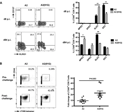
Quantitation of cytokine mRNAs by qPCR.Total RNA was isolated from spleens by use of a SimplyRNA kit (Promega) according to the man-ufacturer’s instructions. One microgram of RNA was used to synthesize cDNA by use of a high-capacity cDNA reverse transcription kit (Thermo Scientific) according to the manufacturer’s instructions. Transcripts for cytokine genes (IL-4, IL-10, IL-12, IFN-, TNF-␣, and IFN-␥) were quan-tified by qPCR with SYBR green master mix (Roche), using an ABI Prism 5700 sequence detection system (Applied Biosystems). The oligonucleo-tide primers used for qPCR are shown inTable 1. Primers for IL-4 (38), IL-12 p40 (39), IL-12 p35 (40), IFN-␥(41), TNF-␣(42), and 18S rRNA (43) were previously published. Primers for IFN-were validated by IDT. Primers for IL-10 were designed by use of the NCBI Primer Blast tool and then validated. Relative fold changes were determined using the threshold cycle (2⫺⌬⌬CT) method, with the 18S rRNA gene used as a reference gene for normalization, as previously described (44). Briefly, amounts of cyto-kine mRNA were normalized to the amount of internal 18S rRNA. The fold change of cytokine mRNA for each infected mouse was calculated by dividing the amount of cytokine mRNA in infected mice by that in unin-fected mice.
Immunofluorescence (IF) assay.Primary mouse embryonic fibro-blast (MEF) cells prepared from 13-day-old B6 mice were pretreated with 500 U/ml recombinant mouse IFN-purified from mammalian cell cul-ture (PBL Interferon Source, Piscataway, NJ) for 24 h. Cells were infected with MPyV at an MOI of 0.01 and cultured with or without IFN-(500 U/ml). At day 3 p.i., cells were fixed at room temperature for 10 min with 2% paraformaldehyde in PBS, stained with the pan-MPyV T antigen-specific F4 MAb (45) followed by goat anti-mouse IgG conjugated to Alexa 594 (Invitrogen), and mounted with Prolong reagent with DAPI (4=,6-diamidino-2-phenylindole) (Invitrogen).
Statistical analyses.Statistical significance was determined by un-paired, two-tailed Student’sttest, assuming equal variance. APvalue of⬍0.05 was considered statistically significant. The 50% effective con-centration of peptide (EC50) for the IFN-␥stimulation assay was calcu-lated according to nonlinear trajectory analysis by using GraphPad Prism software (San Diego, CA).
RESULTS
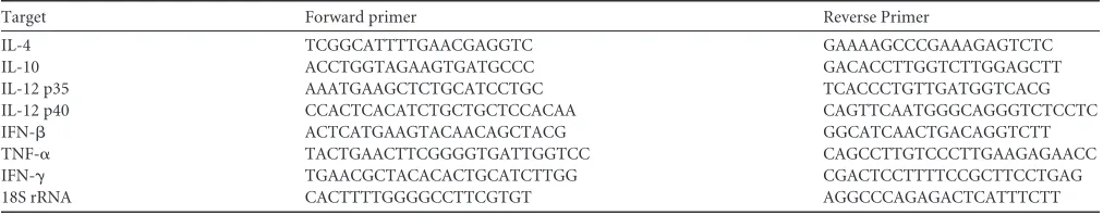
E-to-G variation at position 91 of VP1 changes viral spread and
the magnitude of the MPyV-specific CD8 T cell response.
The
magnitude of the CD8 T cell response correlates with the initial
viral load and duration of infection in various viral infection
mod-els (
46
,
47
). LP strains of MPyV disseminate more extensively than
do SP strains (
5
,
6
). Administration of a low dose of the LP strain
A2 delays the kinetics of expansion, but not the magnitude, of the
MPyV-specific CD8 T cell response (
23
). We therefore asked
whether B6 mice infected with an LP strain (A2) or an SP strain
(RA) showed differences in viral dissemination and in the
magni-tude of the strongly dominant D
b-restricted CD8 T cell response
to amino acids 359 to 368 of the large T (LT) antigen, designated
D
bLT359 (
48
). To control for differences in the noncoding control
region and the few differences in coding regions between strains
A2 and RA, we used site-directed mutagenesis to change the
glu-tamic acid (E) at position 91 of VP1 in A2 to glycine (G), as
pre-viously described (
7
). This A2(91G) mutant virus converted the
parental A2 virus from LP to SP, confirming previous reports (
6
,
7
) that this single amino acid difference in VP1 is solely
responsi-ble for the small-plaque morphology of the RA strain (
Fig. 1A
).
The mutant A2(91G) exhibited
in vitro
growth kinetics similar to
those of A2 and SP strain RA (
Fig. 1B
). However, viral loads in the
spleens and kidneys of mice infected by the RA or A2(91G) virus
showed minimal differences over the course of infection (
Fig. 1C
)
and were over 100-fold lower than those in mice infected by A2
virus during the acute phase of infection. The frequency and
mag-nitude of the D
bLT359-specific CD8 T cell response in mice
in-fected by the RA or A2(91G) virus were significantly lower than
those in mice infected by the A2 virus, but notably, the kinetics of
T cell expansion and contraction in response to infection by each
of these viruses were similar (
Fig. 1D
). To address the possibility
that less replication by A2(91G) may itself be responsible for the
smaller magnitude of the anti-MPyV CD8 T cell response, mice
were inoculated with a 10-fold higher dose of A2(91G) than that of
the A2 strain. We found that the kinetics of viral replication and
numbers of D
bLT359-specific CD8 T cells remained significantly
lower than those for A2-infected mice (data not shown). Given
that low-dose A2 virus inoculation yields a different D
bLT359-specific CD8 T cell response profile (delayed kinetics of expansion
but an equal peak magnitude during acute infection) (
23
), these
results indicated that the tropism difference imparted by the
E-to-G switch at position 91 of VP1 negatively affected the size of the
MPyV-specific CD8 T cell response.
MPyV-specific CD8 T cells recruited by A2(91G) virus
infec-tion exhibit superior funcinfec-tionality.
During persistent infection
with LCMV strain cl13, a high viral load correlates with a loss of
CD8 T cell polycytokine functionality and proliferative potential
(
47
). We next asked whether the 2-log difference in viral loads
during acute infection between the SP viruses RA and A2(91G)
and the LP virus A2 affected the functional competence of the
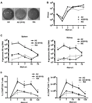
MPyV-specific CD8 T cell response. During the acute (day 8 p.i.)
and persistent (day 60 p.i.) phases of infection, A2(91G)
virus-infected mice showed a lower frequency and number of splenic
D
bLT359-specific CD8 T cells than those for A2 virus-infected
mice, as detected by D
bLT359 tetramers and
ex vivo
LT359
[image:3.585.40.546.77.176.2]pep-tide-stimulated intracellular IFN-
␥
and TNF-
␣
production
TABLE 1Primers used for quantitative RT-PCRTarget Forward primer Reverse Primer
IL-4 TCGGCATTTTGAACGAGGTC GAAAAGCCCGAAAGAGTCTC
IL-10 ACCTGGTAGAAGTGATGCCC GACACCTTGGTCTTGGAGCTT
IL-12 p35 AAATGAAGCTCTGCATCCTGC TCACCCTGTTGATGGTCACG
IL-12 p40 CCACTCACATCTGCTGCTCCACAA CAGTTCAATGGGCAGGGTCTCCTC
IFN- ACTCATGAAGTACAACAGCTACG GGCATCAACTGACAGGTCTT
TNF-␣ TACTGAACTTCGGGGTGATTGGTCC CAGCCTTGTCCCTTGAAGAGAACC
IFN-␥ TGAACGCTACACACTGCATCTTGG CGACTCCTTTTCCGCTTCCTGAG
18S rRNA CACTTTTGGGGCCTTCGTGT AGGCCCAGAGACTCATTTCTT
on November 7, 2019 by guest
http://jvi.asm.org/
(
Fig. 2A
). Comparison of the capacity of LT359-specific CD8 T
cells to coproduce IFN-
␥
and TNF-
␣
(
Fig. 2B
, top and middle
panels), however, revealed that MPyV-specific CD8 T cells
re-cruited by A2(91G) infection had an elevated capacity to
co-produce these cytokines during persistent infection (
Fig. 2B
,
bottom panel). LT359 peptide titration further showed that
MPyV-specific CD8 T cells recruited by A2(91G) infection
dur-ing persistent infection (day 60 p.i.) exhibited a higher antigen
sensitivity (approximately 3-fold, as shown by EC
50values in
the legend to
Fig. 2
), a difference not seen for cells recruited
during acute infection (
Fig. 2C
). It merits highlighting that this
difference in functional avidity by D
bLT359-specific CD8 T
cells, albeit small, was not seen in mice given low- versus
high-dose A2 virus inocula (
23
).
MPyV-specific CD8 T cells generated in response to A2 and
A2(91G) infections also exhibited phenotypic differences
indica-tive of skewed memory T cell differentiation. Memory precursors
and terminally differentiated CD8 T cells can be distinguished
based on their expression of CD127 (IL-7R
␣
) (
49
,
50
) and killer
cell lectin-like receptor G1 (KLRG1) (
51
). Phenotypic dissection
using these markers demarcates CD127
loKLRG1
locells as early
effector cells (EECs), CD127
loKLRG1
hicells as short-lived
ef-fector cells (SLECs), CD127
hiKLRG1
hicells as double-positive
effector cells (DPECs), and CD127
hiKLRG1
locells as memory
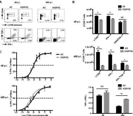
precursor effector cells (MPECs). Using these markers, we
ob-served that acute infection with A2(91G) generated a
signifi-cantly higher frequency of SLECs and a lower frequency of
EECs than did A2 infection. In persistently infected mice,
how-ever, A2(91G) recruited a higher frequency of MPECs and a
lower frequency of SLECs than those with the A2 strain (
Fig.
3A
). The higher virus levels during acute A2 infection appear to
favor differentiation of EECs over SLECs, whereas in
persis-tently infected mice, where levels of A2 and A2(91G) are similar
(
Fig. 1C
), A2 infection biases cells toward SLEC differentiation
and A2(91G) infection tilts them toward MPEC
differentia-tion.
We next tested the prediction that this MPEC bias by
MPyV-specific CD8 T cells in mice persistently infected with A2(91G)
was associated with an augmented recall capability upon antigen
challenge. A2- and A2(91G)-infected mice at day 84 p.i. were thus
infected with a recombinant VSV strain encoding the LT359
epitope (
22
). As shown in
Fig. 3B
, at 5 days postchallenge,
FIG 1E-to-G substitution at position 91 of VP1 limits viral spread and the magnitude of the MPyV-specific CD8 T cell response. (A) Plaque formation at day 6 p.i. by A2, A2(91G), and RA strain viruses on A31 cells. (B) One-step growth curve assay of A2, RA, and A2(91G) in NMuMG cells infected at an MOI of 1. Viral titers were measured by plaque assay at the indicated days p.i. (C) Viral DNAs in spleens and kidneys were measured by qPCR at the indicated days p.i. after A2, RA, or A2(91G) inoculation (1.0⫻106PFU) into hind footpads. (D) Frequencies (left) and numbers (right) of splenic DbLT359 tetramer⫹CD8⫹cells at the indicated days p.i. Data are the means⫾standard errors of the means (SEM) for at least three mice per group and are representative of three experiments. *,P⬍0.05.Qin et al.
on November 7, 2019 by guest
http://jvi.asm.org/
[image:4.585.134.454.63.420.2]D
bLT359-specific CD8 T cells were expanded to a significantly
greater extent in A2(91G)-infected mice than in A2-infected
mice. Taken together, the data show that E-to-G variation at
position 91 of VP1 is associated with superior functionality of
the MPyV-specific CD8 T cell response during persistent
infec-tion.
Inflammatory environments created by A2 versus A2(91G)
infection differentially affect the MPyV-specific CD8 T cell
re-sponse.
Early inflammation during viral infection plays a role in
regulating CD8 T cell proliferation (
17
). Thus, we asked whether
the inflammatory environment associated with A2 infection and
A2(91G) infection affected the proliferation of naive virus-specific
CD8 T cells. We introduced the G-91 VP1 mutation into the
pre-viously characterized MPyV.LT206 virus, an A2 mutant in which
the LT359 peptide sequence was replaced with that of the SV40
LT206 epitope recognized by TCR-I-transgenic CD8 T cells (
22
,
36
). We designated this virus LT206(91G). MPyV.LT206 and
LT206(91G) have replication kinetics similar to those of A2 and
A2(91G), respectively (data not shown). Adoptive transfer of
Thy1.1
⫹TCR-I cells into B6 (Thy1.2) mice prior to infection
re-capitulated the observation made in
Fig. 1D
that peak
accumula-tion of virus-specific CD8 T cells during acute infecaccumula-tion was higher
for MPyV.LT206(E-91) than for LT206(91G) (
Fig. 4A
). Using
CFSE-labeled donor TCR-I cells, we determined that this
higher-magnitude expansion could be accounted for by more efficient
expansion of anti-MPyV CD8 T cells in mice infected by the E-91
VP1 than the G-91 VP1 virus (
Fig. 4B
).
To determine whether the A2 infection-associated
inflamma-tory environment also contributed to more efficient recruitment
of naive virus-specific CD8 T cells, we immunized mice with
LT206 peptide-pulsed BMDCs in the setting of infection by A2 or
A2(91G) virus, both of which lack the D
bLT206 epitope. No
cross-FIG 2A2(91G) infection recruits memory CD8 T cells having higher polycytokine functionality and functional avidity. (A) Representative dot plots of splenic DbLT359 tetramer⫹CD8 T cells surface stained with anti-CD44 and of intracellular costaining with anti-IFN-␥and anti-TNF-␣of anti-CD8 surface-stained spleen cells after LT359 peptide stimulation at days 8 and 60 p.i. (no IFN-␥⫹or TNF-␣⫹cells were detected in the absence of LT359 peptide [data not shown]). (B) Numbers of DbLT359 tetramer⫹, IFN-␥⫹, and IFN-␥⫹TNF-␣⫹CD8 T cells at days 8 and 60 p.i. (top and middle) and ratios of TNF-␣⫹to IFN-␥⫹CD8 T cells (bottom). Values represent the means and SEM for three experiments using 3 or 4 mice per group, *,P⬍0.05; **,P⬍0.005; NS, not significant. (C) LT359 peptide dose-response curves for intracellular IFN-␥staining of splenic CD8 T cells from A2- and A2(91G)-infected mice at day 8 p.i. (top) and day 60 p.i. (bottom). EC50values were as follows: at day 8 p.i., 9.0⫻10⫺10M for A2 and 8.5⫻10⫺10M for A2(91G) (P⫽0.96); and at day 60 p.i., 8.71⫻10⫺10M for A2 and 3.3⫻10⫺10M for A2(91G) (P⫽0.017). Experiments were repeated twice, with 3 mice per group.
on November 7, 2019 by guest
http://jvi.asm.org/
[image:5.585.78.509.66.440.2]reactivity is seen by CD8 T cells specific for the D
bLT206 and
D
bLT359 epitopes (
22
; data not shown). This approach allowed us
to eliminate differences in cognate viral antigen levels resulting
from A2 versus A2(91G) infection. As shown in
Fig. 5
, the
inflam-matory environment associated with A2 infection drove a
signif-icantly larger D
bLT206 CD8 T cell expansion than that with
A2(91G) infection by day 6 p.i. These results raised the possibility
that A2 and A2(91G) elicited different types or amounts of
cyto-kines early in infection that affected the expansion of
MPyV-spe-cific CD8 T cells.
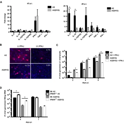
Type I IFN controls MPyV replication
in vitro
and
in vivo.
Using quantitative PCR, we assessed the mRNA levels for
candi-date cytokines (IL-4, IL-10, IL-12, IFN-

, TNF-
␣
, and IFN-
␥
)
potentially elicited early during MPyV infection and compared
the levels of these transcripts in MPyV-infected and uninfected
mice. Of the cytokine transcripts examined at days 3 and 6 p.i.,
IFN-

mRNA levels were elevated in infected mice at both time
points, although they were markedly higher at day 3 p.i.; no
sig-nificant difference was observed in induction of IFN-

transcripts
between A2 and A2(91G) virus-infected mice (
Fig. 6A
).
Interest-ingly, by day 6 p.i., mice infected with A2 virus showed a dramatic
upregulation of transcripts for IL-12 p40, the subunit shared by
IL-12 and IL-23, as well as transcripts for IL-4 and IL-10; strong
induction of transcripts for these cytokines was not seen in
A2(91G)-infected mice (
Fig. 6A
). This difference in profile for
these cytokines between A2 and A2(91G) during acute infection
may influence the effector versus memory differentiation of
MPyV-specific CD8 T cells. To determine whether type I
interfer-ons inhibited MPyV replication, primary mouse embryonic
fibro-blasts (MEFs) were infected with either A2 or mutant A2(91G)
virus in the presence or absence of mouse IFN-

. IFN-

inhibited
infection by both the A2 and A2(91G) viruses. Approximately
15% of MEFs among untreated cells were T antigen
-positive, while
fewer than 1% of cells among IFN-

-treated cells were positive
(
Fig. 6B
). The antiviral effect of IFN-

was confirmed by
enumer-ation of MPyV genome copies by qPCR (
Fig. 6C
). IFN-

-medi-FIG 3A2(91G) infection-recruited CD8 T cells exhibit a skewed CD127hiKLRG1lophenotype and have a larger recall response. (A) At days 8 and 84 p.i., spleen cells were surface stained with DbLT359 tetramers and MAbs to KLRG1 and CD127. (Left) Contour plots representative of 3 to 5 mice per time point and three independent experiments; (right) frequencies of LT359-specific CD8 T cells with the MPEC (KLRG1loCD127hi), DPEC (KLRG1hiCD127hi), SLEC (KLRG1hi CD127lo), and EEC (KLRG1loCD127lo) phenotypes. Values indicate means and SEM. *,P⬍0.05; **,P⬍0.005. (B) At 80 days p.i. with A2 or A2(91G), B6 mice were challenged with rVSV-LT359. (Left) Representative mean frequencies of DbLT359 tetramer⫹CD44⫹CD8 T cells in blood before and 5 days after challenge; (right) pre- versus postchallenge fold expansion, with each dot corresponding to an individual mouse and horizontal lines indicating means⫾SEM. Data were collected from two independent experiments (n⫽4 or 5 mice for each group).Qin et al.
on November 7, 2019 by guest
http://jvi.asm.org/
[image:6.585.94.493.64.429.2]ated antiviral effects were also observed
in vivo
. In spleens of B6
versus IFNAR
⫺/⫺mice infected with A2 or A2(91G), viral levels in
IFNAR
⫺/⫺mice were approximately 1 log higher than those in B6
mice at day 4 p.i. (
Fig. 6D
), a time point preceding detection of
MPyV-specific CD8 T cells (
52
). These results indicate that IFN-I
controls viral replication early in the course of MPyV infection
and that it does so equivalently for infections with A2 and
A2(91G).
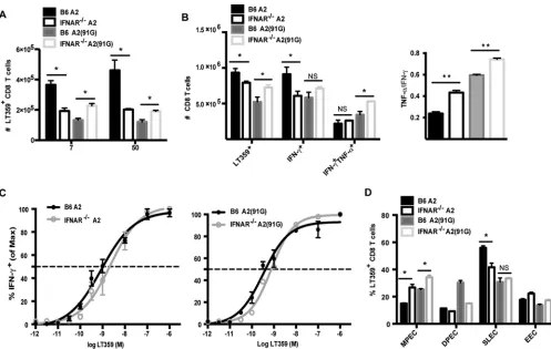
Next, we compared MPyV-specific CD8 T cell responses in B6
versus IFNAR
⫺/⫺mice infected by the A2 or A2(91G) virus. As
shown in
Fig. 7A
and
B
, the absence of IFN-I receptors had
oppo-site effects on the anti-MPyV CD8 T cell responses recruited in
response to A2 and A2(91G) infections. Although the size of the
D
bLT359-specific CD8 T cell response was lower in A2-infected
IFNAR
⫺/⫺mice than B6 mice, the reverse was seen for A2(91G)
infection; this difference was seen in both acutely and persistently
infected mice (
Fig. 7A
and
B
). IFN-I receptor deficiency was
generally associated with an augmented cytokine-producing
capability, which was particularly evident for polycytokine
production by CD8 T cells during persistent infection (
Fig.
7B
). Interestingly, memory CD8 T cells in mice persistently
infected with either A2 or A2(91G) virus exhibited a shift
to-ward decreased viral antigen sensitivity in the absence of IFN-I
receptors (
Fig. 7C
and as shown by EC
50values in the legend to
Fig. 7
), as well as skewed differentiation toward the MPEC
phenotype (
Fig. 7D
). Thus, MPyV infections of wild-type and
IFNAR
⫺/⫺mice, irrespective of the 91E-to-G VP1 tropism
dif-ference, inversely affected the effector function and functional
avidity of the virus-specific CD8 T cells.
DISCUSSION
In this study, we compared the CD8 T cell responses to MPyV
strains with different receptor binding specificities and affinities.
The single-amino-acid E-to-G exchange at position 91 in VP1
causes MPyV virions to bind not only to terminally unbranched
sialic acid true cellular receptors but also to branched-chain sialic
acid “pseudoreceptors,” resulting in attenuation of viral spread
and tumorigenicity (
6
,
9
). We validated that the E-to-G
substitu-tion at VP1 posisubstitu-tion 91 dampened viral replicasubstitu-tion
in vivo
and that
it did so in adult B6 mice, which are highly resistant to MPyV
tumorigenesis (
53
). Here we found that this VP1 difference
lim-ited expansion of MPyV-specific CD8 T cells and endowed
them with superior function (more polycytokine production,
higher functional avidity, and a larger recall response) during
persistent infection. We further found that IFN-I was induced
early during MPyV infection and differentially regulated the
FIG 4Naive MPyV-specific CD8 T cells are recruited more efficiently by infection with A2(91E) virus than by that with A2(91G) virus. (A) Experimental setup. One hundred naive Thy1.1 TCR-I cells were injected i.v. into the tail vein in B6 (Thy1.2) mice 1 day before inoculation with 5⫻105PFU of MPyV.LT206(91E) or MPyV.LT206(91G). The number of TCR-I cells in blood was determined by flow cytometry at day 8 p.i. Data were collected from three experiments with three mice per group. Each dot represents an individual mouse, with horizontal lines indicating means⫾SEM. ***,P⬍0.0005. (B) (Top) Experimental setup. A total of 1⫻106naive TCR-I cells labeled with CFSE were transferred i.v. into B6 mice inoculated 5 days earlier with MPyV.LT206(91E) or MPyV.LT206(91G). (Bottom left) After 40 h, the number of cell divisions of donor TCR-I cells was determined based on CFSE dilution 2 days after cell transfer. (Bottom right) Mean numbers of cell divisions of TCR-I cells (and SEM) for three mice per group from two independent experiments. **,P⬍0.005.on November 7, 2019 by guest
http://jvi.asm.org/
[image:7.585.94.495.66.367.2]magnitude and functionality of virus-specific CD8 T cells in a
manner dependent on the tropism of viruses with an E versus G
at VP1 position 91.
CD8 T cell differentiation is programmed by three major
factors: the strength and duration of peptide major
histocom-patibility complex (MHC)-TCR engagement, the availability
and constellation of costimulatory molecules from
antigen-presenting cells, and the profile of inflammatory cytokines.
Ac-cumulating literature indicates that the pathogen-induced
in-flammatory environment plays a central role in guiding the
pathway for memory CD8 T cell differentiation (
17
,
24
). Our
previous study showed that early virus-associated bystander
events induced by a low-dose inoculum of MPyV A2 generated
virus-specific CD8 T cells with a superior capacity to produce
IFN-
␥
. CD8 T cells primed by low-dose MPyV also expressed
higher levels of CD127 and the antiapoptotic protein Bcl-2,
which are required for memory CD8 T cell survival during
persistent infection (
23
). The low-dose A2 strain inoculation
delayed the kinetics but did not change the magnitude of the
CD8 T cell response compared to high-dose inoculation of the
A2 strain virus (
23
). In sharp contrast, infection with the VP1
mutant A2(91G) virus, which spread poorly compared to the
parental A2(91E) virus, generated an MPyV-specific CD8 T cell
response with the same kinetics as that of the A2(91E) virus,
albeit of lower magnitude. Thus, the lower virus infection levels
and lesser spread by A2(91G) were of themselves insufficient
factors to account for the less efficient recruitment of
anti-MPyV CD8 T cells. Using an adoptive T cell transfer approach
and dendritic cell immunization, we demonstrated that the
early inflammatory environment caused by a change in viral
receptor binding contributed to a different magnitude and
functionality of virus-specific CD8 T cells. IFN-I and IL-12
have been described as a “third signal” to promote full
activa-tion and development of CD8 T cells (
54–56
). Through
cyto-kine profiling, we found that the transcripts of IFN-

were
markedly upregulated during early acute infection by both
A2(91E) and A2(91G) viruses, whereas mRNAs for IL-12
(and/or IL-23) were increased later in acute infection only by
A2(91E) virus.
Type I IFNs are comprised of a family of highly related
mole-cules that program a state of resistance to intracellular pathogens
and serve to alarm cells of both innate and adaptive immunity of
the presence of infections (
57
). In our study, we showed that
MPyV replication was directly inhibited by IFN-

in vitro
and that
viral levels were higher in IFNAR
⫺/⫺mice than in wild-type mice.
IFN-I has been shown to promote IFN-
␥
production by CD4 T
cells through STAT4 activation (
58
), to enhance the terminal
dif-ferentiation of dendritic cells (
59
), and to augment the
prolifera-tion of CD8 T cells (
55
). We showed that the tropism change due
to the E-to-G difference at position 91 of VP1, while associated
with a lower-magnitude CD8 T cell response, was equally capable
of eliciting IFN-

. This discrepancy might be explained by the
timing of exposure of naive CD8 T cells to IFN-I. IFN-I may
mit-igate CD8 T cell proliferation when T cells encounter IFN-I before
engaging their cognate antigen (
60
). Because A2(91G)
dissemi-nates less efficiently than A2, naive anti-MPyV CD8 T cells
re-cruited in response to infection by A2(91G) may bind IFN-I
be-fore TCR activation. Thus, it is conceivable that polyomavirus
VP1 variants that differ in type and/or affinity for host cell
recep-tor glycans may alter the fate of the antiviral CD8 T cell response
by changing when naive T cells engage IFN-I relative to antigen.
This possibility is supported by evidence that innate inflammatory
signals induced by different pathogens differentially dictate the
IFN-I dependence of CD8 T cell expansion and memory
differen-tiation (
24
). An increase in IL-12 (and/or IL-23) during acute
infection by A2(91E) virus may also have promoted
MPyV-spe-cific CD8 T cell expansion and effector differentiation.
IFN-I plays an important role in the effector and memory fate
decision process via regulating the secretion of particular
cyto-kines (e.g., IL-15) (
61–63
) and the expression of the transcription
FIG 5Inflammation associated with A2 and A2(91G) infections differentially promotes CD8 T cell expansion. (Left) Experimental setup. A total of 2⫻105 BMDCs pulsed with LT206 peptide were injected i.v. into the tail vein in B6 mice. Three hours later, mice were inoculated with 1.0⫻106PFU of A2 or A2(91G) in the hind footpads. (Right) The frequency of DbLT206 tetramer⫹CD8 T cells in blood was analyzed at the indicated days p.i. Data were combined from three independent experiments with three mice per group. Values indicate means and SEM. **,P⬍0.005; NS, not significant.Qin et al.
on November 7, 2019 by guest
http://jvi.asm.org/
[image:8.585.95.492.64.271.2]factors T-bet (
51
) and eomesodermin (
64
). IFN-I receptor
signal-ing regulates the expression of CD127 and KLRG1, which are
mol-ecules used to demarcate subsets of effector and memory T cells
(
49
,
51
). Our data showed that the E-to-G change at position 91 of
VP1 shifted CD8 T cell differentiation toward SLECs during acute
infection but toward MPECs during persistent infection. In
con-trast, IFN-I signaling skewed virus-specific CD8 T cell
differenti-ation toward SLECs during persistent infection by A2 virus. The
increased polycytokine functionality by MPyV-specific memory
CD8 T cells in mice lacking IFN-I receptors may be related to their
lower peak expansion and effector differentiation during acute
infection. IFN-I receptor signaling during persistent infection
may also impair memory CD8 T cell functionality, as recently
reported for chronic LCMV infection, where it has been proposed
to limit immunopathology (
65–67
). IFN-I receptor signaling by
CD8 T cells during acute LCMV infection upregulated T-bet and
directed differentiation toward SLECs, while a subset of CD8 T
cells exhibited the MPEC phenotype during chronic LCMV
infec-tion in both IFN-I receptor-sufficient and -deficient mice (
68
).
While a mechanistic understanding of how IFN-I modulates CD8
T cell differentiation remains unknown, our results reinforce the
concept that the impact of IFN-I on memory differentiation is
pathogen dependent.
All MPyV isolates from feral mice have a glutamic acid at VP1
FIG 6Type I interferons inhibit viral replicationin vitroandin vivo. (A) Fold changes of the indicated cytokine mRNAs from spleens of infected mice at days 3 and 6 p.i. normalized to transcripts from spleens of uninfected mice. Data were collected from three independent experiments with three mice per group. Values indicate means and SEM. *,P⬍0.05. (B) MEFs were infected with A2 or A2(91G) virus at an MOI of 0.01, with and without addition of IFN-. MEFs were stained with a pan-T-antigen MAb and counterstained with DAPI at 24 h p.i. Representative percentages of T antigen⫹cells among DAPI⫹cells (⬎250 cells counted) from two experiments are shown. (C) Viral genome copies in the cell lysates used for panel B were quantified by qPCR at the indicated time points. Values indicate means and SEM for triplicates in one experiment. This experiment was repeated twice. (D) Viral loads in spleens of infected B6 and IFNAR⫺/⫺mice were measured by qPCR at days 4 and 7 p.i. Values indicate means and SEM for 4 or 5 mice from each group. *,P⬍0.05.on November 7, 2019 by guest
http://jvi.asm.org/
[image:9.585.76.509.65.499.2]position 91 (
11
). From the perspective of virus-host coevolution,
it is seemingly counterintuitive that mice in the wild harbor MPyV
strains with a VP1 amino acid shared by highly tumorigenic
lab-oratory strains. Tumor induction by MPyV, however, is relegated
to neonatal infection of virus-free colonies of mice of
tumor-sus-ceptible strains or infection of immunodeficient naive adult mice
(
69
). In natural mouse populations, MPyV establishes a silent
per-sistent infection. It has been proposed that differences in
regula-tory sequences among MPyV isolates from feral mice might offset
the viral oncogenicity by changing host range properties of the
virus. The results presented here give the additional explanation
that the relative impairment in virus-specific CD8 T cell immunity
associated with infection by E91-VP1 strains in feral mice favors
higher viral loads and intermouse transmission.
CD8 T cells play an essential role in controlling persistent
infection by polyomavirus (
34
). The high frequency of amino
acid mutations in VP1 close to or at the receptor binding site in
isolates of JCV from PML patients suggests that subtle changes
in binding affinity for sialylated receptors can significantly
af-fect viral pathogenicity (
70
). Recent evidence indicates that
mutations in VP1 may serve to enable escape from neutralizing
JCV antibodies (
71
,
72
). PML nonsurvivors had selectively
im-paired JCV-specific CD8 T cell responses (
16
,
73
). We recently
reported that brain-resident MPyV-specific memory CD8 T
cells expressed higher-affinity TCRs than those of splenic CD8
T cells (
74
). Whether IFN-I is induced by JCV infection and
affects the functional integrity of CD8 T cells in PML patients
in response to VP1 variant viruses is an open question. IFN-I
receptor blockade has been reported to reverse CD8 T cell
ex-haustion and to control chronic LCMV infection (
67
,
75
). Our
study suggests that the IFN-I pathway may similarly warrant
consideration as a therapeutic target for persistent
polyomavi-rus infection.
ACKNOWLEDGMENTS
We thank Ge Jin for excellent technical support and animal handling and Taryn Mockus for a critical review of the manuscript. We thank Ziaur Rahman (Department of Microbiology and Immunology, Penn State Col-lege of Medicine) for generously providing IFNAR⫺/⫺mice. We
acknowl-edge the technical support of Nate Sheaffer, Jade Vogel, and Joe Bednar-FIG 7Type I interferons regulate CD8 T cell proliferation, functionality, and memory differentiation in a virus strain-dependent manner. B6 mice and IFNAR⫺/⫺mice were infected with 1.0⫻106PFU of A2 or A2(91G) virus in the hind footpads. (A) The frequency of DbLT359 tetramer⫹CD8 T cells in blood was measured by flow cytometry at days 7 and 50 p.i. Data were collected from two independent experiments (3 to 5 mice/group). (B) Splenocytes from mice inoculated 55 days earlier with A2 or A2(91G) were surface stained with anti-CD44, anti-CD8␣, and DbLT359 tetramers or stained for surface CD8␣and intracellular IFN-␥and TNF-␣after 5 h of stimulation with LT359 peptide or without peptide (data not shown; used to set gates). (Left) Numbers of splenic DbLT359 tetramer⫹CD8 T cells and LT359 peptide-stimulated IFN-␥⫹and TNF-␣⫹IFN-␥⫹CD8 T cells. (Right) Ratios of TNF-␣⫹to IFN-␥⫹LT359 CD8 T cells. (C) LT359 peptide dose-response curves for intracellular IFN-␥staining of splenic CD8 T cells at day 55 p.i. The EC50for A2-infected B6 mice was 8.87⫻ 10⫺10M, and that for A2-infected IFNAR⫺/⫺mice was 2.25⫻10⫺9M (P⫽0.04). The EC
50for A2(91G)-infected B6 mice was 2.3⫻10⫺
10M, and that for
A2(91G)-infected IFNAR⫺/⫺mice was 7.63⫻10⫺10M (P⫽0.0022). (D) At day 50 p.i., splenic DbLT359 tetramer⫹CD8 T cells were stained for KLRG1 and CD127; designations based on these molecules are described in the legend toFig. 3A. Data are representative of three experiments using 3 or 4 mice/group. Values indicate means and SEM. *,P⬍0.05; **,P⬍0.005; NS, not significant.
Qin et al.
on November 7, 2019 by guest
http://jvi.asm.org/
[image:10.585.42.539.64.379.2]cyk of the Flow Cytometry Core Facility of The Penn State College of Medicine (Hershey, PA).
FUNDING INFORMATION
This work, including the efforts of Aron Lukacher, was funded by HHS | NIH | National Institute of Allergy and Infectious Diseases (NIAID) (AI102543). This work, including the efforts of Aron Lukacher, was funded by HHS | NIH | National Institute of Neurological Disorders and Stroke (NINDS) (NS088367). This work, including the efforts of Eliza-beth Frost, was funded by HHS | NIH | National Institute of Neurological Disorders and Stroke (NINDS) (NS083336).
REFERENCES
1.Neu U, Stehle T, Atwood WJ.2009. The Polyomaviridae: contributions of virus structure to our understanding of virus receptors and infectious entry. Virology384:389 –399.http://dx.doi.org/10.1016/j.virol.2008.12.021. 2.Tsai B, Gilbert JM, Stehle T, Lencer W, Benjamin TL, Rapoport TA.
2003. Gangliosides are receptors for murine polyoma virus and SV40. EMBO J22:4346 – 4355.http://dx.doi.org/10.1093/emboj/cdg439. 3.You J, O’Hara SD, Velupillai P, Castle S, Levery S, Garcea RL,
Benja-min T.2015. Ganglioside and non-ganglioside mediated host responses to the mouse polyomavirus. PLoS Pathog11:e1005175.http://dx.doi.org/10 .1371/journal.ppat.1005175.
4.Bauer PH, Bronson RT, Fung SC, Freund R, Stehle T, Harrison SC, Benjamin TL.1995. Genetic and structural analysis of a virulence deter-minant in polyomavirus VP1. J Virol69:7925–7931.
5.Dubensky TW, Freund R, Dawe CJ, Benjamin TL.1991. Polyomavirus replication in mice: influences of VP1 type and route of inoculation. J Virol65:342–349.
6.Bauer PH, Cui C, Liu WR, Stehle T, Harrison SC, DeCaprio JA, Benjamin TL.1999. Discrimination between sialic acid-containing recep-tors and pseudoreceprecep-tors regulates polyomavirus spread in the mouse. J Virol73:5826 –5832.
7.Freund R, Garcea RL, Sahli R, Benjamin TL.1991. A single-amino-acid substitution in polyomavirus VP1 correlates with plaque size and hemag-glutination behavior. J Virol65:350 –355.
8.Qian M, Tsai B.2010. Lipids and proteins act in opposing manners to regulate polyomavirus infection. J Virol84:9840 –9852.http://dx.doi.org /10.1128/JVI.01093-10.
9.Buch MH, Liaci AM, O’Hara SD, Garcea RL, Neu U, Stehle T.2015. Structural and functional analysis of murine polyomavirus capsid pro-teins establish the determinants of ligand recognition and pathogenicity. PLoS Pathog 11:e1005104.http://dx.doi.org/10.1371/journal.ppat .1005104.
10. Stehle T, Harrison SC.1996. Crystal structures of murine polyomavirus in complex with straight-chain and branched-chain sialyloligosaccharide receptor fragments. Structure 4:183–194.http://dx.doi.org/10.1016 /S0969-2126(96)00021-4.
11. Carroll J, Dey D, Kreisman L, Velupillai P, Dahl J, Telford S, Bronson R, Benjamin T. 2007. Receptor-binding and oncogenic properties of polyoma viruses isolated from feral mice. PLoS Pathog3:e179.http://dx .doi.org/10.1371/journal.ppat.0030179.
12. Virgin HW.2014. The virome in mammalian physiology and disease. Cell 157:142–150.http://dx.doi.org/10.1016/j.cell.2014.02.032.
13. Gorelik L, Reid C, Testa M, Brickelmaier M, Bossolasco S, Pazzi A, Bestetti A, Carmillo P, Wilson E, McAuliffe M, Tonkin C, Carulli JP, Lugovskoy A, Lazzarin A, Sunyaev S, Simon K, Cinque P.2011. Pro-gressive multifocal leukoencephalopathy (PML) development is associ-ated with mutations in JC virus capsid protein VP1 that change its receptor specificity. J Infect Dis 204:103–114. http://dx.doi.org/10.1093/infdis /jir198.
14. Bayliss J, Harrison E, McLean CA.2011. Progressive multifocal leuko-encephalopathy development is associated with mutations in JC virus cap-sid protein VP1 that change the receptor specificity of the virus. J Infect Dis 204:1643–1644.http://dx.doi.org/10.1093/infdis/jir611.
15. Martin-Blondel G, Bauer J, Cuvinciuc V, Uro-Coste E, Debard A, Massip P, Delisle MB, Lassmann H, Marchou B, Mars LT, Liblau RS. 2013. In situ evidence of JC virus control by CD8⫹T cells in PML-IRIS during HIV infection. Neurology81:964 –970.http://dx.doi.org/10.1212 /WNL.0b013e3182a43e6d.
16. Gheuens S, Bord E, Kesari S, Simpson DM, Gandhi RT, Clifford DB,
Berger JR, Ngo L, Koralnik IJ.2011. Role of CD4⫹and CD8⫹T-cell responses against JC virus in the outcome of patients with progressive multifocal leukoencephalopathy (PML) and PML with immune reconsti-tution inflammatory syndrome. J Virol85:7256 –7263.http://dx.doi.org /10.1128/JVI.02506-10.
17. Obar JJ, Jellison ER, Sheridan BS, Blair DA, Pham QM, Zickovich JM, Lefrancois L.2011. Pathogen-induced inflammatory environment con-trols effector and memory CD8⫹T cell differentiation. J Immunol187: 4967– 4978.http://dx.doi.org/10.4049/jimmunol.1102335.
18. Blair DA, Turner DL, Bose TO, Pham QM, Bouchard KR, Williams KJ, McAleer JP, Cauley LS, Vella AT, Lefrancois L. 2011. Duration of antigen availability influences the expansion and memory differentiation of T cells. J Immunol 187:2310 –2321. http://dx.doi.org/10.4049 /jimmunol.1100363.
19. Urban SL, Welsh RM.2014. Out-of-sequence signal 3 as a mechanism for virus-induced immune suppression of CD8 T cell responses. PLoS Pathog 10:e1004357.http://dx.doi.org/10.1371/journal.ppat.1004357.
20. Mueller SN, Ahmed R.2009. High antigen levels are the cause of T cell exhaustion during chronic viral infection. Proc Natl Acad Sci U S A106: 8623– 8628.http://dx.doi.org/10.1073/pnas.0809818106.
21. Wherry EJ, Ahmed R.2004. Memory CD8 T-cell differentiation during viral infection. J Virol78:5535–5545.http://dx.doi.org/10.1128/JVI.78.11 .5535-5545.2004.
22. Wilson JJ, Pack CD, Lin E, Frost EL, Albrecht JA, Hadley A, Hofstetter AR, Tevethia SS, Schell TD, Lukacher AE.2012. CD8 T cells recruited early in mouse polyomavirus infection undergo exhaustion. J Immunol 188:4340 – 4348.http://dx.doi.org/10.4049/jimmunol.1103727. 23. Andrews NP, Pack CD, Vezys V, Barber GN, Lukacher AE.2007. Early
virus-associated bystander events affect the fitness of the CD8 T cell re-sponse to persistent virus infection. J Immunol178:7267–7275.http://dx .doi.org/10.4049/jimmunol.178.11.7267.
24. Thompson LJ, Kolumam GA, Thomas S, Murali-Krishna K. 2006. Innate inflammatory signals induced by various pathogens differentially dictate the IFN-I dependence of CD8 T cells for clonal expansion and memory formation. J Immunol 177:1746 –1754. http://dx.doi.org/10 .4049/jimmunol.177.3.1746.
25. Ahmed R, Hahn CS, Somasundaram T, Villarete L, Matloubian M, Strauss JH.1991. Molecular basis of organ-specific selection of viral vari-ants during chronic infection. J Virol65:4242– 4247.
26. Matloubian M, Kolhekar SR, Somasundaram T, Ahmed R.1993. Mo-lecular determinants of macrophage tropism and viral persistence: impor-tance of single amino acid changes in the polymerase and glycoprotein of lymphocytic choriomeningitis virus. J Virol67:7340 –7349.
27. Wherry EJ, Blattman JN, Murali-Krishna K, van der Most R, Ahmed R. 2003. Viral persistence alters CD8 T-cell immunodominance and tissue distribution and results in distinct stages of functional impairment. J Virol 77:4911– 4927.http://dx.doi.org/10.1128/JVI.77.8.4911-4927.2003. 28. Baca Jones C, Filippi C, Sachithanantham S, Rodriguez-Calvo T,
Eh-rhardt K, von Herrath M.2014. Direct infection of dendritic cells during chronic viral infection suppresses antiviral T cell proliferation and induces IL-10 expression in CD4 T cells. PLoS One9:e90855.http://dx.doi.org/10 .1371/journal.pone.0090855.
29. Brooks DG, Trifilo MJ, Edelmann KH, Teyton L, McGavern DB, Oldstone MB.2006. Interleukin-10 determines viral clearance or persis-tence in vivo. Nat Med12:1301–1309.http://dx.doi.org/10.1038/nm1492. 30. Xiao Z, Casey KA, Jameson SC, Curtsinger JM, Mescher MF.2009. Programming for CD8 T cell memory development requires IL-12 or type I IFN. J Immunol182:2786 –2794.http://dx.doi.org/10.4049/jimmunol .0803484.
31. Keppler SJ, Rosenits K, Koegl T, Vucikuja S, Aichele P.2012. Signal 3 cytokines as modulators of primary immune responses during infections: the interplay of type I IFN and IL-12 in CD8 T cell responses. PLoS One 7:e40865.http://dx.doi.org/10.1371/journal.pone.0040865.
32. Way SS, Havenar-Daughton C, Kolumam GA, Orgun NN, Murali-Krishna K.2007. IL-12 and type-I IFN synergize for IFN-␥production by CD4 T cells, whereas neither are required for IFN-␥production by CD8 T cells after Listeria monocytogenes infection. J Immunol178:4498 – 4505.
http://dx.doi.org/10.4049/jimmunol.178.7.4498.
33. Lukacher AE, Wilson CS.1998. Resistance to polyoma virus-induced tumors correlates with CTL recognition of an immunodominant H-2Dk -restricted epitope in the middle T protein. J Immunol160:1724 –1734. 34. Moser JM, Altman JD, Lukacher AE.2001. Antiviral CD8⫹T cell
re-sponses in neonatal mice: susceptibility to polyoma virus-induced tumors
on November 7, 2019 by guest
http://jvi.asm.org/
is associated with lack of cytotoxic function by viral antigen-specific T cells. J Exp Med193:595– 606.http://dx.doi.org/10.1084/jem.193.5.595. 35. Kemball CC, Lee ED, Vezys V, Pearson TC, Larsen CP, Lukacher AE.
2005. Late priming and variability of epitope-specific CD8⫹T cell re-sponses during a persistent virus infection. J Immunol174:7950 –7960.
http://dx.doi.org/10.4049/jimmunol.174.12.7950.
36. Staveley-O’Carroll K, Schell TD, Jimenez M, Mylin LM, Tevethia MJ, Schoenberger SP, Tevethia SS.2003. In vivo ligation of CD40 enhances priming against the endogenous tumor antigen and promotes CD8⫹T cell effector function in SV40 T antigen transgenic mice. J Immunol171:697– 707.http://dx.doi.org/10.4049/jimmunol.171.2.697.
37. Bender A, Sapp M, Schuler G, Steinman RM, Bhardwaj N. 1996. Improved methods for the generation of dendritic cells from nonprolifer-ating progenitors in human blood. J Immunol Methods196:121–135.
http://dx.doi.org/10.1016/0022-1759(96)00079-8.
38. Le Moine A, Flamand V, Demoor FX, Noel JC, Surquin M, Kiss R, Nahori MA, Pretolani M, Goldman M, Abramowicz D.1999. Critical roles for IL-4, IL-5, and eosinophils in chronic skin allograft rejection. J Clin Invest103:1659 –1667.http://dx.doi.org/10.1172/JCI5504. 39. Malmgaard L, Paludan SR, Mogensen SC, Ellermann-Eriksen S.2000.
Herpes simplex virus type 2 induces secretion of IL-12 by macrophages through a mechanism involving NF-B. J Gen Virol81:3011–3020.http: //dx.doi.org/10.1099/0022-1317-81-12-3011.
40. Liu J, Guan X, Tamura T, Ozato K, Ma X.2004. Synergistic activation of interleukin-12 p35 gene transcription by interferon regulatory factor-1 and interferon consensus sequence-binding protein. J Biol Chem279: 55609 –55617.http://dx.doi.org/10.1074/jbc.M406565200.
41. Hein J, Schellenberg U, Bein G, Hackstein H.2001. Quantification of murine IFN-␥mRNA and protein expression: impact of real-time kinetic RT-PCR using SYBR green I dye. Scand J Immunol54:285–291.http://dx .doi.org/10.1046/j.1365-3083.2001.00928.x.
42. Park SW, Kim M, Kim M, D’Agati VD, Lee HT. 2011. Sphingosine kinase 1 protects against renal ischemia-reperfusion injury in mice by sphingosine-1-phosphate1 receptor activation. Kidney Int80:1315–1327.
http://dx.doi.org/10.1038/ki.2011.281.
43. Soni C, Domeier PP, Wong EB, Shwetank, Khan TN, Elias MJ, Schell SL, Lukacher AE, Cooper TK, Rahman ZS.2015. Distinct and synergistic roles of Fc␥RIIB deficiency and 129 strain-derived SLAM family proteins in the development of spontaneous germinal centers and autoimmunity. J Autoimmun63:31– 46.http://dx.doi.org/10.1016/j.jaut.2015.06.011. 44. Livak KJ, Schmittgen TD.2001. Analysis of relative gene expression data
using real-time quantitative PCR and the 2⫺⌬⌬Ctmethod. Methods25: 402– 408.http://dx.doi.org/10.1006/meth.2001.1262.
45. Pallas DC, Schley C, Mahoney M, Harlow E, Schaffhausen BS, Roberts TM.1986. Polyomavirus small t antigen: overproduction in bacteria, pu-rification, and utilization for monoclonal and polyclonal antibody pro-duction. J Virol60:1075–1084.
46. Akondy RS, Johnson PL, Nakaya HI, Edupuganti S, Mulligan MJ, Lawson B, Miller JD, Pulendran B, Antia R, Ahmed R.2015. Initial viral load determines the magnitude of the human CD8 T cell response to yellow fever vaccination. Proc Natl Acad Sci U S A112:3050 –3055.http: //dx.doi.org/10.1073/pnas.1500475112.
47. Wherry EJ, Blattman JN, Ahmed R.2005. Low CD8 T-cell proliferative potential and high viral load limit the effectiveness of therapeutic vaccina-tion. J Virol 79:8960 – 8968.http://dx.doi.org/10.1128/JVI.79.14.8960 -8968.2005.
48. Kemball CC, Pack CD, Guay HM, Li ZN, Steinhauer DA, Szomolanyi-Tsuda E, Lukacher AE.2007. The antiviral CD8⫹ T cell response is differentially dependent on CD4⫹T cell help over the course of persistent infection. J Immunol 179:1113–1121.http://dx.doi.org/10.4049 /jimmunol.179.2.1113.
49. Schluns KS, Kieper WC, Jameson SC, Lefrancois L.2000. Interleukin-7 mediates the homeostasis of naive and memory CD8 T cells in vivo. Nat Immunol1:426 – 432.http://dx.doi.org/10.1038/80868.
50. Kaech SM, Tan JT, Wherry EJ, Konieczny BT, Surh CD, Ahmed R. 2003. Selective expression of the interleukin 7 receptor identifies effector CD8 T cells that give rise to long-lived memory cells. Nat Immunol 4:1191–1198.http://dx.doi.org/10.1038/ni1009.
51. Joshi NS, Cui W, Chandele A, Lee HK, Urso DR, Hagman J, Gapin L, Kaech SM.2007. Inflammation directs memory precursor and short-lived effector CD8⫹T cell fates via the graded expression of T-bet transcription factor. Immunity27:281–295.http://dx.doi.org/10.1016/j.immuni.2007.07 .010.
52. Lukacher AE, Moser JM, Hadley A, Altman JD.1999. Visualization of polyoma virus-specific CD8⫹T cells in vivo during infection and tumor rejection. J Immunol163:3369 –3378.
53. Freund R, Dubensky T, Bronson R, Sotnikov A, Carroll J, Benjamin T. 1992. Polyoma tumorigenesis in mice: evidence for dominant resistance and dominant susceptibility genes of the host. Virology191:724 –731.
http://dx.doi.org/10.1016/0042-6822(92)90248-N.
54. Curtsinger JM, Lins DC, Mescher MF.2003. Signal 3 determines toler-ance versus full activation of naive CD8 T cells: dissociating proliferation and development of effector function. J Exp Med197:1141–1151.http: //dx.doi.org/10.1084/jem.20021910.
55. Curtsinger JM, Valenzuela JO, Agarwal P, Lins D, Mescher MF.2005. Type I IFNs provide a third signal to CD8 T cells to stimulate clonal expansion and differentiation. J Immunol174:4465– 4469.http://dx.doi .org/10.4049/jimmunol.174.8.4465.
56. Schmidt CS, Mescher MF.1999. Adjuvant effect of IL-12: conversion of peptide antigen administration from tolerizing to immunizing for CD8⫹ T cells in vivo. J Immunol163:2561–2567.
57. Huber JP, Farrar JD.2011. Regulation of effector and memory T-cell functions by type I interferon. Immunology132:466 – 474.http://dx.doi .org/10.1111/j.1365-2567.2011.03412.x.
58. Nguyen KB, Watford WT, Salomon R, Hofmann SR, Pien GC, Mori-nobu A, Gadina M, O’Shea JJ, Biron CA.2002. Critical role for STAT4 activation by type 1 interferons in the interferon-␥response to viral infec-tion. Science297:2063–2066.http://dx.doi.org/10.1126/science.1074900. 59. Luft T, Pang KC, Thomas E, Hertzog P, Hart DN, Trapani J, Cebon J. 1998. Type I IFNs enhance the terminal differentiation of dendritic cells. J Immunol161:1947–1953.
60. Marshall HD, Urban SL, Welsh RM.2011. Virus-induced transient immune suppression and the inhibition of T cell proliferation by type I interferon. J Virol85:5929 –5939.http://dx.doi.org/10.1128/JVI.02516-10.
61. Berard M, Brandt K, Bulfone-Paus S, Tough DF.2003. IL-15 promotes the survival of naive and memory phenotype CD8⫹T cells. J Immunol 170:5018 –5026.http://dx.doi.org/10.4049/jimmunol.170.10.5018. 62. Mattei F, Schiavoni G, Belardelli F, Tough DF.2001. IL-15 is expressed
by dendritic cells in response to type I IFN, double-stranded RNA, or lipopolysaccharide and promotes dendritic cell activation. J Immunol 167:1179 –1187.http://dx.doi.org/10.4049/jimmunol.167.3.1179. 63. Zhang X, Sun S, Hwang I, Tough DF, Sprent J. 1998. Potent and
selective stimulation of memory-phenotype CD8⫹T cells in vivo by IL-15. Immunity8:591–599.http://dx.doi.org/10.1016/S1074-7613(00)80564-6. 64. Martinet V, Tonon S, Torres D, Azouz A, Nguyen M, Kohler A, Flamand V, Mao CA, Klein WH, Leo O, Goriely S. 2015. Type I interferons regulate eomesodermin expression and the development of unconventional memory CD8⫹T cells. Nat Commun6:7089.http://dx .doi.org/10.1038/ncomms8089.
65. Honke N, Shaabani N, Merches K, Gassa A, Kraft A, Ehrhardt K, Haussinger D, Lohning M, Dittmer U, Hengel H, Recher M, Lang PA, Lang KS.2016. Immunoactivation induced by chronic viral infection inhibits viral replication and drives immunosuppression through sus-tained IFN-I responses. Eur J Immunol46:372–380.http://dx.doi.org/10 .1002/eji.201545765.
66. Odorizzi PM, Wherry EJ.2013. Immunology. An interferon paradox. Science340:155–156.http://dx.doi.org/10.1126/science.1237568. 67. Teijaro JR, Ng C, Lee AM, Sullivan BM, Sheehan KC, Welch M,
Schreiber RD, de la Torre JC, Oldstone MB.2013. Persistent LCMV infection is controlled by blockade of type I interferon signaling. Science 340:207–211.http://dx.doi.org/10.1126/science.1235214.
68. Wiesel M, Crouse J, Bedenikovic G, Sutherland A, Joller N, Oxenius A. 2012. Type-I IFN drives the differentiation of short-lived effector CD8⫹T cells in vivo. Eur J Immunol42:320 –329.http://dx.doi.org/10.1002/eji .201142091.
69. Lukacher AE, Ma Y, Carroll JP, Abromson-Leeman SR, Laning JC, Dorf ME, Benjamin TL.1995. Susceptibility to tumors induced by polyoma virus is conferred by an endogenous mouse mammary tumor virus superantigen. J Exp Med181:1683–1692.http://dx.doi.org/10.1084/jem.181.5.1683. 70. Stroh LJ, Maginnis MS, Blaum BS, Nelson CD, Neu U, Gee GV, O’Hara
BA, Motamedi N, DiMaio D, Atwood WJ, Stehle T.2015. The greater affinity of JC polyomavirus capsid for␣2,6-linked lactoseries tetrasaccha-ride c than for other sialylated glycans is a major determinant of infectivity. J Virol89:6364 – 6375.http://dx.doi.org/10.1128/JVI.00489-15. 71. Ray U, Cinque P, Gerevini S, Longo V, Lazzarin A, Schippling S, Martin
R, Buck CB, Pastrana DV.2015. JC polyomavirus mutants escape
anti-Qin et al.
on November 7, 2019 by guest
http://jvi.asm.org/
body-mediated neutralization. Sci Transl Med7:306ra151.http://dx.doi .org/10.1126/scitranslmed.aab1720.
72. Jelcic I, Combaluzier B, Jelcic I, Faigle W, Senn L, Reinhart BJ, Stroh L, Nitsch RM, Stehle T, Sospedra M, Grimm J, Martin R.2015. Broadly neutralizing human monoclonal JC polyomavirus VP1-specific antibod-ies as candidate therapeutics for progressive multifocal leukoencephalop-athy. Sci Transl Med7:306ra150.http://dx.doi.org/10.1126/scitranslmed .aac8691.
73. Khanna N, Wolbers M, Mueller NJ, Garzoni C, Du Pasquier RA, Fux CA, Vernazza P, Bernasconi E, Viscidi R, Battegay M, Hirsch HH, Swiss HIV
Cohort Study.2009. JC virus-specific immune responses in human immu-nodeficiency virus type 1 patients with progressive multifocal leukoencepha-lopathy. J Virol83:4404 – 4411.http://dx.doi.org/10.1128/JVI.02657-08. 74. Frost EL, Kersh AE, Evavold BD, Lukacher AE. 2015. Cutting edge:
resident memory CD8 T cells express high-affinity TCRs. J Immunol195: 3520 –3524.http://dx.doi.org/10.4049/jimmunol.1501521.
75. Wilson EB, Yamada DH, Elsaesser H, Herskovitz J, Deng J, Cheng G, Aronow BJ, Karp CL, Brooks DG.2013. Blockade of chronic type I interferon signaling to control persistent LCMV infection. Science340: 202–207.http://dx.doi.org/10.1126/science.1235208.
on November 7, 2019 by guest
http://jvi.asm.org/