Molecular Evolution and Intraclade Recombination of Enterovirus
D68 during the 2014 Outbreak in the United States
Yi Tan,aFerdaus Hassan,bJennifer E. Schuster,cAri Simenauer,aRangaraj Selvarangan,bRebecca A. Halpin,aXudong Lin,a Nadia Fedorova,aTimothy B. Stockwell,aTommy Tsan-Yuk Lam,dJames D. Chappell,eTina V. Hartert,fEdward C. Holmes,g
Suman R. Dasa
Infectious Diseases Group, J. Craig Venter Institute, Rockville, Maryland, USAa
; Department of Pathology and Laboratory Medicine, Children’s Mercy Hospitals and Clinics, Kansas City, Missouri, USAb
; Department of Pediatrics, Children’s Mercy Hospitals and Clinics, Kansas City, Missouri, USAc
; Centre of Influenza Research, School of Public Health, The University of Hong Kong, Hong Kong, Chinad
; Department of Pathology, Microbiology, and Immunology, Vanderbilt University School of Medicine, Nashville, Tennessee, USAe
; Division of Allergy, Pulmonary, and Critical Care Medicine, Department of Medicine, Vanderbilt University School of Medicine, Nashville, Tennessee, USAf
; Marie Bashir Institute for Infectious Diseases & Biosecurity, Charles Perkins Centre, School of Biological Sciences and Sydney Medical School, The University of Sydney, Sydney, NSW, Australiag
ABSTRACT
In August 2014, an outbreak of enterovirus D68 (EV-D68) occurred in North America, causing severe respiratory disease in
chil-dren. Due to a lack of complete genome sequence data, there is only a limited understanding of the molecular evolution and
epi-demiology of EV-D68 during this outbreak, and it is uncertain whether the differing clinical manifestations of EV-D68 infection
are associated with specific viral lineages. We developed a high-throughput complete genome sequencing pipeline for EV-D68
that produced a total of 59 complete genomes from respiratory samples with a 95% success rate, including 57 genomes from
Kansas City, MO, collected during the 2014 outbreak. With these data in hand, we performed phylogenetic analyses of complete
genome and VP1 capsid protein sequences. Notably, we observed considerable genetic diversity among EV-D68 isolates in
Kan-sas City, manifest as phylogenetically distinct lineages, indicative of multiple introductions of this virus into the city. In
addi-tion, we identified an intersubclade recombination event within EV-D68, the first recombinant in this virus reported to date.
Finally, we found no significant association between EV-D68 genetic variation, either lineages or individual mutations, and a
variety of demographic and clinical variables, suggesting that host factors likely play a major role in determining disease
sever-ity. Overall, our study revealed the complex pattern of viral evolution within a single geographic locality during a single
out-break, which has implications for the design of effective intervention and prevention strategies.
IMPORTANCE
Until recently, EV-D68 was considered to be an uncommon human pathogen, associated with mild respiratory illness. However,
in 2014 EV-D68 was responsible for more than 1,000 disease cases in North America, including severe respiratory illness in
chil-dren and acute flaccid myelitis, raising concerns about its potential impact on public health. Despite the emergence of EV-D68, a
lack of full-length genome sequences means that little is known about the molecular evolution of this virus within a single
geo-graphic locality during a single outbreak. Here, we doubled the number of publicly available complete genome sequences of
EV-D68 by performing high-throughput next-generation sequencing, characterized the evolutionary history of this outbreak in
de-tail, identified a recombination event, and investigated whether there was any correlation between the demographic and clinical
characteristics of the patients and the viral variant that infected them. Overall, these results will help inform the design of
inter-vention strategies for EV-D68.
E
nteroviruses (EVs) are members of the family
Picornaviridae
(order
Picornavirales
) of RNA viruses, with the genus
Entero-virus
comprising 12 species, designated A to H and J, as well as
rhinovirus A to C (
1
). All EVs are characterized by a single
posi-tive-strand RNA genome of approximately 7,500 nucleotides (nt)
in length. To date, five types of
Enterovirus D
(EV-D) species have
been described: EV-D68, together with EV-D70 and EV-D94, is an
important human pathogen (
2–5
), while EV-D111 and EV-D120
were only recently assigned (
4
,
6
,
7
). EV-D68 was first isolated
from hospitalized children with pneumonia and bronchiolitis in
California in 1962 (
8
), and rhinovirus 87 was later reclassified as
EV-D68 based on phylogenetic analysis, cross-neutralization, and
acid sensitivity (
9–11
). The EV-D68 genome includes a single
open reading frame which encodes four structural proteins (VP1
to VP4) and seven nonstructural proteins (2A to 2C and 3A to
3D), a 5
=
untranslated region (UTR) with a hairpin-loop
second-ary structure, and a 3
=
UTR with a poly(A) tract (
1
).
EV-D68 was detected sporadically from the 1970s through the
early 2000s (
12–18
). However, an increased number of infections
by EV-D68 has been reported worldwide during the past decade
(
15
,
16
,
18–33
). Almost all documented EV-D68 cases during this
Received18 September 2015Accepted30 November 2015
Accepted manuscript posted online9 December 2015
CitationTan Y, Hassan F, Schuster JE, Simenauer A, Selvarangan R, Halpin RA, Lin X, Fedorova N, Stockwell TB, Lam TT-Y, Chappell JD, Hartert TV, Holmes EC, Das SR. 2016. Molecular evolution and intraclade recombination of enterovirus D68 during the 2014 outbreak in the United States. J Virol 90:1997–2007.
doi:10.1128/JVI.02418-15.
Editor:S. R. Ross
Address correspondence to Suman R. Das, sdas@jcvi.org.
Copyright © 2016, American Society for Microbiology. All Rights Reserved.
on November 7, 2019 by guest
http://jvi.asm.org/
time were associated with acute respiratory infections, ranging
from mild upper respiratory tract illness and asthma to severe
bronchitis and pneumonia, with the exception of four isolated
cases associated with neurological syndromes, notably, acute
flac-cid myelitis (AFM), in which viruses were isolated from the
cere-brospinal fluid or nasopharyngeal swabs (
12
,
31
,
34
). The 2014
outbreak of EV-D68 was the largest outbreak recorded in North
America, with more than 1,153 confirmed EV-D68 cases in 49
U.S. states and the District of Columbia (
35
). The majority of cases
were associated with severe respiratory illness in children (
36
,
37
)
leading to hospitalization. In addition, there was a surge in
EV-D68 infections associated with AFM and encephalitis (
36
,
38
,
39
).
EV-D68 infections were reported in Europe during the same time
period, with three cases associated with AFM (
40
,
41
).
Previous studies have revealed the presence of three major
clades of EV-D68, designated A, B and C, which have circulated or
cocirculated during different time periods in different geographic
regions (
18
,
38
). Most of the viruses sampled from the 2014
EV-D68 outbreak in the United States have been assigned to a novel
B1 subclade, especially those sampled from AFM cases (
38
). Along
with high rates of nucleotide substitution, recombination is an
important way in which genetic diversity can be generated in
en-teroviruses (
33
). However, no recombination events have yet been
reported in EV-D68, in part reflecting a lack of full-length genome
sequences for analysis (
22
).
There is growing interest in understanding the epidemiology of
EV-D68 in the United States, particularly given its association
with severe disease outcomes during the 2014 outbreak. Indeed,
little is known about the molecular evolution of the virus during a
single outbreak, nor whether differences in clinical manifestation
are associated with specific genetic variants. The most important
limitation here has been the lack of complete genome sequences of
EV-D68 in the public domain, even though such information will
assist the design of effective intervention and prevention strategies
and help formulate modalities of future treatments. To help
de-termine the evolution of EV-D68 within a single geographic
re-gion during a single outbreak, we performed full-length genome
sequencing of viruses sampled from children seen at Children’s
Mercy Hospital—the first hospital reporting the EV-D68
out-break in 2014 —in Kansas City, MO, during August to September
2014 (
36
). As well as determining the extent and pattern of viral
genetic diversity, we screened these genomic data for
recombina-tion and assessed whether there was any associarecombina-tion between
ge-netic variation and specific demographic and clinical features of
the infected patients.
MATERIALS AND METHODS
Study population and patient data collection.Patients, aged 0 to 17
years, admitted to Children’s Mercy Hospital from 1 August to 15 Sep-tember 2014 with a positive test result for enterovirus/rhinovirus by FilmArray respiratory panel assay (Biofire LLC, Idaho) were retrospec-tively tested for the presence of EV-D68 by real-time PCR (37). EV-D68-positive patients admitted to the pediatric intensive care unit (ICU) were age matched with two patients not requiring ICU care. Data were retro-spectively obtained from chart abstraction and entered into a standard-ized data collection instrument. This project was approved by the Chil-dren’s Mercy Hospital Institutional Review Board.
A single-term, previously healthy infant with EV-D68 was enrolled in an infant birth cohort based at Vanderbilt University Medical Center and was included as a preemergent isolate in a region geographically close to Kansas City. This is a population-based birth cohort, and respiratory
ill-ness surveillance was performed every 2 weeks during the winter season. All information was prospectively collected, and parents gave their in-formed consent for study inclusion (42).
RNA extraction and reverse transcription-PCR (RT-PCR).
Extrac-tion of viral RNA was performed at the J. Craig Venter Institute (JCVI), Rockville, MD, with 140l of respiratory samples (nasal swab or nasal aspirates/wash) in transport medium using a QIAamp viral RNA minikit (Qiagen, Hilden, Germany)/ZR96 viral RNA kit (Zymo, Irvine, CA) hy-brid protocol. In brief, specimen lysis was performed in Qiagen buffer AVL in a 96-well deep-well plate. Lysate was transferred to a ZR96 spin plate (Zymo), and samples were processed according to the manufactur-er’s protocol. The cDNA was generated using SuperScript III reverse transcriptase (Thermo Fisher Scientific, MA) from 4l undiluted RNA and either oligo(dT)20or a 1:1 mix of two primers specific to the 3= -terminal region (D68_7333AR and D68_7333BR) (Table 1).
Three independent approaches were taken when performing PCR am-plification of the viral genome. (i) Full-length genome amplicons contain-ing the 3=poly(A) tail (FL-A) were generated from the cDNA generated by oligo(dT)20 using D68_1F and M13-dt18 (reverse primer). (ii) Full-length genome amplification excluding the 3=poly(A) tail (FL) was per-formed using gene-specific RT mix primers and PCR primer D68_1F and a 1:1 mix of D68_7333AR and D68_7333BR. (iii) Generation of complete viral genomes through the amplification of two overlapping amplicons using gene-specific RT was achieved as follows. A small (S) 904-bp ampli-con encompassing the EV-D68 5=UTR was generated directly from RNA using the Qiagen one-step RT-PCR kit (Qiagen) according to the manu-facturer’s protocol using primers D68_1F and D68_904R. A large (L) 6.8-kbp amplicon encompassing the rest of the viral genome was gener-ated from primer D68_536F and a 1:1 mix of D68_7333AR and D68_7333BR. Amplicons were verified on 1% agarose gels or via the QIAxcel advanced (Qiagen) capillary gel electrophoresis DNA screening platform, and excess primers and deoxynucleoside triphosphates (dNTPs) were removed by treatment with exonuclease I (New England BioLabs) and shrimp alkaline phosphatase (Affymetrix, Santa Clara, CA, USA) at 37°C for 60 min, followed by incubation at 72°C for 15 min. Amplicons were quantified using a SYBR green double-stranded DNA (dsDNA) detection assay (SYBR green I nucleic acid gel stain; Thermo Fisher Scientific), and all four amplicons per genome were pooled in equal concentrations.
EV-D68 complete genome sequencing assembly and annotation.
Il-lumina libraries were prepared using the Nextera DNA sample prepara-tion kit (Illumina, Inc., San Diego, CA, USA) with half-reacprepara-tion-mixture volumes as described previously (43,44). For samples requiring extra coverage, the Ion Torrent Personal Genome Machine (PGM) (Thermo Fisher Scientific) was used in addition to Illumina sequencing, in which 100 ng of pooled DNA amplicons was sheared for 7 min and Ion Torrent-compatible barcoded adapters were ligated to the sheared DNA using the Ion Xpress Plus fragment library kit (Thermo Fisher Scientific) to create 400-bp libraries. Sequencing was performed on the Ion Torrent PGM using 316v2 or 318v2 chips (Thermo Fisher Scientific).
Sequence reads were sorted by barcode, trimmed, andde novo assem-bled using CLC bio’sclc_novo_assembleprogram (45). The resulting con-tigs were searched against custom, full-length EV-D68 nucleotide data-bases to identify the closest reference sequence. All sequence reads were then mapped to the selected reference EV-D68 sequence using CLC bio’s
clc_ref_assemble_longprogram (46). At loci where both Illumina and Ion Torrent sequence data agreed on a variation compared with the reference sequence, it was updated to reflect the difference. A final mapping of all next-generation sequences to the updated reference sequences was per-formed with CLC bio’sclc_ref_assemble_longprogram (46). Curated as-semblies were validated and annotated with the viral annotation software Viral Genome ORF Reader (VIGOR) 3.0 (47) before submission to Gen-Bank. VIGOR was used to predict genes, perform alignments, ensure the fidelity of open reading frames, associate nucleotide polymorphisms with amino acid changes, and detect any potential sequencing errors. The
on November 7, 2019 by guest
http://jvi.asm.org/
notation was subjected to manual inspection and quality control before submission to GenBank.
Phylogenetic analysis.We analyzed 59 newly acquired complete
ge-nome sequences of EV-D68 (57 collected in Kansas City during the 2014 outbreak; one preoutbreak sequence collected in Nashville, TN, USA [Vanderbilt University Medical Center], in 2012; and one prototype Fer-mon virus strain purchased from ATCC) together with all EV-D68 sequences available on GenBank (http://www.ncbi.nlm.nih.gov/GenBank/). In total, we utilized two global data sets of EV-D68: a 110-complete-genome data set (59 JCVI-sequenced sequences and 51 background sequences) and a 357-complete-VP1-gene data set (59 JCVI-sequenced sequences and 298 background sequences). Sequences within these two data sets were aligned separately using the MUSCLE program in MEGA 6.0 with manual adjustment (48). Maximum likelihood (ML) phylogenies (49) were in-ferred in MEGA 6.0. A general time reversal (GTR) substitution model with a gamma distribution of among-site rate variation and a proportion of invariable sites (GTR⫹⌫⫹I) was selected as the best-fit model by Mod-elTest in MEGA 6.0 and used in all tree inference methods. In addition, trees were inferred using the Bayesian Markov chain Monte Carlo (BM-CMC) method available in MrBayes version 3.2.5 (50), run for 1⫻108 steps. Trees were sampled every 1⫻104steps, with the first 1,000 trees discarded as burn-in. The robustness of the ML tree was assessed by boot-strap analyses of 1,000 pseudoreplicates and by comparison with the to-pologies sampled in the Bayesian analysis. All phylogenies were rooted with the oldest EV-D68 sequence in GenBank, the Fermon strain, col-lected in 1962 in California, USA (2).
Inferring the events of virus introduction to Kansas City.A total of
5,000 trees were evenly subsampled from the posterior distribution of trees produced during the BMCMC analysis described above using the LogCombiner program within the BEAST package (51). Each sequence was assigned a discrete geographic state— either “Kansas City” or “other place”—according to our data or its record in GenBank. A parsimony procedure (52) was then used to infer ancestral geographic states given these data and hence to determine the frequency of state change from the “other place” character state to the “Kansas City” character state in each tree. Such changes in geographic state are indicative of independent viral introductions into Kansas City. The mean and 95% confidence intervals of the frequency of introduction events were summarized from the counts in the 5,000 subsampled trees.
Analysis of EV-D68 recombination.Potential recombination within
the VP1 and complete genome sequences of EV-D68 was screened using seven methods (RDP, GENECONV, MaxChi, Bootscan, Chimaera, SiScan, and 3Seq) implemented in the Recombination Detection Program version 4.46 (RDP4) (53). Phylogenetic incongruence between different regions and withPvalues of less than 1⫻10⫺4in all methods was taken to represent strong evidence for recombination. To confirm these putative recombination events, we utilized a smaller data set including the recom-binant and the parental strains determined above, employing similarity plots and Bootscan analysis as implemented in Simplot version 3.5.1 (54), with a window size of 300 nucleotides (nt) and a step size of 10 nt. Recom-bination breakpoints were inferred based on the distribution of informa-tive sites supporting the two incongruent tree topologies that maximized the chi-square (2) sum (55). The midpoint of the breakpoint region was used to partition the sequence alignment for separate phylogenetic infer-ences.
Assessing the association between virus phylogeny and the
demo-graphic and clinical features of EV-D68 patients.Demographic and
clin-ical information was available for 57 outbreak samples from Kansas City. To determine whether there was phylogenetic clustering by the age and gender of the patient, we grouped viruses into two age classes (⬍5 years andⱖ5 years old) and by gender separately. Similarly, each patient was classified as either positive or negative for each clinical symptom individ-ually. The clinical symptoms analyzed were (i) presence in pediatric ICU, (ii) medical history of asthma or recurrent wheezing, and (iii) require-ment for ventilation. The strength of association between the phenotypic
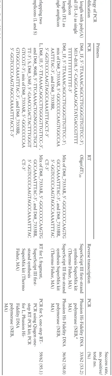
TABLE 1 EV-D68 primers used for three strategies of PCR amplification Strategy of PCR amplification Primers Reverse transcription PCR Success rate, no. positive/ total no. (%) PCR RT Full length with poly(A) tail (FL-A) as single amplicon D68_1F, 5 = -TTAAAACAGCCTTGGGGTTGTTCC-3 = ; M13-dt18, 5 = -CAGGAAACAGCTATGACCGTTTT TTTTTTTTTTTTTT-3 = Oligo(dT) 20 SuperScript III first-strand synthesis SuperMix kit (Thermo Fisher, MA) Phusion Hi-Fidelity DNA polymerase (NEB, a MA) 33/62 (53.2) Full length (FL) as single amplicon D68_1F, 5 = -TTAAAACAGCCTTGGGGTTGTTCC-3 = ; mix of D68_7333AR, 5 = -GGCCCCCAAGTGGCCAA AATTTAC-3 = ,and D68_7333BR, 5 = -GGTCCCCAAGTAGCCAAAATTTACCT-3 = Mix of D68_7333AR, 5 = -GGCCCCCAAGTG GCCAAAATTTAC-3 = ,and D68_7333BR, 5 = -GGTCCCCAAGTAGCCAAAATTTAC CT-3 = SuperScript III first-strand synthesis SuperMix kit (Thermo Fisher, MA) Phusion Hi-Fidelity DNA polymerase (NEB, MA) 36/62 (58.0) Overlapping two amplicons (L and S) L, D68_1F, 5 = -TTAAAACAGCCTTGGGGTTGTTCC-3 = , and D68_904R, 5 = -TTCAAAACTGGNGCCCCTGCT TT-3 = ;S, D68_536F, 5 = -GAACCGACTACTTTGGGT GTCCGT-3 = ;mix of D68_7333AR, 5 = -GGCCCCCAA GTGGCCAAAATTTAC-3 = ,and D68_7333BR, 5 = -GGTCCCCAAGTAGCCAAAATTTACCT-3 = Mix of D68_7333AR, 5 = -GGCCCCCAAGTG GCCAAAATTTAC-3 = ,and D68_7333BR, 5 = -GGTCCCCAAGTAGCCAAAATTTAC CT-3 = RT for L fragment, SuperScript III first-strand synthesis SuperMix kit (Thermo Fisher, MA) PCR for S, one-step RT-PCR using Qiagen one-step RT-PCR kit; PCR for L, Phusion Hi-Fidelity DNA polymerase (NEB, MA) 59/62 (95.1) a NEB, New England BioLabs.
on November 7, 2019 by guest
http://jvi.asm.org/
[image:3.585.65.258.80.724.2]features described above and the EV-D68 phylogeny was determined us-ing two phylogeny-trait association statistics, the parsimony score (PS) and the association index (AI) tests, both of which were implemented in the Bayesian tip association significance testing (BaTS) program (56). A null distribution of these statistics was determined using the posterior distribution of trees obtained from the MrBayes analysis described above.
Nucleotide sequence accession numbers.All sequences generated as
part of this study were submitted to GenBank as part of the BioProject identifiers PRJNA266349 and PRJNA270340 and assigned accession numbersKT347223toKT347280andKT725431.
RESULTS
High-throughput full-length genome sequencing of EV-D68
from the 2014 outbreak.
A total of 62 EV-D68-positive samples
were chosen for complete genome sequencing, including 60
clin-ical samples from the 2014 outbreak collected from Children’s
Mercy Hospital, Kansas City, MO; one preoutbreak sample from
Vanderbilt University Medical Center, Nashville, TN, collected in
2012; and one sample from the American Type Culture Collection
(ATCC), VR-1197. Oligo(dT)
20and/or two primers specific to the
3
=
-terminal region of the virus were used for reverse transcription
(
Table 1
). The general pattern of primer sites and the locations of
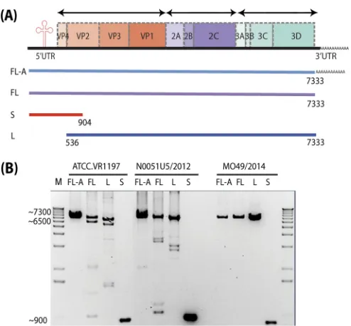
primer targets in EV-D68 genomes are shown in
Fig. 1A
. To
achieve complete genome sequencing, three approaches were
at-tempted from respiratory samples: (i) full-length genome with
poly(A) tail (FL-A), (ii) full-length genome without the poly(A)
tail (FL), and (iii) two overlapping amplicons—large (L) and
small (S) (
Fig. 1A
). Representative PCR products for each method
are shown in
Fig. 1B
. PCR products of the expected size and
com-plete genome assemblies were obtained with variable success rates:
53% and 58% for FL-A and FL, respectively, and 95% (59 out of
62) for the two-amplicon method, which is superior to a recently
published method based on sequence-independent amplification
(
57
). These PCR products were subjected to next-generation
quencing using both the Illumina Mi-Seq and Ion Torrent
se-quencing platforms. In total, we obtained 59 complete genome
sequences of EV-D68, including 57 outbreak samples from Kansas
City, one preoutbreak sample from Nashville, and one historical
sample from ATCC.
Our sequencing resulted in an average of 62,256 reads/sample
with an average coverage of 1,569
⫻
(minimum average coverage
of 219
⫻
and maximum average coverage of 3,324
⫻
). Deep
se-quence analysis revealed an average of 4.54 single nucleotide
poly-morphisms (SNPs) per virus that are present in more than 3% of
reads and hence above the background level of SNPs expected
from reverse transcription, PCR, and sequencing
platform-spe-cific errors (
58
). Together, these new complete genome sequences
doubled the number available on GenBank.
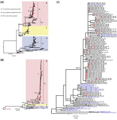
Distinct lineages and multiple introductions of the EV-D68
viruses in Kansas City during the 2014 outbreak.
Phylogenetic
analyses of the EV-D68 VP1 gene and complete genome sequences
revealed that the viruses that circulated in Kansas City during the
2014 outbreak did not form a single monophyletic group, such
that EV-D68 was clearly introduced multiple times into the city
(
Fig. 2
). More broadly, global VP1 phylogenies revealed the
pres-ence of the three major clades of EV-D68 —A, B, and C—which
have spread worldwide (
Fig. 2A
), consistent with previous studies
(
18
,
38
). Clades A and B could be further subdivided into two
subclades each: A1 and A2 and B1 and B2. All clades and subclades
were supported by bootstrap values over 80% in the ML approach
and by posterior probabilities of 1.0 in the BMCMC approach.
All 57 outbreak samples from Kansas City fell into subclade B1.
Within this subclade, it was notable that the Kansas City viruses
fell into two monophyletic groups and were interspersed among
those viruses sampled from different states in the United States
(New York, California, and Colorado) and from different
coun-tries (Canada or France), indicating frequent gene flow between
populations (
Fig. 2C
). However, because of low bootstrap support
values and frequent polytomies, it is difficult to define exact
monophyletic groups of viruses from Kansas City on global VP1
phylogeny. Therefore, we estimated the number of independent
viral introductions into the Kansas City population using a
parsi-mony reconstruction of geographic transitions, utilizing 5,000
subsampled BMCMC topologies to reflect topological
uncer-tainty. From this, we documented an average of 10 independent
introductions of EV-D68 in to Kansas City during the outbreak,
with a 95% confidence interval between 5 and 14 introductions.
The large and well-supported monophyletic group of viruses
in subclade B1 (at the top of
Fig. 2C
) includes 2014 U.S. outbreak
samples collected from New York, Missouri, and California; two
2014 samples from France; and one 2014 sample from Canada.
Again, this phylogenetic pattern is indicative of both the national
and international movement of viruses. Seven Kansas City
se-quences did not fall in this cluster; four were closely related to each
other, while another sequence grouped with a virus from Canada.
The remaining two Kansas City sequences formed separate
branches in the tree, suggesting that they also represent
indepen-dent introductions into the region. The remaining subclade B1
viruses are preoutbreak samples located near the root of the B1
cluster, including four U.S. isolates collected in 2013, a lone 2012
FIG 1High-throughput sequencing of the complete genome of EV-D68. (A)
Schematic representation of the different approaches used to sequence the complete EV-D68 genome. FL-A, full-length genome with poly(A) tail; FL, full-length genome without poly(A) tail; S, small overlapping amplicon; L, large overlapping amplicon. (B) Three examples of the EV-D68 RT PCR prod-uct—2014 outbreak strain MO49/2014, preoutbreak strain Vanderbilt N0051U5/2012, and ATCC strain VR1197—resolved by agarose gel electro-phoresis and visualized by ethidium bromide staining. Sizes of molecular markers (in base pairs) are indicated on the left side of the gel.
on November 7, 2019 by guest
http://jvi.asm.org/
[image:4.585.40.287.62.291.2]isolate from Italy, and, notably, three Chinese isolates sampled in
2011 (
Fig. 2C
). In addition, only five U.S. outbreak sequences
collected in 2014 fell outside subclade B1: two from New York
(NY73 and NY74), one from Kentucky (US.KY.14.18951), and
one from Illinois (US.IL.14.18952), all of which fell in subclade
B2. Sequence US.KY.14.18953 from Kentucky fell into subclade
A2, while the preoutbreak sample collected in Nashville,
Tennes-see, in 2012 fell into subclade A1. Finally, there are two Fermon
strain sequences in our analysis, one downloaded from GenBank
and the prototype Fermon strain that we purchased from ATCC
and sequenced. These two sequences clustered near the root,
re-flecting its sampling date in the 1960s (
Fig. 2A
).
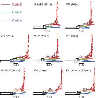
The phylogeny of complete EV-D68 genome sequences
pre-sented the same picture of three major clades circulating
world-FIG 2Evolutionary history of the VP1 and complete genome sequences of EV-D68. (A and B) Global ML phylogenies estimated for the VP1 gene (A) and full
genome sequences (B). Clades and subclades are indicated by colors and described in the trees. The bootstrap support values (percent) from 1,000 ML bootstrap replications and posterior probabilities from BMCMC analyses (10,000 tree samples) are shown for each clade/subclade with separation by a slash. Sequences reported by this study are shown in red and described in the key. Phylogenetic trees were rooted by the oldest EV-D68 sequence in GenBank, the Fermon strain collected in 1962 in California, USA. (C) Magnification of subclade B1 viruses from the VP1 phylogeny (A). Sequences generated in this study are marked by red squares in the tree. Sequences from acute flaccid myelitis (AFM) patients in the United States are marked by black squares. Sequences collected from outside the United States have taxon names in blue. The scale bars represent the numbers of nucleotide substitutions per site.
on November 7, 2019 by guest
http://jvi.asm.org/
[image:5.585.39.529.64.565.2]wide and multiple viral introductions to Kansas City during the
2014 outbreak (
Fig. 2B
and
3
). The nucleotide similarities among
the clades ranged from 90.7% to 92.7%: clade A and clade B,
90.7%; clade A and clade C, 91.6%; and clade B and clade C,
92.7%. Nonstructural genes (2A, 2B, 2C, 3A, 3B, 3C, and 3D)
exhibited levels of heterogeneity similar to those of structural
genes (VP4, VP2, VP3, and VP1), with nucleotide similarity
rang-ing from 96.5% to 97.1%. VP1 is the most variable gene across the
genome as a whole, displaying 96.5% nucleotide similarity.
EV-D68 recombination.
We found no overt evidence of
ho-mologous recombination in the VP1 data set. However, a single
recombinant virus—strain US.KY.14.18951—was identified in
the complete genome data set, supported by
P
values of less than
1
⫻
10
⫺15in all methods. To locate the recombination
break-points, a carefully chosen subset of sequence data (
n
⫽
22),
in-cluding the recombinant strain and the closest parental lineages,
was analyzed using similarity plots and Bootscan analysis in
Sim-plot v3.5.1. This identified one significant recombination
break-point in VP2, at nucleotide positions 1502 to 1576 (supported by
a maximum
2sum of 34.3), that separated the genome into two
nonrecombinant regions (
Fig. 4
) and which was strongly
sup-ported by phylogenetic trees inferred on either side of the
break-point (
Fig. 4C
). Although the Bootscan analysis provided evidence
for a second recombination event in the 3D gene region, this
lacked statistical support. Hence, these data suggest that there was
an intersubclade recombination event between B2 and B1. In
con-trast to subclade B1, which is the main U.S. 2014 outbreak lineage,
the majority of subclade B2 sequences were collected from Asia
and Europe during 2009 to 2014, with the exception of four U.S.
2014 outbreak sequences. Among the latter, one is the
con-firmed recombinant, and another is the potential parental
strain, US.IL.14.18952 (
Fig. 2A
).
No association between viral phylogeny and demographic
and clinical metadata.
We performed two phylogeny-trait
asso-ciation statistics (AI and PS) to identify whether EV-D68 outbreak
strains might exhibit some phylogenetic clustering by disease
se-verity. Notably, however, we found no significant association of
age distribution, gender ratio, or clinical symptomatology with
viral genetic variation (i.e., phylogenetic position) in the Kansas
City cohort (
Table 2
). Hence, phylogenetic clustering by disease
phenotype was not greater than that expected by chance alone.
Similarly, although no EV-D68-associated AFM cases were
re-ported in Kansas City, recently rere-ported EV-D68 sequences
col-lected from AFM patients from Colorado and California did
not form a monophyletic group (
38
) but were interspersed
with sequences of EV-D68 isolates from non-AFM patients
(
Fig. 2C
; sequences from AFM patients marked by black
rect-angle).
DISCUSSION
We developed a high-throughput method to sequence complete
genomes of EV-D68, from which we were able to obtain 59 from
FIG 3Phylogenetic trees of the complete genome and individual genes of EV-D68. Sequences in different clades are colored as described in the key. Phylogenetic
trees inferred for different regions of the EV-D68 genome are indicated in black. Phylogenetic trees were rooted using the oldest EV-D68 sequence (the Fermon strain) available on GenBank, and scale bars represent the numbers of nucleotide substitutions per site.
on November 7, 2019 by guest
http://jvi.asm.org/
[image:6.585.136.450.66.386.2]the major 2014 outbreak, including 57 from Kansas City. This
doubles the number of publicly available genome sequences of
EV-D68 and allowed us to identify both multiple introduction events
into a single community and multiple cocirculating lineages
world-wide, although there was no association between phylogenetic history
and a range of demographic or clinical features. Notably, we also
identified the first recombination event in EV-D68.
Until now, only limited full-length genome sequences of
EV-D68 from the 2014 EV-EV-D68 outbreak in the United States have
been available in the public domain, with most coming from
clin-ical isolates. With our high-throughput sequencing method, we
were able to generate overlapping amplicons from primary
spec-imens to obtain the complete viral genome with a 95% success
rate. The success of this method compared to the single-amplicon
FIG 4Recombination analyses of the full genome of EV-D68. (A) Bootscan analysis. A sliding window of 300 nt was utilized (step size of 10 nt), with
[image:7.585.41.542.68.502.2]neighbor-joining phylogenetic trees with 100 bootstrap replicates inferred in each case. Bootstrap values supporting the clustering of query sequence (i.e., US.KY.14.18951) with different groups of reference sequences (i.e., subclade B1, subclade B2, and others) were recorded. (B) Schematic diagram of the EV-D68 genome. (C) Phylogenies inferred for nonrecombinant fragments identified by Simplot and Bootscan (A). The putative recombinant strain is marked by the black square in the trees. Clades are indicated by the colors described in the key. Phylogenetic trees were midpoint rooted for clarity only.
TABLE 2Results of the phylogeny-trait association tests for particular
demographic and clinical characteristics of EV-D68 patients within Kansas City, MO, 2014
Comparison
No. of cases
(n⫽57) AIastatistic PSbstatistic
Male 39 P⫽0.15 P⫽0.40
Age⬍5 yr 17 P⫽0.08 P⫽0.12
PICUc 18 P⫽0.85 P⫽1
Asthma/wheezing 41 P⫽0.82 P⫽1
Requiring ventilation 8 P⫽0.69 P⫽0.53
aAI, association index. b
PS, parsimony score.
cPICU, pediatric intensive care unit.
on November 7, 2019 by guest
http://jvi.asm.org/
[image:7.585.300.545.621.697.2]approach is most likely due to RNA secondary structure in the 5
=
region of the virus untranslated region that could impact the
effi-ciency of RT-PCR.
Our phylogenetic analyses are notable in that they suggest a
relatively complex molecular evolution of EV-D68 during the
2014 outbreak. Before this outbreak, EV-D68 was rarely reported
and was associated with mild respiratory illness, although small
outbreaks had been documented since 2005 (
15
,
16
,
18–33
).
Pre-vious studies showed that the A, B, and C clades circulated or
cocirculated during different time periods in different geographic
regions (
15
,
16
,
18
,
19
,
21–32
,
59
). We further defined subclades
A1, A2, B1, and B2. In line with our subclade definition, subclades
A1 and B2 were endemic and found in many countries before the
2014 outbreak, including Thailand during 2005 to 2010 (
26
), the
United Kingdom during 2009 to 2010 (
27
), China during 2009 to
2012 (
24
), Philippines from 2008 to 2014 (
21–23
), and the
Neth-erlands from 1994 to 2013 (
15
,
29
). Clade C was relatively
geo-graphically restricted, being reported in Japan during 2005 to 2010
and in Italy during 2010 to 2012 (
16
,
19
,
30
). During the 2014
outbreak, a novel subclade, B1, seems to have rapidly emerged and
was dominant during the U.S. outbreak (
38
,
60
,
61
). During the
same period, subclades A1 and B2 continued to be isolated in
European countries (
40
,
41
,
62–65
). In addition, we found
multi-ple viral introductions from other localities into the single locality
of Kansas City during the 2014 U.S. outbreak despite the relatively
small number of sequences collected (
60
), indicating that EV-D68
exhibits relatively fluid spatial dynamics within the United States.
Our study is noteworthy for the observation of recombination
in EV-D68. Although recombination occurs frequently among
members of the
Enterovirus
genus, with interspecies
recombina-tion documented between rhinovirus A and rhinovirus B and
be-tween EV-A and EV-B (
33
), and intraspecies recombination
within both EV-A71 and EV-B81 (
44
,
66
), it had not been
previ-ously definitely demonstrated to occur in EV-D68. Indeed, a
pre-vious report of phylogenetic incongruence between the 5
=
UTR
and VP1 could not be confirmed since only partial 5
=
UTR and
VP1 sequences were amplified (
22
). In contrast, our analysis
of
⬎
100 complete EV-D68 genomes provided compelling
evi-dence for recombination between outbreak subclade B1 and
en-demic subclade B2 strains, with a breakpoint in VP2. In
recombi-nation events in other enteroviruses, most breakpoints have been
documented between the structural and nonstructural regions, or
within the nonstructural region (
44
,
67
). Interestingly, the
previ-ous report of phylogenetic incongruence between the 5
=
UTR
and VP1 in EV-D68 suggests the possibility of a recombination
event within the structural region (
22
), although this clearly
needs to be confirmed. The recombinant strain identified here,
US.KY.14.18951 from Kentucky, and the potential parental
strain in subclade B2, US.IL.14.18952 from Illinois, were both
isolated in August 2014, at the start of the 2014 outbreak (
68
). In
turn, the occurrence of recombination indicates that multiple
dis-tinct strains cocirculated at the beginning of the outbreak, as is
also suggested by our phylogenetic analysis. The impact of
VP2-centered recombination in EV-D68 evolution remains to be
de-fined.
One of the most striking features of the 2014 outbreak was that
most EV-D68 infections were associated with severe respiratory
diseases in children (
36
,
40
,
41
,
60–65
,
69
). However, we found no
association between age, gender, or clinical symptoms and
differ-ent viral subclades in our Kansas City population. Hence, whether
sequence data can predict the clinical outcome is still unclear, and
our sample size was relatively small. One finding suggestive of an
association between viral sequence diversity and clinical features is
that AFM-associated strains from the United States during the
2014 outbreak were all members of the novel outbreak subclade
B1 (
38
), although we were unable to identify viral genetic
signa-tures of disease severity. Indeed, there are a number of feasigna-tures
that argue against a strong strain basis to virulence. First, of the
four cases of neurological conditions before the 2014 outbreak,
two cases from Kenya (strain HEV126010 in 2010 and strain
HEV199011 in 2011) were associated with viruses from subclade
A1, the endemic subclade (
31
), while sequence information
con-cerning the two U.S. patients is unavailable. Second, although all
U.S. 2014 AFM cases fall in subclade B1 (
38
), two of three
Euro-pean AFM cases (
40
,
41
) are from subclade B2. Third, the subclade
B1-associated AFM sequences do not form a single cluster but
rather are interspersed with those representing non-AFM cases
(
Fig. 2C
). It was recently suggested that a G272 mutation in the 5
=
UTR and a series of amino acid changes in the clade B1
polypro-tein (T291 in VP2, A341 in VP3, N860 in VP1, N927 in 2A, K2005
in 3D, and G1108 at the 2B/2C junction) might have increased
neurovirulence in EV-D68 (
38
). However, G272 was present in all
the clades studied here and the polyprotein mutations were B1
specific, not AFM specific, as they were also found in non-AFM
patients. In addition, two non-B1 AFM sequences, reported
be-fore the U.S. 2014 outbreak, possess none of the polyprotein
mu-tations. Hence, the concentration of AFM cases in subclade B1
may simply reflect the predominance of B1 infections in the
United States (i.e., a founder effect). Indeed, it is pertinent that
EV-D68 viruses could not be isolated from cerebrospinal fluid of
AFM patients from the 2014 U.S. outbreak (
38
).
Although we considered only a single city in the United States
during the outbreak, these results are likely to be applicable to the
spread of EV-D68 in other modern and highly mobile
popula-tions. For an improved understanding of the factors determining
possible spatiotemporal differences in EV-D68 infection and
transmission, continuous global monitoring of the clinical and
molecular epidemiology of EV-D68 by representative surveillance
systems should clearly be a public health priority.
ACKNOWLEDGMENTS
The clinical sample and data collection for this study were supported by a National Institute of Allergy and Infectious Diseases grant (AI U19-AI-095277). The sequencing work was supported by the NIAID/NIH Genomic Centers for Infectious Diseases (GCID) program (U19-AI-110819). Vanderbilt birth cohort and respiratory viral surveillance was supported by NIAID U19-AI-95227. E.C.H. was supported by an NHMRC Australia Fellowship (AF30).
The content is solely the responsibility of the authors and does not represent official views of the National Institutes of Health.
S.R.D., Y.T., R.S., E.C.H., T.V.H., and J.D.C. conceived this study. F.H., J.E.S., R.S., J.D.C., and T.V.H. collected clinical samples and clinical data and performed EV-D68-specific PCR. A.S., X.L., and R.A.H. per-formed RNA extraction, genome amplification, and viral sequencing. N.F. and T.B.S. assembled and analyzed the genomes and finished genome sequences as needed. Y.T., T.T.-Y.L., E.C.H., and S.R.D. analyzed the data. Y.T., T.T.-Y.L., E.C.H., and S.R.D. wrote the manuscript, and all authors reviewed and approved the final version.
We have no conflicts of interest to declare.
on November 7, 2019 by guest
http://jvi.asm.org/
FUNDING INFORMATION
HHS | NIH | National Institute of Allergy and Infectious Diseases (NIAID) provided funding to Yi Tan, Ari Simenauer, Rebecca A Halpin, Xudong Lin, Nadia Fedorova, Timothy Brian Stockwell, and Suman R Das under grant number U19-AI-110819. HHS | NIH | National Institute of Allergy and Infectious Diseases (NIAID) provided funding to Tina V Hartert under grant number AI U19-AI-095277. HHS | NIH | National Institute of Allergy and Infectious Diseases (NIAID) provided funding to Jim Chappell and Tina V Hartert under grant number U19-AI-95227. Depart-ment of Health | National Health and Medical Research Council (NHMRC) provided funding to Edward C Holmes under grant number AF30.
REFERENCES
1.Imamura T, Oshitani H.2015. Global reemergence of enterovirus D68 as
an important pathogen for acute respiratory infections. Rev Med Virol
25:102–114.http://dx.doi.org/10.1002/rmv.1820.
2.Kono R, Sasagawa A, Ishii K, Sugiura S, Ochi M.1972. Pandemic of new
type of conjunctivitis. Lanceti:1191–1194.
3.Kew OM, Nottay BK, Hatch MH, Hierholzer JC, Obijeski JF. 1983.
Oligonucleotide fingerprint analysis of enterovirus 70 isolates from the 1980 to 1981 pandemic of acute hemorrhagic conjunctivitis: evidence for a close genetic relationship among Asian and American strains. Infect Immun41:631– 635.
4.Junttila N, Leveque N, Kabue JP, Cartet G, Mushiya F,
Muyembe-Tamfum JJ, Trompette A, Lina B, Magnius LO, Chomel JJ, Norder H.
2007. New enteroviruses, EV-93 and EV-94, associated with acute flaccid paralysis in the Democratic Republic of the Congo. J Med Virol79:393–
400.http://dx.doi.org/10.1002/jmv.20825.
5.Smura TP, Junttila N, Blomqvist S, Norder H, Kaijalainen S, Paananen
A, Magnius LO, Hovi T, Roivainen M.2007. Enterovirus 94, a proposed
new serotype in human enterovirus species D. J Gen Virol88:849 – 858. http://dx.doi.org/10.1099/vir.0.82510-0.
6.Harvala H, Sharp CP, Ngole EM, Delaporte E, Peeters M, Simmonds P.
2011. Detection and genetic characterization of enteroviruses circulating among wild populations of chimpanzees in Cameroon: relationship with human and simian enteroviruses. J Virol85:4480 – 4486.http://dx.doi.org /10.1128/JVI.02285-10.
7.Harvala H, Van Nguyen D, McIntyre C, Ahuka-Mundeke S, Ngole EM,
Delaporte E, Peeters M, Simmonds P.2014. Co-circulation of
enterovi-ruses between apes and humans. J Gen Virol95:403– 407.http://dx.doi .org/10.1099/vir.0.059048-0.
8.Schieble JH, Fox VL, Lennette EH.1967. A probable new human
picor-navirus associated with respiratory diseases. Am J Epidemiol85:297–310.
9.Blomqvist S, Savolainen C, Raman L, Roivainen M, Hovi T. 2002.
Human rhinovirus 87 and enterovirus 68 represent a unique serotype with rhinovirus and enterovirus features. J Clin Microbiol40:4218 – 4223.http: //dx.doi.org/10.1128/JCM.40.11.4218-4223.2002.
10. Savolainen C, Blomqvist S, Mulders MN, Hovi T.2002. Genetic
clus-tering of all 102 human rhinovirus prototype strains: serotype 87 is close to human enterovirus 70. J Gen Virol83:333–340.http://dx.doi.org/10.1099 /0022-1317-83-2-333.
11. Ishiko H, Miura R, Shimada Y, Hayashi A, Nakajima H, Yamazaki S,
Takeda N.2002. Human rhinovirus 87 identified as human enterovirus
68 by VP4-based molecular diagnosis. Intervirology45:136 –141.http: //dx.doi.org/10.1159/000065866.
12. Khetsuriani N, Lamonte-Fowlkes A, Oberst S, Pallansch MA, Centers
for Disease Control and Prevention.2006. Enterovirus surveillance—
United States, 1970 –2005. MMWR Surveill Summ55(8):1–20.
13. Wang Z, Malanoski AP, Lin B, Long NC, Leski TA, Blaney KM, Hansen
CJ, Brown J, Broderick M, Stenger DA, Tibbetts C, Russell KL, Metzgar D.2010. Broad spectrum respiratory pathogen analysis of throat swabs from military recruits reveals interference between rhinoviruses and ad-enoviruses. Microb Ecol59:623– 634.http://dx.doi.org/10.1007/s00248 -010-9636-3.
14. Oberste MS, Maher K, Schnurr D, Flemister MR, Lovchik JC, Peters H,
Sessions W, Kirk C, Chatterjee N, Fuller S, Hanauer JM, Pallansch MA.
2004. Enterovirus 68 is associated with respiratory illness and shares bio-logical features with both the enteroviruses and the rhinoviruses. J Gen Virol85:2577–2584.http://dx.doi.org/10.1099/vir.0.79925-0.
15. Meijer A, van der Sanden S, Snijders BE, Jaramillo-Gutierrez G, Bont L,
van der Ent CK, Overduin P, Jenny SL, Jusic E, van der Avoort HG,
Smith GJ, Donker GA, Koopmans MP.2012. Emergence and epidemic
occurrence of enterovirus 68 respiratory infections in The Netherlands in 2010. Virology423:49 –57.http://dx.doi.org/10.1016/j.virol.2011.11.021.
16. Ikeda T, Mizuta K, Abiko C, Aoki Y, Itagaki T, Katsushima F,
Katsu-shima Y, Matsuzaki Y, Fuji N, Imamura T, Oshitani H, Noda M,
Kimura H, Ahiko T.2012. Acute respiratory infections due to enterovirus
68 in Yamagata, Japan between 2005 and 2010. Microbiol Immunol56:
139 –143.http://dx.doi.org/10.1111/j.1348-0421.2012.00411.x.
17. Bingjun T, Yoshida H, Yan W, Lin L, Tsuji T, Shimizu H, Miyamura T.
2008. Molecular typing and epidemiology of non-polio enteroviruses iso-lated from Yunnan Province, the People’s Republic of China. J Med Virol
80:670 – 679.http://dx.doi.org/10.1002/jmv.21122.
18. Tokarz R, Firth C, Madhi SA, Howie SR, Wu W, Sall AA, Haq S, Briese T,
Lipkin WI.2012. Worldwide emergence of multiple clades of enterovirus 68.
J Gen Virol93:1952–1958.http://dx.doi.org/10.1099/vir.0.043935-0.
19. Kaida A, Kubo H, Sekiguchi J, Kohdera U, Togawa M, Shiomi M,
Nishigaki T, Iritani N.2011. Enterovirus 68 in children with acute
respi-ratory tract infections, Osaka, Japan. Emerg Infect Dis17:1494 –1497. http://dx.doi.org/10.3201/eid1708.110028.
20. Hasegawa S, Hirano R, Okamoto-Nakagawa R, Ichiyama T, Shirabe K.
2011. Enterovirus 68 infection in children with asthma attacks: virus-induced asthma in Japanese children. Allergy66:1618 –1620.http://dx.doi .org/10.1111/j.1398-9995.2011.02725.x.
21. Imamura T, Fuji N, Suzuki A, Tamaki R, Saito M, Aniceto R, Galang H,
Sombrero L, Lupisan S, Oshitani H.2011. Enterovirus 68 among
chil-dren with severe acute respiratory infection, the Philippines. Emerg Infect Dis17:1430 –1435.http://dx.doi.org/10.3201/eid1708.101328.
22. Imamura T, Suzuki A, Lupisan S, Okamoto M, Aniceto R, Egos RJ, Daya
EE, Tamaki R, Saito M, Fuji N, Roy CN, Opinion JM, Santo AV, Macalalad
NG, Tandoc A, III, Sombrero L, Olveda R, Oshitani H.2013. Molecular
evolution of enterovirus 68 detected in the Philippines. PLoS One8:e74221. http://dx.doi.org/10.1371/journal.pone.0074221.
23. Furuse Y, Chaimongkol N, Okamoto M, Imamura T, Saito M, Tamaki
R, Saito M, Tohoku-RITM Collaborative Research Team, Lupisan SP,
Oshitani H.2015. Molecular epidemiology of enterovirus d68 from 2013
to 2014 in Philippines. J Clin Microbiol53:1015–1018.http://dx.doi.org /10.1128/JCM.03362-14.
24. Lu QB, Wo Y, Wang HY, Wei MT, Zhang L, Yang H, Liu EM, Li TY,
Zhao ZT, Liu W, Cao WC.2014. Detection of enterovirus 68 as one of the
commonest types of enterovirus found in patients with acute respiratory tract infection in China. J Med Microbiol63:408 – 414.http://dx.doi.org /10.1099/jmm.0.068247-0.
25. Xiang Z, Gonzalez R, Wang Z, Ren L, Xiao Y, Li J, Li Y, Vernet G,
Paranhos-Baccala G, Jin Q, Wang J.2012. Coxsackievirus A21,
entero-virus 68, and acute respiratory tract infection, China. Emerg Infect Dis
18:821– 824.http://dx.doi.org/10.3201/eid1805.111376.
26. Linsuwanon P, Puenpa J, Suwannakarn K, Auksornkitti V,
Vichiwat-tana P, Korkong S, Theamboonlers A, Poovorawan Y.2012. Molecular
epidemiology and evolution of human enterovirus serotype 68 in Thai-land, 2006 –2011. PLoS One7:e35190.http://dx.doi.org/10.1371/journal .pone.0035190.
27. Lauinger IL, Bible JM, Halligan EP, Aarons EJ, MacMahon E, Tong
CY.2012. Lineages, sub-lineages and variants of enterovirus 68 in recent outbreaks. PLoS One 7:e36005. http://dx.doi.org/10.1371 /journal.pone.0036005.
28. Renois F, Bouin A, Andreoletti L.2013. Enterovirus 68 in pediatric
patients hospitalized for acute airway diseases. J Clin Microbiol51:640 –
643.http://dx.doi.org/10.1128/JCM.02640-12.
29. Meijer A, Benschop KS, Donker GA, van der Avoort HG.23 October
2014. Continued seasonal circulation of enterovirus D68 in the Nether-lands, 2011–2014. Euro Surveill19:20935.http://dx.doi.org/10.2807/1560 -7917.ES2014.19.42.20935.
30. Piralla A, Girello A, Grignani M, Gozalo-Marguello M, Marchi A,
Marseglia G, Baldanti F.2014. Phylogenetic characterization of
entero-virus 68 strains in patients with respiratory syndromes in Italy. J Med Virol
86:1590 –1593.http://dx.doi.org/10.1002/jmv.23821.
31. Opanda SM, Wamunyokoli F, Khamadi S, Coldren R, Bulimo WD.
2014. Genetic diversity of human enterovirus 68 strains isolated in Kenya using the hypervariable 3=-end of VP1 gene. PLoS One9:e102866.http: //dx.doi.org/10.1371/journal.pone.0102866.
32. Todd AK, Hall RJ, Wang J, Peacey M, McTavish S, Rand CJ, Stanton
JA, Taylor S, Huang QS.2013. Detection and whole genome sequence
on November 7, 2019 by guest
http://jvi.asm.org/
analysis of an enterovirus 68 cluster. Virol J10:103.http://dx.doi.org/10 .1186/1743-422X-10-103.
33. Garcia J, Espejo V, Nelson M, Sovero M, Villaran MV, Gomez J,
Barrantes M, Sanchez F, Comach G, Arango AE, Aguayo N, de Rivera IL, Chicaiza W, Jimenez M, Aleman W, Rodriguez F, Gonzales MS,
Kochel TJ, Halsey ES.2013. Human rhinoviruses and enteroviruses in
influenza-like illness in Latin America. Virol J10:305.http://dx.doi.org/10 .1186/1743-422X-10-305.
34. Kreuter JD, Barnes A, McCarthy JE, Schwartzman JD, Oberste MS,
Rhodes CH, Modlin JF, Wright PF.2011. A fatal central nervous system
enterovirus 68 infection. Arch Pathol Lab Med135:793–796.http://dx.doi .org/10.1043/2010-0174-CR.1.
35. Centers for Disease Control and Prevention.23 March 2015.
Enterovi-rus D68. Centers for Disease Control and Prevention, Atlanta, GA.http: //www.cdc.gov/non-polio-enterovirus/about/ev-d68.html. Accessed 26 August 2015.
36. Midgley CM, Jackson MA, Selvarangan R, Turabelidze G, Obringer E,
Johnson D, Giles BL, Patel A, Echols F, Oberste MS, Nix WA, Watson
JT, Gerber SI.2014. Severe respiratory illness associated with enterovirus
D68 —Missouri and Illinois, 2014. MMWR Morb Mortal Wkly Rep63:
798 –799.
37. Schuster JE, Miller JO, Selvarangan R, Weddle G, Thompson MT, Hassan
F, Rogers SL, Oberste MS, Nix WA, Jackson MA.2015. Severe enterovirus
68 respiratory illness in children requiring intensive care management. J Clin Virol70:77– 82.http://dx.doi.org/10.1016/j.jcv.2015.07.298.
38. Greninger AL, Naccache SN, Messacar K, Clayton A, Yu G, Somasekar
S, Federman S, Stryke D, Anderson C, Yagi S, Messenger S, Wadford D, Xia D, Watt JP, Van Haren K, Dominguez SR, Glaser C, Aldrovandi G,
Chiu CY.2015. A novel outbreak enterovirus D68 strain associated with
acute flaccid myelitis cases in the USA (2012–14): a retrospective cohort study. Lancet Infect Dis 15:671– 682. http://dx.doi.org/10.1016/S1473 -3099(15)70093-9.
39. Messacar K, Schreiner TL, Maloney JA, Wallace A, Ludke J, Oberste MS, Nix
WA, Robinson CC, Glode MP, Abzug MJ, Dominguez SR.2015. A cluster of
acuteflaccidparalysisandcranialnervedysfunctiontemporallyassociatedwithan outbreak of enterovirus D68 in children in Colorado, USA. Lancet385:1662– 1671.http://dx.doi.org/10.1016/S0140-6736(14)62457-0.
40. Pfeiffer HC, Bragstad K, Skram MK, Dahl H, Knudsen PK, Chawla MS,
Holberg-Petersen M, Vainio K, Dudman SG, Kran AM, Rojahn AE.
2015. Two cases of acute severe flaccid myelitis associated with enterovirus D68 infection in children, Norway, autumn 2014. Euro Surveill20:21062. http://dx.doi.org/10.2807/1560-7917.ES2015.20.10.21062.
41. Lang M, Mirand A, Savy N, Henquell C, Maridet S, Perignon R, Labbe
A, Peigue-Lafeuille H.2014. Acute flaccid paralysis following enterovirus
D68 associated pneumonia, France, 2014. Euro Surveill19:20952.http: //dx.doi.org/10.2807/1560-7917.ES2014.19.44.20952.
42. Larkin EK, Gebretsadik T, Moore ML, Anderson LJ, Dupont WD, Chappell
JD, Minton PA, Peebles RS, Jr, Moore PE, Valet RS, Arnold DH, Rosas-Salazar
C, Das SR, Polack FP, Hartert TV, INSPIRE Study.2015. Objectives, design and
enrollment results from the Infant Susceptibility to Pulmonary Infections and Asthma Following RSV Exposure Study (INSPIRE). BMC Pulm Med15:45.http: //dx.doi.org/10.1186/s12890-015-0040-0.
43. Stucker KM, Schobel SA, Olsen RJ, Hodges HL, Lin X, Halpin RA,
Fedorova N, Stockwell TB, Tovchigrechko A, Das SR, Wentworth
DE, Musser JM.2015. Haemagglutinin mutations and glycosylation
changes shaped the 2012/13 influenza A(H3N2) epidemic, Houston, Texas. Euro Surveill 20:21122. http://dx.doi.org/10.2807/1560-7917 .ES2015.20.18.21122.
44. Geoghegan JL, Tan LV, Kuhnert D, Halpin RA, Lin X, Simenauer A,
Akopov A, Das SR, Stockwell TB, Shrivastava S, Ngoc NM, Uyen LTT, Tuyen NT, Thanh TT, Hang VT, Qui PT, Hung NT, Khanh TH, Thinh LQ, Nhan LNT, Van HMT, Viet DC, Tuan HM, Viet HL, Hien TT, Chau NV, Thwaites G, Grenfell BT, Stadler T, Wentworth DE, Holmes
EC, Van Doorn HR.2015. Phylodynamics of enterovirus A71-associated
hand, foot, and mouth disease in Viet Nam. J Virol89:8871– 8879.http: //dx.doi.org/10.1128/JVI.00706-15.
45. CLC bio.2012. White paper.De novoassembly in CLC Assembly Cell
4.0. CLC bio, Waltham, MA.http://www.clcbio.com/files/whitepapers /whitepaper-denovo-assembly-4.pdf.
46. CLC bio.2010. White paper on reference assembly in CLC Assembly Cell
3.0. CLC bio, Aarhus, Denmark.http://www.clcbio.com/wp-content/ uploads/2012/09/white_paper_on_reference_assembly_on_the_CLC _Assembly_Cell.pdf.
47. Wang S, Sundaram JP, Stockwell TB.2012. VIGOR extended to annotate
genomes for additional 12 different viruses. Nucleic Acids Res40:W186 – W192.http://dx.doi.org/10.1093/nar/gks528.
48. Tamura K, Stecher G, Peterson D, Filipski A, Kumar S.2013. MEGA6:
Molecular Evolutionary Genetics Analysis version 6.0. Mol Biol Evol30:
2725–2729.http://dx.doi.org/10.1093/molbev/mst197.
49. Guindon S, Dufayard JF, Lefort V, Anisimova M, Hordijk W, Gascuel
O.2010. New algorithms and methods to estimate maximum-likelihood phylogenies: assessing the performance of PhyML 3.0. Syst Biol59:307–
321.http://dx.doi.org/10.1093/sysbio/syq010.
50. Ronquist F, Teslenko M, van der Mark P, Ayres DL, Darling A, Hohna S,
Larget B, Liu L, Suchard MA, Huelsenbeck JP.2012. MrBayes 3.2: efficient
Bayesian phylogenetic inference and model choice across a large model space. Syst Biol61:539 –542.http://dx.doi.org/10.1093/sysbio/sys029.
51. Drummond AJ, Suchard MA, Xie D, Rambaut A.2012. Bayesian
phy-logenetics with BEAUti and the BEAST 1.7. Mol Biol Evol29:1969 –1973. http://dx.doi.org/10.1093/molbev/mss075.
52. Fitch WM.1971. Evolution of clupeine Z, a probable crossover product.
Nat New Biol229:245–247.http://dx.doi.org/10.1038/newbio229245a0.
53. Martin D, Rybicki E.2000. RDP: detection of recombination amongst
aligned sequences. Bioinformatics16:562–563.http://dx.doi.org/10.1093 /bioinformatics/16.6.562.
54. Lole KS, Bollinger RC, Paranjape RS, Gadkari D, Kulkarni SS,
Novak NG, Ingersoll R, Sheppard HW, Ray SC. 1999. Full-length
human immunodeficiency virus type 1 genomes from subtype C-in-fected seroconverters in India, with evidence of intersubtype recombi-nation. J Virol73:152–160.
55. Robertson DL, Hahn BH, Sharp PM. 1995. Recombination in AIDS
viruses. J Mol Evol40:249 –259.http://dx.doi.org/10.1007/BF00163230.
56. Parker J, Rambaut A, Pybus OG.2008. Correlating viral phenotypes with
phylogeny: accounting for phylogenetic uncertainty. Infect Genet Evol
8:239 –246.http://dx.doi.org/10.1016/j.meegid.2007.08.001.
57. Huang W, Wang G, Zhuge J, Nolan SM, Dimitrova N, Fallon JT.2015.
Whole-genome sequence analysis reveals the enterovirus D68 isolates during the United States 2014 outbreak mainly belong to a novel clade. Sci Rep5:15223.http://dx.doi.org/10.1038/srep15223.
58. Lakdawala SS, Jayaraman A, Halpin RA, Lamirande EW, Shih AR,
Stockwell TB, Lin X, Simenauer A, Hanson CT, Vogel L, Paskel M, Minai M, Moore I, Orandle M, Das SR, Wentworth DE, Sasisekharan
R, Subbarao K.2015. The soft palate is an important site of adaptation for
transmissible influenza viruses. Nature526:122–125.http://dx.doi.org/10 .1038/nature15379.
59. Poelman R, Scholvinck EH, Borger R, Niesters HG, van Leer-Buter C.
2015. The emergence of enterovirus D68 in a Dutch university medical center and the necessity for routinely screening for respiratory viruses. J Clin Virol62:1–5.http://dx.doi.org/10.1016/j.jcv.2014.11.011.
60. Wylie KM, Wylie TN, Orvedahl A, Buller RS, Herter BN, Magrini V,
Wilson RK, Storch GA.2015. Genome sequence of enterovirus D68 from
St. Louis, Missouri, USA. Emerg Infect Dis21:184 –186.http://dx.doi.org /10.3201/eid2101.141605.
61. Zhang T, Ren L, Luo M, Li A, Gong C, Chen M, Yu X, Wu J, Deng
Y, Huang F. 2015. Enterovirus D68-associated severe pneumonia,
China, 2014. Emerg Infect Dis21:916 –918.http://dx.doi.org/10.3201 /eid2105.150036.
62. Bragstad K, Jakobsen K, Rojahn AE, Skram MK, Vainio K,
Holberg-Petersen M, Hungnes O, Dudman SG, Kran AM.2015. High frequency
of enterovirus D68 in children hospitalised with respiratory illness in Nor-way, autumn 2014. Influenza Other Respir Viruses9:59 – 63.http://dx.doi .org/10.1111/irv.12300.
63. Gimferrer L, Campins M, Codina MG, Esperalba J, Martin MD,
Fuen-tes F, Pumarola T, Anton A.2015. First enterovirus D68 (EV-D68) cases
detected in hospitalised patients in a tertiary care university hospital in Spain, October 2014. Enferm Infecc Microbiol Clinhttp://dx.doi.org/10 .1016/j.eimc.2015.01.008.
64. Midgley SE, Christiansen CB, Poulsen MW, Hansen CH, Fischer TK.
2015. Emergence of enterovirus D68 in Denmark, June 2014 to February 2015. Euro Surveill 20:21105. http://dx.doi.org/10.2807/1560-7917 .ES2015.20.17.21105.
65. Reiche J, Bottcher S, Diedrich S, Buchholz U, Buda S, Haas W,
Schweiger B, Wolff T. 2015. Low-level circulation of enterovirus
D68-associated acute respiratory infections, Germany, 2014. Emerg Infect Dis21:837– 841.http://dx.doi.org/10.3201/eid2105.141900.
66. Hu L, Zhang Y, Hong M, Zhu S, Yan D, Wang D, Li X, Zhu Z, Tsewang
on November 7, 2019 by guest
http://jvi.asm.org/
Xu W.2014. Phylogenetic evidence for multiple intertypic recombina-tions in enterovirus B81 strains isolated in Tibet, China. Sci Rep4:6035. http://dx.doi.org/10.1038/srep06035.
67. Guo WP, Lin XD, Chen YP, Liu Q, Wang W, Wang CQ, Li MH, Sun
XY, Shi M, Holmes EC, Zhang YZ.2015. Fourteen types of co-circulating
recombinant enterovirus were associated with hand, foot, and mouth dis-ease in children from Wenzhou, China. J Clin Virol70:29 –38.http://dx .doi.org/10.1016/j.jcv.2015.06.093.
68. Brown BA, Nix WA, Sheth M, Frace M, Oberste MS.2014. Seven strains
of enterovirus D68 detected in the United States during the 2014 severe respiratory disease outbreak. Genome Announc2:e01201-14.http://dx .doi.org/10.1128/genomeA.01201-14.
69. Oermann CM, Schuster JE, Conners GP, Newland JG, Selvarangan
R, Jackson MA.2015. Enterovirus d68. A focused review and clinical
highlights from the 2014 U.S. outbreak. Ann Am Thorac Soc12:775–
781.http://dx.doi.org/10.1513/AnnalsATS.201412-592FR.
on November 7, 2019 by guest
http://jvi.asm.org/