This is a repository copy of
A systematic review of classification systems for pilonidal
sinus
.
White Rose Research Online URL for this paper:
http://eprints.whiterose.ac.uk/146803/
Version: Published Version
Article:
Beal, E.M., Lee, M.J., Hind, D. orcid.org/0000-0002-6409-4793 et al. (3 more authors)
(2019) A systematic review of classification systems for pilonidal sinus. Techniques in
Coloproctology. ISSN 1123-6337
https://doi.org/10.1007/s10151-019-01988-x
[email protected] https://eprints.whiterose.ac.uk/
Reuse
This article is distributed under the terms of the Creative Commons Attribution (CC BY) licence. This licence allows you to distribute, remix, tweak, and build upon the work, even commercially, as long as you credit the authors for the original work. More information and the full terms of the licence here:
https://creativecommons.org/licenses/
Takedown
If you consider content in White Rose Research Online to be in breach of UK law, please notify us by
https://doi.org/10.1007/s10151-019-01988-x
ORIGINAL ARTICLE
A systematic review of classiication systems for pilonidal sinus
E. M. Beal1,4 · M. J. Lee2,4 · D. Hind1,4 · A. P. Wysocki3,4 · F. Yang1 · S. R. Brown2
Received: 17 December 2018 / Accepted: 8 April 2019 © The Author(s) 2019
Abstract
Background Pilonidal sinus disease (PSD) is a simple chronic inlammatory condition resulting from loose hairs forcibly inserted into vulnerable tissue in the natal cleft. It is an acquired disease with a slight familial tendency. There is no agree-ment on optimum treatagree-ment and the multitude of therapeutic options cannot be compared due to the lack of a universally adopted classiication of the disease. The aim of our study was to perform a systematic review of the literature to determine how presentations of PSD are classiied and reported.
Methods A systematic review of the English language literature was undertaken searching studies published after 1980.
Results Eight classiication systems of PSD were identiied. Most classiication systems were based on anatomical pathol-ogy hypotheses. The location and number of sinuses were the main factors deining classiication systems. No articles were retrieved that assessed the validity and/or reliability of the classiication system employed. Furthermore, there was no evi-dence to suggest a correlation between prognosis outcome and subgroup.
Conclusions Based on the evidence available from the literature reviewed we have no recommendations regarding the use of the current classiication of PSD. A well-recognised and practical classiication system to guide clinical practice is required.
Keywords Pilonidal sinus disease · Classiication system · Colorectal surgery
Introduction
Pilonidal sinus disease (PSD) is a common condition afect-ing 26/100,000 of the general population, predominantly young, and employed males [1]. PSD is rarely self-limiting and, therefore, surgery is the mainstay of treatment. Many surgical methods are described including sinus/pit-based procedures, excision with open management, excision and midline closure, and excision with completely of midline lap repair or lap closure which crosses the midline. Adju-vant laser treatment and shaving are also described. Despite
all these options, the recurrence may be as high as 60.4% at 24 months post-surgery [2]. In addition to recurrence, early wound complications such as infection and dehiscence are very common [3–5].
PSD is an umbrella term for a spectrum of abnormali-ties ranging from relatively asymptomatic simple midline pits or sinuses to complex chronically inlamed cavities with multiple istulous tracks to treatment failure. Most patients present with chronic symptoms but a signiicant proportion present with an acute abscess. Diferent stages of the disease may be amenable to diferent treatment strategies. However, there is no universally adopted classiication system for dis-ease appearance.
Classiication
Clinical classiication has a prognostic function. The Prog-nosis Research Strategy (PROGRESS) Group summarizes and proposes a stepwise approach to prognosis research. Ultimately this allows stratiied treatment. In the treatment of PSD, the existing classiication systems are not used in routine practice to guide treatment as clinicians generally * E. M. Beal
[email protected]; [email protected]
1 University of Sheield, The Innovation Centre, 217
Portobello, Sheield S1 4DP, UK
2 Department of General Surgery, Sheield Teaching
Hospitals, Sheield, UK
3 Department of Surgery, Logan Hospital, Meadowbrook,
QLD, Australia
4 Griith Health Centre, Griith University Medical School,
Techniques in Coloproctology
1 3
select treatment methods in accordance with their own expe-rience and training [2]. Previous literature has outlined the prevalence of PSD [6] and evaluated surgical techniques [7] with the use of a classiication system, however, few stud-ies exist evaluating the use of a classiication system for PSD that informs the choice of treatment. Where a classii-cation system is proposed, its predictive properties should be assessed. Properties commonly assessed in classiication systems are internal consistency (i.e., ensuring the items being used are measuring the correct constructs that are being investigated), reliability, measurement error, content validity (including face validity), construct validity (includ-ing structural validity, hypotheses test(includ-ing and cross-cultural validity), criterion validity (i.e., the extent to which a meas-ure is related to an outcome), responsiveness, and interpret-ability as per the COnsensus-based Standards for the selec-tion of health Measurement Instruments group (COSMIN) [8]. This highlights the need for an accessible classiication system that will allow the informed choice of treatment.
The aim of this systematic review was to provide an overview of published classiication systems for PSD and to summarise any analyses of the reliability and validity of each system.
Materials and methods
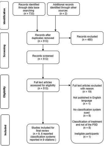
This systematic review of PSD classiications was registered with PROSPERO (Registration number: CRD42018111767). It is reported in accordance with the preferred reporting items for systematic reviews and meta-analysis (PRISMA) guidelines (for PRISMA lowchart see Fig. 1) [9]. The qual-ity of the methods used in each study was assessed against the Consensus-based standards for the selection of health status Measurement Instruments (COSMIN) guidelines [8].
Eligibility criteria
All original studies investigating the ways in which to clas-sify the presentation of PSD were eligible for inclusion. This review focused exclusively on the classiication systems used to describe PSD rather than therapeutic interventions. Systematic reviews, randomized controlled trials, observa-tional studies (such as cohort studies), case–control studies and cross-sectional studies, could be included, when they described details of ways in which to classify PSD. Theoreti-cal articles and empiriTheoreti-cal studies were also included. These included letters to the editor, reports or conference abstracts which proposed classiication systems of PSD. Empirical work included case reports and primary research involving individual patients in which the reliability and validity of the classiication system might be formally tested. Articles pub-lished after 1980 and in English were eligible for inclusion
in the systematic review. Articles were excluded when PSD was present in a body region other than the natal cleft, were not written in the English language or did not speciically describe a classiication system.
Search and information sources
A search of the online databases MEDLINE and EMBASE was completed using keywords to search for articles pub-lished after January 1980. The MeSH search terms and key-words were “pilonidal disease”, “pilonidal sinus”, “SPSD”, “jeep disease” combined with “classiication” or “classify” or “system” or “instrument” or “type” or “prognosis” or “predict” (sample search strategy is presented in “Appendix A”). A basic search of Google Scholar was also conducted. Furthermore, experts in the ield (SRB, APW) were con-sulted for signposting to other relevant literature.
Study selection
Studies were screened for eligibility against the above cri-teria by two reviewers (EMB and FY). Where there was conlict in the assessment this was resolved by DH. Full texts were retrieved for the screening of the eligible studies. If a study was excluded, reasons were recorded. In cases where abstracts met inclusion criteria and a full text version of the study could not be retrieved the authors were contacted requesting further information.
Data collection
The articles identiied were included for full text review if they clearly indicated in the abstract or in the title that they had employed the use of or had proposed a classiication sys-tem or diagnostic tool for PSD. These full text articles were then reviewed to ascertain whether a classiication system had been created.
Data extraction
Results
Study selection
Seven hundred and thirty-three records were screened for eligibility and 512 were assessed for eligibility. After the titles and abstracts were screened, a total of 27 records were assessed by full-text review and nine were included (Fig. 2). Articles were excluded when PSD was present in areas other than the natal cleft [9, 10]. Of the articles included, eight presented novel systems. One abstract was included even
though the full text article could not be retrieved [11]. After conferring, it was decided to retain this study as the abstract detailed the classiication system used which complied with the inclusion criteria. Two articles referred to the same clas-siication system [12, 13]. One of these studies was included after consensus agreement. Among the papers included there were three letters to the editor. The remaining three studies presented longitudinal data on subgroup outcomes and had employed a classiication system. One paper was a theoreti-cal description [14].
[image:4.595.205.546.54.533.2]Techniques in Coloproctology
1 3
Synthesis of results
Components of the classiication systems
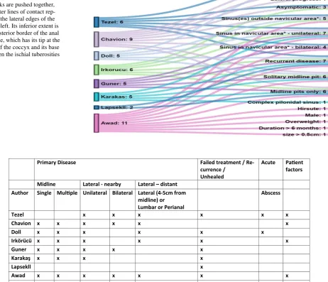
Classiication can be viewed as a logical process of sorting complex data into groups using shared features. The clas-siication systems included produced overlapping dimen-sions as shown in Fig. 3. These were; classiication due to location of sinus (although varying terminology was used) (n = 8) [11, 12, 14–19]; recurrent disease as a single dimension (n = 7) [11, 12, 14–19]; total number of pits/ sinuses and tracks present (n = 4) [15, 16, 18, 19]; pres-ence of abscess or treatment elements such as the need for
drainage (n = 3) [14, 15, 17]; the size of the sinus or lesion (n = 2) [15, 18].
Three of the classiication systems [12, 15, 17] included an asymptomatic sinus as a classiication item in their sys-tems. One classiication system was based almost entirely on measurements (to anus, vertical and transverse disease extent) [14]. Only one classiication system recorded patient as well as sinus characteristics [18]. This was the most detailed classiication system, employing a scoring system based on the following factors: patient characteristics (hir-sute or not, weight, sex), sinus characteristics (location, size, recurrence) and duration of symptoms.
Location of the sinus was present in all classiication systems, although the terminology varied. Tezel’s [17]
Fig. 2 Alluvial diagram of clas-siication systems. *Deinition for navicular area: When the buttocks are pushed together, the outer lines of contact rep-resent the lateral edges of the natal cleft. Its inferior extent is the posterior border of the anal triangle, which has its tip at the apex of the coccyx and its base between the ischial tuberosities [17]
-e R / t n e m t a e r t d e l i a F e s a e s i D y r a m i r P currence / Unhealed
Acute Paent
factors t n a t s i d – l a r e t a L y b r a e n -l a r e t a L e n i l d i M
Author Single Mulple Unilateral Bilateral Lateral (4-5cm from midline) or Lumbar or Perianal
s s e c s b A
Tezel x x x x x x
x x x x x x n o i v a h C
Doll x x x x x x
x x x x x x ü c ü r ö k r I x x x x x r e n u G
Karakaş x x x x
x l l k e s p a L x x x x x x x d a w A
TOTAL 6 6 7 4 5 7 3 4
[image:5.595.73.536.85.482.2] [image:5.595.86.512.305.493.2]classiication was based on the navicular area concept which was deined as “the extent of the natal cleft described by its lateral edges and posterior extent” with the patient in jack-knife position. Sinus openings within and outside the navicular area belong to diferent subgroups. Awad [18] classiied location as either a sinus present in the midline or convex side. Irkörücü [12] deined the region as one or both sides of the natal cleft. Lapsekili [11] classiied the areas as primary pits within a 1 cm area lateral to the intergluteal line, pits outside this area, pits under the imaginary line par-allel to the anal canal in between the anal canal and coccyx. Guner’s [16] system classiied location as midline and or lateral extension on one or both sides. Karakaş [19] divided this region into intergluteal sulcus, gluteal region, lumber region and perianal region. Chavoin [15] reported pits occur-ring in the midline area but did not classify them based on speciic location. The classiication systems of Chavoin [15] and Tezel [17] described symptoms and abscess formation.
In addition to the location of the sinus, Guner [16], Karakaş [19] and Chavoin [15] established that a single pit/ sinus within the intergluteal sulcus could be classiied as a separate category, as this represented mild disease that could be treated diferently from multiple pits/sinuses. All classiication systems, with the exception of Chavoin’s [15], referred to treatment failure (e.g., recurrent disease) as a separate category because the varied presentations afected complex treatment choices.
Using a classiication system to predict severity
Only two groups explored the use of a classiication system in deining severity [17, 19]. The system coined by Guner [16] found a correlation between symptom duration and dis-ease severity. However, this group did not compare the out-comes between disease stages. Awad’s [18] scoring system allowed patients to be divided into three groups according to the score. Treatment was then informed by the authors’ classiication. The patient’s hospital stay had a positive cor-relation with their score and time for wound healing had a negative correlation likely related to the choice of proce-dure (see below). One further publication classiied PSD into mild and severe and indicated what treatments should be performed for each [17]. However, this was not included in the analysis as the author did not describe the diferences between a mild and severe presentation of PSD.
Using a classiication system to guide intervention
Tezel [17], Irkörücü [12, 13], Guner [16] and Chavoin [16] recommended that the management or treatment of PSD should be informed by a classiication system. Tezel [17]
proposed Bascom Cleft Lift for all elective symptomatic patients (types III–V). Irkörücü [13] proposed multiple pro-cedures for each pilonidal disease type. Guner [16] grouped PSD into ive stages. Pit picking was recommended if there were only 1 or 2 midline sinuses, otherwise the Bascom Cleft Lift was used for unilateral disease and the Limberg lap for bilateral disease. In Awad’s [18] scoring system those with low scores were managed by excision and heal-ing by secondary intent, those with intermediate scores underwent excision and tension-free midline closure while these with highest scores underwent excision and unilateral or bilateral rotation laps.
Using a classiication system to predict outcome
Three of the articles were discursive letters to the editor and did not present empirical observations of the outcomes obtained by varying sub groups [12, 17, 19]. Furthermore, Lapsekili [11] presented only cross-sectional observational data. The article by Awad et al. [18] provided longitudi-nal data on outcomes by categories of patients but did not assess whether the classiication items caused varying out-comes for varying severities—3 months recurrence for all procedures and types was 2%. Guner and colleagues [16] highlighted a correlation between disease subgroups and the duration of symptoms. There was no correlation explained in the results and tables with only descriptive statistics out-comes shown, such as medians and ranges. Quinodzo [15] using Chavoin’s classiication system outlined the surgical methods with the shortest recovery time and which surgi-cal methods were more efective with decreased pain scores and shorter recovery time. However, the study measured the outcomes of the whole population rather than comparing the prognosis outcomes according to diferent subgroups. There were no articles that directly evaluated the prognosis of diferent subgroups as categorised by the proposed clas-siication systems.
Validation of classiication systems
Techniques in Coloproctology
1 3
Discussion
There are three potential roles of a classiication system. Two are clinical; predicting prognosis and guiding treatment and the third is primarily for research purposes, allowing more precise comparative studies to be carried out. Our systematic review of classiication systems for PSD found eight classii-cation systems, none of which have been vigorously assessed for their prognostic characteristics, been adopted to guide management or been used routinely in surgical practice or comparative trials. All articles found used judgmental meth-odology to develop their classiication systems. They identi-ied homogeneous categories based on the experience of the investigators i.e., based on researcher practice and obser-vation. In all the articles the cassiication was mainly used to select patients for diferent procedures ranging from pit picking procedures for the lowers stages to lap closures for the most advanced stages of disease. However, no article provided analyses to demonstrate reliability or predictive criterion validity. Three articles list outcomes according to stage without a formal analysis [15, 16, 18]. However, for these articles diferent treatments had been selected for each subgroup, meaning it was impossible to distinguish the efect of the treatment or classiication system from the prognosis of the condition. Despite these drawbacks this review gives some insight into what components may create a classiica-tion that is clinically useful and statistically valid.
Of the included articles almost all used anatomical posi-tion of pits/sinuses and secondary/lateral extensions (n = 7). Position is likely to be important in deining complex and diicult to manage disease. For instance, proximity to the anus is crucial because it impacts on healing [20] and is likely to inluence surgical management for 47% (52/112) of surgeons in a recent survey of clinical practice [21].
Midline disease may consist of tiny openings (pits) or the classical 2–3 mm openings (sinuses). The size of a sinus or lesion was mentioned in two systems included in the review. Undoubtedly there are a group of patients diag-nosed with pilonidal disease who present with a long deep midline wound [20]. These types of patients, usually male, are certainly inappropriate for some surgical interventions (e.g., pit picking) and may respond diferently to diferent surgical strategies. While some patients have only one mid-line opening, the implication of the number of sinuses/pits (e.g., less than or more than 5) is not clear but a high number may dissuade some surgeons from performing a minimally invasive procedure. However, the number of midline sinuses did not matter to 51% of survey respondents and the distance between highest and lowest sinuses did not matter to 55% of respondents [21].
A pilonidal sinus requires a midline site of hair entry (midline primary pit or sinus). Often there is an underlying
cavity (originally termed “cyst”) which may discharge to the skin surface. This represents a lateral or secondary opening. This may present as cavity or healed scar which may be bilateral and is usually cephalad to the highest sinus [20]. Presence of lateral extension is part of seven of the eight classiications surveyed, weather within or outside the navicular area, and is considered as advanced stage and indication for closure with large laps [7, 15–17]. In the survey only 37% of clinicians felt that the presence of a secondary extension did not afect their management while the majority felt that this had important treatment implications weather > 2–4 cm form midline (42%) bilat-eral (10%) or multiple (12%) [21]. The location of second-ary extensions may also afect the type of surgery per-formed e.g., inferior (below coccyx) or superior (lumbar region) although 53% of survey respondents reported that it location below the tip of the coccyx had no treatment implications.
While “recurrences” are all diferent (e.g., depend-ing on prior surgical history), what constitutes recurrent disease remains to be surgically deined. As some pro-cedures are contraindicated (e.g., pit picking), failure of deinitive management needs to be included as a separate category. What distinguishes an unhealed surgical wound from disease recurrence remains to be deined and it is not clear whether failure of deinitive surgical management should be managed in the same way as primary PSD [20]. When asked if a surgical site which has not healed within 3 months be included in a proposed staging system 54% felt that it should but only on its own while 24% felt that it should be classed in the same category as recurrence [21].
Other important indicators are symptoms as they are important factors inluencing decision-making about the type of treatment to use (surgical or non-surgical manage-ment). The absence of symptoms should not be consid-ered in a morphological classiication system because even morphologically extensive disease can be asymptomatic [20]. The presence of an abscess or need for drainage was included in two classiication systems reviewed whilst oth-ers dealt speciically with chronic disease [15, 17]. Local oedema in the presence of an abscess makes assessment of midline disease diicult and the timing and nature of the chosen intervention will difer for acute vs chronic symptoms. The majority of abscesses point away from the midline and the resultant scar would then represent lat-eral/secondary disease extent in a chronic pilonidal sinus. Therefore, it is suggested that leaving acute pilonidal abscess out of classiication would also reduce complexity.
A review is only as reliable as the literature upon which it is based; a limitation of this review is that half of the included papers retained were letters and small observa-tional studies rather than large-scale experimental studies. In addition, none of the articles met the basic criteria so that their quality could not be evaluated using the COS-MIN system [8]. The review, however, does highlight the need for further research into this area and evidences the dearth of clear universally accepted guidance for the clas-siication of PSD. This paper is also the only review of the classiication systems for PSD that the authors are aware of and, therefore, gives an overview of the classiication systems currently in use.
Future research
Various experts in the ield have highlighted the need for a universally acceptable classiication system [21]. With-out such a system comparative trials have an unacceptable potential for selection bias. Bayhan et al. [17] is an example where the authors admit to the weakness of their work due to a lack of a classiication system. This may explain to some extent the multitude of interventions for PSD, with many studies reporting that their authors’ favoured procedure has exceptionally low levels of recurrence [17]. For the average surgeon, the literature is bewildering and it is likely many surgeons simply continue to practice a procedure they are familiar with.
It is possible that a suitably pragmatic classiication sys-tem could be integrated into clinical practice to support treatment decisions and the counselling of patients on likely outcomes. Such a system should be simple to use, relect clinical practice and be meaningful in terms of prognostica-tion. Commonly utilised examples outside cancer surgery do exist (e.g., Goligher’s system for haemorrhoids [22], and Park’s system for istula in ano [23] but few to date have undergone rigorous validity testing. The development of such a system is one component of an ongoing United Kingdom cohort trial on pilonidal disease [24]. Key ele-ments would include variety of midline openings, degree of secondary extent, extent below the level of the coccyx and treatment failure.
Consensus statement
Based upon the available data, it is not possible to recom-mend any system to stratify severity of PSD or guide selec-tion of treatment. Work to deine a valid and reliable clas-siication tool should be a priority. This will allow surgeons to conidently stratify disease and ofer the most appropriate treatments.
Compliance with ethical standards
Conflict of interest The authors declare that they have no conlict of interest.
Ethical approval Systematic Reviews involve a synthesis of published data and not a collection of primary data, therefore they are exempt from the University of Sheield ethical approvals process.
Informed consent For this type of study formal consent is not required.
Open Access This article is distributed under the terms of the Crea-tive Commons Attribution 4.0 International License (http://creat iveco mmons .org/licen ses/by/4.0/), which permits unrestricted use, distribu-tion, and reproduction in any medium, provided you give appropriate credit to the original author(s) and the source, provide a link to the Creative Commons license, and indicate if changes were made.
Appendix A
Search history (15 searches) (MEDLINE via Ovid).
Searches Results
1. Pilonidal sinus/ 1768
2. Pilonidal sinus.mp. 1985
3. Pilonidal disease.mp. 463
4. SPSD.mp. 34
5. jeep disease.mp. 5
6. or/1-5 2050
7. Classiication/ 9755
8. classif$.mp. 1,012,390
9. system$.mp. 3,930,768
10. instrument$.mp. 839,229
11. typ$.mp. 3,100,218
12. prognos$.mp. 759,349
13. predict$.mp 1,439,158
14. or/7-13 8,938,510
15. 6 and 14 289
Result 489
Search history (15 searches) (EMBASE via Ovid).
Searches Results
1. Pilonidal sinus/ 2038
2. Pilonidal sinus.mp. 2098
3. Pilonidal disease.mp. 522
4. SPSD.mp. 46
5. jeep disease.mp. 5
6. or/1-5 2146
7. Classiication/ 311,576
8. classif$.mp. 1,033,086
9. system$.mp. 6,207,585
Techniques in Coloproctology
1 3
Searches Results
11. typ$.mp. 3,652,745
12. prognos$.mp. 969,045
13. predict$.mp 1,864,406
14. or/7-13 11,534,473
15. 6 and 14 444
Result 444
References
1. Søndenaa K, Andersen E, Nesvik I, Søreide JA (1995) Patient characteristics and symptoms in chronic pilonidal sinus disease. Int J Colorectal Dis 10(1):39–42
2. Staufer VK, Luedi MM, Kauf P, Schmid M, Diekmann M, Wief-erich K, et al (2018) Common surgical procedures in pilonidal sinus disease: a meta-analysis, merged data analysis, and com-prehensive study on recurrence. Sci Rep [Internet] 8(1):3058.
http://ovids p.ovid.com/ovidw eb.cgi?T=JS&PAGE=refer ence&D=prem&NEWS=N&AN=29449 548. Accessed 3 Dec 2018
3. Emir S, Topuz Ö, Kanat BH, Bali İ (2014) Sinotomy technique versus surgical excision with primary closure technique in Pilo-nidal sinus disease. Bosn J Basic Med Sci 14(4):263–267 4. Al-Khamis A, McCallum I, King P, Bruce J (2010)
Heal-ing by primary vs secondary intention after surgery for pilo-nidal sinus: a cochrane systematic review and metaanaly-sis. Dis Colon Rectum [Internet]. 53(4):586–7. http://ovids p.ovid.com/ovidw eb.cgi?T=JS&PAGE=refer ence&D=emed1 2&NEWS=N&AN=70180 004. Accessed 3 Dec 2018
5. Petersen S, Koch R, Stelzner S, Wendlandt T-P, Lud-wig K (2002) Primary closure techniques in chronic pilo-nidal sinus: a survey of the results of different surgical approaches. Dis Colon Rectum [Internet]. 45(11):1458–67.
http://ovids p.ovid.com/ovidw eb.cgi?T=JS&PAGE=refer ence&D=med4&NEWS=N&AN=12432 292. Accessed 3 Dec 2018
6. Duman K, Girgin M, Harlak A (2017) Prevalence of sacrococcygeal pilonidal disease in Turkey. Asian J Surg [Internet]. 40(6):434– 7. http://ovids p.ovid.com/ovidw eb.cgi?T=JS&PAGE=refer ence&D=prem&NEWS=N&AN=27188 235. Accessed 3 Dec 2018
7. Zorlu M, Sahiner IT, Zobaci E, Kocak C, Yasti AC, Dola-pci M (2016) Early results with the Mutaf technique: a novel off-midline approach in pilonidal sinus sur-gery. Ann Surg Treat Res [Internet]. 90(5):265–71. http:// ov i d s p . ov i d . c o m / ov i d w e b . c g i ? T = J S & PAG E = r e fe r ence&D=prem&NEWS=N&AN=27186 571. Accessed 3 Dec 2018
8. Mokkink LB, Terwee CB, Patrick DL, Alonso J, Stratford PW, Knol DL et al (2010) The COSMIN checklist for assessing the methodological quality of studies on measurement properties of health status measurement instruments: an international Delphi study. Qual Life Res [Internet]. 19(4):539–49. http://www.ncbi. nlm.nih.gov/pubme d/20169 472. Accessed 22 Oct 2018
9. Patey DH, Scarff RW (1948) Pilonidal sinus in a barber’s hand with observations on postanal pilonidal sinus. Lancet 252(6514):13–14
10. Kushwaha P, Merritt A, Aslam MB (2015) A rare case of vulval pilonidal sinus: incidental diagnosis. BMJ Case Rep [Internet]. 2015. http://ovids p.ovid.com/ovidw eb.cgi?T=JS&PAGE=refer ence&D=med8&NEWS=N&AN=26216 921. Accessed 3 Dec 2018
11. Lapsekill E, Coskun M, Oztas M, Urkan M, Can MF (2013) A classiication proposal for the sacrococcygeal pilonida1 sinus dis-ease (SPSD). Eur Surg Res [Internet]. 50(SUPPL. 1):144. https :// www.karge r.com/Artic le/Pdf/35197 8. Accessed 3 Dec 2018 12. Irkor ucu O (2016) Management for pilonidal
dis-ease: before you compare, use a classification sys-tem. Asian J Surg [Internet]. 39(4):260–1. http:// ov i d s p . ov i d . c o m / ov i d w e b . c g i ? T = J S & PAG E = r e fe r ence&D=med8&NEWS=N&AN=27321 177. Accessed 3 Dec 2018
13. Irkorucu O, Erdem H, Reyhan E (2012) The best therapy for pilonidal disease: which management for which type? World J Surg [Internet]. 36(3):691–2. Available from:
http://ovids p.ovid.com/ovidw eb.cgi?T=JS&PAGE=refer ence&D=med7&NEWS=N&AN=21956 594. Accessed 3 Dec 2018
14. Doll D, Vassiliu P (2018) Another pilonidal classiication— PLLATIN. Pilonidal Sinus J [Internet]. 4(1):1–3. https ://www. pilon idal.com.au/ojs/ojs-3.1.0-1/index .php/PSJ/artic le/view/10. Accessed 31 Oct 2018
15. Quinodoz PD, Chilcott M, Grolleau JL, Chavoin JP, Costagliola M (1999) Surgical treatment of sacrococcygeal pilonidal sinus disease by excision and skin laps: the Toulouse experience. Eur J Surg 165(11):1061–1065
16. Guner A, Cekic AB, Boz A, Turkyilmaz S, Kucuktulu U (2016) A proposed staging system for chronic sympto-matic pilonidal sinus disease and results in patients treated with stage-based approach. BMC Surg [Internet]. 16:18.
http://ovids p.ovid.com/ovidw eb.cgi?T=JS&PAGE=refer ence&D=med8&NEWS=N&AN=27084 534. Accessed 3 Dec 2018
17. Tezel E (2007) A new classification according to navic-ular area concept for sacrococcygeal pilonidal dis-ease. Colorectal Dis [Internet]. 9(6):575–6. http:// ov i d s p . ov i d . c o m / ov i d w e b . c g i ? T = J S & PAG E = r e fe r ence&D=med5&NEWS=N&AN=17573 759. Accessed 3 Dec 2018
18. Awad MM, Elbaset AA, Ebraheem S, Tantawy E, Elhafez MA, Elsayed AM (2009) A scoring system as a method to evaluate pilonidal sinus disease to make an easy decision for its management. Indian J Plast Surg [Internet]. 42(1):43–8.
http://ovids p.ovid.com/ovidw eb.cgi?T=JS&PAGE=refer ence&D=prem&NEWS=N&AN=19881 019. Accessed 3 Dec 2018
19. Karakaş DÖ, Yılmaz İ, Hazer B, Dandin Ö, Sücüllü İ, Sinüs P et al (2017) Congress of Turkish colon and rectal surgery on 19–23. Color Dis [Internet]. 27:65–6. https ://www.journ alage nt.com/ krhd/pdfs/KRHD_27_2_65_66.pdf. Accessed 5 Oct 2018 20. Wysocki AP, Andersson RP, Gips M, Girgin M, Guner A,
Immer-man S, Kanat BH, Kayaalp C, Milone M, Petersen S, Senapati A, Tezel E, Doll D (2018) Towards a classiication for sacrococcy-geal pilonidal disease—Berlin 2017. Pilonidal Sinus J [Internet]. 4(1):5–12. https ://www.resea rchga te.net/publi catio n/32753 7521. Accessed 22 Feb 2019
21. Survey towards pilonidal classiication [Internet]. http://www. pilon idal.com.au/blog/item/surve y-towar ds-pilon idal-class iica tion. Accessed 22 Feb 2019
position statement: diagnosis and treatment of hemorrhoids. Gas-troenterology [Internet]. 126(5):1461–2. http://www.ncbi.nlm.nih. gov/pubme d/15131 806. Accessed 6 Nov 2018
23. Parks AG, Gordon PH, Hardcastle JD (1976) A classiication of istula-in-ano. Br J Surg [Internet]. 63(1):1–12. http://www.ncbi. nlm.nih.gov/pubme d/12678 67. Accessed 6 Nov 2018
24. PIlonidal sinus treatment: studying the options (PITSTOP) [Internet]. https ://www.journ alsli brary .nihr.ac.uk/progr ammes / hta/17170 2/#/. Accessed 25 Feb 2019