S H O R T R E P O R T
Open Access
Occurrence of
Giardia duodenalis
assemblages in farmed long-tailed
chinchillas
Chinchilla lanigera
(Rodentia)
from Romania
C
ă
lin Mircea Gherman, Zsuzsa Kalmár
*, Adriana Györke and Viorica Mircean
Abstract
Background:Giardia duodenalisis a parasitic protist that infects a large number of species, being localized in the small intestine. Two of the eight recognized assemblages have zoonotic potential, but studies regarding their distribution in less important pet or farm species are scarce. Of these species, the long-tailed chinchilla is a host for
Giardiaspp., although data on the spread of infection and assemblages involved are confined. The present work aimed to determine the prevalence ofGiardiainfection and assemblage identification in farmed chinchillas in Romania. A total of 341 fecal samples were collected from 5 farms and microscopically examined using flotation test based on saturated sodium chloride solution. DNA from all positive samples was extracted and identified by PCR targeting thegdhgene.
Results:The overall prevalence ofGiardiainfection was 55.7% (190/341); there was no statistically significant difference (P= 0.25) in prevalence between young animals (58.8%) and adults (52.6%). Assemblages B (151/190), D (33/190) and E (6/190) were identified. Among assemblage B, sub-assemblages BIII (6/151) and BIV (145/151) were determined.
Conclusions:This study demonstrates thatGiardiaspp. infection is highly prevalent in farmed chinchillas from Romania, and the sub-assemblages identified are potentially zoonotic.
Keywords:Farmed long-tailed chinchilla,Chinchilla lanigera,Giardia duodenalis, Prevalence, Assemblages, Romania
Background
The genus Giardia contains six species of aerotolerant anaerobic enteric protozoan parasites isolated from mammals, birds and amphibians [1–4]. Of all these species, three infect mammals, Giardia muris and G.
microti in rodents and G. duodenalis commonly in a
broad range of mammalian hosts [5]. Within G. duode-nalis, eight species assemblages, or genotypes, are cur-rently recognized, named from A to G. The hosts of assemblages A and B of G. duodenalis are the humans and other primates, livestock, domestic carnivores and wild mammals; C and D infect canids, E is common in
hoofed livestock, F is typical for cats, G infects rodents and H was isolated from marine mammals [6]. The most important are zoonotic assemblages A and B, within each of them being isolated by protein polymorphisms or allozyme electrophoresis four sub-assemblages (AI, AII, AIII, AIV and BI, BII, BIII, BIV, respectively) [7,8].
In Romania, limited data exist regarding the prevalence ofGiardiainfection in animals. Recently, the presence of
G. duodenalis was reported in domestic carnivores; the
overall prevalence was 8.5% in dogs and 27.9% in cats [9,10]. Furthermore, assemblages A (AII), B, C (10/60; 16.7%), D (42/60; 70.0%), and E (7/60; 11.7%) have been identified in domestic and wild animals (dogs, cats, foxes, deer, wolves, raccoon dogs and muskrats) [11–13]. Consequently, the study of Giardia spp. in-fection in Romania is a field of high importance. * Correspondence:zsuzsa.kalmar@usamvcluj.ro
Parasitology and Parasitic Diseases Department Cluj-Napoca, University of Agricultural Sciences and Veterinary Medicine Cluj-Napoca, Faculty of Veterinary Medicine, Cluj-Napoca, Romania
The long-tailed chinchillas (C. lanigera) are mountainous and crepuscular animals native to South America. Exten-sively hunted for their fur during the 19th century the spe-cies is now almost extinct in the wild, several colonies being identified only in Chile [14]. Due to their complex social behavior and attractive aspect, chinchillas became increasingly popular as pets across the world. At the same time, because of the softest, longest and finest furs among wild animals, the species became of interest for animal breeders. Farming of chinchilla dates back to 1923, when M. F. Chapman began to raise chinchillas in captivity, being the inception of what has become an industry [15]. Inten-sive farming exposed chinchillas to different pathogens, which are probably less common in the wild animals. Of these, water-borne parasitic diseases, particularly giardiasis, may cause clinical and sanitary problems and lead to pro-duction and economic losses [16]. Currently, there are about 75 chinchilla farms in Romania, with a production of 12,500 animals exported per year. It manifests also an in-creasing trend of chinchillas’ farming, whose debut in Romania dates back about 10 years ago (http://agfcicr.ro/). Due to the increasing number of farmed chinchillas in Romania, and the lack of information on the occurrence and zoonotic potential ofG. duodenalis in these animals, the present study aimed to investigate the prevalence of the infection and preliminary genotyping of the isolates in Romanian chinchilla farms.
Methods
Animals and collection sites
Five farms with an overall stock of 5500 animals were involved in the study. Of these 2200 were breeding ani-mals and the rest were kits and young of different ages. The following abbreviations were used for the farms studied: BM, RG, SB, SM and LU. All farms use the in-tensive growth closed system, but farms BM and RG also buy animals from small farmers who grow chinchillas in polyspecific farms exposed to contact with other species. A total of 341 fecal samples were collected, representing 6.2% of the stock. Of these, 171 samples were from chin-chilla mothers and 170 from young animals (Table1).
Sample processing
Each fecal sample was individually examined by flotation technique using saturated sodium chloride solution (spe-cific gravity 1.28) [17], followed by microscopic examin-ation (light microscopy, magnificexamin-ation: 10×, 20×, 40×) for the identification of Giardia cysts. Briefly, 0.5 g of feces/sample was homogenized with 10 ml of distilled water, filtered and centrifuged at 3000× g for 10 min. The supernatant was discarded, and the sediment con-taining Giardia cysts was transferred to an Eppendorf tube and used for DNA extraction.
DNA extraction and PCR analysis
DNA extraction was performed from Giardia-positive samples, confirmed by microscopic examination, using Isolate Fecal DNA kit (Bioline, London, UK). To increase the specificity of DNA amplification, a semi-nested PCR reaction was performed targeting the glutamate dehydro-genase (gdh) gene in a T100 Thermal Cycler (Bio-Rad, Hercules, USA) [18,19]. The PCR reaction mix contained 2× Red PCR Master mix (Rovalab, Teltow, Germany), 12 pmol of primers, 1 μl of genomic DNA; the reaction profile consisted of 1 cycle of initial denaturation at 95 °C for 5 min, followed by 40 cycles of 30 s each at 94 °C, annealing at 50 °C for 30 s for the primary reaction and 60 °C for secondary reaction, extension at 72 °C for 1 min and final extension at 72 °C for 5 min. Agarose gel (1.5%) electrophoresis stained with SYBR Safe DNA gel stain (Invitrogen, Carlsbad, USA) was performed for the visualization of PCR products.
RFLP
[image:2.595.55.539.603.724.2]For discrimination of all assemblages of G. duodenalis, RFLP analysis was performed using Rsa I and NlaIV (Biolabs, New England, US) restriction enzymes [18]. The amplified fragments were digested in a total volume of 50μl, as recommended by the manufacturer’s instruc-tions, 5 min at 37 °C forRsaI, 1 h forNlaIV, and the re-actions were stopped by 20 min of incubation at 60 °C. The digested products were visualized by electrophoresis on 3% agarose gel.
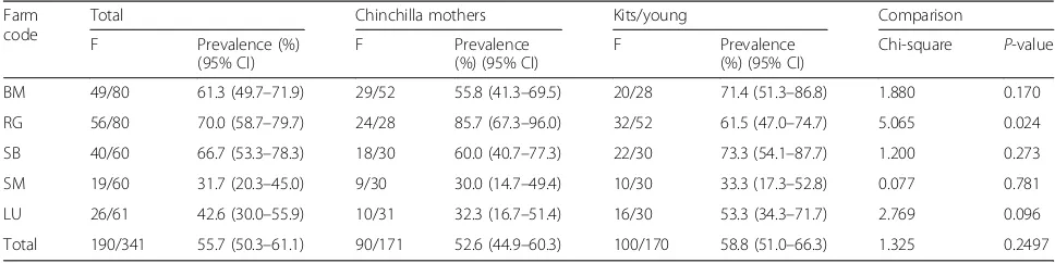
Table 1Prevalence ofG.duodenalisin fecal samples collected from long-tailed chinchillas in farms in Romania
Farm code
Total Chinchilla mothers Kits/young Comparison
F Prevalence (%)
(95% CI)
F Prevalence
(%) (95% CI)
F Prevalence
(%) (95% CI)
Chi-square P-value
BM 49/80 61.3 (49.7–71.9) 29/52 55.8 (41.3–69.5) 20/28 71.4 (51.3–86.8) 1.880 0.170
RG 56/80 70.0 (58.7–79.7) 24/28 85.7 (67.3–96.0) 32/52 61.5 (47.0–74.7) 5.065 0.024
SB 40/60 66.7 (53.3–78.3) 18/30 60.0 (40.7–77.3) 22/30 73.3 (54.1–87.7) 1.200 0.273
SM 19/60 31.7 (20.3–45.0) 9/30 30.0 (14.7–49.4) 10/30 33.3 (17.3–52.8) 0.077 0.781
LU 26/61 42.6 (30.0–55.9) 10/31 32.3 (16.7–51.4) 16/30 53.3 (34.3–71.7) 2.769 0.096
DNA sequencing
The PCR products were purified by using QIAquick PCR purification kit (Qiagen, Hilden, Germany) and sequenced at Macrogen Europe (Amsterdam). Nucleotide sequence data from this study were submitted to the GenBank data-base under the accession numbers MG432793–MG432795.
Statistical analysis
The frequency of Giardia-positive samples, their preva-lence and 95% confidence interval were calculated. The difference in prevalence between age groups and among farms was statistically analyzed by a Chi-square test. Statistical significance was set at aP-value of ≤0.05. All statistical analyses were performed using EpiInfo soft-ware version 3.5.1. (Centers for Disease Control and Pre-vention:http://wwwn.cdc.gov/epiinfo/).
Results
The occurrence ofGiardiaspp.
Giardiacysts were identified in 190 of 341 (55.7%, 95%
CI: 50.3–61.0) fecal samples by microscopic examin-ation. All 190 microscopically identifiedGiardia-positive samples were positive by PCR. General prevalence re-corded the highest value in farm RG (56/80, 70%, 95% CI: 58.7–79.7%) and the lowest in farm SM (19/60, 31.6, 95% CI: 20.3–45.0%) (χ2= 28.83, df= 4, P< 0.001). The infection was somewhat more frequent in young animals (100/170, 58.8%, 95% CI: 51.0–66.3%) compared to mother chinchillas (90/171, 52.6%, 95% CI: 44.9– 60.3%) but the difference was not statistically signifi-cant (χ2= 1.09,df= 1,P= 0.25) (Table1).
Giardiaspp. assemblages
In total, three Giardia assemblages (B, D and E) were found in the chinchilla farms studied. Assemblage B was the most prevalent (151/190, 79.5%), followed by D (33/ 190, 17.4%) and E (6/190, 3.1%). The identified sub-assemblages were BIV (145/190, 76.3%) and BIII (6/190; 3.1%) (Table2).
Sequence analysis of fecal samples confirmed the in-fection with G. duodenalissub-assemblages BIII, BIV, D and E (Table 2). Assemblages B (MG432795) and D (MG432793) were common in all farms in the study, in both age categories, and Assemblage E (MG432794) was identified only in farms BM and RG. No mixed assem-blage infections were detected in animals in this study.
Discussion
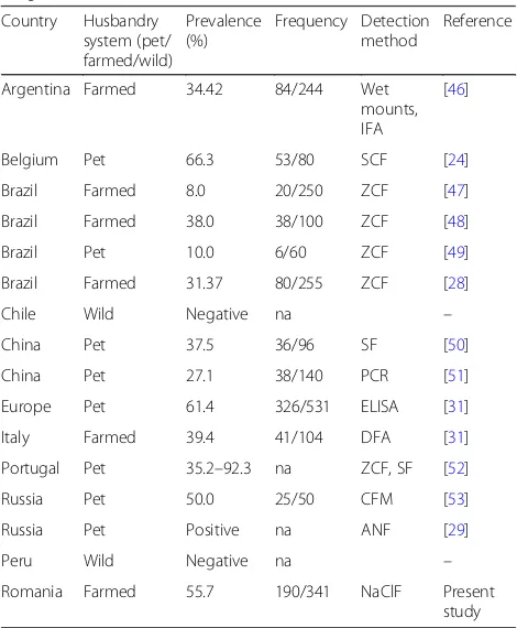
[image:3.595.304.538.120.310.2]The study of intestinal parasites in the long-tailed chinchilla is an important field of interest due to a permanent contact of this pet or farmed animal with humans. Among parasitic diseases identified in this species, giardiasis seems to be the most significant, due to the zoonotic character and in-creased values of prevalence reported worldwide (Table3).
Table 2Assemblages ofG.duodenalisidentified by PCR-RFLP and sequencing targeting thegdhgene in fecal samples of long-tailed chinchillas from farms in Romania
Farm Age Assemblage (RFLP) Assemblage (sequencing) (no. of samples)
NlaIV RsaI BIII BIV D E
BM CM BIII/BIV/E/D BIII; BIV 3 20 4 2
Y BIII/BIV/D BIV 16 4
RG CM BIII/BIV/D/E BIV 19 3 2
Y BIII/BIV/D/E BIV 1 22 7 2
SB CM BIII/BIV/D BIV 15 3
Y BIII/BIV/D BIII; BIV 2 17 3
SM CM BIII/BIV/D BIV 4 5
Y BIII/BIV BIV 10
LU CM BIII/BIV/D BIV 8 2
Y BIII/BIV/D BIV 14 2
Total 6 145 33 6
Abbreviations:CMchinchilla mothers,Yyoung
Table 3Reported prevalence ofGiardiaspp. infection in the long-tailed chinchilla
Country Husbandry system (pet/ farmed/wild)
Prevalence (%)
Frequency Detection method
Reference
Argentina Farmed 34.42 84/244 Wet
mounts, IFA
[46]
Belgium Pet 66.3 53/80 SCF [24]
Brazil Farmed 8.0 20/250 ZCF [47]
Brazil Farmed 38.0 38/100 ZCF [48]
Brazil Pet 10.0 6/60 ZCF [49]
Brazil Farmed 31.37 80/255 ZCF [28]
Chile Wild Negative na –
China Pet 37.5 36/96 SF [50]
China Pet 27.1 38/140 PCR [51]
Europe Pet 61.4 326/531 ELISA [31]
Italy Farmed 39.4 41/104 DFA [31]
Portugal Pet 35.2–92.3 na ZCF, SF [52]
Russia Pet 50.0 25/50 CFM [53]
Russia Pet Positive na ANF [29]
Peru Wild Negative na –
Romania Farmed 55.7 190/341 NaClF Present
study
Abbreviations:ANFammonium nitrate flotation,CFMcombined flotation method,
DFAdirect fluorescent assay,ELISAenzyme-linked immunosorbent assay,IFA
immunofluorescence assay,nanot applicable,NaClFsodium chloride flotation,
[image:3.595.304.539.404.689.2]The prevalence revealed in the present study (55.7%) is slightly increased compared to that reported in farmed chinchilla from other regions (8.0–38.0% in Brazil, 34.4% in Argentina and 39.4% in Italy) but is comparable to those reported in pet animals (10.0–92.3%).
The discrepancies of prevalence recorded in existing studies can be explained by the different diagnostic value of copromicroscopic methods used, determined by the technique, less than the density of supersaturated solu-tions [20]. Moreover, coproscopic techniques have a lower diagnostic value, the prevalence determined by other modern serological or molecular methods (ELISA, IFA, PCR) being 2.6-fold higher in dogs and 3.7-fold higher in cats [21]. As such, we consider that the preva-lence of infection revealed in this study, although high, can be appreciated as undervalued.
Prevalence of Giardia infection is generally influenced by many factors, such as the sensitivity of the diagnostic method used, the peculiarities of the biological cycle of the parasite (the discontinuities of cysts removal), the host, the age of host, the growth system, and the hygiene condi-tions (water, food, bedding) [22]. A variety of factors favor the emergence and transmission of infection in chinchilla populations. These risk factors may differ among pet and farmed chinchilla. Regarding pet chinchilla, participation in shows and contact with other pet animals, such as dogs, cats or other rodents, are significant [23, 24]. In farmed chinchillas, the age of animals, stress, poor husbandry system associated with low quality of water source, over-crowding and close contact with feces seems to act as predisposing factors. Juvenile chinchillas are more sensitive to acquire the infection [25]. Intensive rear-ing in plastic or metal cages, with fecal accumulation underneath and vulnerability of the drinking-water-processing system, favor the contact between animals and cysts of Giardia spp. [26, 27]. Captivity associated with specific stress emphasizes the sensitivity of chinchilla toG.
duodenalis infection, an aspect demonstrated by the
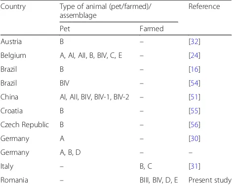
absence ofGiardiaspp. infection in wild animals [28,29]. Chinchillas harbor various assemblages (A, B, C, D and E) of G. duodenalis, representing a potential zoo-notic risk (Table 4). Assemblage B is the most common, being identified in almost all reported studies, except for an axenic isolate ofG.duodenalisfrom Germany [30], in which assemblage A was identified. In our research, RFLP analysis ofG. duodenalis-positive samples revealed a high occurrence of assemblage B isolates grouped into sub-assemblages BIII and BIV, representing the main assemblages involved in chinchilla’s infection. In the present study, no mixed assemblage infections were detected, similar to previous studies [16, 31]. However, our data do differ from those reported in Belgium and Germany, which showed the presence of mixed assem-blage A, B, C and E infections in chinchillas [24,32].
The presence of C and D assemblages typical for ca-nids, and E from hoofed livestock, in chinchillas is quite interesting. In this work, the existence of assemblages E in farms BG and RG can be explained by acquiring animals from farms in which ruminants were also kept, the direct or indirect contact between the two species being possible.
Generally, multiple factors can explain the diversity of assemblages identified across the world. Interspecies transmission is of particular importance for the zoonotic risk of infection, domestic animals being the source of human infection. Reverse or cross-species transmission of different assemblages (BIV, E) has also been demon-strated in areas where humans, primates and livestock overlap in their use of habitat [33]. Interspecific trans-mission is possible between species belonging to differ-ent taxa, from roddiffer-ents to carnivores and from ruminants to humans [34]. It is also proven thatG.duodenalisfrom the North American beaver (Castor canadensis) may infect Mongolian gerbils (Meriones unguiculatus); in this case, the transmission was carried out between two ro-dent species [35]. Transmission ofGiardiaspp. between different species of rodents is also confirmed in other older studies [36]. Nevertheless, Goltz [37] demonstrated thatG. chinchillaefromC. lanigerawere not infective to laboratory mice, rats and guinea pigs. However, the in-terspecies transmission may explain the presence of assemblages D and E in our study, sustained by the existence of guard dogs and small ruminants in the examined farms.
[image:4.595.304.539.110.299.2]Transport vectors can also play a significant role in the transmission of giardiasis [38]. It is confirmed that assem-blage E ofG.duodenalisis carried by flies, increasing the possibility of repeated infection or cross-transmission be-tween sensitive species, by mechanical transmission [39].
Table 4Assemblages ofG. duodenalisidentified in the long-tailed chinchillas
Country Type of animal (pet/farmed)/ assemblage
Reference
Pet Farmed
Austria B – [32]
Belgium A, AI, AII, B, BIV, C, E – [24]
Brazil B – [16]
Brazil BIV – [54]
China AI, AII, BIV, BIV-1, BIV-2 – [51]
Croatia B – [55]
Czech Republic B – [56]
Germany A – [30]
Germany A, B, D – –
Italy – B, C [31]
As a result, despite of the strong host specificity and nar-row host range of assemblage E, which is mostly identified in cloven-hoofed mammals, the involvement of the trans-port hosts can ensure the transmission of this assemblage to captive chinchillas [8].
Water source is also important in the circulation ofG.
duodenalis cysts, giardiasis being recognized as one of
the major waterborne diseases [40]. Although the long-tailed chinchilla is a species adapted to aridity, with low water needs, it prefers the open dish drinker [41]. The best water supply in chinchilla farming is represented by bottled water, free of pathogens and chlorine [42]. Tap and well water are also accepted sources, but they present the risk of contamination with Giardia cysts. Surprisingly, in Romania, bottled water seems to have an increased risk of infection compared with wells or tap water [43]. Feces of different animal species can pollute water sources, shedding cysts into the water supply [44]. These cysts can pass through water treatment, even for pristine or filtered drinking water. Furthermore,Giardia spp. cysts have a demonstrated effective resistance to chlorination [45]. Tap water was the source in studied farms, without an additional water filtration; chlorin-ation and filtrchlorin-ation performed by water plant suppliers being the unique treatments. Combining predisposing factors as interspecific transmission, the possible in-volvement of vectors and deficiencies in water supply, the increased prevalence of G. duodenalis infection in farmed chinchilla from Romania may be explained.
Conclusions
This study revealed the increased prevalence of infection
with G. duodenalis in farmed chinchilla from Romania
and the presence of BIII, BIV, D and E assemblages. Further studies are needed to clarify the zoonotic risk for the owners and workers in chinchilla husbandry.
Abbreviations
BM:Baia Mare; LU: Luncani; RG: Reghin; SB: Bejenaru; SM: Suceava-Morozan
Acknowledgements
The work of VM and AG was carried out under the frame of the USAMV Cluj-Napoca Internal Grant number 6270/2017.
Funding
This work was supported by a grant of the Romanian National Authority for Scientific Research, CNDI-UEFISCDI, project number 110/2012.
Availability of data and materials
Nucleotide sequence data from this study were submitted to the GenBank database under the accession numbers MG432793–MG432895.
Authors’contributions
GCM collected the samples, GA and MV made the flotation and microscopic examinations, KZ performed the DNA extraction, PCR, RFLP and sequencing. All authors read and approved the final manuscript.
Ethics approval and consent to participate Not applicable.
Consent for publication Not applicable.
Competing interests
The authors declare that they have no competing interests.
Publisher’s Note
Springer Nature remains neutral with regard to jurisdictional claims in published maps and institutional affiliations.
Received: 8 June 2017 Accepted: 16 January 2018
References
1. Morrison HG, McArthur AG, Gillin FD, Aley SB, Adam RD, Olsen GJ, et al. Genomic minimalism in the early diverging intestinal parasiteGiardia lamblia. Science. 2007;317:1921–6.
2. Cavalier-Smith T. The excavate protozoan phyla Metamonada Grassé emend. (Anaeromonadea, Parabasalia, Carpediemonas, Eopharyngia) and Loukozoa emend. (Jakobea, Malawimonas): their evolutionary affinities and new higher taxa. Int J Syst Evol Microbiol. 2003;53:1741–58.
3. Lindmark DG. Energy metabolism of the anaerobic protozoonGiardia lamblia. Mol Biochem Parasitol. 1980;1:1–12.
4. Monis PT, Caccio SM, Thompson RC. Variation inGiardia: towards a taxonomic revision of the genus. Trend Parasitol. 2009;25:93–100. 5. Plutzer J, Ongerth J, Karanis P.Giardiataxonomy, phylogeny and
epidemiology: facts and open questions. Int J Hyg Enviro Health. 2010; 213:321–33.
6. Ryan U, Cacciò SM. Zoonotic potential ofGiardia. Int J Parasitol. 2013;43: 943–56.
7. Monis PT, Andrews RH, Mayrhofer G, Ey PL. Genetic diversity within the morphological speciesGiardia intestinalisand its relationship to host origin. Infect Gen Evol. 2003;3:29–38.
8. Feng Y, Xiao L. Zoonotic potential and molecular epidemiology ofGiardia
species and giardiasis. Clin Microbiol Rev. 2011;24:110–40.
9. Mircean V, Györke A, Jarca A, Cozma V. Prevalence ofGiardiaspecies in stool samples by ELISA in household cats from Romania and risk factors. J Fel Med Sur. 2011;13:479–82.
10. Mircean V, Györke A, Cozma V. Prevalence and risk factors ofGiardia duodenalisin dogs from Romania. Vet Parasitol. 2012;184:325–9. 11. Onac D, Oltean M, Mircean V, Jarca A, Cozma V. Occurrence ofGiardia
duodenaliszoonotic assemblages in red foxes from Romania. Sci Parasitol. 2015;16:177–80.
12. Sommer MF, Beck R, Ionita M, Stefanovska J, VasićA, ZdravkovićN, et al. Multilocus sequence typing of canineGiardia duodenalisfrom south eastern European countries. Parasitol Res. 2015;114:2165–74.
13. Gyӧrke A, Kalmar Z, Dumitrache OM, Gherman MC, Mircean V.Giardia duodenalisgenotypes in domestic and wild animals from Romania identified by PCR-RFLP targeting the gdh gene. Vet Parasitol. 2016;217:71–5. 14. Spotorno A, Patton JL. Superfamily Chinchilloidea. In: Patton JL, Pardiňas
UFJ, D’Elia G, editors. Mammals of South America. Rodents. Chicago: University of Chicago Press; 2015, pp. 762–786.
15. Anthony HM. Oldest chinchilla rancher visits chinchilla capital. Madera Daily Tribune. 1967;76:12.
16. Soares RM, de Souza SL, Silveira LH, Funada MR, Richtzenhain LJ, Gennari SM. Genotyping of potentially zoonoticGiardia duodenalisfrom exotic and wild animals kept in captivity in Brazil. Vet Parasitol. 2011;180:344–8. 17. Willis HH. A simple levitation method for the detection of hookworm ova.
Med J Aust. 1921;2:375–6.
18. Read CM, Monis PT, Thompson RC. Discrimination of all genotypes of
Giardia duodenalisat the glutamate dehydrogenase locus using PCR-RFLP. Infect Genet Evol. 2004;4:125–30.
19. Gillhuber J, Pallant L, Ash A, Thompson RC, Pfister K, Scheuerle MC. Molecular identification of zoonotic and livestock-specificGiardia-species in faecal samples of calves in southern Germany. Parasit Vectors. 2013;6:346. 20. Dryden MW, Payne PA, Ridley R, Smith V. Comparison of common fecal
flotation techniques for the recovery of parasite eggs and oocysts. Vet Ther. 2005;6:15–28.
22. Oates SC, Miller MA, Hardin D, Conrad PA, Melli A, Jessup DA, et al. Prevalence, environmental loading, and molecular characterization of
CryptosporidiumandGiardiaisolates from domestic and wild animals along the Central California coast. Appl Environ Microbiol. 2012;78:8762–72. 23. Warburton AR, Jones PH, Bruce J. Zoonotic transmission of giardiasis: a case
control study. Commun Dis Rep CDR Rev. 1994;4:R32–6.
24. Levecke B, Meulemans L, Dalemans T, Casaert S, Claerebout E, Geurden T. MixedGiardia duodenalisassemblage a, B, C and E infections in pet chinchillas (Chinchilla lanigera) in Flanders (Belgium). Vet Parasitol. 2011; 177:166–70.
25. Fehr M. Chinchilla. In: Fehr M, Sassenburg L, Zwart P, editors. Krankheiten der Heimtiere. 6th ed. Hannover: Schluetersche; 2005. p. 183–213. 26. Neringer R, Andersson Y, Eitrem R. A water-borne outbreak of giardiasis in
Sweden. Scand J Infect Dis. 1987;19:85–90.
27. Sudré AP. Carvalho Machado do Couto M, Bergamo do Bomfim TC. Occurrence ofGiardia intestinalisin dairy goats and evaluation of risk factors for infection: research note. R Bras Ci Vet. 2012;19:149–53.
28. Fialho CG, Oliveira RG, Teixeira MC, Marques SMT, Oliveira RG, Oliveira RG, et al. Comparison of protozoan infection between chinchillas (Chinchilla lanigera) from a commercial breeding facility in southern Brazil and chinchillas from a natural reserve in Chile. Parasitol Latinoam. 2008;63:85–7. 29. Sivkova TN. Parasites of wild and domestic chinchilla. In: Proceedings of the
Scientific Conference of the All-Russian Society of Helminthologists RAS "Theory and practice of struggle against parasitic diseases"; 2016, pp. 433–4. 30. Karanis P, Ey PL. Characterization of axenic isolates ofGiardia intestinalis
established from humans and animals in Germany. Parasitol Res. 1998; 84:442–9.
31. Veronesi F, Piergili Fioretti D, Morganti G, Bietta A, Moretta I, Moretti A, Traversa D. Occurrence ofGiardia duodenalisinfection in chinchillas (Chincilla lanigera) from Italian breeding facilities. Res Vet Sci. 2012;93:807–10.
32. Pantchev N, Broglia A, Paoletti B, Globokar Vrhovec M, Bertram A, et al. Occurrence and molecular typing ofGiardiaisolates in pet rabbits, chinchillas, guinea pigs and ferrets collected in Europe during 2006-2012. Vet Record. 2014;175:18.
33. Johnston AR, Gillespie TR, Rwego IB, McLachlan TL, Kent AD, Goldberg TL. Molecular epidemiology of cross-speciesGiardia duodenalistransmission in western Uganda. PLoS Negl Trop Dis. 2010;4:e683.
34. Davies RB, Hibler CP. Animal reservoirs and cross-species transmission of
Giardia. In: Jakubowski W, Hoff JC, editors. Proceedings of a Symposium on Waterborne Transmission of Giardiasis, EPA-600/9-79-001. Cincinnati; 1978. p. 104–27.
35. Faubert GM, Belosevic M, Walker TS, MacLean JD, Meerovitch E. Comparative studies on the pattern of infection withGiardiaspp. in Mongolian gerbils. J Parasitol. 1983;69:802–5.
36. Grant DR, Woo PTK. Comparative studies ofGiardiaspp. in small mammals in southern Ontario. II. Host specificity and infectivity of stored cysts. Can J Zoo. 1978;56:1360–6.
37. Goltz JP. Giardiasis in chinchillas, MSc thesis. Guelph, Ontario, Canada: University of Guelph; 1980.
38. Doiz O, Clavel A, Morales S, Varea M, Castillo FJ, Rubio C, et al. House fly (Musca domestica) as a transport vector ofGiardia lamblia. Folia Parasitol (Praha). 2000;47:330–1.
39. Zhao Z, Dong H, Wang R, Zhao W, Chen G, Li S, et al. Genotyping and subtypingCryptosporidium parvumandGiardia duodenaliscarried by flies on dairy farms in Henan, China. Parasit Vectors. 2014;7:190.
40. Karanis P, Kourenti C, Smith H. Waterborne transmission of protozoan parasites: a worldwide review of outbreaks and lessons learnt. J Water Health. 2007;5:1–38.
41. Hagen K, Clauss M, Hatt JM. Drinking preferences in chinchillas (Chinchilla laniger), degus (Octodon degu) and guinea pigs (Cavia porcellus). J Anim Physiol Anim Nut. 2014;98:942–7.
42. Tapscott B. Standard guidelines for the operation of chinchilla ranches. 1998.http://www.omafra.gov.on.ca/english/livestock/alternat/facts/chinguid. htm. Accessed 15 Sep 2017.
43. JarcăAD. Epidemiological, clinical, laboratory and therapeutic researches in humans and animals giardiasis. PhD Thesis, University of Medicine and Pharmacy "Iuliu Hatieganu", Cluj-Napoca, Romania; 2011.
44. Lippy E. Water supply problems associated with a waterborne outbreak of giardiasis. In: Jakubowski W, Hoff JC. Proceedings of a Symposium on Waterborne Transmission of Giardiasis, EPA-600/9-79-001. Cincinnati; 1978 p. 164–74.
45. Steiner TS, Thielman NM, Guerrant RL. Protozoal agents: what are the dangers for the public water supply? Annu Rev Med. 1997;48:329–40. 46. Martino PE, Bautista EL, Gimeno EJ, Stanchi NO, Radman NE. Fourteen-year
status report of fatal illnesses in captive chinchilla (Chinchilla lanigera). J Appl Anim Res. 2016;45:310–4.
47. Fagundes Gurgel AC, Dos Santos SA, Pacheco De Araújo FA. Protozoan parasites in captive chinchillas (Chinchilla lanigera) raised in the state of Rio Grande do Sul, Brazil. Parasitol Latinoam. 2005;60:186–8.
48. Petroneto BS, Calegari BF, Sansão P, Bonadiman DC, Moulin GN, Silva MA, et al. Intestinal protozoan in chinchillas (Chinchilla lanigera) created in captivity, the region serrana of state of Espirito Santo, Brasil. Acta Vet Bras. 2015;9:65– 70.
49. Alves JG, Silva AS, Monteiro SG. First report ofCryptosporidiumsp. in
Chinchilla lanigerain Brazil. Rev FZVA. 2008;15:186–90.
50. Lv C, Wang H, Qi M, Zhang L. Survey of intestinal parasites in petChinchilla lanigera. Chin Anim Husb Vet Med. 2009;36:176–7.
51. Qi M, Yu F, Li S, Wang H, Luo N, Huang J, et al. Multilocus genotyping of potentially zoonoticGiardia duodenalisin pet chinchillas (Chinchilla lanigera) in China. Vet Parasitol. 2015;208:113–7.
52. Teixeira RSD. Survey of gastrointestinal parasites in two groups ofChinchilla lanigerain northern Portugal. Dissertação de Mestrado. Universidade de Lisboa, Faculdade de Medicina Veterinária, Lisboa, Portugal; 2013. 53. Kurnosova OP. Specific structure and features of distribution of intestinal
protozoa at pets in Moscow. Ros Parazitol Zh. 2013;1:9–15.
54. Souza SLP. Caracterização genética de isolados deGiardiaspp. provenientes de amostras fecais de origem humana e animal. Tese de Doutorado, Faculdade de Medicina Veterinária e Zootecnia, Universidade de São Paulo, São Paulo, Brazil; 2007.
55. Cacciò SM, Beck R, Almeida A, Bajer A, Pozio E. Identification ofGiardiaspecies andGiardia duodenalisassemblages by sequence analysis of the 5.8S rDNA gene and internal transcribed spacers. Parasitology. 2010;137:919–25. 56. Šedinová J, Flegr J, Ey PL, Kulda J. Use of random amplified polymorphic
DNA (RAPD) analysis for the identification ofGiardia intestinalissubtypes and phylogenetic tree construction. J Eukaryot Microbiol. 2003;50:198–203.
• We accept pre-submission inquiries
• Our selector tool helps you to find the most relevant journal
• We provide round the clock customer support
• Convenient online submission
• Thorough peer review
• Inclusion in PubMed and all major indexing services
• Maximum visibility for your research
Submit your manuscript at www.biomedcentral.com/submit