R E S E A R C H
Open Access
Achyranthes bidentata
extract exerts
osteoprotective effects on steroid-induced
osteonecrosis of the femoral head in rats by
regulating RANKL/RANK/OPG signaling
Yini Jiang
1†, Yanqiong Zhang
1†, Weiheng Chen
2, Chunfang Liu
1, Xiaomin Li
1, Danni Sun
1, Zhenli Liu
1, Ying Xu
1,
Xia Mao
1, Qiuyan Guo
1and Na Lin
1*Abstract
Background:Steroid-induced osteonecrosis of the femoral head (steroid-induced ONFH) presents great challenges due to the various effects of steroids on multi-system pathways involved into osteoblast differentiation, osteoblast and osteoclast apoptosis, lipid metabolism, calcium metabolism and coagulation. As one of the most frequently used herbs in Traditional Chinese Medicine formulas that are prescribed for the regulation of bone and mineral metabolism, the therapeutic effects of Achyranthes bidentata on steroid-induced ONFH remain unclear. Thus, the aim of the current study was to verify whether Achyranthes bidentata extract (ABE) can be used to prevent steroid-induced ONFH and to investigate its underlying pharmacological mechanisms.
Methods:Steroid-induced ONFH rat models were established to evaluate the effects of ABE treatment on
osteonecrotic changes and repair processes. Microfocal computed tomography (Micro-CT) was performed to assess the effects of ABE treatment on bone mass, microstructure, and vascularization. Then, the effects of ABE treatment on osteoclast differentiation and bone formation were also evaluatedin vivoandin vitro. In addition, receptor activator of nuclear factor kappa B (RANK), RANK ligand (RANKL), and osteoprotegerin (OPG) expression in sera, femoral heads and bone marrow-derived mesenchymal stem cells (BMSCs) were detected at both protein and mRNA levels.
Results:The ratio of empty lacuna, adipose tissue area, and adipocyte perimeter in the bone marrow were markedly lower in the ABE treatment groups than in the model group. Micro-CT evaluation indicated that ABE treatment could improve the microstructure of the trabecular bone, increase bone mineral density and promote vascularization in steroid-induced ONFH rats. Moreover, ABE treatment inhibited osteoclast differentiation and activated bone formation markers. Interestingly, OPG downregulation, RANK and RANKL upregulation, and an increased ratio of RANKL to OPG in sera and necrotic femoral head could be reversed by ABE treatment, which also effectively inhibited RANKL-induced osteoclast differentiation and regulated RANKL and OPG expression ofin vitro. Conclusion:ABE may prevent steroid-induced ONFH and alleviate steroid-induced bone deterioration by regulating the RANKL/RANK/OPG signaling pathway.
Keywords:Achyranthes bidentataextract, Steroid-induced osteonecrosis, Femoral head, Osteoprotective, RANKL/RANK/OPG signaling pathway
* Correspondence:linna888@163.com
†Equal contributors
1
Institute of Chinese Materia Medica, China Academy of Chinese Medical Sciences, No. 16, Nanxiaojie, Dongzhimennei, Beijing 100700, China Full list of author information is available at the end of the article
Background
Steroid-induced osteonecrosis of the femoral head (ster-oid-induced ONFH) is a serious complication in patients who have received steroids for the treatment of various diseases, including nephrotic syndrome, renal transplant-ation, and systemic lupus erythematosus [1]. As a degen-erative bone disease, it leads to the collapse of the femoral head, which subsequently destroys the hip joint and influ-ences the patient’s activities [2]. Owing to the various ef-fects of steroids on multi-system pathways involved in osteoblast differentiation, osteoblast and osteoclast apop-tosis, lipid metabolism, calcium metabolism, and coagula-tion, it has been challenging to fully elucidate the pathogenesis and etiology of steroid-induced ONFH [3]. According to recent studies, ONFH is caused by the im-pairment of bone cell survival and bone formation, as well as the promotion of osteoclastic resorption and adipocytic differentiation in bone microenvironments [4]. Current treatment for steroid-induced ONFH focuses on prevent-ing irreversible complications, such as biomechanical col-lapse of the femoral head and osteoarthritis of the hip joint. However, these treatments were limited in their abil-ity to enhance bone repair and to prevent collapse of the articular surface and hip arthroplasty [5]. Therefore, novel and efficient agents for the treatment of this disease are needed.
An increasing number of plant-based therapies derived from traditional Chinese medicine (TCM) have been shown to be effective in the treatment of bone injuries and bone-related diseases via strengthening of bones and muscles and easing joints [6]. Achyranthes bidentata, an important medicinal plant of the Amaranthaceae family that has been listed in the Chinese Pharmacopoeia [5], is one of the most frequently used herbs in formulas that are prescribed for the regulation of bone and mineral metab-olism [7]. Achyranthes bidentatais rich in active phyto-chemical compounds including oleanolic acid glycosides, saponins, ecdysterone, ketosteroids, and flavonoids, and produces effects that include invigoration of the liver and kidneys, strengthening of the muscles and bones, promo-tion of blood flow, removal of blood stasis, and increase in longevity [8-10]. Five new oleanolic acid glycosides from Achyranthes bidentata have been reported to inhibit the formation of osteoclasts [11]. Of these compounds, ecdys-terone and daucosterol markedly stimulate proliferation of osteoblast-like UMR106 cells, and ecdysterone increases osteoblastic activity [12]. The flavonoid quercetin, also found inAchyranthes bidentatadecreases osteoclastic dif-ferentiation [13]. Based on these results, we hypothesize thatAchyranthes bidentatamay produce a therapeutic ef-fect on steroid-induced ONFH. Thus, the aim of the current study was to verify whetherAchyranthes bidentata extract (ABE) prevents steroid-induced ONFH and to in-vestigate its underlying pharmacological mechanisms.
Materials and methods
This study was approved by the Research Ethics Committee of the Institute of Chinese Materia Medica, China Academy of Chinese Medical Sciences, Beijing, China. All animals were treated in accordance with the guidelines and regula-tions for the use and care of animals at the Center for La-boratory Animal Care, China Academy of Chinese Medical Sciences.
ABE preparation and analysis
Achyranthes bidentatawas purchased from Beijing Medi-cinal Herbs Co. Ltd. (Beijing, China), and identified and authenticated by Professor Zhenli Liu at the Institute of Basic Theory of Traditional Chinese Medicine, China Academy of Chinese Medical Sciences. Achyranthes bidentata (500 g) was pulverized to a fine powder and boiled twice with 4 L of 80% ethanol for 1 h under reflux. The ethanol extracts were collected and filtered. The fil-trates were concentrated under reduced pressure at 50°C to 500 mL by a concentration of 2 g/mL.
An HPLC method was developed for the quantification of β-ecdysone in the extract. A Waters 2695 instrument equipped with a UV detector at 250 nm was used with a stationary phase of Agilent Zorbax SB-C18 (4.6 × 150 mm, 5 μm) at 30°C and a mobile phase of acetonitrile:0.1% methanoic acid (15:85) (Fisher, Waltham, Massachusetts, USA) running at 1 mL/min. The ABE used in this trial contained 0.0425%β-ecdysone (Figure 1).
Cells and culture system
Bone marrow cells were obtained from the long bones of 4-6-week-old C57 mice (Peking university health science cen-ter, Cat No. SCXK: 2011–0012). Bone marrow cells cultured in the presence of M-CSF (20 ng/ml, PeproTech, Inc., Rocky Hill, NJ, USA) for 3 days to generate bone marrow derived macrophages (BMMs).
Bone marrow cells were obtained from the long bones of 4-6-week-old C57 mice (Peking university health science center, Cat No. SCXK: 2011–0012) by flushing with α -minimum essential medium containing antibiotics (Sigma, St. Louis, MO, USA), and further red blood cells (RBC) were removed with RBC lysis buffer (Sigma, St. Louis, MO, USA). The cells were plated on 90-mm culture dishes (Corning, NY, U.S.A.) and incubated for 3 days in a-MEM containing 15% FCS and antibiotics. Adherent cells were used as bone marrow mesenchymal stem cells (BMSCs).
Animals
Groups and treatment
After 1 week of feeding adaptation, the animals were weighed and randomly divided into 5 groups: control (n =20), model (rats with steroid-induced ONFH, n =25), ABE 10 g/kg (rats with steroid-induced ONFH treated with 10 g/kg ABE, n =20), ABE 15 g/kg (rats with steroid-induced ONFH treated with 15 g/kg ABE, n =20), and ABE 22.5 g/kg (rats with steroid-induced ONFH treated with 22.5 g/kg ABE, n =20). The steroid-induced ONFH rat model was implemented according to previous studies [14,15]. Briefly, methylprednisolone acetate (MPSL, Pfizer Manufacturing, Puurs, Belgium) (21 mg/kg) was injected subcutaneously for 6 weeks to induce osteonecrosis. One hour after the MPSL injection, rats in the ABE 10 g/kg, ABE 15 g/kg, and ABE 22.5 g/kg groups, received ABE dissolved in distilled water by oral gavage at 10 g/kg/d, 15 g/kg/d and 22.5 g/kg/d respectively for 6 weeks. In the control and model groups, the rats received no treatment. The animals were fed a standard diet and allowed free activity.
Tissue sample preparation
Rats from each group were killed 6 weeks after the methyl-prednisolone injection. The rats were anaesthetized with an intravenous injection of trichloroacetaldehyde hydrate (0.3 mL/kg, Sinopharm Chemical Reagent Co., Ltd, China) and were then killed by exsanguination via an aortectomy. Bilateral femora were obtained at the time of death and the left sides were fixed for 3 days in 4% paraformaldehyde
(pH 7.4) to prepare for the microfocal computed tomog-raphy (Micro-CT) and light microscopy examinations. After micro-CT scanning, the bone samples were decalci-fied with ethylenediaminetetraacetic acid (EDTA, 10%, pH 7.4) for 28 days. Samples were sectioned along the cor-onal plane for the proximal one-third and cut along the axial plane in the distal part (condyle). Finally, the speci-mens were embedded in paraffin, cut into 5 mm sections, and stained with hematoxylin and eosin. The right sides were stored at−80°C for western blots and real-time PCR.
Evaluation of steroid-induced ONFH
Osteonecrotic changes and repair processes in steroid-treated rats were observed by histopathological examin-ation using a light microscope 6 weeks after the MPSL injection. The slides were evaluated in a blinded fashion by 3 independent observers. The evaluation criteria for osteo-necrosis were based on the report of Yamamoto et al. [16]. Osteonecrosis was judged to be present when there was ne-crosis of medullary hematopoietic cells or fat cells, empty lacunae, or condensed nuclei in osteocytes. The ratio of empty lacunae (empty lacunae/the total number of osteo-cytes) was calculated for each femoral head using a coronal section taken at the maximal femoral width. Image Pro 6.0 was used for this calculation.
Micro-CT
[image:3.595.59.541.89.370.2]head sample and bone trabeculae. The following parame-ters were calculated: bone volume (BV), bone surface (BS), trabecular bone pattern factor (Tb.Pf), structure model index (SMI), trabecular thickness (Tb.Th), trabecular num-ber (Tb.N), trabecular separation (Tb.Sp), and bone mineral density (BMD).
Quantification and three-dimensional visualization of vessel networks
Femoral head blood vascularization in steroid-treated rats was measured using Micro-CT-based micro-angiography 6 weeks after methylprednisolone injection according to previously reported methods [17,18]. Briefly, rats from each group were anaesthetized as described above and the thoracic and abdomen cavities were opened. A hypoder-mic needle with disposable infusion device was inserted in the ventriculus sinister with ligation of that proximal to the aorta ascendens. The vasculature was flushed with 500 ml heparinized saline (50 U/ml) at 37°C via a dispos-able infusion device. After flushing, 500 ml 4% parafor-maldehyde solution was pumped into the vasculature to fix the tissues and blood vessels. The vasculature was then injected with Microfil based on the manufacturer’s proto-col (Microfil MV-122, Flow Tech, Carver, MA, USA). Animals were then stored overnight at 4°C to ensure polymerization of the contrast agent before microangio-graphy. Bilateral femoral samples were harvested and fixed in 4% paraformaldehyde and 10% EDTA. After perfusion and decalcification, the femoral shaft was fixed in a poly-methylmethacrylate sample tube with its long axis perpen-dicular to the bottom of the tube in preparation for Micro-CT scanning. The scan was perpendicular to the shaft and was initiated from a reference line 10 mm away from the bottom with a scan length of 10 mm.
Hematological examination
To detect the hyperlipidemia-improving effects of prava-statin, blood samples were collected from the abdominal aorta 6 weeks after methylprednisolone administration. The serum levels of total cholesterol (TC), triglycerides (TG), low-density lipoprotein (LDL), high-density lipopro-tein (HDL), apolipoprolipopro-tein A1 (ApoA1), and apolipopro-tein B (ApoB) were determined.
Analysis of Tartrate-resistant acid phosphatase (TRAP), bone-specific alkaline phosphatase (BAP), receptor activator of nuclear factor kappa B (RANK), RANK ligand (RANKL), and osteoprotegerin (OPG) levels in serum and BMSCs
Serum was separated from 5 mL blood samples. BMSCs were cultured for 3 days in the presence of 15% FCS with or without ABE (0.16, 0.8, 4μg/ml, respectively) in 96 well cul-ture plate. Supernatants were obtained and stored at−80°C until use. TRAP, BAP, RANK, RANKL, and OPG were
measured using enzyme linked immunosorbent assay (ELISA) kits (for TRAP: Kamiya Biomedical Company, Seattle, WA; for BAP: Quidel Corp., San Diego, CA; for RANK, RANKL, and OPG: R&D Systems, Minneapolis, MN) that were specific for the rat. The concentration of the reaction product was determined from a standard curve.
Western blot analysis
The protein expression levels of RANK, RANKL, and OPG in the femoral head tissues obtained from rats in different groups were detected by western blot analysis. The western blot protocol and semiquantitative analysis were carried out following the protocol of our previous study [19]. The following antibodies were used: RANK antibody (rabbit antibody, dilution 1:50, Cell Signaling Technology, Inc., Danvers, MA, USA), RANKL antibody (rabbit antibody, dilution 1:100, Millipore Corporation, Billerica, MA, USA), OPG antibody (rabbit antibody, di-lution 1:100, Santa Cruz Biotechnology, Inc., Santa Cruz, CA, USA), and GAPDH antibody (internal control, rabbit polyclonal antibody, dilution 1:200, Santa Cruz Biotechnology, Inc., Santa Cruz, CA, USA).
RNA isolation and real-time PCR
The expression of RANK, RANKL, and OPG in the femoral head tissues was analyzed by real-time PCR. A small cube of trabecular bone (proximal femur) was homogenized, and the total RNA was isolated with TRIzol reagent (Invitrogen, Carlsbad, CA, USA). The extracted RNA was dissolved in RNAse-free distilled water. The quality and quantity of the RNA samples were determined by spectrophotometry, with the ratios of absorbance at 260 nm and 280 nm ranging from 1.8 to 2.0. Next, 3 mg of total RNA was reverse-transcribed into cDNA using a High-Capacity cDNA Kit (Takara Bio Inc., Tokyo, Japan) according to the manufac-turer’s instructions. The specific transcripts were quantified by quantitative real-time PCR using the QuantiTect SYBR Green PCR Kit (Takara Bio Inc., Tokyo, Japan) and analyzed with an ABI 7500 real-time PCR system (Applied Biosys-tems, Foster City, CA, USA). Gene-specific primers used for RANK, RANKL, OPG and GAPDH were listed in Table 1. The mRNA levels of RANK, RANKL, and OPG were nor-malized to GAPDH mRNA levels. PCR was performed as 40 cycles at 94°C for 15 s, 55°C for 30 s, and 72°C for 30 s. The relative mRNA expression was calculated using the comparative CT method.
TRAP Staining
15% fetal calf serum (FCS), M-CSF (20 ng/ml), RANKL (100 ng/ml, PeproTech) and/or ABE (0.16, 0.8, 4μg/ml, respectively) in 96 well culture plates (Corning, MA, USA). Six days later, cells were fixed and stained by TRAP staining kit (Sigma Alrich, USA) according to the manufacturer’s protocol. The images were taken with a digital camera attached to the microscope. TRAP posi-tive multinucleated cells (>3 nuclei) were scored as osteoclast-like cells. The number of TRAP-positive cells was counted using an eyepiece graticule at a magnification of 100 and the results expressed as the number of cells per cm2.
Cell viability assay
BMM cells were seeded in 96-well plates and incubated cultured in the presence of M-CSF and RANKL with or without different concentrations of ABE (0.16, 0.8, 4μg/ml, respectively) for 24 h. BMSCs were cultured with or with-out different concentrations of ABE (0.16, 0.8, 4μg/ml, re-spectively) for 24 h. After drug treatment, cells were washed twice with phosphate-buffered saline (PBS; pH 7.4), and then cell viability was determined by 3-(4,5-Dimethylthiazol-2-yl)-2,5-diphenyltetrazolium bromide (MTT) method using Cell Titer 96® Non-Radioactive Cell Proliferation Assay (Promega, Madison, USA) according to the manufacturer’s instructions. All absorbance at 570 nm were measured with a microplate reader.
Statistical analysis
SPSS version 13.0 software (SPSS Inc., Chicago, IL, USA) and SAS version 9.1 software (SAS Institute, Cary, NC, USA) were used for statistical analysis. All experiments were performed in triplicate. Continuous variables were expressed as mean ± standard deviation. For comparisons of means among multiple groups, one-way ANOVAs followed by LSD tests were performed. Differences were considered sta-tistically significant when P <0.05.
Results
ABE treatment reduces histopathological changes in rats with steroid-induced ONFH
To evaluate the effect of ABE treatment on steroid-induced ONFH, the osteonecrotic changes and repair processes of rats in each group were histopathologically observed. Com-pared with the control group, there was an accumulation of bone marrow cell debris found in ONFH lesions in the model group, while ABE treatment dramatically attenuated this change in rats with steroid-induced ONFH (Figure 2A). In addition, the ratio of empty lacunae in the bone trabecu-lae of the model group was significantly greater than that of the control group (P <0.01, Figure 2A and B), and was de-creased by ABE treatment in a dose-dependent manner (Figure 2B). Moreover, the adipose tissue area and adipo-cyte perimeter in the bone marrow, which were dramatic-ally increased in rats with steroid-induced ONFH, were dose-dependently reduced by ABE treatment (Figure 2B).
ABE treatment improves the microstructure of the trabecular bone and increases bone mineral density (BMD) in rats with steroid-induced ONFH
As shown in Figure 3A–G, the bone volume/tissue volume (BV/TV, P <0.01), trabecular thickness (Tb.Th, P <0.01), trabecular bone pattern factor (Tb.Pf ), and trabecular number (Tb.N, P <0.01) were significantly re-duced, while the trabecular separation (Tb.Sp, P <0.01) and structure model index (SMI, P <0.01) were signifi-cantly increased in rats with steroid-induced ONFH when compared with controls. ABE treatment protected rats from steroid-induced effects on the levels of the above microstructural parameters (Figure 3A–G).
To determine whether ABE treatment increases the bone mass of rats with steroid-induced ONFH, BMD values were measured. As shown in Figure 3H, the rats with steroid-induced ONFH showed markedly reduced BMD in the femoral head compared with the control rats (P <0.01). The BMD values in the rats treated with 10.0–22.5 g/kg ABE were increased in a dose-dependent manner compared to those without drug treatment (P <0.01).
ABE treatment enhances femoral head neovascularization in rats with steroid-induced ONFH
The blood vessel microarchitecture of each group was reconstructed in 3 dimensions for presentation. Com-pared with the control, both the number and the thick-ness of vessels in necrotic lesions of the femoral head of rats with steroid-induced ONFH were markedly reduced, and vasculatures were not visible, while the samples in the ABE 10 g/kg group showed some capillary vessels, and the samples in the ABE 15–22.5 g/kg groups showed intensive vascular architecture (Figure 4A).
[image:5.595.57.293.100.226.2]Quantitatively, Figure 4B showed that ABE treatment dose-dependently increased vessel thickness, percent of
Table 1 Primer sequences used in this study
Primer name Sequence (5’-3’)
RANK Forward GTC TGC AGC TCT TCC CTG AC
Reverse GAG GAG CAG GAC GAT GAG AC
RANKL Forward ACC AGC ATC AAA ATC CCA AG
Reverse TTT GAA AGC CCC AAA GTA CG
OPG Forward GTT CTT GCA CAG CTT CAC CA
Reverse AAA CAG CCC AGT GAC CAT TC
GAPDH Forward ACC CTA AGG CCA ACC GTG AAA AG
vessel volume, vessel volume, and vessel surface of the femoral heads of rats with steroid-induced ONFH.
ABE treatment improves hyperlipidemia in rats with steroid-induced ONFH
Blood chemistry data showed that steroid hormone admin-istration (model group) induced marked hyperlipidemia. Steroid administration significantly elevated TG (Figure 5B), TC (Figure 5A), LDL (Figure 5C), ApoA1 (Figure 5E) and ApoB (Figure 5F) levels, but significantly decreased HDL levels (Figure 5D). Administration of 10–22.5 g/kg ABE dose-dependently improved hyperlipidemia by decreasing TG (P <0.01, Figure 5B), TC (P <0.01, Figure 5A), LDL (P <0.05, Figure 5C), ApoA1 (P <0.05, Figure 5E), and ApoB (P <0.05, Figure 5F) levels, and increasing HDL levels (P <0.05, Figure 5D).
ABE treatment inhibits osteoclast differentiation and activates bone formation markers in rats with steroid-induced ONFH
To confirm the effect of ABE on the number of osteoclasts, the femoral head sections were stained with TRAP. Only TRAP-positive multinucleated cells located at the bone sur-face within the bone destruction were considered to be oste-oclasts (Figure 6A). Compared with steroid-induced ONFH model rats, the numbers of osteoclasts in the areas of bone destruction were significantly decreased in ABE-treated rats with a tendency for dose-dependence (P <0.01, Figure 6B).
In line with histological observations, serum TRAP activ-ity level was significantly increased in rats with steroid-induced ONFH compared to control rats (0.77 ± 0.34 U/L vs. 2.57 ± 1.13 U/L, P <0.01, Figure 6C). After ABE treat-ment, serum TRAP activity levels were severely reduced in a dose-dependent manner (Figure 6C). In contrast, serum BAP activity levels decreased to 55.07 ± 21.43 U/L in rats with steroid-induced ONFH, which were significantly lower than control rat levels (90.38 ± 34.12 U/L, P <0.01, Figure 6D). After the ABE treatment, the serum BAP activ-ity level was markedly increased in a dose-dependent man-ner (Figure 6D).
ABE treatment regulates RANKL/RANK/OPG signaling in rats with steroid-induced ONFH
[image:6.595.60.537.91.312.2]dependently increased (Figure 7A). In addition, ABE treat-ment significantly reduced transcript abundance of RANKL and RANK, but increased abundance of OPG, in the fem-oral heads of rats with steroid-induced ONFH (P <0.05, Figure 7B).
In accordance with quantitative real-time RT-PCR results, changes in RANKL, RANK, and OPG protein expression were reversed by 10–22.5 g/kg ABE in a dose-dependent manner (P <0.01, Figure 7C). Furthermore, ABE markedly
reduced the ratio of RANKL to OPG in the sera and femoral heads of rats with steroid-induced ONFH in a dose-dependent manner (P <0.01, Figure 7D).
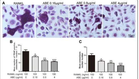
ABE treatment inhibits RANKL-induced osteoclast differentiation in BMMs
[image:7.595.56.541.91.540.2]ABE (0.16 ~ 4μg/ml) into the same cultures showed dose-dependent inhibition of osteoclast formation as measured by the TRAP positive multinucleated cells (Figure 8A and B). Notably, osteoclast-like cells in cultures that were treated with ABE exhibited morphological differences from model osteoclast-like cells, containing fewer numbers of nuclei compared to osteoclast-like cells in model (Figure 8C). ABE without the presence of RANKL did not stimulate osteoclast development (data not shown).
ABE treatment regulates the expression of RANKL and OPG in BMSCs
Compared with model group, doses of 0.16 ~ 4 μg/ml ABE significantly reduced the expression of RANKL, and enhanced the expression of OPG in supernatant of BMSCs with a dose-related manner (Figure 9). More interestingly, ABE treatments markedly decreased the ratio of RANKL to OPG. MTT assay also showed that the anti-osteoclastogenic effect of ABE was not attribut-able to cellular toxicity (Figure 10).
Discussion
Excessive steroid treatment induces bone microstructure integrity loss that leads to femoral head collapse and osteoarthritis, and eventually to the need for total hip re-placement [20]. Due to the uncertain pathophysiology of
steroid-induced ONFH, the best approach to prevent the progression of this disease remains unknown. A large number of kidney-tonifying agents have been used in TCM to treat bone diseases for thousands of years. Among them, ABE has been demonstrated to prevent bone loss [21]. However, its potential role in the treat-ment of steroid-induced ONFH remains unknown. In the current study, the main findings were: 1) ABE atten-uated steroid-induced ONFH by reducing osteonecrotic changes and bone marrow adipogenesis; 2) ABE im-proved the microstructure of the trabecular bone and increased BMD in the femoral head of rats with steroid-induced ONFH; 3) ABE enhanced femoral head neovas-cularization and improved the hyperlipidemic state of rats with steroid-induced ONFH; 4) ABE inhibited osteoclast differentiation and improved bone formation by regulating RANKL/RANK/OPG signaling in rats with steroid-induced ONFH; 5) ABE inhibited RANKL-induced osteoclast differentiation in BMMs and regu-lated the expression of RANKL and OPG in BMSCs.
[image:8.595.58.546.91.356.2]Figure 6ABE inhibits osteoclast differentiation in rats with steroid-induced ONFH. (A)Tartrate-resistant acid phosphatase (TRAP) stained sections from the femoral head of control, model, and ABE-treated rats.(B)The number of osteoclasts (multinucleated TRAP-positive cells) in the femoral head of control, model, and ABE-treated rats.(C)Serum TRAP activity in the serum of control, model, and ABE-treated rats.(D)Serum BAP activity in the serum of control, model, and ABE-treated rats. Data are presented as the mean ± S.D. (n =20 for control, n =25 for model, n =20 for ABE 10 g/kg, ABE 15 g/kg, and 22.5 g/kg groups). # and ##: P <0.05 and P <0.01, respectively, in comparison with the control group. * and **: P <0.05 and P <0.01, respectively, in comparison with the model group. The arrow heads indicate osteoclasts.
lumbar region and knees, and flaccidity of limbs [22]. With regard to bone diseases, in 2010 He et al. [23] reported that the n-butanol-soluble fraction of Achyranthes bidentata root prevented bone loss in ovariectomized rats and may have potential as an alternative treatment for osteoporosis. In 2012, Zhang et al. [21] reported that ABE treatment im-proved biomechanical bone quality through modification of BMD and trabecular microarchitecture without hyperplas-tic effects on the uterus, therefore ABE might be a potential alternative treatment for postmenopausal osteoporosis.
The current study was designed to systematically evalu-ate the therapeutic effects of ABE on steroid-induced
[image:10.595.58.538.88.503.2]improved the trabecular microarchitecture, in part by re-storing trabecular connectivity through increasing trabecu-lar thickness (TbTh), while reducing trabecutrabecu-lar separation (TbSp), which is consistent with the increase in BMD. These results suggest that ABE could prevent the loss of bone mass induced by excessive steroid treatment.
Because impeded blood flow through the femoral head is implicated in the pathogenesis of steroid-induced ONFH, we applied a novel Micro-CT-based micro-angiography technique to visualize and quantify new blood vessel forma-tion and vascularizaforma-tion in the femoral head of the rat.
[image:11.595.57.543.91.367.2]Recent studies have demonstrated that this technique is quantitative and effective for assessing vascularization [17,18]. Consistent with the improvement produced by ABE treatment on the microstructure of the trabecular bone and BMD, we observed a significant increase in blood vessel volume, vessel surface, percentage of vessel volume, and ves-sel thickness in the ABE-treated groups, suggesting a dose-dependent increase in vascularization of the femoral heads in this rat model. These findings imply that ABE treatment may produce an environment conducive to bone formation by generating a blood supply for bone reconstruction. Figure 8ABE inhibits RANKL-induced osteoclastdifferentiation inbone marrow derived macrophages (BMMs). (A)BMMs were cultured in the presence of M-CSF and RANKL withor without different concentrations of ABE (0.16, 0.8, 4μg/ml, respectively). Six days post-culture, cells were fixed with 4% paraformadehyde followed by TRAP staining. Representative images of TRAP staining of osteoclast-like cells from one of the three experiments are shown. B-C: Quantitative analysis show the mean number of TRAP-positive osteoclast-like cells(B), mean osteoclast-like cells nuclei numbers(C). Data are represented as the mean ± SD of three independent experiments. *P < 0.05, **P < 0.01 and ***P < 0.001 significantly different from Model group.
[image:11.595.59.539.597.686.2]Bone development and maintenance is controlled by the dynamic balance between bone formation by osteoblasts and bone resorption by osteoclasts [24]. The main cause of steroid-induced ONFH is excessive bone resorption that exceeds the rate of bone formation, leading to loss of bone mass. In this context, we observed the effects of ABE treat-ment on the number of osteoclasts, as well as its regulatory effects on TRAP and BAP, which are markers of osteo-blastic bone formation and osteoclastic bone resorption, re-spectively. Our data showed that ABE treatment reduced numbers of osteoclasts and suppressed serum TRAP, but increased serum BAP, implying that ABE may inhibit osteo-clastogenesis while promoting osteoblastogenesis in rats with steroid-induced ONFH.
Accumulating studies have indicated that osteoclast-mediated bone destruction is regulated by the RANKL/ RANK/OPG signaling pathway [25,26]. RANK and its lig-and RANKL are crucial regulators of osteoclast differenti-ation. Under physiological conditions, RANKL is expressed in osteoblasts and activated T cells [27], and triggers osteo-clast maturation and bone resorption by binding with RANK on osteoclasts. As a soluble decoy receptor for RANKL, OPG is expressed by osteoblasts and inhibits bone resorption by binding with RANKL, which prevents RANKL binding to RANK [28]. Under pathological conditions, the RANKL/RANK/OPG signaling pathway plays a crucial role in the process of bone destruction [29,30]. Bone resorption is regulated locally by the balance between RANKL and OPG. In the current study, we observed down-regulation of OPG, up-regulation of RANK and RANKL, and an in-creased ratio of RANKL to OPG in the sera and the necrotic femoral head, which were reversed by ABE treatment. Since RANKL is essential and sufficient for the differentiation of osteoclast precursors into mature osteoclasts in the presence of M-CSF [31,32], our data confirmed the inhibitive effects of ABE on RANKL-induced osteoclast formation from BMMs. Moreover, BMSCs and BMMs are originated from
bone marrow, and RANKL and OPG expression in BMSCs can regulate osteoclast development and function [33,34]. To clarify the anti-osteoclastogenicaction mechanism of ABE, the RANKL and OPG protein expression levels de-rived from BMSCs were further detected. As a result, our data showed that ABE treatment could reduce RANKL ex-pression and enhance OPG exex-pression in BMSCs. Thus, both in vivo and in vitro evidence suggest the regulatory ef-fects of ABE treatment on the RANKL/RANK/OPG signal-ing pathway, which has been considered as a potential target for the prevention of bone destruction in steroid-induced ONFH patients.
In conclusion, our data offer for the first time evidence that ABE prevents steroid-induced ONFH and alleviates steroid-induced bone deterioration by regulat-ing the RANKL/RANK/OPG signalregulat-ing pathway. Thus, ABE should be considered a potential candidate drug for the treatment of steroid-induced ONFH.
Competing interests
The authors declare that they have no competing interests.
Authors’contributions
NL participated in study design and coordination, material support for obtained funding, and supervised study. YN and YZ: performed the data analysis, designed the experimental validation and drafted the manuscript. WC, CL, XL, DS, XM and QG: carried out parts of the experiments. ZL and YX: carried out the HPLC-MS analysis. All authors read and approved the final manuscript.
Acknowledgements
This study was supported by grants from the National Major Scientific and Technological Special Project for“Significant New Drugs Creation” (No. 2013ZX09301307) and the National Natural Science Foundation of China (No. 81173417).
Author details
1Institute of Chinese Materia Medica, China Academy of Chinese Medical
Sciences, No. 16, Nanxiaojie, Dongzhimennei, Beijing 100700, China.
2Wangjing Hospital, China Academy of Chinese Medical Sciences, Beijing
100102, China.
[image:12.595.59.540.90.219.2]Received: 30 October 2014 Accepted: 18 November 2014
References
1. Wang XS, Zhuang QY, Weng XS, Lin J, Jin J, Qian WW:Etiological and clinical analysis of osteonecrosis of the femoral head in Chinese patients.Chin Med J (Engl)2013,126:290–295.
2. Moriya M, Uchiyama K, Takahira N, Fukushima K, Yamamoto T, Hoshi K, Itoman M, Takaso M:Evaluation of bipolar hemiarthroplasty for the treatment of steroid-induced osteonecrosis of the femoral head.
Int Orthop2012,36:2041–2047.
3. Aimaiti A, Wufuer M, Wang YH, Saiyiti M, Cui L, Yusufu A:Can bisphenol A diglycidyl ether (BADGE) administration prevent steroid-induced femoral head osteonecrosis in the early stage?Med Hypotheses2011,77:282–285. 4. Mikami T, Ichiseki T, Kaneuji A, Ueda Y, Sugimori T, Fukui K, Matsumoto T:
Prevention of steroid-induced osteonecrosis by intravenous administration of vitamin E in a rabbit model.J Orthop Sci2010,15:674–677.
5. Powell C, Chang C, Naguwa SM, Cheema G, Gershwin ME:Steroid induced osteonecrosis: an analysis of steroid dosing risk.Autoimmun Rev2010, 9:721–743.
6. Li J, Qi H, Qi LW, Yi L, Li P:Simultaneous determination of main phytoecdysones and triterpenoids in radix achyranthis bidentatae by highperformanceliquid chromatography with diode array-evaporative light scattering detectors and mass spectrometry.Anal Chim Acta2007, 596:264–272.
7. Committee of National Pharmacopoeia:Pharmacopoeia of the People’s Republic of China.Beijing: Chemical Industry Press; 2005:49.
8. Chen Q, Liu Z, He JH:Achyranthes bidentat polysaccharide enhances immune response in weaned piglets.Immunopharmacol Immunotoxicol
2009,31:253–260.
9. Han SB, Lee CW, Yoon YD, Lee JH, Kang JS, Lee KH, Yoon WK, Lee K, Park SK, Kim HM:Prevention of arthritic inflammation using an oriental herbal combination BDX-1 isolated from achyranthes bidentat and atractylodes japonic.Arch Pharm Res2005,28:902–908.
10. Cheng J, Di LQ, Shan JJ, Zhao XL, Kang A, Bi XL, Li JS:Studies on effects of Achyranthes bidentata on tongsaimai pellets main active ingredients chlorogenic acid, isoliquiritin, harpagoside and glycyrrhizin in vivo pharmacokinetics.Zhongguo Zhong Yao Za Zhi2014,39:1502–1508. 11. Li JX, Hareyama T, Tezuka Y, Zhang Y, Miyahara T, Kadota S:Five new
oleanolic acid glycosides from Achyranthes bidentata with inhibitory activity on osteoclast formation.Planta Med2005,71:673–679. 12. Gao XY, Wang DW, Li FM:Determination of ecdysterone in Achyranthes
bidentata BL. and its activity promoting proliferation of osteoblast-like cells.Yao Xue Xue Bao2000,35:868–870.
13. Wattel A, Kamel S, Prouillet C, Petit JP, Lorget F, Offord E, Brazier M: Flavonoid quercetin decreases osteoclastic differentiation induced by RANKL via a mechanism involving NF kappa B and AP-1.J Cell Biochem
2004,15:285–295.
14. Bitto A, Polito F, Burnett B, Levy R, Di Stefano V, Armbruster MA, Marini H, Minutoli L, Altavilla D, Squadrito F:Protective effect of genistein aglycone on the development of osteonecrosis of the femoral head and secondary osteoporosis induced by methylprednisolone in rats.
J Endocrinol2009,201:321–328.
15. Han N, Yan Z, Guo CA, Shen F, Liu J, Shi Y, Zhang Z:Effects ofp-glycoprotein on steroid-induced osteonecrosis of the femoral head.Calcif Tissue Int2010, 87:246–253.
16. Yamamoto T, Irisa T, Sugioka Y, Sueishi K:Effects of pulse
methylprednisolone on bone and marrow tissues: corticosteroid-induced osteonecrosis in rabbits.Arthritis Rheum1997,40:2055–2064.
17. Sun Y, Feng Y, Zhang C, Cheng X, Chen S, Ai Z, Zeng B:Beneficial effect of autologous transplantation of endothelial progenitor cells on steroid-induced femoral head osteonecrosis in rabbits.Cell Transplant2011,20:233–243. 18. Sun Y, Feng Y, Zhang C:The effect of bone marrow mononuclear cells on
vascularization and bone regeneration in steroid-induced osteonecrosis of the femoral head.Joint Bone Spine2009,76:685–690.
19. Lin N, Liu C, Xiao C, Jia H, Imada K, Wu H, Ito A:Triptolide, a diterpenoid triepoxide, suppresses inflammation and cartilage destruction in collagen-induced arthritis mice.Biochem Pharmacol2007,73:136–146. 20. Kerachian MA, Seguin C, Harvey EJ:Glucocorticoids in osteonecrosis of the
femoral head: a new understanding of the mechanisms of action.
J Steroid Biochem Mol Biol2009,114:121–128.
21. Zhang R, Hu S, Li C, Zhang F, Gan H, Mei Q:Achyranthes bidentata root extract prevent OVX-induced osteoporosis in rats.J Ethnopharmacol2012, 139:12–18.
22. Hu J, Qi YX, Li QX, Shan BE:The research of extract of achyranthes bidentata blume anti-tumor activity.Chin J Microbiol Immunol2005, 25:415–418.
23. He C, Hui R, Tezuka Y, Kadota S, Li J:Osteoprotective effect of extract from Achyranthes bidentata in ovariectomized rats.J Ethnopharmacol2010, 127:229–234.
24. Teitelbaum SL:Bone resorption by osteoclasts.Science2000, 289:1504–1508.
25. Eriksen EF, Mosekilde L, Melsen F:Trabecular bone remodeling and bone balance in hyperthyroidism.Bone1985,6:421–428.
26. Oursler MJ, Landers JP, Riggs BL, Spelsberg TC:Oestrogen effects on osteoblasts and osteoclasts.J Ann Med1993,25:361–371.
27. Bai YD, Yang FS, Xuan K, Bai YX, Wu BL:Inhibition of RANK/RANKL signal transduction pathway: a promising approach for osteoporosis treatment.
Med Hypotheses2008,71:256–258.
28. Bezerra MC, Carvalho JF, Prokopowitsch AS, Pereira RM:RANK, RANKL and osteoprotegerin in arthritic bone loss.Braz J Med Biol Res2005,38:161–170. 29. Geusens P:The role of RANK ligand/osteoprotegerin in rheumatoid
arthritis.Ther Adv Musculoskelet Dis2012,4:225–233.
30. Ho TY, Santora K, Chen JC, Frankshun AL, Bagnell CA:Effects of relaxin and estrogens on bone remodeling markers, receptor activator of NF-kB ligand (RANKL) and osteoprotegerin (OPG), in rat adjuvant-induced arthritis.Bone2011,48:1346–1353.
31. Lacey DL, Timms E, Tan HL, Kelley MJ, Dunstan CR, Burgess T, Elliott R, Colombero A, Elliott G, Scully S, Hsu H, Sullivan J, Hawkins N, Davy E, Capparelli C, Eli A, Qian YX, Kaufman S, Sarosi I, Shalhoub V, Senaldi G, Guo J, Delaney J, Boyle WJ:Osteoprotegerin ligand is a cytokine that regulates osteoclast differentiation and activation.Cell1998,93:165–176. 32. Xu J, Tan JW, Huang L, Gao XH, Laird R, Liu D, Wysocki S, Zheng MH:
Cloning, sequencing, and functional characterization of the rat homologue of receptor activator of NF-kappaB ligand.J Bone Miner Res
2000,15:2178–2186.
33. Shao B, Yu Y, Fu X, Xue H, Qi M, Shuai Y, Zhou Z, Jin Y, Yang D:RANKL and OPG expression in bone marrow-derived mesenchymal stem cells of ovariectomied promotes osteoclast development and enhances its function.Xi Bao Yu Fen Zi Mian Yi Xue Za Zhi2013,29:1262–1266. 34. Mbalaviele G, Jaiswal N, Meng A, Cheng L, Van Den Bos C, Thiede M:
Human mesenchymal stem cells promote human osteoclast differentiation from CD34+ bone marrow hematopoietic progenitors.
Endocrinology1999,140:3736–3743.
doi:10.1186/s12967-014-0334-7
Cite this article as:Jianget al.:Achyranthes bidentataextract exerts osteoprotective effects on steroid-induced osteonecrosis of the femoral head in rats by regulating RANKL/RANK/OPG signaling.Journal of Translational Medicine201412:334.
Submit your next manuscript to BioMed Central and take full advantage of:
• Convenient online submission
• Thorough peer review
• No space constraints or color figure charges
• Immediate publication on acceptance
• Inclusion in PubMed, CAS, Scopus and Google Scholar
• Research which is freely available for redistribution