R E S E A R C H A R T I C L E
Open Access
Serotype influences on dengue severity: a
cross-sectional study on 485 confirmed
dengue cases in Vitória, Brazil
Creuza Rachel Vicente
1*, Karl-Heinz Herbinger
2, Günter Fröschl
1,2, Camila Malta Romano
3,
Aline de Souza Areias Cabidelle
4and Crispim Cerutti Junior
5Abstract
Background:Dengue is caused by a RNA virus of the familyFlaviviridae, which presents four serotypes (DENV-1 to DENV-4) capable of inducing hemorrhage. The purpose of this study was to evaluate the influence of serotype on the outcome of dengue.
Methods:This cross-sectional study included data from dengue cases with serotyping results that occurred between 2009 and 2013 in Vitória, Espírito Santo, Brazil. Data were accessed through the Information System for Notifiable Diseases. Chi-square test, Fisher exact test, Mann–WhitneyUtest, and logistic regression were performed to assess associations between different serotypes and dengue severity, while considering gender and age.
Results:The sample consisted of 485 laboratory confirmed dengue cases, of which 46.4 % were females, with median age of 26 years. Regarding overall samples, 77.3 % were caused by DENV-1, 16.1 % by DENV-4, 6.4 % by DENV-2, and 0.2 % by DENV-3. Severe dengue affected 6.6 % of all cases, of which 32.3 % of the cases caused by DENV-2, 6.4 % of those caused by DENV-4, 4.5 % of those caused by DENV-1, and none of those caused by DENV-3. Severe dengue was found to be seven times more frequent among cases of DENV-2 than among those of the other serotypes.
Conclusions:The present study found that cases of DENV-2 had a higher proportion of severe dengue than among those of DENV-1 and DENV-4. Consequently, early detection of serotypes circulating in the territory could be an important approach to prevent increasing numbers of severe outcomes during dengue outbreaks by predicting the health support needed for early diagnoses and treatment of dengue cases.
Keywords:Dengue virus, Severe dengue, Signs and symptoms
Background
Dengue is a disease caused by a RNA virus of the family Flaviviridae, caused by four serotypes (1, DENV-2, DENV-3, and DENV-4) which are genetically and an-tigenically distinct. The virus is transmitted to humans by a bite from an infected mosquito of species Aedes aegypti and Aedes albopictus [1]. The clinical spectrum of dengue ranges from asymptomatic to severe presenta-tions, with signs of increased vascular permeability, homeostasis disorders, and organ impairment. All
serotypes are capable of inducing hemorrhage [2]. Deter-minants of dengue severity are not clear. Assumptions regarding pathogenesis focus on interactions between the dengue virus and its human host [3]. Virus strain, immune response to previous dengue infection, and gen-etic background of the host are factors that interfere in the propensity for worse outcomes [3]. Regarding den-gue serotypes, their structural particularities affect the pathogenesis [4], since viral genetic components deter-mine the virulence and ability to infect [5]. The magni-tude of viral replication [4] also affects the severity of dengue [6–10]. Thereby, serotypes that tend to have higher replication rates induce a faster production of antibodies, causing more severe outcomes [7]. Therefore,
* Correspondence:Rachel.Vicente@lrz.uni-muenchen.de
1Center for International Health, Medical Center of the University of Munich,
Leopoldstraße 7, 80802 Munich, Germany
Full list of author information is available at the end of the article
the purpose of this study is to evaluate the influence of serotypes on clinical manifestations and outcomes of dengue.
Methods
Study design
A cross-sectional study was conducted using data ob-tained through the Information System for Notifiable Diseases (SINAN) on dengue cases that occurred in Vitória, Espírito Santo, Brazil, between 2009 and 2013. Vitória provides health care services with total public fi-nancing through the Unique Health System, which in-cludes ambulatory care in the 28 health centers and in the two emergency units of the city, as well as laboratory services through the local public laboratory. Private health care plays a supplemental role in the health sys-tem. The municipality is a highly endemic area of den-gue, with the four serotypes circulating in a period of five years and causing successive epidemics. More than 10 % of cases reported in the city presented specific la-boratory tests for dengue confirmation, above the rec-ommendation of the Brazilian Ministry of Health, in part due to the active surveillance by the Epidemio-logical Surveillance Service. In Brazil, suspected dengue cases must be reported to the Epidemiological Surveil-lance Service. In case of severe dengue, reporting has to be performed within 24 h. The data documented in the SINAN originate from standard report forms filled by physicians. The forms contain the description of clinical manifestations and the criteria of severe dengue. In addition, the Epidemiological Surveillance Service, which receives the report, controls the information which is documented on the report forms and the clinical condi-tion of the patient during follow-up by contacting med-ical staff responsible for attending hospitalized cases, or the patient directly. Among the cases registered in SINAN (n= 30,027), all 485 (1.6 %) with information on serotyping were included in the analysis: 403 (83.1 %) with serotype determined by viral isolation and 82 (16.9 %) determined by polymerase chain reaction (PCR). Viral isolation was performed by inoculating cell cultures of Aedes albopictus (C6/36) and using indirect immunofluorescence method based on the reaction of the antibody specific for each of the four dengue sero-types with marking by fluorochrome, according to the technique established by Gubler et al. [11]. The PCR technique utilized was the reverse-transcriptase-polymerase chain reaction (RT-PCR), with amplification of cDNA obtained from dengue virus RNA, using spe-cific initiators of DENV serotypes, following the tech-nique described by Lanciotti et al. [12]. Both tests were performed in the period of viremia, before emergence of warning signs, minimizing the probability of selection bias. The patients submitted to these tests were selected
systematically in sentinel sites from the public health sector, for surveillance purpose which did not influence the patient care. Among the cases included, data on gen-der, age, serotype, dengue outcome, and clinical manifes-tations were collected and evaluated.
Definitions
The criteria of the Brazilian Ministry of Health were used to classify the cases as dengue fever (DF) and se-vere dengue (SD) [13]. Characterization of DF was deter-mined by the presence of acute febrile illness lasting up to seven days, accompanied by at least two of the follow-ing signs or symptoms: headache, retro-orbital pain, my-algia, arthrmy-algia, malaise, or rash [13]. The classification of SD combines the entities of dengue with complication (DWC) and dengue hemorrhagic fever (DHF). DWC cases demonstrated at least one of the following mani-festations: neurological disorders (manifested by delir-ium, drowsiness, coma, depression, irritability, psychosis, dementia, amnesia, meningeal signs, paresis, paralysis, polyneuropathy, Reye syndrome, Guillain-Barré syn-drome, and encephalitis), cardiac disorders (manifested by heart failure and myocarditis associated with myocar-dial depression, reduction in fraction ejection, and cardio-genic shock), hepatic failure (indicated by hepatomegaly, elevated level of hepatic enzymes and icterus), thrombocytopenia equal to or less than 20,000/mm3, gastrointestinal bleeding, cavity effusion (pleural effusion, pericardial effusion, ascites, checked by ultrasound or radiography), total leukocyte count equal to or less than 1,000/mm3or death. DHF cases displayed all of the fol-lowing characteristics: fever or recent history of fever for up to seven days, thrombocytopenia demonstrated by platelet count equal or less than 100,000/mm3, signs of hemorrhage (demonstrated by positive tourniquet test, hematuria, petechiae, menorrhagia, gingival bleeding, epi-staxis, gastrointestinal bleeding), and signs of plasma leak-age (hemoconcentration demonstrated by 20 % increase in the hematocrit over the baseline at admission or 20 % drop in hematocrit after appropriate treatment, cavity ef-fusion, or hypoproteinemia) [13]. The age groups were classified as follows: children (0 to 9 years), adolescents (10 to 19 years), adults (20 to 59 years), and elderly (60 to 83 years).
Statistical Analysis
performed to adjust for gender, age group, and serotype to avoid confounding by these independent variables, considering men, elderly, and DENV-4 as reference cat-egories. The choice of the reference category is based on the hypothesis and on the difference to be measured. In this case, the hypothesis is that DENV-2 increases the probability of severity. The nearest category in terms of frequency is DENV-4, what makes it the candidate for being the reference. The dependent variable was the clinical progress of dengue with dichotomous values: dengue fever and severe dengue.
Ethical considerations
The Research Ethics Committee of the Health Sciences Center at Federal University of Espírito Santo (opinion number 881,909) and the Ethics Committee of the University of Munich (opinion number 231–15) ap-proved the study protocol. Application of ethical con-sent form to patients was not considered necessary, since data were collected from the national Informa-tion System for Notifiable Diseases. The anonymity of patients and confidentiality of the secondary data is ensured by the researchers and institutions involved in the study. In addition, in this study no individual data is being presented. This approach has been supported by the above named ethics committees in Brazil and Germany.
Results
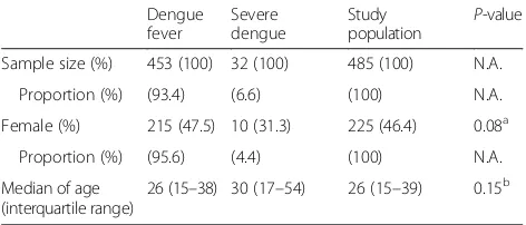
The study population comprised 485 dengue cases, with a proportion of 46.4 % females and a median age of 26 years. Among them, 6.6 % presented SD. Among the cases with SD, the proportion of females was lower and the age was higher, although these differences were not significant (Table 1).
DENV-1 (77.3 %) was the predominant serotype, followed by DENV-4 (16.1 %) and DENV-2 (6.4 %). As only one dengue case was caused by DENV-3 (0.2 %), which occurred in a man who presented DF, DENV-3 could not be included in the analyses. Regarding SD, 4.5 % of cases with DENV-1, 32.3 % of cases with DENV-2, and 6.4 % of cases with DENV-4 presented this form of the disease. Cases caused by DENV-2 were more
associated with SD (P-value < 0.01), whereas cases caused by DENV-1 presented a lower proportion of SD (P-value < 0.01) (Table 2).
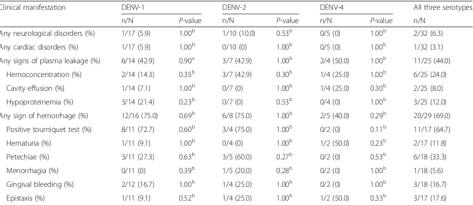
In logistic regression, the proportion of SD among cases of DENV-2 was significantly higher than the SD among cases of DENV-4: adjusted OR = 7.42; 95 % CI 2.21–24.93. Cases caused by DENV-1 presented less SD than cases of DENV-4, although the difference was not significant (adjusted OR = 0.65; 95 % CI 0.23–1.88). Re-garding demographic characteristics evaluated in logistic regression, the results were the following: the OR ad-justed for females was 0.43 (95 % CI 0.19–1.01), consid-ering males as a reference category, and taking elderlies as the reference category for age group, the OR adjusted for children was 0.19 (95 % CI 0.02–1.92), the OR ad-justed for adolescents was 0.48 (95 % CI 0.12–1.87), and the OR adjusted for adults was 0.26 (95 % CI 0.07–0.96). Positive tourniquet test and petechiae were the most common signs of hemorrhage in cases caused by DENV-1 and DENV-2. Hemoconcentration was the most com-mon sign of plasma leakage in cases caused by DENV-2, and hypoproteinemia in cases caused by DENV-1. Cases caused by DENV-4 had hemoconcentration and cavity effusion as the main signs of plasma leakage, and hematuria and epistaxis as the main signs of hemorrhage. No clinical manifestation was significantly associated to any of the serotypes (Table 3).
Discussion
[image:3.595.305.539.574.724.2]This cross-sectional study assessed the influence of dif-ferent serotypes of dengue virus on the clinical out-comes of 485 laboratory cases of dengue infection detected in Vitória, Brazil, between 2009 and 2013. The results of the present study demonstrated that infections caused by DENV-2 were more associated with SD. In this study, gender and age did not influence the associa-tions between the serotypes and the presentation of SD.
Table 1Demographic characterization of study population
Dengue fever
Severe dengue
Study
population P -value Sample size (%) 453 (100) 32 (100) 485 (100) N.A.
Proportion (%) (93.4) (6.6) (100) N.A. Female (%) 215 (47.5) 10 (31.3) 225 (46.4) 0.08a
Proportion (%) (95.6) (4.4) (100) N.A. Median of age
(interquartile range)
26 (15–38) 30 (17–54) 26 (15–39) 0.15b
a
Pearson chi-square test.bMann-WhitneyUtest
Table 2Proportion of cases with dengue fever and with severe dengue among patients diagnosed with different dengue serotypes
Dengue fever
Severe dengue
Study
population P -value Sample size (%) 453 (100) 32 (100) 485 (100) N.A.
Proportion (%) (93.4) (6.6) (100) N.A. DENV-1 (%) 358 (79.0) 17 (53.1) 375 (77.3) <0.01a
Proportion (%) (95.5) (4.5) (100) N.A. DENV-2 (%) 21 (4.6) 10 (31.3) 31 (6.4) <0.01a
Proportion (%) (67.7) (32.3) (100) N.A. DENV-3 (%) 1 (0.2) 0 1 (0.2) 1.00b DENV-4 (%) 73 (16.1) 5 (15.6) 78 (16.1) 0.94a Proportion (%) (93.6) (6.4) (100) N.A.
a
[image:3.595.56.292.622.723.2]Previous studies confirm the present findings, showing an increased proportion of severe outcomes [14], such as DHF [2, 7, 15, 16], and dengue shock syndrome (DSS) [15, 17] in infections caused by DENV-2. Introduction of DENV-2 was a determinant factor for the emergence of SD in different global regions [3], and epidemics with a high number of severe hemorrhagic cases had DENV-2 as the predominant serotype [18, 19].
DENV-2 apparently played a crucial role in the emer-gence of severe cases in Vitória, considering the epi-demiological overview seen in the period analyzed. In the municipality, the years with higher proportion of SD were 2009 (n= 5; 22.7 %), 2010 (n= 125; 23.4 %), and 2011 (n= 268; 10.9 %), when considering cases with la-boratory confirmation. In the time series analyzed, 2009, 2010 and 2011 were the only years when DENV-2 was detected. In 2009, DENV-2 was the only serotype de-tected, but unfortunately, the number of laboratory tests performed was low in this period. Thereby, the restricted number of SD with laboratory confirmation impairs the link of this year with an increasing severity, especially that related to DENV-2 circulation. In 2010, DENV-2 was isolated from 52.9 % of samples with serotyping and was responsible for 88.9 % (n= 8) of severe cases with serotyping, providing evidence for its relevance in the emergence of SD during the epidemic.
The mechanism involved in DENV-2 virulence is not clear. One possible factor is the stimulatory effect of DENV-2 on nitric oxide production, causing toxic and inflammatory effects, inducing apoptosis in host cells [20]. Another factor responsible for the enhanced patho-genicity of DENV-2 is its efficient replication [7]. As a consequence, DENV-2 infections presented high viral load [14].
In the DENV-2 cases of the sample analyzed, milder hemorrhagic manifestations, such as petechiae and posi-tive tourniquet test, were more common than serious ones, such as hematemesis, melena, hematuria, menor-rhagia and epistaxis, as was found in a previous study [19]. It was not possible to generate evidence for severe manifestations being linked to DENV-2, such as plasma leakage [15, 21], cavity effusion [2], hypovolemic shock, internal hemorrhage [19], liver dysfunction [22], thrombocytopenia [21, 23] and hemoconcentration [21]. However, ten cases of SD caused by DENV-2 were present and larger sample sizes would be necessary to establish a statistically sound relation between DENV-2 and clinical manifestations.
DENV-3 was anteriorly related to severe forms of dengue [24], including DHF [16] and DSS [22, 25] and to other severe manifestations [25, 26], such as liver in-volvement [15, 22]. However, no conclusion can be drawn from the present study, as only one case was detected.
[image:4.595.58.539.98.307.2]The present results suggest that those infected by DENV-1 evolved less frequently to severe outcomes. Less severe cases were linked to DENV-1 in previous in-vestigations as well [22, 24], and plasma leakage was rarely observed [21]. In 2011, DENV-1 was isolated from 96.7 % (n= 355/367) of cases with serotyping in Vitória and was responsible for 88.2 % (n= 15/17) of the severe cases where serotyping was performed. Additionally, 2011 met other conditions related to increasing severity. In that period, there was a co-circulation of the sero-types DENV-1 and DENV-2, and probably a consider-able number of secondary dengue infections occurred after years with wide circulation of DENV-2. Even with these two factors, DENV-1 presented better outcomes Table 3Clinical manifestations of severe dengue according to serotype
Clinical manifestation DENV-1 DENV-2 DENV-4 All three serotypes n/N P-value n/N P-value n/N P-value n/N
Any neurological disorders (%) 1/17 (5.9) 1.00b 1/10 (10.0) 0.53b 0/5 (0) 1.00b 2/32 (6.3)
Any cardiac disorders (%) 1/17 (5.9) 1.00b 0/10 (0) 1.00b 0/5 (0) 1.00b 1/32 (3.1)
Any signs of plasma leakage (%) 6/14 (42.9) 0.90a 3/7 (42.9) 1.00b 2/4 (50.0) 1.00b 11/25 (44.0)
Hemoconcentration (%) 2/14 (14.3) 0.35b 3/7 (42.9) 0.30b 1/4 (25.0) 1.00b 6/25 (24.0)
Cavity effusion (%) 1/14 (7.1) 1.00b 0/7 (0) 1.00b 1/4 (25.0) 0.30b 2/25 (8.0)
Hypoproteinemia (%) 3/14 (21.4) 0.23b 0/7 (0) 0.53b 0/4 (0) 1.00b 3/25 (12.0)
Any sign of hemorrhage (%) 12/16 (75.0) 0.69b 6/8 (75.0) 1.00b 2/5 (40.0) 0.29b 20/29 (69.0)
Positive tourniquet test (%) 8/11 (72.7) 0.60b 3/4 (75.0) 1.00b 0/2 (0) 0.11b 11/17 (64.7)
Hematuria (%) 1/11 (9.1) 1.00b 0/4 (0) 1.00b 1/2 (50.0) 0.23b 2/17 (11.8)
Petechiae (%) 3/11 (27.3) 0.63b 3/5 (60.0) 0.27b 0/2 (0) 0.53b 6/18 (33.3)
Menorrhagia (%) 0/11 (0) 0.39b 1/5 (20.0) 0.28b 0/2 (0) 1.00b 1/18 (5.6)
Gingival bleeding (%) 2/12 (16.7) 1.00b 1/4 (25.0) 1.00b 0/2 (0) 1.00b 3/18 (16.7)
Epistaxis (%) 1/11 (9.1) 0.52b 1/4 (25.0) 1.00b 1/2 (50.0) 0.33b 3/17 (17.6)
a
Pearson chi-square test;b
than other serotypes. Similarly to the present findings, Balmaseda et al. (2006) found DENV-1 to be associated with milder hemorrhagic manifestations, such as positive tourniquet test and petechiae [19]. The present study did not capture the presence of gastrointestinal symp-toms in DENV-1, as reported by Thomas et al. [21].
There are discordant results in other studies. Fox et al. (2011) showed that DENV-1 and DENV-2 cases had similar chances to progress to DHF [27]. However, their study included only hospitalized cases, which may have introduced a bias. Yung et al. (2015) demonstrated that patients with DENV-1 had a higher chance of develop-ing DHF than those with DENV-2 in Sdevelop-ingapore. The DENV-2 Cosmopolitan genotype circulating in that period in Singapore [23] was different from the DENV-2 American/Asian genotype, circulating in Vitória [28], af-fecting the disease presentation. There is an association between the introduction of the Asian genotype in the Americas and the emergence of hemorrhagic cases. Up until 2003, all DENV-2 isolated from DHF cases on the American continent belonged to Asian genotype [29].
Cases with DENV-4 infection did not present in-creased or lower association with SD in the sample eval-uated. Previous studies demonstrated that infections caused by DENV-4 presented less severe clinical mani-festations [16, 21] and lower viral titers than other sero-types [30]. In Vitória, DENV-4 was detected for the first time in 2012. In 2013, DENV-4 was circulating in a highly susceptible population and caused the largest epi-demic ever registered in Vitória, with more than 19,000 cases reported. Despite the impressive incidence, consid-ering laboratory confirmed cases, SD affected only 7.6 % of cases in 2012 and 10.6 % of cases in 2013. Both years presented a lower occurrence of SD than 2009, 2010 and 2011, indicating the limited capacity of DENV-4 to cause SD. Halsey et al. (2012) showed the relation of DENV-4 and hemorrhagic cutaneous manifestations [31]. In the present study, no case with petechiae and positive tour-niquet test was found in DENV-4 cases. However, the presence of only five severe cases caused by this serotype impaired the analysis on clinical manifestations.
The difference observed in serotypes association with severity is a concern in face of the recent approval of the first dengue vaccine in Brazil, since DENV-2 presented an efficacy of 42.3 % (varying from 14 % to 61.1 %) the lowest compared to other serotypes. [32]. A phase III trial for the vaccine has been conducted in Vitória since June 2011. However, no case of DENV-2 has been de-tected in the population since the beginning of the trial. Therefore, so far it is not possible to perceive the effect of the vaccination in this site in a scenario with DENV-2 circulation, regarding protection against dengue infec-tion, emergence of severe cases and demand for hospi-talizations. This issue will most likely be clarified in
future, with the vaccine implementation and surveillance information.
The present study had some limitations. Potential in-fluencing factors, such as secondary dengue infection, the sequence of serotypes responsible for the secondary infection [33], or co-morbidity, which could contribute to dengue severity, have not been assessed. As data col-lection was performed for surveillance purpose, it was dependent on professional precision by the involved health care workers when recording clinical information. Consequently, data on age and clinical manifestations were missing and could have been inaccurately docu-mented. However, standardization of the report forms and the control of documented information by the Epi-demiological Surveillance Service minimize information bias caused by misclassification. Determination of sero-type was conducted in 485 systematically selected cases, who attended sentinel sites aiming at surveillance of cir-culating serotypes, which did not relate to patient care. Between 2009 and 2013, Vitória reported 30,027 sus-pected cases of dengue fever. In 1.6 % of them, serotyp-ing was performed. Furthermore, the collection of blood occurred in the viremic phase, before the emergence of warning signs. As this was a test requested by the sur-veillance service during this period, it is unlikely that these cases were selected based on their clinical manifes-tations. Despite the fact that the study includes only 1.6 % of all dengue cases in the catchment area reported in the period, its sample was large enough to have suffi-cient power to detect the association between serotype and severe dengue. Dengue serotypes present genetic variations and some genotypes have a higher association with SD. Molecular studies of dengue in Vitória are ne-cessary to define the genotypes circulating and their re-lation with severe epidemics.
Future investigations with prospective approaches in hyperendemic sites or multicenter settings could con-tribute to elucidate the role of different factors that in-fluence the progression to severe dengue. Some of the factors that should be included are related to the virus, such as serotypes and genotypes, and others are related to the human hosts, such as demographic characteris-tics, co-morbidities and immunological status. Thereby, an integrated analysis of these factors could contribute to understand the complexity influencing severe dengue outcome.
Conclusions
permanently and intensively, since the early detection of serotypes circulating could be an effective tool to antici-pate the number of severe outcomes during dengue out-breaks. With this information, health systems can be prepared to provide early detection of dengue cases, en-suring the follow up of patients and adequate treatment, preventing worse outcomes.
Abbreviations
DF, dengue fever; DHF, dengue hemorrhagic fever; DSS, dengue shock syndrome; DWC, dengue with complications; SD, severe dengue
Acknowledgements
The authors would like to acknowledge the Health Department of Vitória, for providing data and logistic support; the Coordination for the Improvement of Higher Education Personnel (CAPES), for awarding a grant (process 9589-13-9) for the first author; The Brazilian National Council for Scientific and Technological Development (CNPq), for financing the project (process 482261/2010-2), the Federal Ministry for Economic Cooperation and Development (BMZ) and the German Academic Exchange Services (DAAD) through the Excellence Centers for Exchange and Development (EXCEED), for funding the CIHLMUCenter for International Health at Medical Center of the University of Munich, Germany.
Funding
The Coordination for the Improvement of Higher Education Personnel (CAPES) (grant number 9589-13-9 for CRV) and the Brazilian National Council for Scientific and Technological Development (CNPq) (grant number 482261/ 2010-2 for CCJ) provided scholarship and finance for the conduct of this study. The funding body did not influence in any manner the design of the study, the data collection, the analysis and interpretation of data or the writing of the manuscript.
Availability of data and materials
Aggregated data is entirely presented in this publication. Individual data will not be shared following the ethical principle of confidentiality in the use of patient data.
Authors’contributions
CRV participated in conception and design, analysis and interpretation of data and drafting the manuscript. KH, GF, CMR and CCJ participated in conception and design, interpretation of data and critical review. ASAC participated in acquisition of data and critical review. All authors read and approved the final manuscript.
Authors’information
CRV (master in Public Health [Epidemiology] and Ph.D. candidate in International Health at the Center for International Health, Medical Center of the University of Munich).
KH (medical doctor [MD], specialist in occupational medicine and tropical medicine, Magister Public Health, Master of Science in International Health, assistant professor [Privatdozent, PD] in tropical medicine and travel medicine at the University of Munich, Germany).
GF (medical doctor [MD], specialist in internal medicine, tropical medicine and infectious disease medicine, Master of Science in International Health). CMR (Scientific Researcher at Institute of Tropical Medicine from University of São Paulo - USP).
ASAC (nurse, responsible for dengue surveillance at the Coordination of Epidemiological Surveillance, Health Department of Vitória).
CCJ (Associate Professor at the Federal University of Espírito Santo teaching epidemiology and infectious diseases to Undergraduate and Postgraduate levels of education).
Competing interests
All authors declare that they have no competing interests.
Consent for publication Not applicable.
Ethics approval and consent to participate
The Research Ethics Committee of the Health Sciences Center at Federal University of Espírito Santo (opinion number 881,909) and the Ethics Committee of the University of Munich (opinion number 231–15) approved the study protocol. Application of ethical consent form to patients was not necessary, since data were collected from a data bank, ensuring the anonymity of patients and confidentiality of the secondary data by the researchers and institutions involved in the study.
Author details
1Center for International Health, Medical Center of the University of Munich,
Leopoldstraße 7, 80802 Munich, Germany.2Department of Infectious Diseases and Tropical Medicine, Medical Center of the University of Munich, Leopoldstraße 5, 80802 Munich, Germany.3Institute of Tropical Medicine,
University of São Paulo, Avenida Eneas de Carvalho Aguiar 470, 05403-000 São Paulo, Brazil.4Coordination of Epidemiological Surveillance, Health Department of Vitória, Avenida Marechal Mascarenhas de Moraes 1185, 29017-010 Vitória, Brazil.5Department of Social Medicine, Federal University
of Espírito Santo, Avenida Marechal Campos 1468, 29040-090 Vitória, Brazil.
Received: 18 November 2015 Accepted: 27 June 2016
References
1. World Health Organization. Dengue: guidelines for diagnosis, treatment, prevention and control. Geneva: World Health Organization Press; 2009. 2. Fried JR, Gibbons RV, Kalayanarooj S, Thomas SJ, Srikiatkhachorn A, Yoon IK,
et al. Serotype-specific differences in the risk of dengue hemorrhagic fever: an analysis of data collected in Bangkok, Thailand from 1994 to 2006. PLoS Negl Trop Dis. 2010;4(3):e617.
3. Dussart P, Baril L, Petit L, Beniguel L, Quang LC, Ly S, et al. Clinical and virological study of dengue cases and the members of their households: the multinational DENFRAME project. PLoS Negl Trop Dis. 2012;6(1):e1482. 4. Leitmeyer KC, Vaughn DW, Watts DM, Salas R, de Chacon IV, Ramos C, et al.
Dengue virus structural differences that correlate with pathogenesis. J Virol. 1999;73(6):4738–47.
5. Pongsiri P, Themboonlers A, Poovorawan Y. Changing pattern of dengue virus serotypes in Thailand between 2004 and 2010. J Health Popul Nutr. 2012;30(3):366–70.
6. Murgue B, Deparis X, Chungue E, Cassar O, Roche C. Dengue: an evaluation of dengue severity in French Polynesia based on an analysis of 403 laboratory-confirmed cases. Trop Med Int Health. 1999;4(11):765–73. 7. Vaughn DW, Green S, Kalayanarooj S, Innis BL, Nimmannitya S, Suntayakorn
S, et al. Dengue viremia titer, antibody response pattern, and virus serotype correlate with disease severity. J Infect Dis. 2000;181(1):2–9.
8. Libraty DH, Young PR, Pickering D, Endy TP, Kalayanarooj S, Green S, et al. High circulating levels of the dengue virus nonstructural protein NS1 early in dengue illness correlate with the development of dengue hemorrhagic fever. J Infect Dis. 2002;186(8):1165–8.
9. Wang WK, Chao DY, Kao CL, Wu HC, Liu YC, Li CM, et al. High levels of plasma dengue viral load during defervescence in patients with dengue hemorrhagic fever: implications for pathogenesis. Virology.
2003;305(2):330–8.
10. Wang WK, Chen HL, Yang CF, Hsieh SC, Juan CC, Chang SM, et al. Slower rates of clearance of viral load and virus-containing immune complexes in patients with dengue hemorrhagic fever. Clin Infect Dis. 2006;43(8):1023–30. 11. Gubler DJ, Kuno G, Sather GE, Velez M. Oliver A Mosquito cell cultures and
specific monoclonal antibodies in surveillance for dengue viruses. Am J Trop Med Hyg. 1984;33(1):158–65.
12. Lanciotti RS, Calisher CH, Gubler DJ, Chang GJ, Vorndam AV. Rapid detection and typing of dengue viruses from clinical samples by using reverse transcriptase-polymerase chain reaction. J Clin Microbiol. 1992;30(3):545–51.
13. Brazilian Ministry of Health. Dengue: diagnóstico e manejo clínico: adulto e criança. 4th ed. Brasília: Brazilian Ministry of Health; 2013.
14. Thomas L, Verlaeten O, Cabié A, Kaidomar S, Moravie V, Martial J, et al. Influence of the dengue serotype, previous dengue infection, and plasma viral load on clinical presentation and outcome during a dengue-2 and dengue-4 co-epidemic. Am J Trop Med Hyg. 2008;78(6):990–8. 15. Kalayanarooj S, Nimmannitya S. Clinical and laboratory presentations of
16. Nisalak A, Endy TP, Nimmannitya S, Kalayanarooj S, Thisayakorn U, Scott RM, et al. Serotype-specific dengue virus circulation and dengue disease in Bangkok, Thailand from 1973 to 1999. Am J Trop Med Hyg. 2003;68(2):191–202.
17. Huy NT, Giang TV, Thuy DHD, Kikuchi M, Hien TT, Zamora J, et al. Factors associated with dengue shock syndrome: a systematic review and meta-analysis. PLoS Negl Trop Dis. 2013;7(9):e2412.
18. Guzmán MG, Kouri GP, Bravo J, Soler M, Vazquez S, Morier L. Dengue hemorrhagic fever in Cuba, 1981: a retrospective seroepidemiologic study. Am J Trop Med Hyg. 1990;42(2):179–84.
19. Balmaseda A, Hammond SN, Pérez L, Tellez Y, Saborío SI, Mercado JC, et al. Serotype-specific differences in clinical manifestations of dengue. Am J Trop Med Hyg. 2006;74(3):449–56.
20. Valero N, Mosquera J, Añez G, Levy A, Marcucci R, de Mon MA. Differential oxidative stress induced by dengue virus in monocytes from human neonates, adult and elderly individuals. PLoS One. 2013;8(9):e73221. 21. Thomas L, Najioullah F, Besnier F, Valentino R, Césaire JRR, Cabié A. Clinical
presentation of dengue by serotype and year of epidemic in Martinique. Am J Trop Med Hyg. 2014;91(1):138–45.
22. Kumaria R. Correlation of disease spectrum among four Dengue serotypes: a five years hospital based study from India. Braz J Infect Dis. 2010;14(2):141–6. 23. Yung CF, Lee KS, Thein TL, Tan LK, Gan VC, Wong JGX, et al. Dengue
serotype-specific differences in clinical manifestation, laboratory parameters and risk of severe disease in adults, Singapore. Am J Trop Med Hyg. 2015;92(5):999–1005.
24. Corwin AL, Larasati RP, Bangs MJ, Wuryadi S, Arjoso S, Sukri N, et al. Epidemic dengue transmission in southern Sumatra, Indonesia. Trans R Soc Trop Med Hyg. 2001;95(3):257–65.
25. Passos MNP, Santos LMJG, Pereira MRR, Casali CG, Fortes BPMD, Valencia LIO, et al. Clinical differences observed in patients with dengue caused by different serotypes in the epidemic of 2001/2002, occurred in Rio de Janeiro. Rev Soc Bras Med Trop. 2004;37(4):293–5.
26. Endy TP, Nisalak A, Chunsuttiwat S, Libraty DH, Green S, Rothman AL, et al. Spatial and temporal circulation of dengue virus serotypes: a prospective study of primary school children in Kamphaeng Phet, Thailand. Am J Epidemiol. 2002;156(1):52–9.
27. Fox A, Hoa LNM, Simmons CP, Wolbers M, Wertheim HFL, Khuong PT, et al. Immunological and viral determinants of dengue severity in hospitalized adults in Ha Noi, Viet Nam. PLoS Negl Trop Dis. 2011;5(3):e967.
28. Dettogni RS, Louro ID. Phylogenetic characterization of dengue virus type 2 in Espírito Santo, Brazil. Mol Biol Rep. 2012;39(1):71–80.
29. Rico-Hesse R. Microevolution and virulence of dengue viruses. Adv Virus Res. 2003;59:315–41.
30. Gubler DJ, Suharyono W, Tan R, Abidin M, Sie A. Viraemia in patients with naturally acquired dengue infection. Bull World Health Organ. 1981;59(4):623–30.
31. Halsey ES, Marks MA, Gotuzzo E, Fiestas V, Suarez L, Vargas J, et al. Correlation of serotype-specific dengue virus infection with clinical manifestations. PLoS Negl Trop Dis. 2012;6(5):e1638.
32. Villar L, Dayan GH, Arredondo-García JL, Rivera DM, Cunha R, Deseda C, et al. Efficacy of a tetravalent dengue vaccine in children in Latin America. N Engl J Med. 2015;372(2):113–23.
33. Guzman MG, Kouri G. Dengue and dengue hemorrhagic fever in the Americas: lessons and challenges. J Clin Virol. 2003;27(1):1–13.
• We accept pre-submission inquiries
• Our selector tool helps you to find the most relevant journal • We provide round the clock customer support
• Convenient online submission • Thorough peer review
• Inclusion in PubMed and all major indexing services • Maximum visibility for your research
Submit your manuscript at www.biomedcentral.com/submit