R E S E A R C H A R T I C L E
Open Access
In vivo
expression of innate immunity markers in
patients with
mycobacterium tuberculosis
infection
Pantelis Constantoulakis
1, Eftihia Filiou
1, Nikoletta Rovina
2*, George Chras
2, Aggeliki Hamhougia
3, Simona Karabela
4,
Adamandia Sotiriou
2, Charis Roussos
2, Nikolaos Poulakis
5Abstract
Background:Toll-like receptors (TLRs), Coronin-1 and Sp110 are essential factors for the containment of
Mycobacterium tuberculosisinfection. The purpose of this study was to investigate thein vivo expression of these molecules at different stages of the infection and uncover possible relationships between these markers and the state of the disease.
Methods:Twenty-two patients with active tuberculosis, 15 close contacts of subjects with latent disease, 17 close contacts of subjects negative for mycobacterium antigens and 10 healthy, unrelated to patients, subjects were studied. Quantitative mRNA expression of Coronin-1, Sp110, TLRs-1,-2,-4 and -6 was analysed in total blood cellsvs
an endogenous house-keeping gene.
Results:The mRNA expression of Coronin-1, Sp110 and TLR-2 was significantly higher in patients with active tuberculosis and subjects with latent disease compared to the uninfected ones. Positive linear correlation for the expression of those factors was only found in the infected populations.
Conclusions:Our results suggest that the up-regulation of Coronin-1 and Sp110, through a pathway that also includes TLR-2 up-regulation may be involved in the process of tuberculous infection in humans. However, further studies are needed, in order to elucidate whether the selective upregulation of these factors in the infected patients could serve as a specific molecular marker of tuberculosis.
Background
In the majority of patients, tuberculosis (TB) is typically found as an airborne disease due to droplet nuclei infec-tion. In the immuno-competent host, lungs are most commonly affected inM. Tuberculosis (MTB) infection, with estimates of 80-87% of lung involvement in sub-jects with active disease. A similar estimate of 70-90% has also been observed in immuno-compromised hosts, such as those with human immuno-deficiency virus infection (HIV) [1]. A person in close contact with a smear-positive patient is at maximum risk of being infected. Studies have shown that the infection rate among close contacts ranges from 25-50%, even in the worst over-crowded and sub-standard conditions, while among those infected only a 10% will progress to disease [2]. Thus, a great portion of close contacts remains
uninfected after exposure and is probably due to the host’s innate immune system.
The effective antimicrobial action of monocytes/ macrophages against MTB infection consists of a well-organized antimicrobial machinery, with a large arma-mentarium of cytotoxic molecules, including Toll-like receptors, phagosome-lysosome function and the recently described tuberculosis resistance genes that are considered integral parts of this mechanism [3,4].
Toll-like receptors (TLRs) are a family of proteins that play a key role in the innate immune response to infectious agents through their ability to discriminate conserved microbial structures, known as pathogen-associated molecular patterns (PAMPs), from self. TLRs recognition of PAMPs, such as lipopolysaccharide (LPS), initiates signal transduction through the NF-kB pathway. Nuclear translocation of NF-kB induces transcription of pro-inflammatory cytokine genes essential to mounting a protective immune response and host defence [5]. TLRs’ stimulation in macrophages has been shown to
* Correspondence: nikrovina@med.uoa.gr
2
National and Kapodistrian University of Athens, Pulmonary Department,
“Sotiria”District Chest Diseases Hospital; Athens, Greece Full list of author information is available at the end of the article
promote phagosomal maturation via activation of p38 MAP kinase in a MyD88 dependent signalling pathway [6,7]. Pathogen associated molecular pattern signatures for pathogenic mycobacteria include, CpG DNA, 19 kDa lipoprotein, lipoarabidomannan and mannosylated phos-photidylinositol (PIM) [8]. TLRs have been reported to participate in the interaction of pathogenic mycobacteria or in their extracellular products in mice and humans. In particular, TLR-2, in association with TLR-1, TLR-6 and TLR-4, has been implicated as a receptor involved in the recognition of mycobacterial antigens and acti-vation of macrophages and dendritic cells (DCs) [9]. Furthermore, TLRs are essential in the killing process of intracellular mycobacteria by human macrophages through the induction of the antimicrobial peptide cathelicidin [10]. In addition, it has recently been shown that TLRs’non-synonymous polymorphisms are significantly associated with tuberculosis disease in humans [11].
Pathogenic mycobacteria have evolved a unique strat-egy to survive intracellularly within macrophages. They modulate phagosome maturation to prevent the fusion of endosomes with lysosomes. The block of phagolyso-some biogenesis has been proposed to play a central role in the pathogenesis of the mycobacterium [12]. The prolonged survival ofM. tuberculosiswithin their mam-malian host cells suggested that these pathogens, apart from producing virulence factors such as phosphatase SapM and protein kinase PknG, have evolved mechan-isms to utilise host molecules for their own survival [3,13]. An analysis of the protein composition of myco-bacterial phagosome showed the exclusive presence of a protein that was strongly retained on phagosomes har-bouring live mycobacteria [13,14]. This protein, initially named TACO (for tryptophan-aspartatecontaining coat protein) is now referred as Coronin-1. Coronin-1 is a member of the WD repeatcontaining protein family of coronins. The founding member of this protein family was identified in the amoebaDictyostelium discoideum
and is involved in the regulation of actin-based dynamics [15].
In mammalian cells up to seven coronin isoforms have been identified and were thought to regulate F-actin-dependent cytoskeleton [16]. Recent data indicate that Coronin-1 is essential for the survival of live mycobac-teria into monocytes/macrophages, because this protein arrests the maturation of phagosome to phagolysosome. Coronin-1 is specifically restricting phagosomes contain-ing live mycobacteria from entercontain-ing the lysosomal path-way by regulating a calcium- dependent signalling process, which activates the calcium-dependent phos-phatase calcineurin. This enzyme is responsible for the blocking effect of phagosome-lysosome fusion. In the absence of Coronin-1, calcineurin cannot be activated
which results in lysosomal transfer and death of interna-lized mycobacteria. Similar results were also retrieved with calcineurin blockers, such as cyclosporine A or FK506 [17]. The prolonged retention of Coronin-1 in phagosome during active infection has been attributed to a secreted mycobacterial lipoamide dehydrogenase C (LpdC), which retains Coronin-1 on the phagosomal membrane [18] and is produced only by live BCG and
M. tuberculosis.
Also, recent data have shown that inbred mice expres-sing the Ipr1(intracellular pathogen resistance 1) gene in the sst1 locus were protected against infection by
M. tuberculosisandM. bovis. The up-regulation ofIpr1
after infection byM. tuberculosis limits the infection in tuberculosis resistant mice by switching the cell death pathway of the infected macrophages from necrosis to apoptosis [19]. Sp110 is the closest human homologue of Ipr1gene and is located on chromosome 2 (2q37,1). Sp110 is a component of the nuclear body, a multi-pro-tein complex assumed to participate in the regulation of gene transcription. This gene is very important for monocytes’ differentiation, apoptosis and activation including the response to pathogens [20,21]. Sp110 inhi-bits vesicular stomatitis and influenza virus replication. It also confers resistance to human Foamy virus, while gene polymorphisms or mutations have been associated with susceptibility to hepatitis C, immunodeficiency virus and hepatic veno-occlusive disease [22,23]. In this respect, Sp110 may also have a role in human tubercu-losis infection.
Studies conducted so far, support the idea that intra-cellular factors like Coronin-1, Sp110 and TLRs play a decisive role in the progression of TB infection, at least in experimental tuberculosis. Hence, it is possible, that the expression of these molecules somehow regulates the intracellular fate ofM. tuberculosisthrough a com-plicated cascade of up- and down-regulations. However, studies regarding the expression of these intracellular molecules in humans are lacking. We hypothesized that during active infection, because of the turnover of the infected monocytes and the following haematogenous spread of the infection, these markers could be increased in patients with TB.
performed according to the CDC and the literature recommendations in all circumstances in which the TST is currently used, including contact investigations, eva-luation of recent immigrants, and sequential-testing sur-veillance programs for infection control (e.g., those for healthcare workers) [24-26].
We demonstrated striking changes in the expression of Coronin-1, Sp110 and TLR-2, between the TB-infected population (actively and latently TB-infected) com-pared to the uninfected controls, whereas the levels of the other TLRs did not differ significantly. Our data suggest that Coronin-1, Sp110 and at least Toll-like receptor-2 molecules are involved in the infectious pro-cess of tuberculosis. However, their significance and their precise mechanism in human tuberculous infection deserve further investigation.
Methods
Patients and samples
Twenty two patients with pulmonary tuberculosis (TB) (group A) were recruited at the “Sotiria Hospital”, Athens (Greece). Patients’characteristics are shown in Table 1. Sixteen patients had pulmonary cavitary disease and or parenchymal TB, four patients pulmonary TB and TB lymphadenitis and two patients had concomi-tantly pulmonary disease associated with TB pericarditis and TB pleural effusion, respectively. The diagnosis of tuberculosis was confirmed by smear and culture posi-tive for M. Tuberculosisfrom the respective biological specimens.
Fifteen volunteers from the close contacts of patients, that were tested positive for Quantiferon-TB (QFT), after an extensive examination to exclude tuberculosis, were considered to have LTBI and constituted the group B. Seventeen volunteers from the close contacts, that were tested negative for Quantiferon-TB in two consecutive examinations (8-10 weeks apart from each other) were considered not infected (group C). QFT testing was used for diagnosis of LTBI because of its greater specificity compared to intrademal Mantoux test according to the recent CDC recommendations for the
investigation of contacts with an infectious case of tuberculosis [27].
The exposure time for close contacts identification, i.e the average daily exposure in hours multiplied by the days of exposure, during the last trimester preceding the tuberculosis diagnosis was also estimated according these CDC guidelines [27]. This estimation of exposure has also been validated in studies correlating the time exposure to an index case with the performance of the interferon gamma releasing assays (IGRAS) test [28]. The mean value of the average exposure time for close contacts with latent TB infection (QFN positive) was 665.6 hour-days, while for the close contacts negative for latent TB infection (QFN negative) was 648.82 hour-days.
Finally, ten healthy volunteers with no exposure to TB, matched for the age and gender were included, after tested negative for QFT.
Informed consent was obtained from all patients, close contacts to patients and control subjects, and the study was approved by the Ethical Review Committee of “Sotiria Hospital”.
Blood collection, RNA isolation, quantitative RT-PCR Whole blood (2.5-5 ml) was drawn before or within the first week of anti-TB treatment and was immediately processed to prevent unwanted changes in RNA profile. Total RNA was extracted from 106 human peripheral blood nucleated cells (PBMCs) using the QIAamp® RNA Blood mini kit (QIAGEN, GmBH), following the manu-facturer’s instructions. The RNA samples were stored in 40μl of RNAse free distilled water at -40°C. The relative expression of Coronin-1, Sp110, TLR-1, TLR-2, TLR-4 and TLR-6 mRNAs was evaluated by quantitative real time reverse transcription PCR with the one step RT-PCR kit Light Cycler® RNA Master HybProbe (ROCHE). Transcripts ofb2 microglubin were initially quantified as
endogenous RNA of reference gene to normalize each sample. However, because of the previously described instability of the above mentioned mRNA during MTB infection [29,30], we repeated the experiments using the human acidic ribosomal protein (HuPO) as a reference gene, that has been reported to remain stable during tuberculosis infection [31,32]. Indeed, we noticed (data not shown) a significant difference in our results regard-ing at least two of the genes analyzed. The results pre-sented in this study refer to the human acidic ribosomal protein HuPO as the control gene. Taqman primers & probe combinations used for TLRs were designed according to a previous publication [33]. TIB MOLBIOL Syntheselabor, GmbH designed and produced the pri-mers and probes used for detection of b2 microglubin
[image:3.595.57.289.658.731.2](forward primer: CCAgCAgAgAATggAAAgTC, reverse primer: gATgCTgCTTACATgTCTCg, probe FL:
Table 1 Characteristics of the patients with active TB (group A), of the close contacts with latent TB infection (group B), of the close contacts without latent TB infection (group C), and of the control subjects (group D)
Group (N) Age (ys)
Mean ± SD
SEX M/F
QUANTIFERON Positive/Negative
A [22] 43 ± 3 19/3 18/4
B [15] 43 ± 5 12/3 15/0
C [17] 35 ± 3 9/8 0/17
TTCTTCAgTAAgTCAACTTCAATgTCggA-FL, probe LC: LC640-ATgAAACCCAgACACATAgCAATTCAg– PH), Coronin-1 (forward primer:CAATCCggTACTTT-gAgATCA, reverse primer: gCTTgTAgAACCTggCgA, probe FL: CTCCTTggAACTgAACATggAgAgA-FL, probe LC: LC640-AgTgCAggAAAggggCCTCg–PH), Sp110 (forward primer: TCAgAggAgATCATTgATggC, reverse primer: CACTTggAgCTTCTCTTggAT, probe FL: AAAgAggTCCCAgAAgACgCCT–FL, probe LC: LC640-gTACACCACgAAgggTCACACAAggg–PH) and HuPO mRNA (forward primer: gCTTCCTggAgggTgTC, reverse primer: CCAAgAAggCCTTgACCTT, probe FL: CgAgTCCTggCCTTgTCTgTggA-FL, probe LC: LC640-CggATTACACCTTCCCACTTgCTg–PH). All primers and probes were stored at a concentration of 20 μM and 10 μM, respectively. In b2 microglubin, Coronin-1
and Sp110 reactions, 0.5 μM of each forward and reverse primer as well as 0.2 μM of the probes were used per 20 μl reaction, while in Toll-like receptors reactions the concentration of primers and probe were 0.5 and 0.1μM, respectively. The PCR conditions were as follows: 61°C for 20 min (RT), 95°C for 1 min (DENATURATION), 95°C for 3 sec, 54°C(50°C for SP110/HuPO and 48°C for coronin) for 12 sec, 72°C for 10 sec (45 cycles), 95°C for 0 sec, 50°C(48°C for SP110/ HuPO and 46°C for coronin) for 30 sec, gradually up to 80°C for 0 sec (MELTING CURVE), 40°C for 30 sec (COOLING).
Data analysis
The Wilcoxon test for the difference of dependent and the Mann-Whitney-Wilcoxon test for the difference of independent means of the variables examined were used for the analysis in the present study. Correlation between the expressions of the variables examined within the different groups was calculated according to Spearman cofactor for linear correlation. The analysis was contacted using SPSS program version 16.0 for Windows.
Results
Effect of tuberculosis infection on the expression of immunity markers
The comparison of mRNA levels (divided by the expres-sion of the reference gene expresexpres-sion) of the different intracellular molecules examined between the various groups of our study is shown in Table 2 and separately for each factor in Figures 1, 2. Compared with the unin-fected population (groups C and D), whole blood from patients (group A) and from latently infected subjects (group B) had significantly increased levels (p < 0.05)of mRNA encoding Coronin-1, Sp110 and TLR-2. In con-trast, expression of Coronin-1, Sp110 and TLR-2 mRNAs were not significantly different between the
patients (group A) and the close contacts with latent infection (group B). TLR-1 mRNA was not significantly increased in TB-patients (group A) vs uninfected subjects (group C and D), while TLR-4 and TLR-6 mRNAs were significantly increased (p < 0.05) only between actively infected patients (group A) and qcontrols (group D).
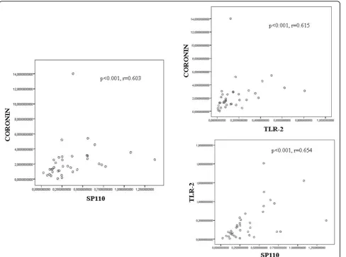
Correlation between the markers’expression in PBMCs with the infection state
According to the Spearman cofactor for linear correla-tion, there was a positive correlation between Coronin-1 and Sp110 (r = 0.603), Coronin-1 and TLR-2 (r = 0.615) and between Sp110 and TLR-2 (r = 0.654) expression in the patients group (group A) and close contacts group with LTBI (group B) (Figure 3). By contrast, no signifi-cant correlation was found between the expression of these markers in the uninfected group of close contacts (group C) and in the control subjects (group D) (data not shown).
Discussion
In this study, we observed an increased expression of TLR-2, Coronin-1, and Sp110 mRNA levels in PBMCs of patients with tuberculosis and in close contacts of patients that had latent tuberculosis infection. Further-more, we found an increased expression of TLR-2 mRNA in patients with tuberculosis compared to the close contact group without LTBI, whereas there was no significant difference in the expression of TLR-2 mRNA between patients and close contacts with LTBI. These findings indicate that TLR-2 Coronin-1, and Sp110 are probably involved in the infection process of human tuberculosis.
repertoire of TLR-2 ligands, such as, 19-kDa lipoprotein (LpqH), lipoproteins LprA (Rv1270) and LprG (Rv1411c). Lipomannan and phosphatidyl-myo-inositol mannoside (PIM) are also TLR-2 agonists, which inter-act with TLR-2 to initiate cellular inter-activation. The 19-kDa lipoprotein (LpqH), secreted by MTB, was the first MTB ligand, which has been shown to interact specifi-cally with TLR-2 to induce TNF-aand nitric oxide pro-duction in both murine and human macrophages. It is also a major inducer of interleukin-12 production in human monocytes [38]. Furthermore, it has been shown that TLR-2 is essential for the intracellular mycobacter-ial killing in macrophages, through the production of the antimicrobial peptide cathelicidin, an effect mediated by a vitamin-D related pathway [10]. However, studies in mice have shown that TLR-2 is not essential for host resistance against tuberculosis infection since the TLR-2-deficient mice were not compromised in their ability to induce Th1 immunity. On the contrary, it did exhibit exaggerated immuno-pathology. Additionalin vitro stu-dies have shown that engagement of TLR-2 with MTB
ligands induces inhibition of macrophage MHC class-II antigen presentation [39] and also blocks macrophage responsiveness to IFN-g[40,41]. These diverse data indi-cate that further studies are needed to elucidate whether TLR-2 is beneficial or harmful in humans in regard to tuberculosis.
The increased expression of Coronin-1, a protein that specifically inhibits the intracellular killing of live myco-bacteria in mononuclear cells, is probably due to a spe-cific subset of inactivated immature monocytes, which act as a Trojan horse for carrying mycobacteria from the inoculation sites to the other compartments of the cell body. However, although occult haematogenous dis-semination is thought to occur following initial infection of humans with MTB, there is no direct evidence of this in the present study. The results observed here are rather consistent with recirculation of activated mono-cytes from the site of the infection.
[image:5.595.58.543.97.209.2]This represents the well known process of haemato-genous spread of tubercle bacilli during MTB infection via blood vessels and lymphatics, which involves the
Table 2 Primers and probes sequences for Real Time RT-PCR ofb2microglubin, Coronin-1, HuPO and SP110 mRNA
mRNA Forward primer Reverse primer Probe FL Probe
b2
microglubin
CCAgCAgAgAATggAAAgTC gATgCTgCTTACATgTCTCg TTCTTCAgTAAgTCAACTTCAATgTCggA-FL
LC640-ATgAAACCCAgACACATAgCAATTCAg– PH
Coronin-1 CAATCCggTACTTTgAgATCA gCTTgTAgAACCTggCgA CTCCTTggAACTgAACATggAgAgA-FL LC640-AgTgCAggAAAggggCCTCg–PH SP110 TCAgAggAgATCATTgATggC CACTTggAgCTTCTCTTggAT AAAgAggTCCCAgAAgACgCCT–FL
LC640-gTACACCACgAAgggTCACACAAggg– PH
HuPO gCTTCCTggAgggTgTC CCAAgAAggCCTTgACCTT CgAgTCCTggCCTTgTCTgTggA-FL
LC640-CggATTACACCTTCCCACTTgCTg–PH
[image:5.595.59.539.494.682.2]continuous turnover of macrophages between tubercu-lous lesions and the blood, a process that peaks during the phase of active infection [1,42]. Furthermore, during MTB infection, activated macrophages ingesting micro-organisms generate phagosomes that mature progres-sively along with the endocytic pathway, leading to fusion with late endosomes and ultimately to the forma-tion of lysosomes [43,44]. As a result, the proper orches-tration of these events leads to the destruction of the pathogen in phagolysosomes and the initiation of the appropriate innate immune response [45,46]. In striking contrast, the failure of phagosomes to fuse with lyso-somes is a frequent finding following macrophage inges-tion of Mycobacterium tuberculosis (MTB) and the variant M. bovis BCG, enabling these organisms to reside safely in vacuoles that support their survival and replication [31,32,47]. Since Coronin-1 is up-regulated in patients with tuberculosis infection (i.e. patients and LTBI close contact group) it is possible that Coronin-1 may have a role as a marker of MTB infection before the occurrence of the disease. Until now the diagnosis
of tuberculous infection is based mostly on immunologi-cal tests such as the tuberculin skin test and the inter-feron gamma releasing assays (IGRAS). However, these tests are informative about the immunological status of an individual to mycobacterial antigens and are not helpful in distinguishing an active infection; therefore specific markers of the infection status are highly desir-able [48,49].
[image:6.595.58.541.87.426.2]recently reported that Sp110 expression promotes the replication of intracellular pathogens, as inA. phagocyto-philum, an obligate intracellular tick-borne pathogen that causes human granulocytic anaplasmosis (HGA) in HL-60 cells, while silencing of Sp110 expression by RNA interference results in decreased levels of infection [53]. Thus, the role and function of Sp110 in human pulmonary tuberculosis needs further investigation.
In our study, we found a strong correlation between Coronin-1 and TLR-2 expression in nucleated blood cells of infected patients compared to the non-infected groups. Recent data point out a link between TLR-2 and calcineurin, which is the final effector molecule of pha-gosome-lysosome fusion in the Coronin-1 pathway. Activation of TLRs generally leads to nuclear transloca-tion of the transcriptransloca-tion factor NF-B, a critical compo-nent within many proinflammatory pathways, [16] including those associated with chemokine gene expres-sion [10]. Although TLRs share the ability to activate
NF-B, cross talk with other signalling pathways is one mechanism by which activation of specific TLRs may lead to different patterns of gene expression [35]. Another key signalling molecule of the immune response, calcineurin, has been reported to have a func-tional interaction with NF-B [54]. In two recently pub-lished studies it has been suggested that calcineurin may play a predominant role in the mediation of TLR-2-sti-mulated chemokine responses in murine myoblasts and in human airway epithelial cells, [55,56] indicating that Coronin-1 is a down-stream molecule in the TLR-2 pathway cytokine activation. In this regard it is possible that TLR-2 signalling pathway mediates the Coronin-1 expression and thus facilitates the intacellular survival of
Mycobacterium tuberculosis in human PBMCs. How-ever, recent studies suggest a negative relation between TLR-2 and Coronin-1, because TLR-2 is essential for the transformation of the inactive vitamin-D3 to its
[image:7.595.58.547.88.450.2]which is a bactericidal substance for mycobacteria [10]. Furthermore, pre-treatment of human monocytes with vitamin- D3/retinoic acid, down-regulated the expression
of Coronin-1 and inhibited the entry and intracellular survival of M. tuberculosiswithin the monocytes [57]. A recent report from Japan also suggested a functional counteraction between TLR-2 and Coronin-1 in macro-phages infected withMycobacterium leprae[58]. Thus, although the data in this report regarding the TLR-2 and Coronin-1 interplay in M. tuberculosis infection need further elucidation, there is actual evidence to sup-port a possible positive and negative feedback functional relation between them.
The strong correlation between Sp110 with TLR-2 expression in PBMCs in infected patients compared to the non-infected groups suggests that Sp110 expression is mediated through a TLR-2 pathway. TLRs activation generally leads to nuclear translocation of the transcrip-tion factor NF-B. Sp110 is a member of the nuclear body (NB) components that functions as a nuclear hor-mone receptor transcriptional co-activator [19]. Sp110 and other NB-associated proteins, induced by type I (a/ b) and type II (g) interferons (IFNs), play a role in IFN response and virus replication. Sp110 expression is induced in human peripheral blood leukocytes and spleen but not in other tissues [22]. These data indicate that the expression of Coronin-1 and Sp110 is probably related to a TLR-2 activation pathway. However, further studies are needed to clarify this assumption. A compar-ison with other well-known genes associated with immune activation (e.g. TNF) might better elucidate this hypothesis.
Conclusions
In conclusion, in the present work we have shown that mRNA levels of Coronin-1, Sp110 and TLR-2 are increased in patients with tuberculosis and in close con-tacts of patients with latent tuberculosis infection. Although our patient cohort sample was not particularly large our findings suggest that these molecules are essential for the disease process. However, further stu-dies are needed to investigate the clinical significance of these markers inM. tuberculosisinfection.
Acknowledgements
The authors would like to thank Aggeliki Hamhougia for supporting the statistical analysis and review of the article.
Author details
1Locus Medicus, Molecular Pathology and Genetics Dept., Athens, Greece. 2National and Kapodistrian University of Athens, Pulmonary Department,
“Sotiria”District Chest Diseases Hospital; Athens, Greece.3Mathematics Dept.,
University of Athens; Athens, Greece.4National Center of Tuberculosis, Microbiology Dpt,“Sotiria”District Chest Diseases; Athens, Greece.51st
Pulmonary Department,“Sotiria”District Chest Diseases Hospital. Athens, Greece.
Authors’contributions
PC conceived the idea and designed the study, made the initial analysis and interpretation of the results and has been involved in drafting and revising the manuscript. EF had a significant contribution in the molecular analysis of patient samples and approved the version to be published. NR had a significant contribution in the acquisition of patient data and has been involved in drafting and revising the manuscript. CG and AS had significant contribution in acquisition of patient data, and approved the final version of the article. SK had a significant contribution in acquisition and
microbiological analysis of patient data, and has given final approval for the version to be published. AH made the statistical analysis and helped in the interpretation of the results. CR edited the manuscript and has given final approval of the version to be published. NP conceived the idea and designed the study. He had the main contribution in the interpretation of the results, drafted and revised the version of the manuscript to be published.
Competing interests
All authors of this paper declare that they have no financial or other potential conflicts of interest concerning the subject of this manuscript.
Received: 7 February 2010 Accepted: 18 August 2010 Published: 18 August 2010
References
1. Kreider EM, Rossman DM:Pulmonary tuberculosis.Tuberculosis & Non tuberculous mycobacterial infectionsSchlossberg D , 5 2006, 177-189. 2. Dutt A, Schlossberg D:Epidemiology and host factors.Tuberculosis & Non
tuberculous mycobacterial infectionsSchlossberg D , 5 2006, 1-17. 3. Pieters J:Mycobacterium tuberculosis and the Macrophage: Maintaining
a Balance.Cell Host & Microbe2008,3:399-406.
4. Martino A:Mycobacteria and innate cells: critical encounter for immunogenicity.J Biosci2008,33:137-144.
5. Akira S, Uematsu S, Takeuchi O:Pathogen recognition and innate immunity.Cell2006,124:783-801.
6. Doyle ES, O’Connell MR, Miranda AG, Vaidya SA, Chow EK, Liu PT, Suzuki S, Suzuki N, Modlin RL, Yeh WC, Lane TF, Cheng G:Toll-like receptors induce a phagocytic gene program through.J Exp Med2004,199:81-90. 7. Blander MJ, Medzhitov R:Regulation of phagosome maturation by signals
from toll-like receptors.Science2004,304:1014-1018.
8. Ryffel B, Fremond C, Jacobs M, Parida S, Botha T, Schnyder B, Quesniaux V:
Innate immunity to mycobacterial infection in mice: critical role for toll-like receptors.Tuberculosis2005,85:395-405.
9. Salgame P:Host innate and Th1 responses and the bacterial factors that control Mycobacterium tuberculosis infection.Curr Opin in Immunol2005,
17:374-380.
10. Liu PT, Stenger S, Li H, Wenzel L, Tan BH, Krutzik SR, Ochoa MT, Schauber J, Wu K, Meinken C, Kamen DL, Wagner M, Bals R, Steinmeyer A, Zügel U, Gallo RL, Eisenberg D, Hewison M, Hollis BW, Adams JS, Bloom BR, Modlin RL:Toll-like Receptor Triggering of a Vitamin D-Mediated Human Antimicrobial Response.Science2006,311:1170-1173.
11. Ma X, Liu Y, Gowen BB, Graviss EA, Clark AG, Musser JM:Full-exon resequencing reveals Toll like receptors variants contribute to human tuberculosis disease.PLos ONE2007,2(12):e1318.
12. Armstrong JA, Hart PD:Phagosome-lysosome interactions in cultured macrophages infected with virulent tubercle bacilli. Reversal of the usual non fusion pattern and observations on bacterial survival.J Exp Med1975,142:1-16.
13. Ferrari G, Langen H, Naito M, Pieters J:A coat protein on phagosomes involved in the intracellular survival of mycobacteria.Cell1999,
97:435-447.
14. Hasan Z, Schlax C, Kuhn L, Lefkovits I, Young D, Thole J, Pieters J:Isolation and characterization of the mycobacterial phagosome segregation from the endosomal/lysosomal pathway.Mol Microbiol1997,24:545-553. 15. de Hostos EL:The coronin family of actin-associated proteins.Trends Cell
Biol1999,9:345-350.
16. Uetrecht AC, Bear JE:Coronins: the return of the crown.Trends Cell Biol 2006,16:421-426.
Mycobacteria in Macrophages is mediated by Coronin 1-Dependent Activation of Calcineurin.Cell2007,130:37-50.
18. Deghmane AE, Soualhine H, Bach H, Sendide K, Itoh S, Tam A, Noubir S, Talal A, Lo R, Toyoshima S, Av-Gay Y, Hmama Z:Lipoamide
dehydrogenase mediates retention of Coronin-1 on BCG vacuoles, leading to arrest in phagosome maturation.J Cell Sci2007,
120:2796-2806.
19. Pan H, Yan BS, Rojas M, Shebzukhov YV, Zhou H, Kobzik L, Higgins DE, Daly MJ, Bloom BR, Kramnik I:Ipr1 gene mediates innate immunity to tuberculosis.Nature2005,434:767-772.
20. Bloch DB, Nakajima A, Gulick T, Chiche JD, Orth D, de La Monte SM, Bloch KD:Sp110 localizes to the PML-Sp100 nuclear body and may function as a nuclear hormone receptor transcriptional coactivator.Mol Cell Biol2000,20:6138-6146.
21. Castrillo A, Tontonoz P:Nuclear receptors in macrophage biology: at the crossroads of lipide metabolism and inflammation.Annu Rev Cell Dev Biol 2004,20:455-480.
22. Regad T, Chelbi-Alix MK:Role and fate of PML nuclear bodies in response to interferon and viral infections.Oncogene2001,20:7274-7286. 23. Saito T, Ji G, Shinzawa H, Okumoto K, Hattori E, Adachi T, Takeda T,
Sugahara K, Ito JI, Watanabe H, Saito K, Togashi H, Ishii K, Matsuura T, Inageda K, Muramatsu M, Kawata S:Genetic variations in humans associated with differences in the course of hepatitis C.Biochem Biophys Res Commun2004,317:335-341.
24. Fietta A, Meloni F, Cascina A, Morosini M, Marena C, Troupioti P, Mangiarotti P, Casali L:Comparison of a whole-blood interferon-gamma assay and tuberculin skin testing in patients with active tuberculosis and individuals at high or low risk ofMycobacterium tuberculosis infection.Am J Infect Control2003,31:347-53.
25. Mazurek GH, LoBue PA, Daley CL, Bernardo J, Lardizabal AA, Bishai WR, Iademarco MF, Rothel JS:Comparison of a whole-blood interferon gamma assay with tuberculin skin testing for detecting latent
Mycobacterium tuberculosisinfection.JAMA2001,286:1740-7.
26. Brock I, Weldingh K, Lillebaek T, Follmann F, Andersen P:Comparison of a new specific blood test and the skin test in tuberculosis contacts.Am J Respir Crit Care Med2004,170:65-9.
27. Guidelines for the Investigation of Contacts of Persons with Infectious Tuberculosis Recommendations from the National Tuberculosis Controllers Association and CDC MMWR. 2005,54(RR-15). 28. Lalvani A, Pathan AA, Durkan H, Wilkinson KA, Whelan A, Deeks JJ,
Reece WH, Latif M, Pasvol G, Hill AV:Enhanced contact tracing and spatial tracking ofMycobacterium tuberculosisinfection by numeration of antigen specific T cells.Lancet2001,357:2017-21.
29. D’Souza CD, Cooper AM, Frank AA, Ehlers S, Turner J, Bendelac A, Orme IM:
A novel onclassicb2-Microglobulin - restricted mechanism influencing early lymphocyte accumulation and subsequent resistance to tuberculosis in the lung.Am J Respir Cell Mol Biol2000,23:188-193. 30. Schaible UE, Collins HL, Priem F, Kaufmann SH:Correction of the iron
overload defect inb2-Microglobulin knockout mice by lactoferrin abolishes their increased susceptibility to tuberculosis.J Exp Med2002,
196:1507-1513.
31. Hestvik AL, Hmama Z, Av-Gay Y:Mycobacterial manipulation of the host cell.FEMS Microbiol Rev2005,29:1041-1050.
32. Kusner DJ:Mechanisms of mycobacterial persistence in tuberculosis.Clin Immunol2005,114:239-247.
33. Zarember KA, Godowski PJ:Tissue expression of human Toll-Like receptors and differential regulation of Toll-Like receptor mRNAs in leukocytes in response to microbes, their products and cytokines.The Journal of Immunology2002,168:554-561.
34. Bhatt K, Salgame P:Host Innate Immune Response to Mycobacterium tuberculosis.J Clin Immunol2007,27:347-362.
35. Takeda K, Akira S:Toll-like receptors in innate immunity.Intern Immunol 2005,17:1 14.
36. Chang JS, Huggett JF, Dheda K, Kim LU, Zumla A, Rook GA:
Mycobacterium tuberculosis induces selective up-regulation of TLRs in the mononuclear leukocytes of patients with active pulmonary tuberculosis.J Immunol2006,176:3010-3018.
37. Fenhalls G, Squires GR, Stevens-Muller L, Bezuidenhout J, Amphlett G, Duncan K, Lukey PT:Associations between Toll-like receptors and interleukin-4 in the lungs of patients with tuberculosis.Am J Respir Cell Mol Biol2003,29:28-38.
38. Brightbill HD, Libraty DH, Krutzik SR, Yang RB, Belisle JT, Bleharski JR, Maitland M, Norgard MV, Plevy SE, Smale ST, Brennan PJ, Bloom BR, Godowski PJ, Modlin RL:Host defence mechanisms triggered by microbial lipoproteins through toll-like receptors.Science1999,
285:732-736.
39. Noss EH, Pai RK, Sellati TJ, Radolf JD, Belisle J, Golenbock DT, Boom WH, Harding CV:Toll-like receptor 2-dependent inhibition of macrophage class II MHC expression and antigen processing by 19-kDa lipoprotein of
Mycobacterium tuberculosis.J Immunol2001,167:910-918.
40. Fortune SM, Solache A, Jaeger A, Hill PJ, Belisle JT, Bloom BR, Rubin EJ, Ernst JD:Mycobacterium tuberculosisinhibits macrophage responses to IFN-gamma through myeloid differentiation factor 88-dependent and -independent mechanisms.J Immunol2004,172:6272-6280.
41. Banaiee N, Kincaid EZ, Buchwald U, Jacobs WR Jr, Ernst JD:Potent inhibition of macrophage responses to IFN-gamma by live virulent
Mycobacterium tuberculosisis independent of mature mycobacterial lipoproteins but dependent on TLR2.J Immunol2006,176:3019-27. 42. Dannenberg MA Jr:Macrophage turnover, division and activation within
developing, peak and“healed”tuberculosis lesions produced in rabbits by BCG.Tuberculosis2003,83:251-260.
43. Desjardins M, Huber LA, Parton RG, Griffiths G:Biogenesis of
phagolysosomes proceeds through a sequential series of interactions with the endocytic apparatus.J Cell Biol1994,124:677-688.
44. Vieira OV, Botelho RJ, Grinstein S:Phagosome maturation: aging gracefully.Biochem J2002,366:689-704.
45. Desjardins M, Houde M, Gagnon E:Phagocytosis: the convoluted way from nutrition to adaptive immunity.Immunol Rev2005,207:158-165. 46. Chan J, Flynn J:The immunological aspects of latency in tuberculosis.
Clin Immunol2004,110:2-12.
47. Deretic V, Singh S, Master S, Harris J, Roberts E, Kyei G, Davis A, de Haro S, Naylor J, Lee HH, Vergne I:Mycobacterium tuberculosis inhibition of phagolysosome biogenesis and autophagy as a host defence mechanism.Cell Microbiol2006,8:719-727.
48. Tuberculosis clinical diagnosis and management of tuberculosis and measures for its prevention and control. Clinical Guideline 33. National Institute for Health and Clinical Excellence 2006.
49. Thye T, Browne EN, Chinbuah MA, Gyapong J, Osei I, Owusu-Dabo E, Niemann S, Rüsch-Gerdes S, Horstmann RD, Meyer CG:No associations of human pulmonary tuberculosis with Sp110 variants.J Med Genet2006,
43(7):e32[http://www.jmedgenet.com/cgi/content/full/43/7/e32)]. 50. Szeszko JS, Healy B, Stevens H, Balabanova Y, Drobniewski F, Todd JA,
Nejentsev S:Resequencing and association analysis of the SP110 gene in adult pulmonary tuberculosis.Hum Genet2007,121:155-160.
51. Babb C, Keet EH, van Helden PD, Hoal EG:SP110 polymorphisms are not associated with pulmonary tuberculosis in a South African population.
Hum Genet2007,121:521-522.
52. Tosh K, Campbell SJ, Fielding K, Sillah J, Bah B, Gustafson P, Manneh K, Lisse I, Sirugo G, Bennett S, Aaby P, McAdam KP, Bah-Sow O, Lienhardt C, Kramnik I, Hill AV:Variants in the SP110 gene are associated with genetic susceptibility to tuberculosis in West Africa.Proc Natl Acad Sci USA2006,
103:10364-10368.
53. de la Fuente J, Manzano-Roman R, Blouin EF, Naranjo V, Kocan KM:Sp110 transcription is induced and required by Anaplasma phagocytophilum for infection of human promyelocytic cell.BMC Infect Dis2007,
207:110-113.
54. Trushin SA, Pennington KN, Algeciras-Schimnich A, Paya CV:Protein kinase C and calcineurin synergize to activate IkappaB kinase and NF-kappaB in T lymphocytes.J Biol Chem1999,274:22923-22931.
55. Boyd JH, Divangahi M, Yahiaoui L, Gvozdic D, Qureshi S, Petrof BJ:Toll-like Receptors Differentially Regulate CC and CXC Chemokines in Skeletal Muscle via NF-B and Calcineurin.Infection and immunity2006,
74:6829-6838.
56. Waters V, Sokol S, Reddy B, Soong G, Chun J, Prince A:The effect of cyclosporin A on airway cell proinflammatory signaling and pneumonia.
Am J Respir Cell Mol Biol2005,33:138-144.
57. Anand PK, Kaul D, Sharma M:Synergistic action of vitamin D and retinoic acid restricts invasion of macrophages by pathogenic mycobacteria.J Microbiol Immunol Infect2008,41:17-25.
Pre-publication history
The pre-publication history for this paper can be accessed here: http://www.biomedcentral.com/1471-2334/10/243/prepub
doi:10.1186/1471-2334-10-243
Cite this article as:Constantoulakiset al.:In vivoexpression of innate
immunity markers in patients withmycobacterium tuberculosisinfection.
BMC Infectious Diseases201010:243.
Submit your next manuscript to BioMed Central and take full advantage of:
• Convenient online submission
• Thorough peer review
• No space constraints or color figure charges
• Immediate publication on acceptance
• Inclusion in PubMed, CAS, Scopus and Google Scholar • Research which is freely available for redistribution