R E S E A R C H A R T I C L E
Open Access
Opportunistic parasitoses among Egyptian
hemodialysis patients in relation to CD4+
T-cell counts: a comparative study
Amany I. Shehata
1, Faika Hassanein
2and Rashad Abdul-Ghani
3,4*Abstract
Background:Some reports are available on the prevalence of opportunistic parasitoses among hemodialysis (HD) patients, yet there is a paucity of data on the association of CD4+ T-cell counts with such infections. Therefore, this study aimed to determine the prevalence of intestinal parasites andToxoplasma gondiiin relation to CD4+ counts among HD patients in Alexandria, Egypt.
Methods:A comparative cross-sectional study was conducted on 120 HD patients and 100 apparently healthy individuals between December 2014 and January 2016. Data and samples (stool and blood) were collected from the participants after obtaining their informed consent. Stool samples were examined for parasites after concentration and staining, EDTA-blood samples were used for CD4+ counting by flow cytometry, and sera were analyzed for
anti-ToxoplasmaIgM and IgG antibodies.
Results:A significantly higher prevalence rate of intestinal parasitoses was found among HD patients compared to apparently healthy individuals (52.5% vs. 12.0%, respectively), with absence of helminths.Cryptosporidiumspecies (32.5%),
B. hominis(24.2%) and microsporidia (11.7%) were the most frequent parasites among HD patients, whileB. hominis(13.0%),Cryptosporidiumspecies (11.0%) andG. lamblia(4.0%) were the most frequent parasites among their counterparts. Statistically significant differences in parasite infection rates between patients and their counterparts were found forCryptosporidiumspecies,B. hominisand microsporidia. However, parasite species were not significantly associated with diarrhea. On the other hand, the overallT. gondiiseroprevalence rate among HD patients was significantly higher than that among their counterparts (33.3% vs. 8%, respectively). HD patients with CD4 + counts < 200 cells/μl were twice more exposed to intestinal parasitoses compared to those with counts≥200 cells/ μl, but the difference was not statistically significant. However, low CD4+ counts were significantly associated with higher rates ofCryptosporidiumspecies, microsporidia andT. gondii.
Conclusions:Intestinal parasitoses andT. gondiiinfection rates are significantly higher among Egyptian HD patients compared to apparently healthy individuals, withCryptosporidiumspecies,B. hominis, microsporidia andT. gondiibeing the most frequent parasites. CD4+ counts < 200 cells/μl are significantly associated withCryptosporidiumspecies, microsporidia andT. gondiiamong HD patients. Therefore, regular screening of HD patients for opportunistic parasites is recommended.
Keywords:Intestinal parasitosis,Toxoplasma gondii, Hemodialysis, CD4+ Tcell, Egypt
© The Author(s). 2019Open AccessThis article is distributed under the terms of the Creative Commons Attribution 4.0 International License (http://creativecommons.org/licenses/by/4.0/), which permits unrestricted use, distribution, and reproduction in any medium, provided you give appropriate credit to the original author(s) and the source, provide a link to the Creative Commons license, and indicate if changes were made. The Creative Commons Public Domain Dedication waiver (http://creativecommons.org/publicdomain/zero/1.0/) applies to the data made available in this article, unless otherwise stated.
* Correspondence:rashadqb@yahoo.com
3
Department of Medical Parasitology, Faculty of Medicine and Health Sciences, Sana’a University, Sana’a, Yemen
4Tropical Disease Research Center, Faculty of Medicine and Health Sciences,
University of Science and Technology, Sana’a, Yemen
Background
Renal failure is an immunosuppressive condition that makes patients more prone to infections, including those caused by opportunistic protozoan parasites. Patients undergoing hemodialysis (HD) suffer from humoral and cell-mediated immune defects and have disturbances in acquired immune response to a variety of antigens [1, 2]. End-stage renal failure (ESRF) leads to impaired cell-mediated immunity as a result of lymphopenia and dys-function of cluster of differentiation 4 (CD4+) T cells [1]. Moreover, uremia-associatedpro-inflammatory conditions developed in ESRD patients can lead to irreversible pre-mature aging of T-cell compartment that contributes to the cell-mediated defects and susceptibility to infectious agents [3,4]. On the other hand, HD induces apoptosis of T cells, decreased phagocytic capacity of neutrophils and monocytes along with abnormal production of pro-inflammatory cytokines by monocytes because of their dir-ect contact with artificial membranes in HD machines [5– 8]. In addition, frequent blood transfusions to patients with chronic renal failure (CRF) can lead to the develop-ment of antibodies against human leukocyte antigens, which can form immune complexes that interfere with cellular immune responses [9,10].
Opportunistic parasitic infections, including those with Cryptosporidium species, Cystoisospora belli, Cyclospora cayetanensis, Blastocystis hominis, Toxoplasma gondii and microsporidia, have been documented to cause serious complications or even death among immuno-compromized patients, including those undergoing HD [11–13]. In this context, humoral and cell-mediated im-munosuppression can increase the susceptibility to and worsen the outcome of parasitic infections through increasing the acquisition of infection and the clinical severity of disease [14].
In Egypt, ESRD has been estimated to affect 264 to 414 per million population [15]. Despite the availability of a number of reports on opportunistic parasitoses among HD patients in Egypt [16–18], there is a paucity of data on the association of CD4+ counts with such infections. Therefore, the present study was conducted to determine the prevalence of intestinal parasites and T. gondiiamong HD patients in Alexandria governorate, Egypt in relation to CD4+ counts compared to appa-rently healthy individuals.
Methods
Study design, subjects and ethical considerations
A comparative cross-sectional study was conducted on 120 HD patients from different hospitals and 100 appa-rently healthy individuals in Alexandria, Egypt from December 2014 to January 2016. According to hospital records, patients were screened for human immunodefi-ciency virus (HIV) on admission and before the start of
HD sessions, and those HIV-positive were referred to special hospitals. HD patients included in the study had no other immunosuppressive conditions, were not undergoing an immunosuppressive therapy and had not received antiparastitic treatment the month preceding the study. On the other hand, apparently healthy individuals of matched age, sex and residence were included provided that they had no kidney problems or immunosuppressive conditions, were not undergoing an immunosuppressive therapy and had not received antiparasitic treatment the month preceding the study.
Data and sample collection
Data on the gender, age, residence, education, occupa-tion, presentation with diarrhea and CD4+ count of study participants were collected using a pre-designed questionnaire after obtaining their informed consent to voluntarily participate in the study. Fresh stool samples were collected into clean, pre-labelled containers on two successive sessions for HD patients, where all patients were subjected to three sessions a week, and on two alternate days for apparently healthy individuals. Patients were classified as diarrheic if they had reported the passage of loose or liquid stools three times or more per day or if they were suffering from passing more frequent loose bowel movements than normal each day [19], either on the day of sample collection or in the 2 weeks prior to sample collection.
Approximately 5 ml of venous blood was collected from the patients on the day of the collection of first stool samples by nurses of the centers for patients and by one of the researchers for the apparently healthy indi-viduals. The collected blood was divided equally (2.5 ml each) into a plain test tube and an EDTA-tube. The blood in plain tubes was left to clot, and the sera were then separated by centrifugation at 3000 rounds per mi-nute for 5 min., dispensed into pre-labelled Eppendorf tubes and stored at −20 °C until the detection of anti-Toxoplasmaantibodies. The EDTA-anticoagulated blood was used for counting CD4+ cells. Stool and sera were transferred to the Parasitology Laboratory of the High Institute of Public Health, Alexandria University for processing and examination. However, EDTA-blood samples were transferred within a maximum of 2 h to the Hematology Laboratory of the Clinical Pathology Department, Faculty of Medicine, Alexandria University for CD4+ cell counting.
Parasitological investigations
concentrated by formalin-ethyl acetate sedimentation con-centration technique and examined for protozoan cysts and helminthic ova [23]. A smear was then prepared from the sediment and stained by the modified Ziehl-Neelsen technique for detecting coccidian oocysts [24, 25]. The identification and differentiation of intestinal parasites were based on the diagnostic criteria and bench aids provided by the World Health Organization [26].
CD4+ T-cell counting
EDTA-blood samples were immediately processed for counting CD4+ cells according to standard procedures [27], using the Attune™ NxT Flow Cytometer (Thermo Fisher Scientific, Waltham, MA, USA). Because CD4+ counts show variability in the absence of HIV infection [28], we used a threshold level of 200 CD4+ count/μl to study the association of low CD4+ count with parasito-ses because the counts below 200/μl is considered the severe level of immunosuppression that can be asso-ciated with opportunistic infection [29].
Detection of anti-Toxoplasmaantibodies
Anti-Toxoplasma IgM and IgG antibodies were detected in sera using enzyme-linked immunosorbent assay kits (BioCheck Inc., South San Francisco, CA, USA) according to the manufacturer’s instructions. The cut-off points for IgM and IgG seropositivity were 1 IU/ml and 32 IU/ml, respectively, where no sera showed borderline reactivity.
Statistical analysis
Data were entered, edited and analyzed using IBM SPSS Statistics, version 20.0 (IBM Corp., Armonk, NY, USA). Categorical variables were presented as frequencies and percentages and compared using chi-square test or Fisher’s exact test, whichever suitable, while continuous variables were presented as means with standard de-viations and compared using t-test. Differences and associations were considered statistically significant at Pvalues < 0.05.
Results
Characteristics of the study subjects
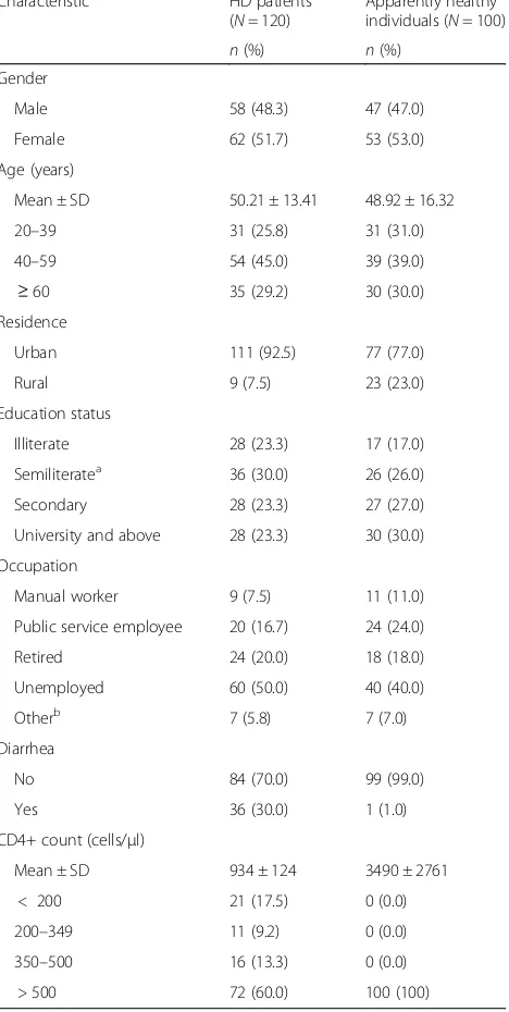
Table1shows the detailed characteristics of HD patients and the comparison group. The mean age and CD4+ count of HD patients was 50 ± 13.41 years (range: 20– 80) and 934 ± 124 cells/μl, respectively. About a third of the HD patients presented with diarrhea (30%; 36/120) and 17.5% (21/120) had CD4+ counts of < 200 cells/μl. On the other hand, the mean age and CD4+ count of the apparently healthy individuals were 48.92 ± 16.32 years (range: 20–80) and 3490 ± 2761 cells/μl, respectively. In addition, all apparently healthy indivi-duals had CD4+ counts of > 500 cells/μl and only one patient (1.0%) presented with diarrhea (Table1).
Comparison between intestinal parasitoses among HD patients and apparently healthy counterparts
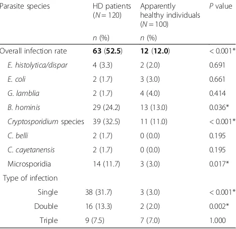
[image:3.595.306.540.116.581.2]The overall prevalence of intestinal parasitoses was significantly higher (P< 0.001) among HD patients com-pared to their apparently healthy counterparts (52.5% vs. 12.0%, respectively). Among HD patients,Cryptosporidium species (32.5%), B. hominis (24.2%) and microsporidia (11.7%) were the most frequent parasite species, while E. coli, G. lamblia, C. belli and C. cayetanensis (1.7% each) were the least frequent species. On the other
Table 1Characteristics of HD patients and their apparently healthy counterparts in Alexandria (2014–2016)
Characteristic HD patients (N= 120)
Apparently healthy individuals (N= 100)
n(%) n(%)
Gender
Male 58 (48.3) 47 (47.0)
Female 62 (51.7) 53 (53.0)
Age (years)
Mean ± SD 50.21 ± 13.41 48.92 ± 16.32
20–39 31 (25.8) 31 (31.0)
40–59 54 (45.0) 39 (39.0)
≥60 35 (29.2) 30 (30.0)
Residence
Urban 111 (92.5) 77 (77.0)
Rural 9 (7.5) 23 (23.0)
Education status
Illiterate 28 (23.3) 17 (17.0)
Semiliteratea 36 (30.0) 26 (26.0)
Secondary 28 (23.3) 27 (27.0)
University and above 28 (23.3) 30 (30.0)
Occupation
Manual worker 9 (7.5) 11 (11.0)
Public service employee 20 (16.7) 24 (24.0)
Retired 24 (20.0) 18 (18.0)
Unemployed 60 (50.0) 40 (40.0)
Otherb 7 (5.8) 7 (7.0)
Diarrhea
No 84 (70.0) 99 (99.0)
Yes 36 (30.0) 1 (1.0)
CD4+ count (cells/μl)
Mean ± SD 934 ± 124 3490 ± 2761
< 200 21 (17.5) 0 (0.0)
200–349 11 (9.2) 0 (0.0)
350–500 16 (13.3) 0 (0.0)
> 500 72 (60.0) 100 (100)
HDHemodialysis,SDStandard deviation;a
includes those barely able to read and write,b
hand, B. hominis (13.0%), Cryptosporidium species (11.0%) and G. lamblia (4.0%) were the most frequent parasite species among apparently healthy individuals.-None of the helminthic parasites was detected among HD patients or apparently healthy individuals. Differences between the two groups were statistically significant for the infection rates with B. hominis, Cryptosporidium-species and microsporidia. Regarding the types of infection, there were statistically significant differences between HD patient and apparently healthy individuals in single infections (31.7% vs. 3.0%, respectively) and double infections (13.3% vs. 2.05, respectively). However, there was no statistically significant difference between the HD patients and apparently healthy individuals in triple infec-tions, being 7.5% vs. 7.0%, respectively (Table 2). On the other hand, no statistically significant association was found between individual parasite species and diarrhea among HD patients (Table3).
T. gondiiseroprevalence among HD patients and apparently healthy counterparts
Table 4 shows that the overallT. gondii seroprevalence was significantly higher (P< 0.001) among HD patients compared to apparently healthy counterparts (33.3% vs. 8%, respectively). Anti-ToxoplasmaIgM antibodies were detected in four cases of HD patients (two alone and two with IgG) and in two cases of apparently healthy individuals (one alone and one with IgG). However, a significantly higher (P< 0.001) IgG seroprevalence rate
was found among HD patients compared to apparently healthy individuals (31.0% vs. 7.0%, respectively).
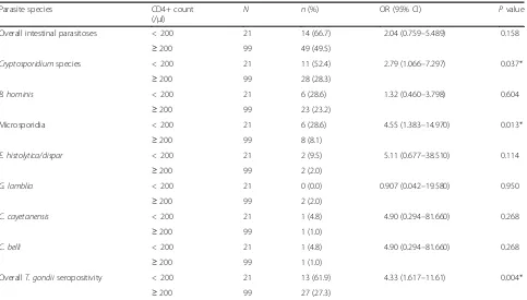
Prevalence of intestinal parasitoses andT.gondiiamong HD patients in relation to CD4+ counts
Table 5 shows that HD patients with CD4+ counts < 200 cells/μl were twice more exposed to intestinal parasitoses compared to those with CD4+ counts ≥ 200 cells/μl, but the difference was not statistically significant (OR = 2.041; 95% CI = 0.759–5.489, P= 0.158). However, low CD4+ count was significantly associated with a higher infection rate with Cryptosporidium species (52.4% vs. 28.3%), where patients with CD4+ counts < 200 cells/μl were at about 2.8-fold higher risk of infection than those with CD4+ counts≥200 cells/μl (OR = 2.79; 95% CI = 1.066– 7.297,P= 0.037). In addition, low CD4+ count was sig-nificantly associated with a higher infection rate with microsporidia (28.6% vs. 8.1%), where patients with counts < 200 cells/μl were at about 4.5-fold higher risk of infection than those with counts≥200 cells/μl (OR = 4.55; 95% CI = 1.066–7.297, P= 0.037). On the other
[image:4.595.305.538.110.300.2]hand, Table 4 shows that low CD4+ count was
Table 2Prevalence of intestinal parasitoses among HD patients and apparently healthy individuals in Alexandria, Egypt (2014–2016)
Parasite species HD patients (N= 120)
Apparently healthy individuals (N= 100)
Pvalue
n(%) n(%)
Overall infection rate 63(52.5) 12(12.0) < 0.001*
E. histolytica/dispar 4 (3.3) 2 (2.0) 0.691
E. coli 2 (1.7) 3 (3.0) 0.661
G. lamblia 2 (1.7) 4 (4.0) 0.414
B. hominis 29 (24.2) 13 (13.0) 0.036*
Cryptosporidiumspecies 39 (32.5) 11 (11.0) < 0.001*
C. belli 2 (1.7) 0 (0.0) 0.195
C. cayetanensis 2 (1.7) 0 (0.0) 0.195
Microsporidia 14 (11.7) 3 (3.0) 0.017*
Type of infection
Single 38 (31.7) 3 (3.0) < 0.001*
Double 16 (13.3) 2 (2.0) 0.002*
Triple 9 (7.5) 7 (7.0) 1.000
HDHemodialysis; *, statistically significant atP< 0.05
Table 3Association of parasite species with diarrhea among HD patients in Alexandria, Egypt (2014–2016)
Parasite speciesa N Presentation with diarrhean(%)
Pvalue
Cryptosporidiumspecies
Yes 39 12 (30.8) 0.530
No 81 24 (29.6)
B. hominis
Yes 29 8 (27.6) 0.469
No 91 28 (30.8)
Microsporidia
Yes 14 4 (28.6) 0.586
No 106 32 (30.2)
E. histolytica/dispar
Yes 4 2 (50.0) 0.384
No 116 34 (29.3)
a
[image:4.595.57.291.497.724.2]Parasite species detected in diarrheic cases were only included;NNumber examined;nNumber presented with diarrhea
Table 4Seroprevalence of T. gondiiamong HD patients and apparently healthy counterparts in Alexandria, Egypt (2014–2016)
T. gondii seropositivity
HD patients (N= 120)
Apparently healthy individuals (N= 100)
Pvalue
n(%) n(%)
Overall 40(33.3) 8(8.0) < 0.001*
IgM 4 (3.3) 2 (2.0) 1.000
IgG 38 (31.7) 7 (7.0) < 0.001*
[image:4.595.300.541.645.724.2]significantly associated with a higherT. gondii seroposi-tivity rate (61.9% vs. 27.3%), where patients with counts < 200 cells/μl were at more than fourfold higher risk of infection than those with counts≥200 cells/μl (OR = 4.33; 95% CI = 1.617–11.610,P= 0.004).
Discussion
Opportunistic parasitic infections are a major cause of morbidity and mortality in immunocompromized patients, particularly those with low CD4+ counts [11–13]. Increased blood urea levels in patients with ESRD could lead to a weakened immune system and increased risk of morbidity and mortality associated with such opportunistic infections [30]. On the other hand, HD patients are more prone to such opportunistic infections, where repeated HD decreases CD4+ counts compared to predialysis and healthy controls [31]. Up to the best of our knowledge, the present study is the first to compare infection rates with intestinal parasites and T. gondii between HD patients and apparently healthy individuals in relation to CD4+ counts in Alexandria, Egypt.
The overall prevalence rate of intestinal parasitoses among HD patients was significantly higher than their apparently healthy counterparts (52.5% vs. 12.0%, respec-tively). Lower infection rates among HD patients were reported from Brazil (45.1%), Turkey (43.7%) and Iran
(11.9–30.7%) [13,32–34]. Compared to the finding of the present study, higher infection rates withCryptosporidium species (40.0%), E. histolytica (14.0%) and G. lamblia (12.0%) have been recently reported among CRF patients undergoing HD in Upper Egypt [16]. The differences in the prevalence rates of infections between HD patients in the present study (north of Egypt) and those in the latter study (south of Egypt) could be attributed, among others, to differences in environmental, sanitary and hygienic fac-tors. However, the reasons for such geographical diffe-rences in the prevalence of parasites among HD patients require further investigations.
[image:5.595.58.541.108.383.2]In line with the findings of the present study, Cryp-tosporidiumspecies (26.4%) and B. hominis (24.5%) were the most prevalent intestinal parasites among Brazilian HD patients [12]. Among the controls, however, B. hominis was the most prevalent species while Cryptosporidium-species was not detected [12]. Another Brazilian study also reported B. hominis as the most prevalent parasite species among HD patients (20.1%) followed by Endo-limax nana(16.3%), whileCryptosporidiumspecies was re-ported among 4.7% of patients [31]. Similarly, B. hominis(4.2–14.1%) and Cryptosporidium species (11.5%) were the most common parasite species among Iranian HD patients [13, 34–36]. In addition, B. hominis (23.9%) followed by G. lamblia (8.5%) were the most prevalent parasite species among
Table 5Association of CD4+ counts with intestinal parasitoses andT. gondiiinfection among HD patients in Alexandria, Egypt (2014–2016)
Parasite species CD4+ count
(/μl)
N n(%) OR (95% CI) Pvalue
Overall intestinal parasitoses < 200 21 14 (66.7) 2.04 (0.759–5.489) 0.158
≥200 99 49 (49.5)
Cryptosporidiumspecies < 200 21 11 (52.4) 2.79 (1.066–7.297) 0.037*
≥200 99 28 (28.3)
B. hominis < 200 21 6 (28.6) 1.32 (0.460–3.798) 0.604
≥200 99 23 (23.2)
Microsporidia < 200 21 6 (28.6) 4.55 (1.383–14.970) 0.013*
≥200 99 8 (8.1)
E. histolytica/dispar < 200 21 2 (9.5) 5.11 (0.677–38.510) 0.114
≥200 99 2 (2.0)
G. lamblia < 200 21 0 (0.0) 0.907 (0.042–19.580) 0.950
≥200 99 2 (2.0)
C. cayetanensis < 200 21 1 (4.8) 4.90 (0.294–81.660) 0.268
≥200 99 1 (1.0)
C. belli < 200 21 1 (4.8) 4.90 (0.294–81.660) 0.268
≥200 99 1 (1.0)
OverallT. gondiiseropositivity < 200 21 13 (61.9) 4.33 (1.617–11.61) 0.004*
≥200 99 27 (27.3)
Turkish ESRF patients undergoing HD, while low rates of Cryptosporidium species, E. histolytica and microsporidia (2.1% each) were detected among patients but not the controls [33]. The variations in the prevalence of intestinal parasitoses among HD patients could be partly explained by the differences in the geographical dis-tribution of parasites at the community level as a result of environmental, climatic and sanitary differences in addition to differences in hygienic and behavioral fac-tors at the individual level. In addition, the role of the immune status and duration of HD as well as the method of stool examination in infection rate differences could not be ruled out.
In the present study, approximately a third of HD patients infected with the opportunistic parasites (Cryptosporidium species, B. hominis or microsporidia) had diarrhea, even though with no statistically significant association. Although the role of B. hominis in human disease is controversial [37], it can be a cause of diarrhea in patients on regular HD and renal transplant recipients [38, 39]. The lack of a statistically significant association between individual parasite species and presentation with diarrhea among HD patients in the present study is in contrast to the recent finding among CRF patients under-going HD in Upper Egypt, where a statistical significance was found between Cryptosporidium species and diar-rhea [16]. It also disagrees with that reported among Saudi patients undergoing HD [40], whereCryptosporidium species, microsporidia and G. lamblia were significantly higher among diarrheic patients compared to diarrheic controls. However, it is in agreement with that reported among Brazilian HD patients, where no association was found among individual parasite species and diarrhea [32]. In fact, the lack or presence association of HD with diar-rhea among patients from different countries could be explained by differences in the level of immunosup-pression, which determines the presence and severity of diarrhea. It is noteworthy that the differences in the inci-dence of gastrointestinal symptoms, including diarrhea, among CRF patients undergoing dialysis could be largely attributed to a number of factors such as the levels of uremic toxins, presence of metabolic co-morbidities, in-take of medications and psychosocial factors [41,42]. On the other hand, absence of helminths among HD patients in the present study is consistent with an earlier study among immunocompromized patients from Alexandria [43].
The overall T. gondiiseroprevalence rate among appro-ximately a third of HD patients in the present study is lower than those previously reported among HD patients (61.7%) and renal transplant recipients (70.0%) in Alexan-dria [17]. Similarly, it is lower than the rates reported from Turkey (56.0–76.5%) [44,45], Mexico (56.7%) [46] and Iran (56.7–73.7%) [47–49]. The anti-Toxoplasma IgM
seroprevalence among HD patients in the present study (3.3%) is lower than those reported among Egyptian pa-tients on regular HD (16.7%) and renal transplant recipients (24.1%) [50]. In addition, it is lower than those (7.8–13.5%) reported among Iranian patients undergoing regular HD [49, 51]. In general, the seroprevalence rate of anti-Toxo-plasma IgM among HD patients in the present study is somewhat close to the rates reported from Turkey (1.7%) and Iran (2.0%) [44,47]. However, comparisons can be diffi-cult due to a number of factors such as the duration of HD and the severity of renal damage. Because of the diffi-culty in adopting molecular techniques for the diag-nosis ofT. gondiiinfection in resource-limited countries, serological detection of anti-Toxoplasmaantibodies is still the most commonly used approach for the screening of the infection among immunocompromized patients [52]. However, a major limitation of the present study is the in-ability to confirm whether the three cases seropositive for both IgM and IgG were acute infections because IgM may persist for years [53, 54]. A good approach to the real-time serodiagnosis ofT. gondiiinfection that distinguishes between acute and chronic infections is the measurement of IgG avidity [55]. In this regard, the low avidity of spe-cific anti-Toxoplasma IgG indicates a recent primary infection [56]. In addition, development of kits using immunoreactive proteins and multi-epitope antigens has been suggested for improving the diagnosis of T. gondiiinfection [57–59]. The highT. gondiiseronegativ-ity rate among HD patients in the present study indicates that the majority of HD patients had not been exposed to infection and are susceptible to the risk of acute infection.
the mean CD4+ counts in Toxoplasma-seropositive and -seronegative patients and that most IgG-seropositive patients had CD4+ counts of < 100/μl. Similarly, CD4+ counts of≤100/μl were also found to be significantly asso-ciated with IgG seropositivity among HIV-positive patients without neurological complication from Nigeria [63].
One of the primary limitations of the present study comes from its observational design and that the prevalence of intestinal parasitoses was determined at a single point in time. The temporal relationship of the duration of HD with CD4+ counts and, hence, prevalence of parasites was not studied in the present study. However, this study reveals an absence of hel-minthic parasites that are usually detected by routine stool examinations as well as a high prevalence of Cryptosporidium species, B. hominis and microspori-dia that are only detected after staining with special-ized stains. This, in turn, necessitates the importance of considering the special request for such staining procedures by physicians as part of the routine diag-nostic tests for HD patients. Another limitation is the inability to confirm serodiagnosis because no IgG avidity testing was done and single serum samples were tested.
Conclusions
Intestinal parasitoses and T. gondiiinfection rates are sig-nificantly higher among Egyptian HD patients compared to apparently healthy individuals. Moreover,Cryptosporidium species, B. hominis, microsporidia and T. gondii are the most frequently detected opportunistic parasites among HD patients. The significantly higher past exposure to T. gondii infection among HD patients compared to their counterparts can pose these patients to the risk of infection reactivation because of their immuno-compromized state. In addition, the high rate of T. gondiiseronegativity can threaten their lives as a result of the possibility of exposure to infection during HD. CD4+ cell counts < 200 cells/μl can be significantly associated with a higher infection rate with Cryptosporidium spe-cies, microsporidia and T. gondii among Egyptian HD patients. Therefore, regular screening of HD patients for opportunistic intestinal parasites using specialized staining techniques and serological testing forT. gondii are recommended as part of the overall healthcare of such patients. Further large-scale studies on parasitic infections and associated risk factors among HD tients and other categories of immunocompromized pa-tients are recommended.
Abbreviations
CD4+:Cluster of differentiation 4; CI: Confidence interval; CRF: Chronic renal failure; EDTA: Ethylenediaminetetraacetic acid; ESRF: End-stage renal failure; HD: Hemodialysis; HIV: Human immunodeficiency virus; IgG: Immunoglobulin G;
IgM: Immunoglobulin M; OR: Odds ratio; SD: Standard deviation; SPSS: Statistical Packages for Social Sciences
Acknowledgements
The authors thank the staff members and patients for their cooperation and participation while conducting the study.
Authors’contributions
AIS and FH designed the study, implemented the laboratory investigations and analyzed the results. RA interpreted the results and drafted the manuscript. All authors revised and approved the final version of the manuscript submitted to the journal.
Funding
This study received no funding.
Availability of data and materials
Data of this study are available from the corresponding author upon reasonable request.
Ethics approval and consent to participate
The study protocol was reviewed and approved by the Ethics Committee of the Faculty of Medicine, Alexandria University, Egypt (Serial No. 0304310). In addition, written informed consent was signed or thumb-printed by the HD patients and apparently healthy individuals after explaining them the pur-pose of the study.
Consent for publication
Not applicable.
Competing interests
The authors declare that they have no competing interests.
Author details
1
Department of Tropical Health, High Institute of Public Health, Alexandria University, Alexandria, Egypt.2Department of Microbiology and Immunology, Faculty of Pharmacy and Drug Manufacturing, Pharos University, Alexandria, Egypt.3Department of Medical Parasitology, Faculty of Medicine and Health Sciences, Sana’a University, Sana’a, Yemen.4Tropical Disease Research Center, Faculty of Medicine and Health Sciences, University of Science and Technology, Sana’a, Yemen.
Received: 26 April 2019 Accepted: 20 May 2019
References
1. Girndt M, Sester U, Sester M, Kaul H, Kohler H. Impaired cellular immune function in patients with end-stage renal failure. Nephrol Dial Transplant. 1999;14(12):2807–10.
2. Eleftheriadis T, Antoniadi G, Liakopoulos V, Kartsios C, Stefanidis I. Disturbances of acquired immunity in hemodialysis patients. Semin Dial. 2007;20(5):440–51.
3. Meijers RW, Litjens NH, de Wit EA, Langerak AW, van der Spek A, Baan CC, et al. Uremia causes premature ageing of the T cell compartment in end-stage renal disease patients. Immun Ageing. 2012;9(1):19.
4. Meijers RW, Betjes MG, Baan CC, Litjens NH. T-cell ageing in end-stage renal disease patients: assessment and clinical relevance. World J Nephrol. 2014;3(4):268–76.
5. Tetta C, Camussi G, Turello E, Salomone M, Aimo G, Priolo G, et al. Production of cytokines in hemodialysis. Blood Purif. 1990;8(6):337–46. 6. Morita Y, Yamamura M, Kashihara N, Makino H. Increased production of
interleukin-10 and inflammatory cytokines in blood monocytes of hemodialysis patients. Res Commun Mol Pathol Pharmacol. 1997;98(1):19–33.
7. Muniz-Junqueira MI, Braga Lopes C, Magalhaes CA, Schleicher CC, Veiga JP. Acute and chronic influence of hemodialysis according to the membrane used on phagocytic function of neutrophils and monocytes and pro-inflammatory cytokines production in chronic renal failure patients. Life Sci. 2005;77(25):3141–55.
end-stage renal disease patients under hemodialysis and rhEPO therapies. Ren Fail. 2011;33(2):138–43.
9. Akgul SU, Ciftci HS, Temurhan S, Caliskan Y, Bayraktar A, Tefik T, et al. Association between HLA antibodies and different sensitization events in renal transplant candidates. Transplant Proc. 2017;49(3):425–9.
10. Rees L, Kim JJ. HLA sensitisation: can it be prevented? Pediatr Nephrol. 2015;30(4):577–87.
11. Mohammadi Manesh R, Hosseini Safa A, Sharafi SM, Jafari R, Bahadoran M, Yousefi M, et al. Parasites and chronic renal failure. J Renal Inj Prev. 2014;3(4):87–90.
12. Gil FF, Barros MJ, Macedo NA, Junior CG, Redoan R, Busatti H, et al. Prevalence of intestinal parasitism and associated symptomatology among hemodialysis patients. Rev Inst Med Trop Sao Paulo. 2013;55(2):69–74. 13. Omrani VF, Fallahi S, Rostami A, Siyadatpanah A, Barzgarpour G, Mehravar S,
et al. Prevalence of intestinal parasite infections and associated clinical symptoms among patients with end-stage renal disease undergoing hemodialysis. Infection. 2015;43(5):537–44.
14. Evering T, Weiss LM. The immunology of parasite infections in immunocompromised hosts. Parasite Immunol. 2006;28(11):549–65. 15. El-Arbagy AR, Yassin YS, Boshra BN. Study of prevalence of end-stage renal
disease in Assiut governorate, upper Egypt. Menoufia Med J. 2016;29:222–7. 16. El-Kady AM, Fahmi Y, Tolba M, Hashim AA, Hassan AA.Cryptosporidium
infection in chronic kidney disease patients undergoing hemodialysis in Egypt. J Parasit Dis. 2018;42(4):630–5.
17. Hamza H, El-Taweel H, Abou-Holw S, Khalil S, Wagdy E.Toxoplasma gondii seropositivity in renal patients: rate, pattern, predictors and related morbidity. J Egypt Soc Parasitol. 2015;45(1):7–15.
18. Ali MS, Mahmoud LA, Abaza BE, Ramadan MA. Intestinal spore-forming protozoa among patients suffering from chronic renal failure. J Egypt Soc Parasitol. 2000;30(1):93–100.
19. World Health Organization. Diarrhoeal disease: key facts. (2017).https:// www.who.int/en/news-room/fact-sheets/detail/diarrhoeal-disease. Accessed 9 May 2019.
20. Moura H, Schwartz DA, Bornay-Llinares F, Sodre FC, Wallace S, Visvesvara GS. A new and improved“quick-hot gram-chromotrope”technique that differentially stains microsporidian spores in clinical samples, including paraffin-embedded tissue sections. Arch Pathol Lab Med. 1997;121(8):888–93. 21. Garcia L, Smith J, Fritsche T. Selection and use of laboratory procedures for
the diagnosis of parasitic infections of the gastrointestinal tract. Washington, DC: ASM Press; 2003.
22. Garcia L. Practical guide to diagnostic parasitology. Washington, DC: ASM Press; 2009.
23. Young KH, Bullock SL, Melvin DM, Spruill CL. Ethyl acetate as a substitute for diethyl ether in the formalin-ether sedimentation technique. J Clin Microbiol. 1979;10(6):852–3.
24. Henriksen SA, Pohlenz JF. Staining of cryptosporidia by a modified Ziehl-Neelsen technique. Acta Vet Scand. 1981;22(3–4):594–6. 25. Garcia LS, Bruckner DA, Brewer TC, Shimizu RY. Techniques for the recovery
and identification ofCryptosporidiumoocysts from stool specimens. J Clin Microbiol. 1983;18(1):185–90.
26. World Health Organization. Bench aids for the diagnosis of intestinal parasites. Geneva: WHO; 1994.
27. Picot J, Guerin CL, Le Van Kim C, Boulanger CM. Flow cytometry: retrospective, fundamentals and recent instrumentation. Cytotechnol. 2012;64(2):109–30.
28. Maini MK, Gilson RJ, Chavda N, Gill S, Fakoya A, Ross EJ, et al. Reference ranges and sources of variability of CD4 counts in HIV-seronegative women and men. Sex Transm Infect. 1996;72(1):27–31.
29. World Health Organization. Interim WHO clinical staging of HVI/AIDS and HIV/AIDS case definitions for surveillance: African region. No. WHO/HIV/ 2005.02. Geneva: WHO; 2005.
30. Kato S, Chmielewski M, Honda H, Pecoits-Filho R, Matsuo S, Yuzawa Y, et al. Aspects of immune dysfunction in end-stage renal disease. Clin J Am Soc Nephrol. 2008;3(5):1526–33.
31. Lisowska KA, Debska-Slizien A, Jasiulewicz A, Heleniak Z, Bryl E, Witkowski JM. Hemodialysis affects phenotype and proliferation of CD4-positive T lymphocytes. J Clin Immunol. 2012;32(1):189–200.
32. Kulik RA, Falavigna DL, Nishi L, Araujo SM.Blastocystissp. and other intestinal parasites in hemodialysis patients. Braz J Infect Dis. 2008;12(4):338–41. 33. Karadag G, Tamer GS, Dervisoglu E. Investigation of intestinal parasites in
dialysis patients. Saudi Med J. 2013;34(7):714–8.
34. Rasti S, Hassanzadeh M, Hooshyar H, Momen-Heravi M, Mousavi SGA, Abdoli A. Intestinal parasitic infections in different groups of immunocompromised patients in Kashan and Qom cities, Central Iran. Scand J Gastroenterol. 2017;52(6–7):738–41.
35. Barazesh A, Fouladvand M, Tahmasebi R, Heydari A, Fallahi J. The prevalence of intestinal parasites in hemodialysis patients in Bushehr, Iran. Hemodial Int. 2015;19(3):447–51.
36. Seyrafian S, Pestehchian N, Kerdegari M, Yousefi HA, Bastani B. Prevalence rate ofCryptosporidiuminfection in hemodialysis patients in Iran. Hemodial Int. 2006;10(4):375–9.
37. Chen TL, Chan CC, Chen HP, Fung CP, Lin CP, Chan WL, et al. Clinical characteristics and endoscopic findings associated withBlastocystis hominis in healthy adults. Am J Trop Med Hyg. 2003;69(2):213–6.
38. Hayashi M, Inamori M, Goto K, Akiyama T, Fujita K, Ikeda I, et al. Blastocystis hominisinfection in patient with regular dialysis. J Gastroenterol. 2006;41(6):605–6.
39. Rao K, Sekar U, Iraivan KT, Abraham G, Soundararajan P.Blastocystis hominis--an emerging cause of diarrhoea in renal transplant recipients. J Assoc Physicians India. 2003;51:719–21.
40. Thagfan FA, Sanad MM, Al Olayan EM, Al Hoshani NI. Intestinal parasitic infections in relation to diarrhea and CD4 T-cell count among Saudi patients with chronic renal insufficiency. In: International Conference on Agricultural, Ecological and Medical Sciences (AEMS). Phuket: IICBEE; 2015. 41. Strid H, Simrén M, Johansson AC, Svedlund J, Samuelsson O, Björnsson ES. The prevalence of gastrointestinal symptoms in patients with chronic renal failure is increased and associated with impaired psychological general well-being. Nephrol Dial Transplant. 2002;17:1434–9.
42. Dong R, Guo ZY. Gastrointestinal symptoms in patients undergoing peritoneal dialysis: multivariate analysis of correlated factors. World J Gastroenterol. 2010;16:2812–7.
43. El-Diffrawy M, Neanaa H, Eissa M, Sadaka H, Nomir A. Study of parasitic infections in immunocompromised patients in Haematology Department at Main University Hospital, Alexandria. Exp Pathol Parasitol. 2002;10:85–92. 44. Yazar S, Demirtas F, Yalcin S, Yaman O, Tokgoz B, Utas C, et al.
Anti-Toxoplasma gondiiantibodies in haemodialysis patients with chronic renal failure. Yonsei Med J. 2003;44(2):288–92.
45. Ocak S, Duran N, Eskiocak AF, Aytac H. Anti-Toxoplasma gondiiantibodies in hemodialysis patients receiving long-term hemodialysis therapy in Turkey. Saudi Med J. 2005;26(9):1378–82.
46. Alvarado-Esquivel C, Liesenfeld O, Torres-Castorena A, Estrada-Martinez S, Urbina-Alvarez JD, Ramos-de la Rocha M, et al. Seroepidemiology of Toxoplasma gondiiinfection in patients with vision and hearing
impairments, cancer, HIV, or undergoing hemodialysis in Durango, Mexico. J Parasitol. 2010;96(3):505–8.
47. Foroutan M, Rostami A, Majidiani H, Riahi SM, Khazaei S, Badri M, et al. A systematic review and meta-analysis of the prevalence of toxoplasmosis in hemodialysis patients in Iran. Epidemiol Health. 2018;40:e2018016. 48. Dorri M, Dabirzadeh M, Maroufi Y, Afshari M, Badri Chokamy M. Prevalence
of anti-ToxoplasmaIgG and IgM in hemodialysis patients comparing to healthy individuals in Sistan area. Iran J Nephropharmacol. 2017;6(2):106–9. 49. Ebrahim Zadeh A, Bamedi T, Etemadi S, Shahrakipour M, Saryazdipour K.
Toxoplasmosis as a complication of transfusion in hemodialysis patients. Iran J Ped Hematol Oncol. 2014;4(1):22–5.
50. Aufy SM, Mahgoub AM, Saadi MG, Adel Elmallawany M. Serological detection ofToxoplasma gondiiin chronic renal failure patients and renal transplant recipients. J Egypt Soc Parasitol. 2009;39(3):943–50.
51. Saki J, Khademvatan S, Soltani S, Shahbazian H. Detection of toxoplasmosis in patients with end-stage renal disease by enzyme-linked immunosorbent assay and polymerase chain reaction methods. Parasitol Res. 2013;112(1):163–8.
52. Murat JB, Hidalgo HF, Brenier-Pinchart MP, Pelloux H. Human toxoplasmosis: which biological diagnostic tests are best suited to which clinical situations? Expert Rev Anti-Infect Ther. 2013;11(9):943–56.
53. Montoya JG. Laboratory diagnosis ofToxoplasma gondiiinfection and toxoplasmosis. J Infect Dis. 2002;185(Suppl 1):S73–82.
54. Dhakal R, Gajurel K, Pomares C, Talucod J, Press CJ, Montoya JG. Significance of a positiveToxoplasmaimmunoglobulin M test result in the United States. J Clin Microbiol. 2015;53(11):3601–5.
56. Hedman K, Lappalainen M, Seppäiä I, Mäkelä O. Recent primary Toxoplasmainfection indicated by a low avidity of specific IgG. J Infect Dis. 1989;159(4):736–40.
57. Sonaimuthu P, Fong MY, Kalyanasundaram R, Mahmud R, Lau YL. Sero-diagnostic evaluation ofToxoplasma gondiirecombinant rhoptry antigen 8 expressed inE. coli. Parasit Vectors. 2014;7:297.
58. Hajissa K, Zakaria R, Suppian R, Mohamed Z. Design and evaluation of a recombinant multi-epitope antigen for serodiagnosis ofToxoplasma gondii infection in humans. Parasit Vectors. 2015;8:315.
59. Wang Y, Yin H. Research advances in microneme protein 3 ofToxoplasma gondii. Parasit Vectors. 2015;8:384.
60. Nsagha DS, Njunda AL, Assob NJC, Ayima CW, Tanue EA, Kibu OD, et al. Intestinal parasitic infections in relation to CD4(+) T cell counts and diarrhea in HIV/AIDS patients with or without antiretroviral therapy in Cameroon. BMC Infect Dis. 2016;16:9.
61. Sucilathangam G, Palaniappan N, Sreekumar C, Anna T. Serological survey of toxoplasmosis in a district in Tamil Nadu: hospital-based study. Indian J Med Res. 2013;137(3):560–3.
62. Anuradha B, Preethi C. Seroprevalence ofToxoplasmaIgG antibodies in HIV positive patients in and around Khammam, Telangana state. J Clin Diagn Res. 2014;8(9):DL01–2.
63. Osunkalu VO, Akanmu SA, Ofomah NJ, Onyiaorah IV, Adediran AA, Akinde RO, Onwuezobe IA. Seroprevalence ofToxoplasma gondiiIgG antibody in HIV-infected patients at the Lagos University teaching hospital. HIV AIDS (Auckl). 2011;3:101–5.
Publisher’s Note