R E S E A R C H
Open Access
Molecular identification and antigenic
characterization of a merozoite surface
antigen and a secreted antigen of
Babesia
canis
(BcMSA1 and BcSA1)
Mo Zhou
1†, Shinuo Cao
1,2†, Yuzi Luo
1, Mingming Liu
1, Guanbo Wang
1, Paul Franck Adjou Moumouni
1,
Charoonluk Jirapattharasate
1, Aiko Iguchi
1, Patrick Vudriko
1, Mohamad Alaa Terkawi
1, Mario Löwenstein
3,
Angela Kern
3, Yoshifumi Nishikawa
1, Hiroshi Suzuki
1, Ikuo Igarashi
1and Xuenan Xuan
1*Abstract
Background:Babesia canisis an apicomplexan tick-transmitted hemoprotozoan responsible for causing canine babesiosis in Europe and west Asia. Despite its importance, there is no known rapid diagnostic kit detection ofB. canisinfection in dogs. The present study identified two novel antigens ofB. canisand used the recombinant antigens to establish a rapid, specific and sensitive serodiagnostic technique for detection ofB. canisinfection.
Methods:A complementary DNA (cDNA) expression library was constructed from the mRNA ofB. canisand
immunoscreened usingB. canis-infected dog sera. The cDNAs encoding a merozoite surface antigen and a secreted antigen protein were identified and designated as BcMSA1 and BcSA1, respectively. The recombinant BcMSA1 and BcSA1 (rBcMSA1 and rBcSA1) expressed inEscherichia coliwere purified and injected into mice for production of anti-sera. The native proteins were characterized by Western blot analysis and immunofluorescence. Furthermore, indirect enzyme-linked immunosorbent assays (iELISA) and rapid immunochromatographic tests (ICT) based on rBcMSA1 or rBcSA1 were established and evaluated to test specific antibodies in consecutive plasma samples from twoB. canis-infected dogs.
Results:Antiserum raised against rBcMSA1 and rBcSA1 recognized the 39 kDa and 44 kDa native proteins by Western blot analysis, respectively. In addition, immunofluorescence and confocal microscopic observations revealed that BcMSA1 was found on the surface of parasites. However, BcSA1 localized in the matrix of the merozoites. The ELISA and ICT based on rBcMSA1 or rBcSA1 could detect specific antibodies in consecutive plasma samples from twoB. canis-infected dogs. They showed no cross-reactions against the serum samples collected from dogs experimentally infected with closely related parasites.
Conclusion:Taken together, the current results indicated that the rBcMSA1 and rBcSA1 are promising
serodiagnostic antigens for developing iELISA and ICT to detectB. canisinfection. To our knowledge, this study is the first to report BcMSA1 and BcSA1 as potential antigenic proteins for serodiagnosis ofB. canisinfection in dogs. Keywords:Babesia canis, Canine babesiosis, BcMSA1, BcSA1, ELISA, Immunochromatographic tests
* Correspondence:gen@obihiro.ac.jp †Equal contributors
1National Research Center for Protozoan Diseases, Obihiro University of
Agriculture and Veterinary Medicine, Obihiro, Hokkaido 080-8555, Japan Full list of author information is available at the end of the article
Background
Babesia canisis an apicomplexan tick-transmitted proto-zoan responsible for causing piroplasmosis in dogs [1]. In many regions of Europe and west Asia, this parasite has been reported as the important and frequent causative agent of canine babesiosis [2, 3]. A number of new endemic areas of
B. canisinfection had been reported in European countries, recently [4]. Babesia canis is transmitted by Dermacentor
reticulatusticks. The symptoms ofB. canisinfection include: anorexia, lethargy, jaundice, fever, anemia and lymphadenop-athy [5]. Currently, control of canine babesiosis relies on drug therapy and limited vector control measures. However, drug therapy often relieves symptoms of the infection without clearing the parasites from the dog’s system. Therefore, prompt diagnosis of B. canis-infected dogs is important to prevent the disease.
Microscopic examination of Giemsa-stained blood smears is still the reasonably sensitive tool for the diagnosis of canine babesiosis during acute infection. This depends on detection of intraerythrocytic Babesia organisms, but it is difficult to detect the parasites during the chronic stage. Serodiagnostic methods are useful for detecting subclinical infection with significantly low level of parasitemia. In gen-eral, indirect immunofluorescence assay (IFAT) and indirect enzyme-linked immunosorbent assay (iELISA) are com-monly used diagnostic methods to detect chronicB. canis infections [6–8]. The above tests are highly sensitive al-though use of whole parasite antigen derived from infected erythrocytes possibly yields false-positive results with closely related parasites due to cross reaction [9]. By comparison, iELISA and immunochromatographic test (ICT) using recombinant antigens have advantages because they are relatively stable and have higher specificity than parasite-infected erythrocyte antigen-based tests. Although several iELISA and ICT using purified recombinant antigens have been established for a range of protozoan diseases [10, 11], no recombinant antigen-based iELISA and ICT are available for the serodiagnosis of B. canis-infected dogs. Therefore, identification and characterization of novel B. canis antigens are urgently needed for devel-oping high quality recombinant antigen-based diag-nostic tests for detection of B. canis infection.
Based on the above background, we constructed a complementary DNA (cDNA) library of B. canis and serologically screened the cDNA expression library. A merozoite surface antigen (BcMSA1) and a secreted antigen of B. canis(BcSA1) were identified and charac-terized and their potential as candidates for serodiagno-sis was evaluated by both iELISA and ICT.
Methods
Parasites and experimental animals
A field strain ofB. caniswas isolated from a clinical case located at the Germany-Austria border and stored under
liquid nitrogen. The species was verified by PCR assay for specific discrimination ofB. canis, Babesia rossiand
Babesia vogeli as described previously [12]. Two one-year-old splenectomized beagle dogs (Nihon Nosan, Japan) were intravenously injected with 1 × 10⁷B. canis -infected erythrocytes. Upon attaining 5–8 % parasitemia, blood was collected from the two dogs into tubes con-taining potassium ethylenediaminetetraacetic acid (EDTA) anticoagulant, and stored at -80 °C for total RNA extraction. Another two nonsplenectomized beagle dogs were intravenously infected with 1 × 10⁷ B. canis -infected erythrocytes. The parasitemia of each dog was examined every day and the blood was collected for pre-paring the consecutive plasma samples. Giemsa-stained thin blood films were prepared daily to monitor the parasitemia in infected dogs. The blood samples from two nonsplenectomized dogs were serially collected until 80 or 222 days post-infection (dpi) by using vacuum tubes with EDTA anticoagulant. The plasma was sepa-rated from RBC by centrifugation at 1,000g for 10 min, both portions were harvested and stored at approxi-mately -70 °C until use.
Ethical approval
Handing of experimental animals was carried out in accordance with the guide for the care and use of la-boratory animals of the National Institutes of Health. The research protocol was approved (Permit Number: 201109–5) by the Animal Care and Use in Research Committee Promulgated by Obihiro University of Agriculture and Veterinary Medicine, Japan. During this study, all animal surgeries were performed under sodium pentobarbital anesthesia and all efforts were made to minimize suffering.
Construction of cDNA expression library
Total RNA was extracted from 36 ml B. canis-infected dog erythrocytes by acid guanidinium thiocyanate phe-nol chloroform extraction methods as described previ-ously [13]. The polyadenylated mRNA was isolated by Oligotex-dT 30 (JSR and Nippon Roche, Japan) from 1.15 mg of total RNA. The mRNA was precipitated with 3 M sodium acetate and 99.5 % ethanol, after that the mRNA was washed with 70 % ethanol. Next, using 10 ug mRNA, double stranded complementary DNA was synthesized by primers containing Xho I and oligo (dT) sequence from ZAP-cDNA kit, and ligated with Uni-ZAP XR vector. Subsequently the ligation products were packaged by using ZAP-cDNA express GigapackIII Gold cloning kit (Stratagene, USA) in vitro. The cDNA library titer was determined by incubating 10μl serial dilutions of the recombinant bacteriophage with 200 μl of the XL1-Blue cells (OD600= 0.5) at 37 °C. The titer of cDNA
pfu/ml = (number of plaques × dilution factor × 103 μl/ ml)/(μl of diluted phage plated).
Serological analysis of cDNA expression library
The B. canis-infected dog sera were used to immune-screen the cDNA expression library. The cDNA library was plated at a density of 1.5 × 10⁴plaque-forming units (PFUs) per 100 cm2square plate on a total of 20 plates. The phage plaques were overlaid with nitrocellulose mem-branes and incubated with the B. canis-infected dog serum following instruction of the PicoBlue Immunoscre-ening Kit (Stratagene, USA). Briefly, the nitrocellulose membranes were blocked with 1 % bovine serum albu-mins (BSA) diluted in Tris-buffered saline (TBS) and then incubated in a 1:100 dilution of anti-B. canis antibodies for 2 h. After washing 5 times with TBS containing 0.05 % Tween20 (TBST) for 5 min, the membranes were treated with 1:2000 diluted alkaline phosphatase conjugate goat anti-dog IgG (Bethyl Laboratories Inc., Montgomery, TX, USA) for 1 h. Then the membranes were washed 4 times with TBST and once with TBS. For color development, the membranes were soaked in the fresh substrate (0.3 % nitroblue tetrazolium (NBT) and 5-bromo-4-chloro-3-indolyl phosphate (BCIP) (Roche, Switzerland) and kept in the dark for 2 min. The membranes were then rinsed with TBS, dried at room temperature and the positive plaques were identified on the plate. In order to isolate the single plaque, all the positive plaques were rescreened three times, and the positive plaques were selected. Finally, the cDNA inserts in the lamda vectors were converted into phagemid vectors after in vivo excision.
DNA sequencing of BcMSA1 and BcSA1 cDNA
The plasmid inserts were rescued in pBluescript SK (pBSK) by in vivo excision technique using ExAssist Interference-Resistant Helper Phage with theE. coliSOLR strain follow-ing the manufacturer’s instructions (Stratagene, USA). The recombinant plasmids were sequenced using M13 forward, reverse and internal primers, respectively. The sequence was analyzed and assembled by using Genetyx software ver-sion 7.0. The sequence data of the BcMSA1 and BcSA1 was submitted to the GenBank with the accession numbers KR134351 and KR134352, respectively. The amino acid sequences of BcMSA1 and BcSA1 were analyzed by a gly-cosylphosphatidylinositol (GPI) anchor predictor (FragAn-chor, http://navet.ics.hawaii.edu/∼fraganchor/NNHMM/ NNHMM.html). The secondary structure of BcMSA1 amino acid sequence was predicted by the SOSUI system (http://harrier.nagahama-i-bio.ac.jp/sosui/).
Southern blot analysis
Genomic DNA ofB. canis was extracted from parasite-infected erythrocytes for Southern blotting. Five micro-grams of DNA was used for each restriction digestion
analysis using enzymes that did not cut the probe-specific sequence of BcMSA1 (BamHI, SalI) and BcSA1 (BamHI,SacI). Furthermore, the DNA was also digested with the restriction enzymes that cut a single site within the probe-specific sequence of BcMSA1 (PacI,KpnI and
NaeI) and BcSA1 (NheI and KpnI), respectively. The digested DNA samples were submitted to 1 % (w/v) agar-ose gel electrophoresis. Then, the fractionated DNA was transferred to Hybond-N+ nylon membrane (Amersham-Buchler, Germany). The BcMSA1 and BcSA1 probes that were PCR-amplified using specific primers were directly labeled with alkaline phosphatase (GE Healthcare Bio-Science Co.) and hybridized following the manufacturer’s instructions. The filters were pre-hybridized at 56 °C for 6 h, and hybridization was carried out at 56 °C for 12 h in a hybridization oven with labeled probes of full-length of BcMSA1 and BcSA1, respectively. The blots were washed for several times. Chemiluminescence signals were gener-ated using the CDP-Star detection reagent (GE Healthcare Bio-Science Co.).
Cloning, expression and purification of rBcMSA1 and rBcSA1 inE. coli
The cDNA fragment of BcMSA1 lacking signal peptide and truncated gene of BcSA1 was amplified, respectively, using two pairs of primers: 5′-CGGGATCCGAAAACA
CTATACTTTTATCC-3′ and 5′-CGGTCGAC TTATT
AAAGTTTAGGAGAAGCAGCAGT-3 for BcMSA1
gene; 5′-CGGAATTCCAATCAACAAGCAGCCAG-3′,
and 5′-CGGTCGACCTAGTTGATTCATTCTTA-3′ for
BcSA1 gene. After restriction enzyme digestion, the PCR products were ligated into the pGEX-4 T vector that was digested by the same restriction enzymes. After ligation, the plasmid was transformed intoE. coli. The transformed col-onies were cultured in LB broth medium with ampicillin sodium (100 μg/ml) at 37 °C, when the OD600 value
reached 0.5, isopropylthio-beta-D-galactosid (IPTG) was added into logarithmically growing bacterial culture to in-duce expression of the recombinant BcMSA1 and BcSA1 for 6 h. After centrifugation, the bacteria were harvested and re-suspended in the lysis buffer (100 mM sodium phosphate, pH 8.0; 10 mM Tris-Cl, pH 8.0), and then son-icated for 10 min. Glutathione S-transferase (GST)-tagged recombinant proteins were incubated with glutathione-Sepharose 4B beads (Amersham Pharmacia Biotech, USA) at 4 °C for overnight. The recombinant proteins were re-covered and the GST tag was cleaved and removed by thrombin protease. Finally, the concentrations of purified protein samples were detected by the modified Lowry pro-tein assay kit (Thermo Scientific, USA).
Production of anti-BcMSA1 and anti-BcSA1 sera
of purified rBcMSA1-GST or rBcSA1-GST in an equal volume of Freund’s complete adjuvant (Difco Laborator-ies, USA) for the primary immunization, respectively. Two booster immunizations were given at 14 day intervals by i.p. using 100 μg of the same protein emulsified in Freund’s incomplete adjuvant. Ten days after the last booster shot, the whole blood was collected and serum was harvested and stored at -30 °C.
Sodium dodecyl sulphate polyacrylamide gel
electrophoresis (SDS-PAGE) and Western blot analysis
The recombinant BcMSA1 and BcSA1 proteins were verified by 10 % SDS-PAGE. Furthermore, mouse anti-rBcMSA1 and anti-rBcSA1 sera were used to detect the native BcMSA1 and BcSA1 fromB. canis-infected eryth-rocytes and plasma by Western blot analysis. The B.
canis-infected erythrocytes and plasma (10-time concen-trated) obtained from infected splenectomized dogs with 8 % parasitemia were sonicated in a loading buffer and were heated at 100 °C for 10 min. Then the proteins were size-separated by electrophoresis in 10 % SDS-PAGE. Afterwards, the proteins were transferred onto a nitrocellulose membrane for 1 h at 60 mA and blocked in 5 % PBS skim-milk for 2 h at room temperature. The blots were incubated with anti-BcMSA1 and anti-BcSA1 polyclonal antibody (1:100) for 1 h. After washing 5 times, the membranes were incubated with horseradish peroxidase conjugated anti-mouse IgG (1:2,500) for 1 h at room temperature. The peroxide activity was visual-ized with 0.05 % (w/v) 3,3′-diaminobenzidine tetrahy-drochloride in 50 Mm Tris–HCl buffer (pH 7.2) containing hydrogen peroxide for 2 min. To determine the antibody responses against BcMSA1 and BcSA1 in
B. canis-infected dogs, the rBcMSA1 and rBcSA1 pro-teins were subjected to SDS-PAGE analysis. After elec-trophoresis, the proteins were transferred onto the membrane and probed with 100 times diluted B. canis -infected dog sera and pre--infected dog sera, respectively. The blots were used for western blotting as described above.
Immunofluorescent antibody test (IFAT) and confocal laser microscopy observations
Thin blood smears were prepared fromB. canis-infected red blood cells. The smears were air-dried, and fixed in 30 % acetone-70 % methanol solution for 30 min at -30 ° C. The fixed smears were incubated with anti-rBcMSA1 and anti-rBcSA1 sera raised in mice (1:200 dilutions) in 5 % skim milk-PBS for 1 h at 37 °C, respectively. After washing three times with 1 % PBS-Tween 20 (PBST), the smears were incubated with Alexa-Fluor 488-conjugated goat anti-mouse immunoglobulin G (IgG) (Molecular Probes, USA) for another 1 h at 37 °C. The slides were washed three times with PBST,
rinsed with PBS and the parasite nucleus was stained with a solution containing propidium iodide (PI) (6.25 mg/ml, Wako, Japan) and RNase A (50 mg/ml, Qiagen, Germany) for 20 min at 37 °C. Finally, the slides were mounted in a Dako cytomation fluorescent mount-ing medium (Dako, Carpenteria, CA., USA) and examined under a confocal laser scanning microscope (TCSNT, Leica, Germany).
Indirect enzyme-linked immunosorbent assay (iELISA) based on rBcMSA1 and rBcSA1
Purified GST-rBcMSA1, GST-rBcSA1 and GST (control) were diluted in a coating buffer (0.05 M carbonate-bicarbonate buffer, pH 9.6) to a final concentration of 2 μg/ml. The microtiter plates (Thermo Scientific) were coated with each protein overnight at 4 °C and blocked with 3 % (w/v) skim milk solution for 1 h at 37 °C. After washing, the plates were incubated with 200 times di-luted serum samples from dogs experimentally infected withNeospora caninum, B. gibsoni,B. rossi, B. vogeli, B.
canis, 30 canine serum samples from non-endemic area and sequential serum samples from the infected dogs, respectively. The plates were washed 3 times with PBST and incubated with horseradish peroxidase (HRP) conju-gated anti-dog IgG (1:4000) for 1 h at 37 °C. After wash-ing, the enzyme reaction was developed with 2, 2-azinobis (3-ethylbenzthiazolinesulfonic acid) (ABTS) (Sigma, USA). The optical density (OD) was measured at wavelength of 415 nm and the cut-off values were defined as the mean value plus 5 × standard deviations of the mean OD value.
Immunochromatographic test (ICT) based on rBcMSA1 and rBcSA1
Serum samples
Canine sera used for ELISA and ICT were as follows: 12 sera from dogs experimentally infected with B. canis,3 sera from dogs experimentally infected with B. vogeli, 3 sera from dogs experimentally infected with B. rossi, 3 sera from dogs experimentally infected withB. gibsoni, 3 sera from dogs experimentally infected with Neospora
caninum, 30 sera from specific-pathogen-free (SPF) dogs and 30 non-endemic control canine serum samples were collected from non-endemic area in Osaka, Japan, serial serum samples (from 0 to 222 days post-infection) from two dogs experimentally infected withB. canis.
Results
Isolation and identification of the cDNA encoding BcMSA1 and BcSA1
Approximately 20,000 plaques from the B. canis cDNA expression library were screened with theB. canis-infected dog serum. A total of 41 immunoreactive recombinant phage plaques were identified after immunoscreening. These homogeneous plaques expressing the antigens were selected and excised to the pBluescript SK phagemid. Ten of 41 immunoreactive recombinant phage plaques shared
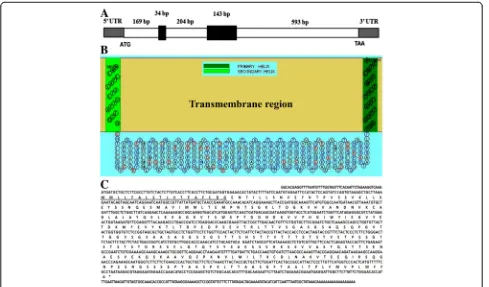
the same sequence, which showed no match with other genes by Blast. The full length of the BcMSA1 cDNA se-quence has a single open reading frame of 966 nucleotides encoding a polypeptide of 321 amino acids. The theoret-ical isoelectric point and molecular mass for the mature protein are 4.93 and 33.599 kDa, respectively. Genomic analyses indicated that BcMSA1 gene contains two introns and three extrons (Fig. 1a). The predicted transmembrane region of BcMSA1 was determined according to the com-puted algorithm (Fig. 1b). Kyte-Doolittle’s hydropathy ana-lysis indicated that the amino acid sequence of BcMSA1 has a hydrophilic core region with a good antigenic index, which suggested that the antigen could be a good candi-date for detection ofB. canisantibodies (data not shown). GPI anchor prediction indicated that three of highly prob-ably GPI sequences were involved in BcMSA1. One GPI sequence located from 297–321 of this amino acid sequence, and another two located from 304–321 and 305–321, respectively. Furthermore, another novel gene named BcSA1 was also identified from these 41 immuno-reactive recombinant phage plaques. The partial length of BcSA1 contained 1,255 nucleotides encoding a polypep-tide with 417 amino acid residues (Fig. 1c). The isoelectric
[image:5.595.56.545.399.686.2]point and molecular mass for the mature protein without signal peptide were 8.77 and 44.06 kDa, respectively. Un-like BcMSA1, the BcSA1 had not predicted GPI-anchored domains in its amino acid sequence.
Southern blot analysis
Southern blot analysis was performed to determine the copy number of BcMSA1 and BcSA1 gene, respectively. A probe derived from cDNA clone BcMSA1 was strongly hybridized to the B. canis DNA fragments, as shown in Fig. 2a. Treatment with the restriction enzymes (BamHI and SalI) that do not cut within the sequence consistent with the probe produced a single band (Fig. 2a, lanes 1, 2). However, two bands were ob-served after treatment with enzymes (PacI, KpnI and
NaeI) that cut a single position with the probe sequence (Fig. 2a, lanes 3–5). These results revealed that the gen-omic DNA of B. canis contains a single-copy gene encoding BcMSA1. Similar result was also observed for BcSA1 cDNA with the restriction enzymes that cut once within the gene (Fig. 2b, lanes 3, 4) and those that did not cut with the gene (Fig. 2b, lanes 1, 2). These results also indicate that the BcSA1 gene exists as a single copy inB. canisgenome.
Expression, purification and Western blot analysis of rBcMSA1 and rBcSA1
The amplified BcMSA1 and BcSA1 genes were cloned into the prokaryotic expression vector pGEX-4 T-1, re-spectively. Both BcMSA1 and BcSA1 were expressed in
E. coli as soluble GST-fusion proteins with molecular
mass of approximately 64 kDa (Fig. 3a, lane 1; Fig. 3c, lane 1). Western blot analysis showed that serum anti-bodies from dogs experimentally infected with B. canis could react with both of the recombinant fusion proteins (Fig. 3a, lane 3; Fig. 3c, lanes 4, 5). In contrast, no reac-tion of the serum was obtained with the GST protein (Fig. 3a, lane 4; Fig. 3c, lane 6).
Characterization of the native BcMSA1 and BcSA1 ofB.
canis
Mouse anti-rBcMSA1 and anti-rBcSA1 polyclonal sera were prepared and used to identify native BcMSA1 and BcSA1 inB. canisparasites by Western blot analysis and confocal laser scanning microscopy. As shown in Fig. 3b the 39 kDa band was detected with mouse anti-rBcMSA1 sera in B. canis-infected erythrocyte lysate (Fig. 3b, lane 1). There was no reaction for the normal erythrocyte lysate with the anti-rBcMSA1 serum (Fig. 3b, lane 2). On the other hand, the predicted 44 kDa band was detected by mouse anti-rBcSA1 sera in the plasma from B. canis-infected dog during acute stage of infec-tion (Fig. 3d, lane 1). Furthermore, the mouse anti-rBcMSA1 serum did not react with the plasma of B.
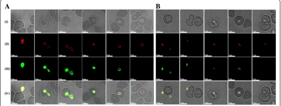
canis-infected dog and the mouse anti-rBcSA1 serum did not react with B. canis-infected erythrocyte lysate in Western blot analysis (data not shown). Native protein localization studies by IFAT revealed that BcMSA1 and BcSA1 possibly localizes on the cell surface (Fig. 4a) and within the cytoplasm of the parasite (Fig. 4b), respectively.
Evaluation of the serodiagnostic potential of rBcMSA1 and rBcSA1 by iELISA
The rBcMSA1- and rBcSA1-based ELISA experiments were conducted to investigate whether these recombin-ant proteins could be used for diagnosis of B. canis infection in dogs. The cut-off values of BcMSA1-ELISA and BcSA1-ELISA were calculated using 30 SPF canine sera; these were 0.149 and 0.110, respectively (Fig. 5c, d). As shown in Fig. 5c and d, all the serum samples from dogs infected with B. canis were positive for these two antigens, whereas, all 30 canine serum samples from non-endemic area (N) were negative. The serum samples from B. rossi-infected dogs (Br), B. vogeli-infected dogs (Bv), B. gibsoni-infected dogs (Bg) and N. caninum -in-fected dogs (Nc) were negative for the antibodies against BcMSA1 and BcSA1. The sensitivity of these assays were tested with sequential sera obtained from two dogs ex-perimentally infected with B. canis, both serologically negative prior to infection. For the BcMSA1-ELISA, the IgG titers increased in one dog on day 9 post-infection and in another dog on day 11 post-infection. Both dogs were serologically positive until 80 days post-infection (Fig. 5a, b). Towards day 80, the dogs already entered
the chronic phase of the disease, evidenced by
[image:6.595.57.291.486.654.2]recovering hematocrit value (data not shown) and sig-nificantly low levels of parasitemia. In contrast, specific antibodies against BcSA1 were detected on day 24 post-infection in one dog and on day 21 post-post-infection in an-other dog. Interestingly, the two dogs were serologically positive for BcSA1 throughout the sampling period.
Evaluation of the serodiagnostic potential of rBcMSA1 and rBcSA1 by ICT
The performance of rBcMSA1 and rBcSA1 as rapid and simple diagnostic antigens for detection of B. canis in-fection in dogs was evaluated using ICT. Serum sam-ples from experimentally infected dogs with closely related parasites (B. rossi, B. vogeli, B. gibsoni and N.
caninum) were used to determine the specificity of ICT based on rBcMSA1 and rBcSA1. Only the serum samples from B. canis-infected dogs were positive in the ICT, the other serum samples and SPF dog sera were negative (Fig. 6). As shown in Fig. 6, the specific antibodies to BcMSA1 and BcSA1 could be detected from day 9 and day 21, respectively. The antibodies against BcMSA1 and BcSA1 were detectable until 80 days post-infection, which was consistent with the results of the iELISA.
Discussion
Canine babesiosis frequently caused byBabesia canisis an emerging infectious disease in central Europe [14, 15]. A
Fig. 3SDS-PAGE and immunoblot analysis of recombinant and native BcMSA1 and BcSA1.aThe 10 % SDS-PAGE stained with Coomassie blue: recombinant BcMSA1 fused with GST (lane 1) and GST (lane 2). Western blot analysis of recombinant protein: recombinant BcMSA1 (lane 3) and GST (lane 4) probed withB. canis-infected dog serum;bWestern blot analysis of native BcMSA1.B. canis-infected erythrocyte lysate (lane 1) and normal canine erythrocyte lysate (lane 2) probed with anti-rBcMSA1 mouse serum;cThe 10 % SDS-PAGE stained with Coomassie blue: recombinant BcSA1 fused with GST (lane 1), recombinant BcSA1 fused without GST (lane 2) and GST (lane 3). Western blot analysis of recombinant protein: recombinant BcMSA1 (lane 4), recombinant BcSA1 fused without GST (lane 5) and GST (lane 6) probed withB. canis-infected dog serum; dWestern blot analysis of native BcMSA1. The plasma from a dog infected withB. canis(lane 1) and a normal dog (lane 2) probed with anti-rBcMSA1 mouse serum
[image:7.595.58.539.87.192.2] [image:7.595.56.540.502.685.2]number of new endemic regions of this disease have been reported in European countries and beyond [16–19]. Canine babesiosis also represents an important veterinary medical problem, since to date treatment and diagno-sis of this disease is still limited and the current vac-cine is derived from culture supernatants [20–22]. Therefore, it is critical to have sensitive and reliable tests for screening the B. canis positive population for prompt intervention. In this regard, we attempted to identify novel antigens to develop reliable, rapid and sensitive diagnostic methods for B. canis infections. A cDNA expression library was constructed from B.
canis merozoites mRNA and serologically screened with B. canis-infected serum, and novel BcMSA1 and BcSA1 were isolated and identified in this study.
Homology blast result indicated that BcMSA1 shared no significant homology with any of the apicomplexan parasites, which was important to confer a degree of specificity when used as a diagnostic probe. A 39 kDa native protein ofB. canis merozoites was recognized in Western blot analysis. The molecular mass of this native protein was higher than predicted. The difference in the molecular mass could be attributed to glycosylation
modification since two predicted N-glycosylation sites were identified in BcMSA1 motif by the NetNGlyc pre-dictor or the PROSITE pattern PS00001 (data not shown). Furthermore, BcMSA1 was identified as a sur-face protein ofB. canismerozoites. Generally, the mero-zoite surface proteins of Babesiaspp. are thought to be the main targets for the host immune responses. For the
Babesia spp., merozoite surface proteins are usually in-volved in the merozoite invasion of host erythrocytes and provide potential targets for vaccine and serodiagnosis [23–26]. Therefore, BcMSA1 might be a potential candi-date for diagnostic antigen and vaccines for diagnosis and prevention ofB. canisinfection, respectively. However, the merozoite surface antigens ofBabesiaspp. are genetically diverse among the different isolates [24, 25]. Thus, more sequences of BcMSA1 from different regions should be identified for further study.
Sequence analysis of BcSA1 revealed that this antigen contained a partial of signal peptide sequence, and BcSA1 shared 25 % amino acid identity with B. gibsoni secreted antigen 1 (BgSA1) in previous report [27]. Fur-thermore, a 44 kDa native protein was detected by Western blot, and this was consistent with the molecular
[image:8.595.57.539.87.367.2]mass of BcSA1 without signal peptide. Therefore, the results indicated that BcSA1 may represent a secreted protein ofB. canis. Secreted proteins have been proved as suitable sources of antigens for the detection of parasites infection in previous studies [27–31]. Therefore, identifi-cation of BcSA1 will be very useful in the development of effective diagnostic methods for the detection of circulat-ing antigens and antibodies durcirculat-ing B. canis infection. In addition, another unexpected 69 kDa band was observed in the 10-time concentratedB. canis-infected dog plasma from both infected and non-infected dogs in the Western blot analysis, which might be dog IgG cross-reacted with the second antibody to give an unspecific band.
To determine the diagnostic potential of the rBcMSA1 and rBcSA1 in the serological assays, the recombinant proteins were used as antigens in iELISAs. The results of specificity analysis indicated that both BcMSA1 and BcSA1 were specific for B. canis infection. This makes the two antigens become suitable candidates for diagno-sis of B. canis infection by iELISA. On the other hand, BcMSA1-ELISA and BcSA1-ELISA could detect the spe-cific antibody as early as 9 and 22 days post-infection, respectively. Moreover, the antigen-antibody interactions were still detectable until the chronic stage of infection with both antigens. These results indicated that both
rBcMSA1 and rBcSA1 can detect chronicB. canis infec-tion in dogs and the BcMSA1 can detectB. canisduring early infection. The ability of BcMSA1 and BcSA1 to induce strong humoral immunity in B. canis-infected dogs necessitates further evaluation of the recombinant antigens as vaccine candidates againstB. canisinfection.
Immunochromatographic test (ICT) is a one-step rapid diagnostic method, which is suitable for various clinical environment and can be completed in a short time (10–15 min) [32, 33]. Therefore, in this study, we established ICT methods for detection of B. canis infection. The ICT based on rBcMSA1 or rBcSA1 could detect the specific antibodies from dogs withB. canis in-fection. These results suggest that rBcMSA1 and rBcSA1 could be candidate antigens for the development of ICT to detectB. canisinfection.
Conclusion
In this study, aB. caniscDNA expression library was con-structed and immunoscreened usingB. canis-infected dog sera. This study was the first to identify and characterize two novel antigens of B. canis encoding a merozoite surface antigen (BcMSA1) and a secreted protein (BcSA1) as potential serodiagnostic antigens. Our results demon-strated that both, rBcMSA1 and rBcSA1, were highly
[image:9.595.57.538.89.324.2]sensitive and specific for serodiagnosis of B. canis infec-tion. Taken together, rBcMSA1 and rBcSA1 could be the first promising serodiagnostic antigens based on iELISA and ICT for detectingB. canisinfection in dogs.
Competing interests
This study was partly funded by a grant from Megacor Diagnostik GmbH (Austria) and a patent was registered (European patent EP 2626365 A1). ML and AK are employees of Megacor Diagnostik GmbH. While we collaborate with the employees of the above company, they did not influence the design and execution of this research in any way to alter the integrity of the results.
Authors’contributions
Conceived and designed the experiments: MZ SC ML AK YN HS II XX. Performed the experiments: MZ SC YL ML GW PFAM CJ. Analyzed the data: MZ SC AI MAT. Contributed reagents/materials/analysis tools: MZ SC ML AK YN HS II XX AI MAT. Wrote the manuscript: MZ SC PV YN HS II XX. All authors read and approved the final version of the manuscript.
Acknowledgements
This work was supported by Megacor Diagnostik GmbH and a Grant-in-Aid for Scientific Research from the Ministry of Education, Culture, Sports, Science, and Technology, Japan.
Author details
1
National Research Center for Protozoan Diseases, Obihiro University of Agriculture and Veterinary Medicine, Obihiro, Hokkaido 080-8555, Japan.
2
Harbin Veterinary Research Institute, CAAS-Michigan State University Joint Laboratory of Innate Immunity, State Key Laboratory of Veterinary Biotechnology, Chinese Academy of Agricultural Sciences, Maduan Street 427, Nangang District, Harbin 150001, PR China.3Megacor Diagnostik GmbH,
Hoerbranz, Vorarlberg A-6912, Austria.
Received: 8 September 2015 Accepted: 18 April 2016
References
1. Ano H, Makimura S, Harasawa R. Detection ofBabesiaspecies from infected dog blood by polymerase chain reaction. J Vet Med Sci. 2001;63(1):111–3. 2. Cacciò SM, Antunovic B, Moretti A, Mangili V, Marinculic A, Baric RR,
Slemenda SB, Pieniazek NJ. Molecular characterisation ofBabesia canis canis andBabesia canis vogelifrom naturally infected European dogs. Vet Parasitol. 2002;106(4):285–92.
3. Carret C, Walas F, Carcy B, Grande N, Précigout E, Moubri K, Schetters TP, Gorenflot A.Babesia canis canis, Babesia canis vogeli, Babesia canis rossi: differentiation of the three subspecies by a restriction fragment length polymorphism analysis on amplified small subunit ribosomal RNA genes. J Eukaryot Microbiol. 1999;46(3):298–303.
4. Kubelová M, Sedlák K, Panev A, Siroký P. Conflicting results of serological, PCR and microscopic methods clarify the various risk levels of canine babesiosis in Slovakia: A complex approach toBabesia canisdiagnostics. Vet Parasitol. 2013;191(3–4):353–7.
5. Schetters TP, Moubri K, Kleuskens J, Scholtes NC, Gorenflot A. Different Babesia canisisolates, different diseases. Parasitology. 1997;115(Pt 5):485–93. 6. Weiland G, Reiter I. Methods for serological response toBabesia. In: Ristic, M.
(Ed.), Babesiosis of Domestic Animals and Man. CRC Press, Boca Raton, Florida, pp. 143–158
7. Yamane I, Thomford JW, Gardner IA, Dubey JP, Levy M, Conrad PA. Evaluation of the indirect fluorescent antibody test for diagnosis ofBabesia gibsoniinfections in dogs. Am J Vet Res. 1993;54(10):1579–84.
8. Birkenheuer AJ, Levy MG, Breitschwerdt EB. Development and evaluation of a seminested PCR for detection and differentiation ofBabesia gibsoni(Asian genotype) andB. canisDNA in canine blood samples. J Clin Microbiol. 2003; 41(9):4172–7.
9. Tebele N, Skilton RA, Katende J, Wells CW, Nene V, McElwain T, Morzaria SP, Musoke AJ. Cloning, characterization, and expression of a 200-kilodalton diagnostic antigen ofBabesia bigemina. J Clin Microbiol. 2000;38(6):2240–7.
10. Böse R, Jorgensen WK, Dalgliesh RJ, Friedhoff KT, de Vos AJ. Current state and future trends in the diagnosis of babesiosis. Vet Parasitol. 1995;57(1–3):61–74.
11. Cao S, Luo Y, Aboge GO, Terkawi MA, Masatani T, Suzuki H, Igarashi I, Nishikawa Y, Xuan X. Identification and characterization of an interspersed repeat antigen ofBabesia microti(BmIRA). Exp Parasitol. 2013;133(3):346–52. 12. Duarte SC, Linhares GF, Romanowsky TN, da Silveira Neto OJ, Borges LM.
Assessment of primers designed for the subspecies-specific discrimination amongBabesia canis canis,Babesia canis vogeliandBabesia canis rossiby PCR assay. Vet Parasitol. 2008;152(1–2):16–20.
13. Chomczynski P, Sacci N. Single step method of RNA isolation by acid guanidium thiocyanate-phenol-chloroform extraction. Anal Biochem. 1987;162(1):156–9. 14. Navarro C, Reymond N, Fourie J, Hellmann K, Bonneau S. Prevention of Babesia canisin dogs: efficacy of a fixed combination of permethrin and fipronil (Effitix®) using an experimental transmission blocking model with infected Dermacentor reticulatus ticks. Parasit Vectors. 2015;8:32. 15. Imre M, Farkas R, Ilie M, Imre K, Hotea I, Morariu S, Morar D, DărăbuşG.
Seroprevalence ofBabesia canisinfection in clinically healthy dogs from western Romania. J Parasitol. 2013;99(1):161–3.
16. Pennisi MG, Caprì A, Solano-Gallego L, Lombardo G, Torina A, Masucci M. Prevalence of antibodies againstRickettsia conorii,Babesia canis,Ehrlichia canis, andAnaplasma phagocytophilumantigens in dogs from the Stretto di Messina area (Italy). Ticks Tick Borne Dis. 2012;3(5–6):315–8.
17. Halos L, Lebert I, Abrial D, Danlois F, Garzik K, Rodes D, Schillmeier M, Ducrot C, Guillot J. Questionnaire-based survey on the distribution and incidence of canine babesiosis in countries of Western Europe. Parasite. 2014;21:13. 18. Schaarschmidt D, Gilli U, Gottstein B, Marreros N, Kuhnert P, Daeppen JA,
Rosenberg G, Hirt D, Frey CF. QuestingDermacentor reticulatusharbouring Babesia canisDNA associated with outbreaks of canine babesiosis in the Swiss Midlands. Ticks Tick Borne Dis. 2013;4(4):334–40.
19. Criado-Fornelio A, Martinez-Marcos A, Buling-Saraña A, Barba-Carretero JC. Molecular studies onBabesia,TheileriaandHepatozoonin southern Europe. Part I Epizootiological aspects. Vet Parasitol. 2003;113(3–4):189–201. 20. Schetters TP, Kleuskens J, Carcy B, Gorenflot A, Vermeulen A. Vaccination
against largeBabesiaspecies from dogs. Parassitologia. 2007;49 Suppl 1:13–7. 21. Schetters TP, Kleuskens JA, Scholtes NC, Gorenflot A, Moubri K, Vermeulen AN. Vaccination of dogs against heterologousBabesia canisinfection using antigens from culture supernatants. Vet Parasitol. 2001;100(1–2):75–86. 22. Schetters TP, Kleuskens JA, Scholtes NC, van de Crommert J, Krijnen E, Moubri K, Gorenflot A, Vermeulen AN. Onset and duration of immunity againstBabesia canisinfection in dogs vaccinated with antigens from culture supernatants. Vet Parasitol. 2006;138(1–2):140–6.
23. Goo YK, Aboge GO, Terkawi MA, Jia H, Yamagishi J, Sunaga F, Namikawa K, Cha SY, Jang HK, Kim S, Nishikawa Y, Xuan X. Four promising antigens, BgP32, BgP45, BgP47, and BgP50, for serodiagnosis ofBabesia gibsoni infection were classified asB. gibsonimerozoite surface protein family. Parasitol Int. 2012;61(2):364–8.
24. LeRoith T, Berens SJ, Brayton KA, Hines SA, Brown WC, Norimine J, McElwain TF. TheBabesia bovismerozoite surface antigen 1 hypervariable region induces surface-reactive antibodies that block merozoite invasion. Infect Immun. 2006;74(6):3663–7.
25. Berens SJ, Brayton KA, Molloy JB, Bock RE, Lew AE, McElwain TF. Merozoite surface antigen 2 proteins ofBabesia bovisvaccine breakthrough isolates contain a unique hypervariable region composed of degenerate repeats. Infect Immun. 2005;73(11):7180–9.
26. Xuan X, Larsen A, Ikadai K, Tanaka T, Igarashi I, Nagasawa H. Expression of Babesia equimerozoite antigen 1 in insect cells by recombinant baculovirus and evaluation of its diagnostic potential in an enzyme-linked
Immunosorbent assay. J Clin Microbiol. 2001;39(2):705–9.
27. Jia H, Zhou J, Ikadai H, Matsuu A, Suzuki H, Igarashi I, Fujisaki K, Xuan X. Identification of a novel gene encoding a secreted antigen 1 ofBabesia gibsoniand evaluation of its use in serodiagnosis. Am J Trop Med Hyg. 2006;75(5):843–50.
28. Luo Y, Terkawi MA, Jia H, Aboge GO, Goo YK, Cao S, et al. A double antibody sandwich enzyme-linked immunosorbent assay for detection of secreted antigen 1 ofBabesia microtiusing hamster model. Exp Parasitol. 2012;130(2):178–82.
29. Valentin A, Precigout E, L’Hostis M, Carcy B, Gorenflot A, Schrevel J. Cellular and humoral immune responses induced in cattle by vaccination with Babesia divergensculture-derived exoantigens correlate with protection. Infect Immun. 1993;61(2):734–41.
31. Schetters TP, Strydom T, Crafford D, Kleuskens JA, van de Crommert J, Vermeulen AN. Immunity againstBabesia rossiinfection in dogs vaccinated with antigens from culture supernatants. Vet Parasitol. 2007;144(1–2):10–9. 32. Jia H, Liao M, Lee E, Nishikawa Y, Inokuma H, Ikadai H, Matsuu A, Igarashi I,
Xuan X. Development of an immunochromatographic test with recombinant BgSA1 for the diagnosis ofBabesia gibsoniinfection in dogs. Parasitol Res. 2007;100(6):1381–4.
33. Hujakka H, Koistinen V, Eerikainen P, Kuronen I, Mononen I, Parviainen M, Lundkvist A, Vaheri A, Närvänen A, Vapalahti O. New
immunochromatographic rapid test for diagnosis of acutePuumalavirus infection. J Clin Microbiol. 2001;39(6):2146–50.
• We accept pre-submission inquiries
• Our selector tool helps you to find the most relevant journal
• We provide round the clock customer support
• Convenient online submission
• Thorough peer review
• Inclusion in PubMed and all major indexing services • Maximum visibility for your research
Submit your manuscript at www.biomedcentral.com/submit