CORRELATION OF PLASMA OSTEOPONTIN WITH
RADIOLOGICAL GRADING IN PATIENTS WITH
OSTEOARTHRITIS IN THE KNEE JOINT
Dissertation submitted for
M.D. BIOCHEMISTRY BRANCH – XIII
DEGREE EXAMINATION
THE TAMILNADU DR. M.G.R. MEDICAL
UNIVERSITY
CHENNAI – 600 032
TAMIL NADU
BONAFIDE CERTIFICATE
This to certify that this dissertation work entitled “CORRELATION
OF PLASMA OSTEOPONTIN WITH RADIOLOGICAL GRADING IN
PATIENTS WITH OSTEOARTHRITIS IN THE KNEE JOINT” is the
original bonafide work done by Dr.P.Nirmaladevi, Post Graduate Student,
Institute of Biochemistry, Madras Medical College, Chennai under our direct
supervision and guidance.
Prof. Dr.R.Chitraa, MD., (Guide) Prof. Dr.K.Ramadevi, MD.,
Professor, Director and Professor,
Institute of Biochemistry, Institute of Biochemistry,
Madras Medical College, Madras Medical College,
Chennai- 600 003. Chennai- 600 003.
Dean
Madras Medical College and
Rajiv Gandhi Government General Hospital,
DECLARATION
I, Dr.P.Nirmaladevi, solemnly declare that the dissertation titled
“CORRELATION OF PLASMA OSTEOPONTIN WITH
RADIOLOGICAL GRADING IN PATIENTS WITH
OSTEOARTHRITIS IN THE KNEE JOINT” is the bonafide work done by
me at Institute of Biochemistry, Madras Medical College under the expert
guidance and supervision of Prof. Dr.R.Chitraa, M.D., Professor, Institute of
Biochemistry, Madras Medical College. The dissertation is submitted to the
Tamil Nadu Dr. M.G.R Medical University towards partial fulfillment of
requirement for the award of M.D., Degree (Branch XIII) in Biochemistry.
Place: Chennai
SPECIAL ACKNOWLEDGEMENT
The author gratefully acknowledges and sincerely thanks Professor
Dr.R.Vimala, M.D., Dean, Madras Medical College and Rajiv Gandhi
Government General Hospital, Chennai, for granting her permission to utilize
ACKNOWLEDGEMENT
The author expresses her warmest respects and profound gratitude to
Dr.K.Ramadevi, M.D., Director and Professor, Institute of Biochemistry,
Madras Medical College, Chennai, for her academic enthusiasm and for
facilitating my research work in the institute.
The author express her heartfelt gratitude to her guide and supervisor
Dr.R.Chitraa, M.D., Professor, Institute of Biochemistry, Madras Medical
College, Chennai, for her intellectual and valuable guidance, unfailing support,
encouragement and continuous inspiration throughout the period of her study.
The inspiring interaction I had with her during the course of my study helped
me to enrich this work. The author feels greatly privileged to work under her
able guidance.
The author in particular, is extremely thankful to Professor Dr.N.Deen
M.Ismail, MS(Orth)., D.Orth., Director I/C,& Professor, Institute of
Orthopaedics & Traumatology, Madras Medical College and Rajiv Gandhi
Government General Hospital, Chennai, for granting permission to obtain
blood samples from the patients.
The author expresses her thanks to the Professors Dr. I.Periyandavar
M.D., Dr.V.Amudhavalli M.D., and Dr.K.Pramila M.D., Associate Professors
Madras Medical College, for their guidance, encouragement, insightful
comments and suggestions.
The author expresses her warm respects and sincere thanks to her
co-guide, Dr.C.Shanmugapriya M.D Assistant Professor, Institute of biochemistry,
Madras Medical College for her guidance and support. The author expresses
her warm respects and sincere thanks to Assistant Professors,
Dr.V.G.Karpagavalli, Dr.C.Mythili, Dr.V.Ananthan, Dr.S.Siva, Dr.
B.SudhaPresanna, Dr.Menaka Shanthi, Institute of biochemistry, Madras
Medical College, for their valuable suggestions regarding the practical issues of
research which is something beyond the textbooks. I appreciate their interest
and effort for improvising my work.
The author expresses warm respects to the members of the Institutional
Ethical committee for approving the study. The author expresses her special
thanks to all laboratory staffs and DMLT students, Institute of biochemistry,
for their timely co-operation and assistance during the study.
The author expresses her special thanks to her co-PGs Dr.S.Michael
Rajam Geetha, Dr.S.Anandhi and Dr.M. Divya for their constructive criticism
and unconditional support. She also thanks all her colleagues in the institute,
for their constant encouragement throughout the study period.
The author is greatful to the Statisticians, Mr. Ravanan D.Ramanujam,
The author is indebted to the patients from whom blood samples were
collected for conducting the study. The author expresses her special thanks to
all the Assistant Professors, Institute of Orthopaedics and all MS Orthopaedics
post graduates especially Dr.Sivaraj and Dr. Somasundaram for their timely
help and co-operation during sample collection.
I am thankful to my husband, Dr. S.Rajkumar, my daughter R.Yazhini
and my in-Laws for their support and co-operation. The author also thanks her
parents and sister and brother for their help.
Above all, I thank the Almighty for providing me this opportunity,
CONTENTS
SI.
NO TITLE PAGE No.
1 INTRODUCTION 1
2 REVIEW OF LITERATURE 3
3 AIMS & OBJECTIVES 52
4 MATERIALS & METHODS 53
5 STATISTICAL ANALYSIS 69
6 RESULTS 70
7 DISCUSSION 80
8 CONCLUSION 85
9 LIMITATION OF THE STUDY 86
10 SCOPE FOR FURTHER STUDIES 87
11 BIBLIOGRAPHY
ABBREVIATIONS
1. OA – Osteoarthritis
2. MMP-13 – Matrix Metalloproteinase-13
3. IL – Interleukin
4. TNF – Tumour Necrosis Factor
5. BMI – Body Mass Index
6. COMP – Cartilage Oligomeric Matrix Protein
7. VDR – Vitamin D Receptor
8. CD – Cluster of Differentiation
9. DDR2 – Discoidin –domain containing receptor
10. MEK/ERK – Mitogen activated protein kinases/ Extracellular
signal- regulated kinases
11. ATP – Adenosine triphosphate
12. PP – Pyrophosphate
13. CPPD – Calcium pyrophosphate dehydrate
14. TIMP – Tissue inhibitors of metalloproteinases
15. TGF – Transforming growth factor
16. BMP – Bone Morphogenetic Proteins
17. TPA – Tissue plasminogen activator
18. Hif2alpha – Hypoxia- inducible factor 2alpha
19. ADAMTS – A disintegrin and metalloproteinase with
thrombospondin motif
21. IHH – Indian hedgehog signalling pathway
22. miR – micro- RNA
23. Rho/ROCK – Rho associated protein kinase
24. iNOS – inducible isoform of nitric oxide synthase
25. PGE2 – Prostaglandin E2
26. ecNOS – endothelial cell nitric oxide synthase
27. KL – Kellgren- Lawrence grading system
28. CTX-II – Carboxy- Terminal telepeptides of Type-II collagen
29. CRP – C Reactive Protein
30. OPN – Osteopontin
31. SIBLING – Small Integrin-Binding Ligand N-linked
Glycoprotein
32. VDRE – Vitamin-D response element
33. VSMC – Vascular smooth muscle cell
34. NF-κB – Nuclear factor kappa-light-chain-enhancer of
activated B cells
35. HA – Hyaluronic acid
36. ECM – Extracellular Matrix
37. RHAMM – Receptor for HA mediated motility
CORRELATION OF PLASMA OSTEOPONTIN WITH RADIOLOGICAL
GRADING IN PATIENTS WITH OSTEOARTHRITIS IN THE KNEE JOINT
ABSTRACT:
Objectives and aims of the study:
To correlate plasma osteopontin level with radiological grade in patients with
knee osteoarthritis (OA) and to correlate plasma osteopontin level with serum
hyaluronic acid in patients with knee OA, thereby to assess, if osteopontin contributes
to the pathogenesis of the degenerative process of OA by stimulating MMP13 and
increase hyaluronic acid levels in serum.
Materials and methods:
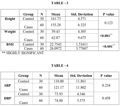
60 patients with varying grades of radiological evidence of OA in the knee
joint and 30 healthy subjects as controls were enrolled in the study. Anteroposterior
knee radiographs, in standing position were taken to determine the disease severity of
the affected knee joint. The radiographic grading of OA in the knee joint was
performed by using the Kellgren–Lawrence grading (K/L). Osteopontin levels in the
plasma and hyaluronic acid levels in the serum were measured using enzyme-linked
immunosorbent assay and compared.
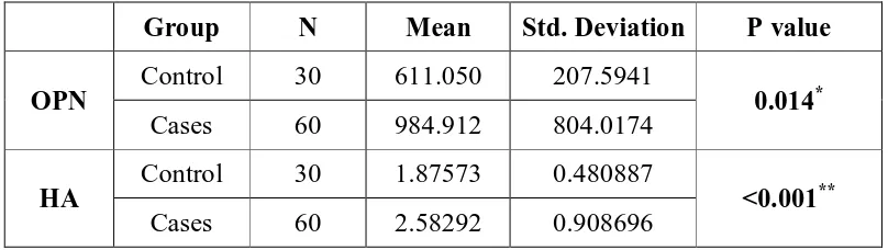
Results:
The mean plasma osteopontin concentration of the knee OA patients was
significantly higher compared with that of healthy controls (984.91+/-804 pg/mL vs
611.05+/-207.59 pg/mL, p=0.014). The plasma osteopontin levels significantly
acid concentration of the knee OA patients was significantly higher compared with
that of healthy controls (2.58+/−0.90ng/mL vs 1.87+/−0.48 ng/mL). The serum
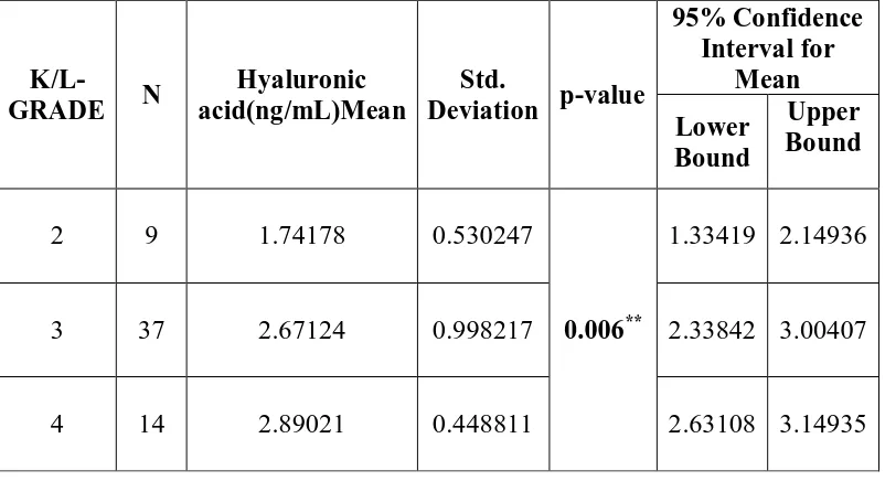
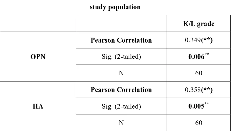
hyaluronic acid level significantly correlated with K/L grades (r=0.358, p=0.005). The
plasma osteopontin and serum hyaluronic acid levels were compared in relation to
radiological K/L grades 2, 3, 4 among the cases and was found to have statistically
significant higher concentrations as the grade increased with p= 0.022 for osteopontin
and p=0.006 for hyaluronic acid.
Conclusion:
Osteopontin in plasma and Hyaluronic acid in serum are related to progressive
joint damage in knee OA. Hence in the present study statistically significant increase
in concentration of both osteopontin & hyaluronic acid with respect to radiological
grade and a positive correlation between osteopontin and K/L grade & hyaluronic acid
and K/L grade implies that osteopontin has a significant role in activating MMP-13
causing degradation of articular cartilage and release of HA into the circulation in OA.
Hence osteopontin and hyaluronic acid can be used in combination as biomarkers to
assess the severity of the disease.
Keywords:
1
INTRODUCTION
Osteoarthritis(OA) is a “universal disorder” affecting both sexes and all
races and is the commonest of all joint diseases. OA is a “strongly age –related
disorder defined by focal lesions of the articular cartilage, combined with a
hypertrophic reaction in the subchondral bone and new bone formation at the
joint margins with chronic nonspecific synovial inflammation”. OA is a
chronic degenerative joint disease characterised by progressive destruction of
articular cartilage with varying degrees of severity within a given joint.
To identify patients with a high risk for destructive OA and to monitor
drug efficacy, most sensitive techniques other than “plain x-rays” are required.
Hence for investigation and monitoring of patients with OA, specific and
sensitive biochemical markers which reflect abnormalities in the turnover of
bone, cartilage and synovial tissues may be useful.
“Osteopontin” is one of the major noncollageneous bone matrix
proteins produced by various cells like activated T cells, macrophages,
osteoblasts and chondrocytes. Osteopontin may be involved in the pathogenesis
of osteoarthritis, at the molecular level1, contributing to progressive
degeneration of articular cartilage.
Osteopontin stimulates MATRIX METALLOPROTEINASE 13
(MMP-13), which causes bone destruction by degrading the major component
2 The present study is undertaken:
1. to unravel the role of plasma osteopontin levels as a biomarker for OA
and to correlate the plasma concentration of osteopontin in patients with
primary knee OA with the radiological grading.
2. to understand the contribution of osteopontin in accelarating the
pathogenesis of OA, anticipated to enhance serum hyaluronic acid
levels.
3
REVIEW OF LITERATURE
INTRODUCTIONOA is a “chronic degenerative disease of the joints characterised by
progressive softening and disintegration of articular cartilage, hypertrophy of
bone at the margins (osteophytes), cyst formation, subchondral sclerosis and a
range of morphological and biochemical alterations of the capsule and synovial
membrane6 of the joint”. Usually, it is distributed asymmetrically and
localised to only one part of any joint and is often associated with “abnormal
loading rather than frictional wear and tear”.
OA is neither primarily an inflammatory disorder nor purely a
degenerative disorder. OA is a dynamic phenomenon which shows features of
both destruction and repair. Cartilage softening and disintegration lead to
hyperactive new bone formation and remodelling. The secondary factors which
influence the progress of this disorder include appearance of calcium
containing crystals in the joint, ischemic changes leading to osteonecrosis in
the subchondral bone in elderly people causing joint instability.
OA is a disease process that affects the entire joint that includes
cartilage, synovial membrane, subchondral bone, ligaments, and peri -articular
muscles. OA arises from both systemic and local factors (biochemical mediated
events) producing a condition with definable morphological and clinical
4
OA may be classified into primary of unknown etiology and secondary
with identifiable cause. Common causes of secondary OA include –
1. Metabolic (calcium crystal deposition, acromegaly)
2. Traumatic (joint injury)
3. Anatomic (congenital hip dislocation)
4. Inflammatory disorder (septic arthritis, ankylosing spondylitis).
Usually, secondary OA arises due to inflammatory cause with release of
degenerative enzymes from synovium attributing to mechanical attrition of
biomechanically altered extracellular matrix.
ETIOLOGY
The factors affecting degree of risk of developing OA are age, gender,
obesity, joint location, genetic predisposition.
1. AGE – the most common risk factor which strongly correlate with OA.
Chondrocytes undergo age-related decrease in mitotic and synthetic
activity. They show a decreased response to anabolic growth factors and
synthesize less uniform large aggregating proteoglycans with fewer link
proteins. Age is an independent risk factor as it predisposes articular
chondrocytes to apoptosis, because in aged cartilage there is higher level of
expression of pro -apoptotic genes (Fas, Fas L, caspase - 8, p 53)3.
2. GENDER – Women develop OA twice more commonly than men after the
5
receptors are found in articular chondrocytes of humans8 and human growth
plate chondrocytes. Wluka & colleagues9 reported that women using long
term ESTROGEN REPLACEMENT THERAPY (ERT) presented with
more knee cartilage than controls.
3. OBESITY – Another important risk factor for OA is obesity. Increased risk
of OA knee is associated with increased body mass index (BMI) in both
men and women. In women due to more total body fat, they tend to develop
OA more commonly than men. This is because adipose tissue is a
metabolically active contributor to inflammatory cascades leading to
increased synthesis of pro-inflammatory cytokines- IL-1&6, TNF, leptin ,
adiponectin , resistin4,5. Hence by reducing BMI, symptoms and
radiological progression can be reduced.
4. JOINT LOCATION – OA most commonly occur in weight-bearing joints
and joint specific age related viability in articular cartilage explains why
OA is more common in knee and hip, than ankle, as age advances.
5. GENETIC PREDISPOSITION – Genetic contribution to the
pathogenesis of OA is difficult to analyse because of the prevalence in the
general population and extensive clinical heterogeneity. Increased risk of
OA has been associated with multiple gene variations6 caused by mutations
in genes coding for the various types of collagen. The types of collagen
expressed in cartilage includes types II, IV, V, VI and CARTILAGE
6
VITAMIN D RECEPTOR (VDR) plays a vital role in controlling bone
mineral density and appears to be associated with a twofold risk of knee
OA7. Loughlin & colleagues provided evidence that the IL-1 gene cluster
harbours susceptibility to knee OA.
Studies of differential gene expression help to elucidate the pathogenesis
for arriving with newer therapies and helps in
1. Identification of unique biomarkers for diagnosis and management of
OA.
2. Identification of candidate susceptibility genotypes like polymorphic
variations of cytokines or growth factors that may predispose to disease
progression6.
PATHOGENESIS
CHANGES IN OSTEOARTHRITIS
Morphologic changes
In early OA articular cartilage becomes irregular and roughened, in the
synovial tissues, superficial clefts become apparent. As disease progresses
clefts deepen and there is an increase in the surface irregularities and articular
cartilage ulcerates exposing the underlying bone. On further progression of
disease, the joint articulates on exposed bone causing thickening of bone and
7
Early Reparative, Proliferative & Hypertrophic changes
In healthy cartilage, chondrocytes are quiescent but proliferate in
clusters in the early stages of OA, associated with, expression of high levels of
matrix proteins like type II collagen, aggrecan, stem cell markers and markers
of hypertrophic differentiation 10. Chondrocyte clusters are thought to
contribute to the pathogenesis of OA and its progression through release of
matrix degrading enzymes, inflammatory cytokines and growth factors which
further affect the surrounding chondrocytes and joint tissues11.
Osteophyte formation
Osteophytes are newly formed fibrocartilage and bone. They are
commonly formed at the peripheral margins of joints between the cartilage and
periosteum interface. Osteophytes contribute to the stability of joints12, 13. As
OA progresses the abundant osteophytes formed can limit movement and
become painful.
Hypo-cellularity
In aging cartilage there is a reduction in cell number due to reduced
synthesis, which is an important contributing factor to the initiation and
progression of OA. Apoptosis in chondrocytes can be triggered by factors
which are involved in initiation and progression of arthritis viz., mechanical
damage or injury, changes in cell matrix interactions, oxidative stress due to
nitric oxide or reactive oxygen species, impaired mitochondrial function, signal
8
accelerated to complete apoptosis. To prevent secondary OA following injury,
inhibition of apoptosis by interfering with caspase activation following injury,
as a chondro protective intervention is being explored. Hence autophagy can be
protective in cartilage and its reduction in OA corresponds with an increase in
the release of apoptotic markers14.
ALTERATIONS IN CARTILAGE MATRIX METABOLISM
In early OA, there is a significant increase in the water content of
articular cartilage which causes the tissues to swell and cause biomechanical
alterations in these tissues. This suggests that there has been weakening of
collagen network. Type II collagen fibres have smaller diameter than that in
normal cartilage and the normal tight weave in the mid zone is slackened and
distorted15 in OA.
In later stages of OA within the extracellular matrix, type I collagen
concentration increases and the proteoglycan concentration falls to less than or
equal to 50 per cent, with less aggregation and shorter glycosaminoglycan side
chains16, 17. The ratio of chondroitin-4-sulfate to chondroitin-6-sulfate increases
and that of keratan sulphate concentration decreases reflecting the synthesis by
chondrocytes of a proteoglycan profile which is typical of immature cartilage.
As the disease progresses the proteoglycan concentration in the cartilage
diminishes progressively18.
The first step in cartilage degradation is the decrease in density of
9
density opens up the cartilage porosity, increases the permeability to
collagenases/ proteases, exposing collagen fibrils. This initiates a vicious cycle
of positive feedback loops which further promote cartilage degradation. For
example, epitopes on collagen become accessible to the cell surface DDR2
receptor which increases MMP13 production by the activation of the
Ras/Raf/MEK/ERK and p38 signal cascades20. The partially digested matrix
has a cytokine like activity that enhances inflammatory response and promotes
matrix degradation. The destruction of collagenous cartilage is thought to be
irreversible.
Calcium crystals are commonly found in the cartilage of the elderly and
crystal arthropathy often coexists with OA. PYROPHOSPHATE (PP) is
produced from adenosine triphosphate (ATP) by the exoenzyme nucleoside
pyrophosphohydrolase21. Synovial fluid of OA patients showed high levels of
PP which directly correlate with the severity of joint damage22. Normal adult
cartilage secretes little of PP but the young or proliferating chondrocytes are
the major source of PP. This increased PP secretion in OA cartilage might
indicate the increased chondrocyte metabolic activity towards the matrix
repair22. The CALCIUM PYROPHOSPHATE DEHYDRATE (CPPD) may
alter the biomechanical properties of the cartilage extracellular matrix which
leads to cartilage breakdown. Hemochromatosis (hemosiderin), Wilson’s
disease (copper), gouty arthritis (monosodium urate crystals), the CPPD crystal
deposition disease are examples of condition which may alter the cartilage
10
thereby increasing the stiffness of the tissue and precipitating the development
of OA.
Metabolic changes
Early OA is characterised by increased synthesis of proteoglycans,
hyaluronate, collagen, noncollagenous proteins and cell replication18, 22. The
activation of chondrocytes is thought to be an attempt to repair the cartilage
matrix which yields a matrix of inferior quality which is more susceptible to
degradation23. Both anabolic and catabolic processes increase as cells attempt
to repair and maintain tissue integrity and it is this imbalance between synthesis
and degradation which is thought to be important in the pathogenesis of OA22.
In later stages of OA, there is a decrease in cell number and in the
synthesis of matrix with lowered quality along with the inability to blend the
hyaluronic acid 16 , 24. In addition there is an activation of matrix degrading
enzymes with an overall decrease in concentration of enzyme inhibitors such
as, TISSUE INHIBITORS of METALLOPROTEINASES (TIMP), in later
stages of OA. The complex interaction between matrix synthesis and
degradation explains why OA is slowly progressive, static by morphologic
criteria, finally resulting in the overall degradation of cartilage matrix.
CONTRIBUTING FACTORS FOR OSTEOARTHRITIS
A. Anabolic factors and cartilage repair
i. TRANSFORMING GROWTH FACTOR – BETA (TGF –β) – It is
11
cartilage homeostasis by enhancing stem cell chondrogenesis to increase
the pool of cells available for cartilage synthesis, thereby increase
matrix production in existing chondrocytes.
TGF-β increases synthesis of anti - catabolic factors such as TIMPs that
inhibit activation of latent proteinases in cartilage. TGF-β attenuates the
cellular response to inflammatory cytokines (IL-1, TGF- β, TNF) 25. In
aging cells TGF-β can have opposing effects by activating MMP-13 and
inducing terminal hypertrophic differentiation in chondrocytes26.
ii. BONE MORPHOGENETIC PROTEINS (BMP) – structurally
related to TGF-β, activate different set of receptors and intracellular
signalling molecules. It influences all stages of embryonic
chondrogenesis. Recent genetic evidence reports that, impaired BMP
signalling with the utmost progress for BMP-14, affects OA
susceptibility27, 28. There is a reduction in BMP- 7 in OA cartilage with
possible regulation occurring through both inhibitory microRNA29 and
promoter methylation30. Supplementation of BMP-7 has reduced
arthritis in experimental animals and has been proven safe in phase – I
clinical trial. BMP can also enhance terminal differentiation and
hypertrophy in chondrocytes, the processes that are hallmarks of OA
12
B. Catabolic factors and cartilage degradation
Cartilage remodelling and new matrix synthesis involves a degree of
proteolysis which occurs via induction of an array of proteases mainly matrix
metalloproteinases (MMPs). IL-1, TNF stimulate the synthesis and secretion of
proteases and MMPs in OA19 (Figure-1). IL -1 is synthesized by chondrocytes
as an autocrine activity and by mononuclear cells and synovial lining cells in
the inflamed joint. IL -1and TNF stimulate the latent forms of the enzymes viz.,
collagenase, stromelysin and gelatinase, including aggrecanase and TISSUE
PLASMINOGEN ACTIVATOR (TPA) 31. Plasminogen either enters the
matrix by diffusion from synovial fluid or synthesized by the chondrocytes.
TPA converts plasminogen to plasmin, a serine proteinase which activates
latent cartilage degrading enzymes. HYPOXIA- INDUCIBLE FACTOR 2
alpha (Hif2α) is gaining increasing attention as a downstream mediator of IL-1
and TNF induced cartilage degradation32, 33. In OA cartilage Hif2 α is a
transcription factor that is strongly up regulated which directly induces the
expression of many cartilage degrading enzymes like MMPs- 1, 3, 9, 12 and A
DISINTEGRIN AND METALLOPROTEINASE with
THROMBOSPONDIN motif (ADAMTS-4) and ADAMTS-5 indirectly. A
high level of Hif2α decreases the protective role of autophagy and increases the
extent of cell death and activates the RUNT- RELATED TRANSCRIPTION
FACTOR 2 (RUNX2) and INDIAN HEDGE HOG pathway which further
contribute to cartilage matrix degradation. The different classes of proteinases
13
Classes of Proteinases (Metalloproteinases, Serine and Cysteine proteases,
Aggrecanases)
In OA there is a marked increase in the synthesis and secretion of matrix
degrading enzymes by chondrocytes18,34. There are four classes of proteases
grouped by the catalytic mechanisms of peptide bond cleavage
1. Metalloproteinases
2. Cysteine proteinases
3. Serine proteinases
4. Aspartyl proteinases
Out of these the first three have clearly defined roles in the degradation
of cartilage during the progression of OA.
Metalloproteinases
Metalloproteinases have enzymatic site which requires zinc metal ion
for activity. Cartilage contains two families of metalloproteinases – ADAMTSs
and MMPs. Early cartilage degeneration in OA is due to metalloproteinase
enzymatic activity. Both families of metalloproteinases are up-regulated and
highly expressed in OA cartilage at sites of lesion. As the MMPs and
ADAMTSs play major role in the degradation of cartilage extracellular matrix
and these are now acting as candidate targets for disease modification.
The control of metalloproteinase activity is complex in OA with
regulation occurring at three different levels – synthesis and secretion,
14
stimulation with cytokine and growth factor signalling metalloproteinases
transcription is induced and its stability and translation are regulated by
microRNAs. For example, microRNA -27b regulates MMP-13 expression36.
The microRNA-140 levels are decreased in OA which reduces its repression of
ADAMTS-537. IL-1 and TGF-beta control these microRNAs, which are
involved in the pathogenesis of OA by affecting transcript stability and protein
translocation. Once translated into proteins all MMPs are expressed as inactive
zymogens which require further processing for complete proteolysis. Most
MMPs contain N-terminal pro-domain that blocks or inhibits the catalytic site.
The primary MMP activators are serine and cysteine dependent proteases
(plasminogen or pro-protein convertases and cathepsin B respectively) as well
as membrane-type MMPs38. Activated MMPs then can be inactivated
non-specifically by α2 macroglobulin and more specifically by the tissue inhibitor
of metalloproteinase.
Based on their substrate specificities MMPs have been divided into three
groups.
1. Collagenases – cleave all three chains of native triple helical
collagen.
2. Gelatinases – cleave denatured collagen
3. Stromelysins – have broader substrate specificities39.
There is considerable overlap in substrate between these classifications
MMP-15
13 (collagenase-3) all are capable of cleaving aggrecan core protein40. In
addition to collagen, MMPs can degrade other cartilage extracellular matrix.
MMPs can rapidly destroy cartilage completely if combined with plasmin
which has the capability of activating many MMPs.
Collagenases
Collagenases cleave typically, the triple helical collagen allowing
further degradation by other proteases. The first irreversible step in the
pathogenesis of OA is the degradation of collagen which significantly reduces
the mechanical properties of cartilage. The best studied MMPs capable of
cleaving native collagen are MMP-13 and MMP-1 of which, MMP-13 may be
the most important in OA because it preferentially degrades type II collagen41.
Expression of MMP-13 is greatly increased in OA42 and overall collagenase
activity markedly increases in human OA cartilage cultures, suggesting that it
is a major progressive factor in OA by degrading the cartilage matrix 43. The
resultant collagen fragments may be susceptible to further cleavage by other
enzymes like MMP-2 (gelatinaseA), MMP-3, MMP-9 (gelatinaseB) and
cathepsin B (a cysteine proteinase).
Aggrecanases
The aggrecanases belong to a family of extracellular proteases known as
ADAMTS44. The two major aggrecanases involved in cartilage degradation are
ADAMTS-4, predominantly associated with aggrecan degradation in human
16
aggrecan is highly resistant to proteases but a glutamate-alanine bond within
the extended region between G1 and G2 is susceptible to proteolysis to further
degradation. ADAMTS-4 and ADAMTS-5 activity is detected in joint capsule
and synovium and may be up-regulated in synovium through posttranslational
processing. In addition several MMPs are also capable of cleaving aggrecan in
vitro (1, 2, 3, 7, 8, 9, 13,
MMP-28 ). ADAMTS-7 and ADAMTS-12 both bind to and degrade COMP – a
prominent non-collagenous protein in cartilage and the latter is up-regulated in
OA cartilage48, 49.
Several lysosomal enzymes that cleave both hyaluronic acid and
chondroitin-6-sulfate have been implicated in OA progression .In the human
genome there are about 6 or 7 potential hyaluronidases of which
hyaluronidase-1, hyaluronidase-2, hyaluronidase-3 are likely to be active in
cartilage50. The decrease in chondroitin sulphate chain length in OA cartilage,
may be due to digestion by synovial fluid hyaluronidase which diffuse into the
matrix as the permeability increases. The concentration of hyaluronic acid in
OA cartilage is low in spite of the increased rate of synthesis of hyaluronic
acid. These degradative enzymes disrupt the proteoglycan aggregate. The early
change that occurs due to the MMP induced tissue degradation is the thinning
of collagen fibres, loosening of the tight collagen network and the consequent
17
Enzyme Inhibitors (Tissue Inhibitor of Metalloproteinases, Plasminogen
Activator Inhibitor – 1)
The balance between the active and the latent enzymes is controlled by
two enzyme inhibitors – tissue inhibitor of metalloproteinases (TIMP) and
plasminogen activator inhibitor -1 (PAI- 1)51. Under the regulation of TGF-β
the synthesis of these two enzymes are increased51. If these enzyme
concentrations decrease along with active enzyme concentration then the
matrix degradation increases. Genes that showed increased expression in OA
are MMP-2, MMP-9, MMP-13, MMP-16, MMP-28, ADAMTS-2, 12, 14, 16
and TIMP-3. Genes with decreased expression in OA are MMP-1, 3, 10 ,
ADAMTS-1, 5, 9, 15 and TIMP1 and 452. These explain the complexity of the
events that occur in the extracellular matrix regarding regulation of tissue
degrading enzymes.
ALTERATIONS IN MATRIX SYNTHESIS
The changes in the extracellular matrix that occur in OA comes
from the animal models. Initially there is an increase in water content in OA
cartilage due to loss of elasticity of collagen network causing the hydrophilic
proteoglycans to swell more than normally51. In early stage of OA the
proteoglycan concentration may increase and the cartilage becomes thicker
than normal and there is increased staining for proteoglycans51. At the same
time water content also increases and the newly synthesized proteoglycans
contain a higher proportion of chondroitin sulphate and lower proportion of
18
change in extracellular matrix occurs before fibrillation. As OA progresses
ulcerations develop in the cartilage. Proteoglycan loss is accompanied by a
decrease in ability to aggregate, persistence in abnormal glycosaminoglycan
composition and also a decrease in chondroitin sulphate chain length. Once
proteoglycan loss reaches a critical threshold level water content which initially
increased falls below normal53.
BIOMECHANICS AND DISEASE MECHANISMS OF
OSTEOARTHRITIS
Biomechanical changes
Two biomechanical theories of pathogenesis of OA suggest that
mechanical stress injure chondrocytes, release the degrading enzymes, initiate
the damage of the collagen network, ultimately causing the breakdown of the
matrix.
In OA cartilage the breakdown of the extracellular matrix leads to:
- loss of elasticity and compressive stiffness resulting in greater mechanical
stress on chondrocytes.
- an increase in hydraulic permeability results in loss of interstitial fluid
during compression and increase diffusion of solutes through the matrix.
Alterations in the inflammatory synovial fluid cause the disruption of
normal fluid-film joint lubrication and loading dynamics54, 55. Joint friction,
lubrication and contact mechanics are negatively affected by the loss of
19
RESPONSE OF CARTILAGE TO MECHANICAL INJURY
Mechanical injuries often result in secondary OA58, 59. After injury,
articular cartilage produces a repair tissue with neither the original structure nor
the properties of normal cartilage. Chondrocytes in areas surrounding an
injured zone are unable to regenerate repair tissue with similar structure,
function and biomechanical properties of normal hyaline cartilage53. The lack
of regenerative power in the articular cartilage is the most common finding.
The regenerative fibrous tissue and fibrocartilage must have originated from
the undifferentiated mesenchymal tissue arising from synovium, bone marrow
and the superficial layer of articular cartilage60.
The reparative process of the avascular cartilage is not appreciated
significantly, whereas the healing process in vascularized tissue, presents with
three main phases viz., necrosis, inflammation and repair58, 61. In response to
injury, cartilage undergoes initial phase of necrosis but cell death is minimal
because chondrocytes are relatively insensitive to hypoxia58, 61. The
inflammatory response is largely absent and the repair phase is limited due to
vascularity. In lesions that do not cross tidemark, i.e., partial-thickness injuries,
the burden of repair falls on chondrocytes – intrinsic repair61. Adult
chondrocytes have little potential for replication and intrinsic repair. In lesions
that cross the tidemark, extrinsic repair via differentiation and proliferation of
20
The articular cartilage injury can be divided into three categories:
1. micro-damage or repetitive trauma to the cells and matrix
2. partial thickness or superficial injuries or chondral fractures that do not
penetrate the sub-chondral plate
3. osteochondral (full thickness or deep penetrating) injuries that extend
through the tidemark and into the underlying subchondral bone58,61.
Trauma induces the release of proinflammatory factors (IL-1, TNF,
Nitric oxide) and degrading enzymes which alter the material properties of the
cartilage – cartilage matrix thins and subchondral bone stiffens accelerating the
degenerative process53.
In lesions that do not cross the tidemark within 48-72 hours, the
surviving chondrocytes increase the synthesis of extracellular matrix and type
II collagen accompanied by cell proliferation and formation of clusters but this
is transient and it falls back to normal level resulting in suboptimal repair53,61.
Chondrocytes proliferating on the border of injured zone do not migrate into
the defect and remain unfilled by the newly synthesized matrix53.
Lesions that cross the tidemark disrupt the underlying subchondral plate
and elicit the three phase repair response like the vascularized tissues. Hence
inflammatory process occurs and that stimulates a repair response. This helps
in the process of fibro cartilaginous repair. The mesenchymal stem cells origin
has been determined to be the underlying bone and they progressively
21
cartilage and bone matrices. After 6-8 weeks of injury, the repair tissue
contains high proportion of chondrocyte like cells consisting of proteoglycans
and type II collagen with less amount of type-I collagen whereas cells in the
deeper layers of the defect differentiate into osteoblasts and undergo
enchondral ossification to heal the subchondral bone defect. Finally there is a
shift in the synthesis of collagen from type II to type I as the regenerative tissue
undergoes a transformation to a more fibro cartilaginous repair. After one year
of injury the repair tissue consists of a mixture of fibro cartilage and hyaline
cartilage and 20-40% of type –I collagen62. Fibro cartilaginous repair is
susceptible to early degenerative changes as it lacks the biomechanical
properties to withstand normal physiologic joint loads62.
MECHANISM OF TRANSDUCTION AND GENE EXPRESSION
Chondrocytes respond to mechanical stimuli via several regulatory
pathways (e.g., transcription, translation, posttranslational modification,
vesicular transport). In load-induced injury the superficial zone is more
vulnerable than the middle and deep zones63. Normal stimuli help chondrocytes
to maintain the extracellular matrix and abnormal stimuli disrupts this balance.
Mechanism of transduction influences the molecular structure of newly
synthesized matrix molecules and the biomechanical tissue properties64. In
chondrocytes cell matrix interactions via integrins are believed to be one of the
important mediators in mechanotransduction. α5β1 integrin acts as
22
initiates a signal cascade which involves stretch-activated ion channels, actin
cytoskeleton and focal adhesion complex molecules. This result in an anabolic
response manifested by increased aggrecan and decreased MMP-3 expression.
Mechanical stimulation activates Rho and Rho kinase pathways that are linked
to the changes in the actin cytoskeleton65, 66. Stimulation of Rho/ROCK
pathway is an anabolic stimulus which leads to nuclear translocation and
activation of Sox9 – a “master regulator” of cartilage gene expression. Indian
hedgehog (IHH) protein is a key signalling molecule which controls
chondrocyte proliferation and differentiation. It also acts as an essential
mediator of mechanism of transduction in cartilage: IHH protein expression by
chondrocytes was shown to be induced by cyclic mechanical stress.
Integrins and integrin associated signalling pathways are partly
regulated by mechanical stimulation which involves activation of plasma
membrane apamin-sensitive calcium activated potassium channels resulting in
membrane hyperpolarization after cyclic mechanical stimulation. Following
mechanical stimulation in normal cartilage chondrocytes exhibit membrane
hyperpolarization to cyclic pressure induced strain where as in OA cartilage
chondrocytes exhibit membrane depolarization and no changes in aggrecan or
MMP-3 messenger RNA67. The different signalling pathways responding to
mechanical stimulation in healthy chondrocytes in comparison with OA, affects
the disease outcome.
Fluid flow is sensed by chondrocytes in addition to cell and matrix
23
deformation of chondrocytes as well as the cells around the matrix, stimulated
protein and proteoglycan synthesis68.
ABNORMALITIES OF BONE
Osteophyte Formation
Osteophytes are bony proliferations at the joint margins and in the floor
of cartilage lesions that are responsible for the pain and restriction of joint
movement in OA. Human osteophytes synthesize cartilage with significant
amounts of type I collagen and nonaggregating proteoglycans69. Osteophytes
are formed by penetration of blood vessels into the basal layers of degenerating
cartilage or due to abnormal healing of stress fractures in subchondral
trabeculae near the joint margins69. Subchondral cysts that occur in OA maybe
created by entry of synovial fluid under pressure through defects in the
cartilage or may occur in necrotic areas of the subchondral bone69. The
increased venous pressure caused by the remodelled trabeculae and the cysts
may contribute for some of the pain in OA. Glucocorticoids and
immobilization have been shown to decrease the size and prevalence of
osteophytes in OA of experimental models69.
SUBCHONDRAL BONE SCLEROSIS
Early in OA increased remodelling and hardening of subchondral bone
becomes evident even before the loss of cartilage thickness is evident
radiologically70. The increased calcification results in the thinning of cartilage
24
combination with subchondral bone sclerosis the altered mechanical
environment and rapid bone remodelling may be contributing factors in
cartilage degradation and OA pathogenesis.
BONE MARROW LESIONS
Studies suggest that bone marrow lesions are associated with OA. They
also contribute to the pain felt by OA patients. The study authors suggest that it
“remains unclear whether bone marrow lesions precede, accompany or follow
cartilage damage and volume loss in OA”71. Unlike cartilage, bone- marrow do
not show a permanent structural change in OA. Several reports suggest that
these lesions either resolve or regress but it is commonly observed that the
bone marrow lesion scores increases over time72. It has been postulated that
oedema-like lesions are less severe and reversible whereas more advanced
fibrotic and necrotic lesions are not73. There is a possibility that synovial fluid
entering the subchondral bone marrow through intra-articular defects may alter
the growth factor and cytokine environment that affects bone turnover. These
findings suggest that there may be an altered biomechanical property of the
subchondral tissues, which in turn would affect the biomechanical stress
25
ROLE OF INFLAMMATORY MEDIATORS IN DISEASE
PROGRESSION
IL-1, IL-6, TNF and other classic inflammatory cytokines are elevated
in the serum of patients with knee OA74. These cytokines auto-catalytically
stimulate their own production, and thereby induce chondrocytes to produce
chemokines, proteases, eicosanoids (prostaglandins and leukotrienes) and nitric
oxide. Within cartilage the action of these inflammatory mediators is
predominantly to activate catabolic pathways, promote cellular apoptosis and
inhibit matrix synthesis. Though OA is not considered as an inflammatory
response, “inflammatory” mediators from the affected tissues perpetuate
disease progression and hence may represent potential targets for disease
modification. Inflammatory molecules that are produced by articular cartilage
include cytokines, chemokines, proteinases, nitric oxide, TGF-β, hyaluronic
acid, prostaglandins, F-spondin.
NITRIC OXIDE
The chondrocytes in response to pro-inflammatory cytokines produce a
major catabolic factor – Nitric oxide, produced by inducible isoform of nitric
oxide synthase (iNOS) . Evidence suggests that the over production of nitric
oxide by chondrocytes plays an important role in the perpetuation of cartilage
destruction in OA. Without stimulation by cytokines the normal cartilage
usually do not produce nitric oxide or express iNOS, but in OA cartilage they
spontaneously produce large amounts of nitric oxide. iNOS is also upregulated
26
The multiple effects exerted by nitric oxide on the chondrocytes to
promote the articular cartilage degradation include the following.
1. Inhibition of synthesis of collagen and proteoglycan
2. Activation of the metalloproteinases
3. Increase in susceptibility to injury by other oxidants
4. Apoptosis
F-SPONDIN
F-spondin is a neuronal extracellular matrix glycoprotein that appears to
regulate cartilage degradation through the TGF-β and PGE2 pathways. One of
the recent articles suggests that addition of F-spondin in vitro to OA cartilage
tissue led to increased levels of production of PGE2 as well as accelerated
collagen degradation and reduced proteoglycan synthesis both of which are
dependent on these two molecules77.
ALTERATIONS IN BONE
Nitric oxide seems to have a role in OA as it contributes to the alteration
in the subchondral bone. The endothelial cell nitric oxide synthase (ecNOS),
endothelial isoform expressed in bone, seems to play a key role in regulating
osteoblast activity and bone formation. Along with prostaglandins, ecNOS
promote bone formation and suppress bone resorption. In bone cells IL-1 and
TNF induce iNOS to produce nitric oxide which then potentiates the bone loss.
Anabolic growth factors like IGF-1 and TGF-β are highly expressed in
27
on bone scintigraphy identify OA joints that are more likely to progress by
radiographic criteria and/or to require surgical intervention over a 5 year
period.
ALTERATIONS IN SYNOVIAL TISSUE
Synovial effusion and synovial lining inflammation have emerged as
another key feature of OA pathophysiology. The localized inflammation of the
synovium suggests that localized proliferation and inflammatory changes in the
synovium occur in up to 50% of patients with OA. The activated synovium
produce proteases and cytokines accelerating the damage to adjacent cartilage.
The clinical symptoms and signs in OA reflect synovial inflammation in
the joints. The histologic changes in the synovium include hypertrophy and
hyperplasia with an increase in the number of lining cells along with scattered
foci of lymphocytes in the sub lining tissue. The synovial inflammation in OA
is confined to areas adjacent to pathologically damaged cartilage and bone.
These activated synovium release proteinases and cytokines, accelerating the
destruction of adjacent cartilage.
Metalloproteinases that degrade the cartilage are produced not only by
the cartilage but also by the synovium. The mechanical or enzymatic
destruction of cartilage enhance the release of cartilage breakdown products
from the articular surface and provoke the release of collagenase and other
28
breakdown products is thought to result in vascular hyperplasia and
mononuclear cell infiltration in the synovial membrane in OA.
Recent studies of arthroscopic specimens of patients with early OA
revealed that synovial tissues in early OA had higher levels of IL-1β ,TNF and
increased mononuclear cell infiltration when compared with late OA78 , that are
likely to be the contributors to the degradation cascade. Reports suggest that
there is an increase in the number of immune cells in the synovial tissue,
including B cells and activated T lymphocytes. Evidence suggests that patients
with OA express cellular immunity to the C1 domain and cartilage
29 CLINICAL FEATURES
The joints commonly affected by OA include knee, hand, hip and spine;
may be symptomatic or asymptomatic with only radiological change. Patients
with OA describe pain in the joint that is worse with activity, with limited
morning stiffness (<30min), pain and stiffness with rest. In OA in affected
joints there is often bony enlargement and crepitus on examination and
concomitant reduction in range of motion. Complaints of pain may be more or
less than the expected on the basis of structural damage79, 80.
SYMPTOMS AND SIGNS IN KNEE OA:
SYMPTOMS:
1. Pain – Knee OA is characterized by the insidious onset of pain with
limited range of motion. They frequently describe pain and limitation
with walking, transferring as from seated to standing, especially stair
climbing. They are associated with a sensation of instability or “giving
out” at the knee.
2. Stiffness – Due to stiffness, loose bodies in the joint space or meniscal
lesions, there is a “locking” sensation at the knee joint.
3. Effusions – Effusions are usually non inflammatory that is without
redness. Sometimes when large they are associated with popliteal bursa
enlargement (Baker’s cyst). In knee OA pain over the anserine bursa or
greater trochanter may be related to altered biomechanics81.
4. Deformity and loss of function – The most common mal-alignment
30
as a risk factor for progression82. Deformity may result from capsular
contracture or joint instability. Difficulty in climbing stairs and
restriction of walking distance eventually makes the patient to seek
medical help.
SIGNS
1. Joint swelling – May be the first sign due to an effusion. Scars suggest
previous abnormalities and muscle wasting denotes longstanding
dysfunction.
2. Deformity - The most common deformity in knee – varus deformity.
3. Local Tenderness – Pain may be elicited by palpation of the medial or
lateral joint line or both.
4. Crepitus – Patients with knee OA often have crepitus during passive
movements and bony enlargement.
5. Other signs – include flexion deformities or joint instability.
In women an early modifiable risk factor for knee progression is
quadriceps weakness83,84 and in late stages there may be apparent muscle
atrophy85,86. Alterations in proprioception and vibratory sense have been found
to be associated with knee OA, although the relation of these factors to pain
and progression is still unclear87,88.
The most important cause that is often overlooked is patella-femoral
OA that can strongly contribute to pain and disability at the knee.
Patella-femoral joint OA is often located anteriorly and is characterized by pain with
31 DIAGNOSTIC TESTING
OA is mostly diagnosed clinically, rarely laboratory testing is used.
The purpose of additional diagnostic testing is to exclude the potentially
treatable underlying conditions such as metabolic or inflammatory
arthropathies. Before initiation of pharmacologic therapy in OA complete
blood count, glucose, creatinine, and liver function tests should be obtained.
Evaluation for hypothyroidism and hemochromatosis may be required in cases
where there is prominent involvement of the metacarpophalangeal joints.
SYNOVIAL FLUID
The synovial fluid in OA is normal or mildly inflammatory, appearing
clear/ colourless to slightly yellowish. The leukocyte cell count is less than or
equal to 2000 cells/cu.mm that is less than 2cells across 10 high-power fields89.
32 IMAGING: Conventional Radiography
General Considerations
Conventional radiography is a relatively inexpensive easily
available technique useful to confirm the diagnosis and exclude other causes,
when there is clinical uncertainty regarding the diagnosis. Joint space
narrowing, sclerosis, osteophytes and cysts of subchondral bone90 are seen in
radiographs of joints affected by OA.
Imaging of the knees should be bilateral and in weight bearing
(standing position) and should generally be antero-posterior (AP) for clinical
purposes. The Kellgren-Lawrence (K/L) grading system remains the most
commonly used one for research purposes91. KL grades range from 0 to 4 and a
grade of 2 is generally considered diagnostic of OA.
KELLGREN AND LAWRENCE CLASSIFICATION
Grade 1: Doubtful narrowing of joint space and possible osteophytic lipping
Grade 2: Definite osteophytes and possible narrowing of joint space
Grade 3: Definite narrowing of joint space, moderate multiple osteophytes,
some sclerosis and possible deformity of bone contour
Grade 4: Marked narrowing of joint space, large osteophytes, severe sclerosis
and definite deformity of bone contour - joint displacement.
33 IMAGING: Advanced Modalities
MRI is now being increasingly used in OA research as a means
to obtain information about structural changes earlier in the disease process
before findings are apparent on conventional radiographs. Bone marrowlesions
identified on knee MRI have been shown to correlate with pain, meniscal
lesions, bone attrition and progressive cartilage damage92. Ultrasound may
have a role at the bedside in detecting small effusions, identifying early
cartilage changes, differentiating inflammatory from non-inflammatory
arthropathies and as a therapeutic adjunct to allow more accurate aspirations
and placement of intra-articular injections93.
BIOMARKER OF OSTEOARTHRITIS
The targets fixed in a biomarker research in OA include the following.
1. Early detection before irreversible damage occurs
2. Predicting OA progression
3. Monitoring response to therapeutic intervention
The OA Biomarkers Network funded by the National Institute of Health
established the “BIPED” biomarker classification with five separate categories
of surrogate markers: burden of disease, investigative, prognostic, efficacy of
intervention and diagnostic94.
The burden of disease and prognosis for hip and knee OA biomarkers
34
The major structural components that are unique to cartilage are type II
collagen and aggrecan. The proteins COMP, cartilage link protein, matrilin,
minor collagens (types I, V, VI, IX, & XI), cartilage intermediate layer protein
(CILP) and hyaluronic acid are other additional constituents of articular
cartilage. In normal healthy cartilage there is relatively a slow turnover rate of
these molecules but in OA it is characterized by enhanced synthesis and
enzymatic degradation of most of these molecules. Therefore recently much
focus has been on detecting biomarkers of cartilage matrix synthesis and
degradation in OA94. In addition to markers of turnover and matrix
degradation, recent advances in the proteomics and microRNA have enabled
detection of new OA biomarkers using screening of serum constituents in
arthritis patients95. The slow progression of primary OA is causing the
researchers to refocus the search for OA biomarkers on secondary OA which is
more rapidly progressing after acute injury96.
The researchers suggested that a single measurement of serum
hyaluronic acid or short term increases in urine CTXII would identify patients
at greatest risk for progression of OA97. Inflammatory markers such as CRP are
moderately but significantly increased in early knee OA and can be predictive
of OA that will progress over time94.
COMP, a non-collagenous extracellular matrix protein is synthesized
both by cartilage and synovium along with TGF-β, stimulation being in
35
often higher with more rapidly progressive joint damage98. COMP is one of the
useful serum marker of OA, lacking specificity and have high variations in
serum which necessitates the use of additional markers, for correlation. The
development of specific reagents to detect degradation products of COMP may
increase its utility as an OA biomarker99. Serum COMP levels indicate a
specific stage of OA.
OSTEOPONTIN
Structure of osteopontin
OSTEOPONTIN (OPN) – a recently discovered ubiquitous
glycoprotein, secreted by various cells of the body has been found to
mineralize the bone matrix by anchoring to bone cells. OPN plays an important
role in migration of neutrophil and degranulation of the mast cells. OPN is one
of the newly emerging biomarker with diverse physiological role in our body.
OPN is found both in body fluids as well as extracellular matrix. OPN is an
acidic, phosphorylated sialic acid rich calcium binding glycoprotein with 314
36
osteoblast cell. The human gene mapped for the osteopontin is on
chromosome4q. The receptor protein contains an Arg-Gly-Asp sequence which
acts as a cell adhesion sequence and recognises various integrins101. OPN is
synthesized in fibroblasts102, pro-osteoblasts, osteoblasts or osteoclastic cell in
bone and found in kidney, specialized epithelial tissues like uterine epithelial
cell, smooth muscle cell103, skeletal muscle myoblasts104, dentritic cells and
macrophages105. OPN, a member of the “small integrin-binding ligand N
-linked glycoprotein (SIBLING) family” is known as “early T cell activation-1
protein (Eta-1)”.
Experimental evidence suggests that hypocalcemia and
hypophosphatemia lead to an increase in OPN transcription, translation and
secretion106 by stimulating the proximal tubule of the kidney to produce
calcitriol, due to the presence of vitamin-D response element (VDRE), in the
OPN gene promoter. It has been identified that extracellular inorganic
phosphate is one other modulator of OPN expression107.
Bone remodelling
The important role of OPN in bone is anchoring the osteoclasts to the
bone mineral matrix108. The organic constituents like type I collagen,
osteonectin, osteocalcin, alkaline phosphatase and OPN constitute about 20%
of the dry weight of the bone.
OPN is considered essential for the initiation of the process by which
37
cells present in the osteoid matrix and forms a bridge (Latin-Pons) between
cells and the mineral in the matrix100 and hence was named as
“OSTEOPONTIN”.
Renal stone formation
In humans the primary composition of urinary stones are calcium salts.
Most individuals do not form stones even though normal urine is
supersaturated with calcium oxalate. This suggests that there are some factors
that inhibit urinary stone formation. UROPONTIN, the urinary form of OPN
has been shown to reduce growth109 and aggregation110 of calcium oxalate
crystals .They block the binding of the crystals to the renal epithelial cells111.
Calcium oxalate monohydrate (COM) growth and aggregation is
inhibited by UROPONTIN. OPN in addition favours the formation of calcium
oxalate dehydrate (COD) which is less adherent to the renal epithelial cells than
COM112.
Immune function
Thrombin cleavage modifies the full length OPN (OPN-FL) and this
exposes the non-cryptic sequence SVVYGLR on the cleaved form of protein
known as OPN-R. The cleaved OPN exposes epitope for the integrin receptors
of α4β, α9β4 and α9β1113. These integrin receptors are present in immune cells
such as mast cells108, neutrophils114 and T cells. These are also expressed by
38
OPN is reported to act as an immune modulator in various ways:
1. The chemotactic property of OPN helps in promoting cell
recruitment to inflammatory sites
2. Acts as an adhesion protein and help in cell attachment and wound
healing
3. OPN mediates cell activation and cytokine production
4. Regulation of apoptosis by promoting cell survival
OPN is important for neutrophil migration in vitro and plays a role in
migration/degranulation of mast cell116, profoundly a macrophage chemotactic
39 Cell activation
OPN inhibits production of T “helper”-2 cytokine IL-10, enhancing T
“helper”-1 response. OPN influences cell mediated immunity and enhances B
cell immunoglobulin production and proliferation101.
Apoptosis
In many circumstances OPN acts as an important anti-apoptotic factor.
On exposure to harmful stimuli OPN blocks the activation induced cell death of
macrophage and T cells as well as fibroblasts and endothelial cells118, 119. In
inflammatory colitis OPN prevents non programmed cell death120.
Role in vascular smooth muscle cell (VSMC) remodelling
OPN is a matrix molecule whose expression is increased dramatically
by angiotensin II. OPN has been shown to exert an important effect on VSMC
growth121. OPN induces both formation of new intima after injury and medial
thickening without injury suggesting that it plays a role in development of
vascular remodelling after angioplasty in vivo. OPN is one of the most highly
induced proteins at sites of epithelial injury. In atherosclerotic lesions OPN
appears to promote early inflammatory mechanism associated with macrophage