JOURNAL OFVIROLOGY, Apr. 2004, p. 3763–3776 Vol. 78, No. 7
0022-538X/04/$08.00⫹0 DOI: 10.1128/JVI.78.7.3763–3776.2004
Copyright © 2004, American Society for Microbiology. All Rights Reserved.
A Peptide from Autoantigen La Blocks Poliovirus and Hepatitis C
Virus Cap-Independent Translation and Reveals a Single Tyrosine
Critical for La RNA Binding and Translation Stimulation
Raquel E. Izumi, Saumitra Das,† Bhaswati Barat, Santanu Raychaudhuri,
and Asim Dasgupta*
Department of Microbiology, Immunology and Molecular Genetics, UCLA School of Medicine, University of California—Los Angeles, Los Angeles, California 90095
Received 24 July 2003/Accepted 25 November 2003
La, a 52-kDa autoantigen in patients with systemic lupus erythematosus, was one of the first cellular proteins identified to interact with viral internal ribosome entry site (IRES) elements and stimulate poliovirus (PV) and hepatitis C virus (HCV) IRES-mediated translation. Previous results from our laboratory have shown that a small, yeast RNA (IRNA) could selectively inhibit PV and HCV IRES-mediated translation by sequestering the La protein. Here we have identified an 18-amino-acid-long sequence from the N-terminal “La motif” which is required for efficient interaction of La with IRNA and viral 5ⴕ untranslated region (5ⴕ-UTR) elements. A synthetic peptide (called LAP, for La peptide) corresponding to this sequence (amino acids 11 to 28) of La was found to efficiently inhibit viral IRES-mediated translation in vitro. The LAP efficiently enters Huh-7 cells and preferentially inhibits HCV IRES-mediated translation programmed by a bicistronic RNA in vivo. The LAP does not bind RNA directly but appears to block La binding to IRNA and PV 5ⴕ-UTR. Competition UV cross-link and translation rescue experiments suggested that LAP inhibits IRES-mediated translation by interacting with proteins rather than RNA. Mutagenesis of LAP demonstrates that single amino acid changes in a highly conserved sequence within LAP are sufficient to eliminate the translation-inhibitory activity of LAP. When one of these mutations (Y23Q) is introduced into full-length La, the mutant protein is severely defective in interacting with the PV IRES element and consequently unable to stimulate IRES-mediated translation. However, the La protein with a mutation of the next tyrosine moiety (Y24Q) could still interact with PV 5ⴕ-UTR and stimulate viral IRES-mediated translation significantly. These results underscore the importance of the La N-terminal amino acids in RNA binding and viral RNA translation. The possible role of the LAP sequence in La-RNA binding and stimulation of viral IRES-mediated translation is discussed.
The majority of eukaryotic mRNAs contain 5⬘cap and are
translated by a cap-dependent mechanism that depends on
interaction of the 40S ribosomal subunit with the 5⬘cap
struc-ture (reviewed in reference 28). In contrast, the RNAs of picornaviruses, hepatitis C virus (HCV, a flavivirus), cricket paralysis virus, and a few cellular mRNAs are translated by a distinct mechanism that differs greatly from cap-dependent translation (24, 38, 47, 52, 55, 69, 71; reviewed in references 26, 33, and 37). It is now well established that these RNAs recruit
ribosomes not through the 5⬘end but by virtue of the presence
of internal ribosome entry sites (IRES) within the 5⬘
untrans-lated region (5⬘-UTR). RNA secondary and tertiary structures
present within the 5⬘-UTR play an important role in ribosome
recruitment (22, 31, 39, 59, 66, 68).
An understanding of the mechanisms of IRES-mediated translation has come about in recent years due mainly to in vitro studies of viral IRES elements. In particular, an in vitro assay for the formation of 48S preinitiation complexes has demonstrated that the majority of viral IRES elements do not require eukaryotic initiation factor 4E (eIF-4E), the
cap-bind-ing protein, for complex formation. In addition, various viruses require different combinations of other canonical initiation factors; the encephalomyocarditis virus (EMCV) IRES, for example, requires eIF-4A and a portion of eIF-4G for 48S complex formation, while the HCV IRES does not require either of these factors (56, 57). Although these studies have shed light on the minimum required factors for viral IRES-mediated translation, it is apparent that transacting factors play an important role in modulating IRES activity. Cellular proteins such as La, PTB, PCBP2, nucleolin, and unr have been shown to interact with viral IRES elements and stimulate IRES-mediated translation (1, 3, 5, 8, 9, 23, 27, 32, 35, 50). It has been hypothesized that the transacting proteins may act as RNA chaperones, stabilizing IRES secondary and tertiary structures to allow efficient translation to take place (6).
La, a 52-kDa autoantigen in patients with systemic lupus erythematosus, was one of the first cellular proteins identified to interact with IRES elements and stimulate poliovirus (PV) and HCV IRES-mediated translation (1, 2, 7, 50, 65). Addi-tional evidence for the involvement of the La protein in viral IRES-mediated translation came from studies using a small yeast RNA (IRNA), which was shown to selectively inhibit PV and HCV IRES-mediated translation in vitro (14, 16–18). IRNA has been shown to sequester the La protein, and the IRNA-mediated inhibition of PV and HCV IRES-mediated translation can be reversed by the exogenous addition of the
* Corresponding author. Mailing address: Department of Microbi-ology, Immunology and Molecular Genetics, UCLA School of Medi-cine, Los Angeles, CA 90095. Phone: (310) 8649. Fax: (310) 206-3865. E-mail: dasgupta@ucla.edu.
† Present address: Department of Microbiology and Cell Biology, Indian Institute of Science, Bangalore 560012, India.
3763
on November 8, 2019 by guest
http://jvi.asm.org/
completely restored the selective inhibition of HCV translation (62). Taken together, these results provide evidence that La may be needed for efficient translation of PV and HCV in vivo. The La protein has also been implicated recently in the trans-lation of several cellular mRNAs, including the X-linked in-hibitor of apoptosis protein and BiP mRNA, that are trans-lated through IRES elements (29, 30, 42). La also interacts with the human immunodeficiency virus type 1 (HIV-1) TAR sequence and relieves translational repression by TAR of a downstream reporter gene (64). Similarly, La has been shown to alleviate translation inhibition from the EMCV IRES im-parted by surplus polypyrimidine tract-binding protein (41).
The majority of the La protein is localized in the nucleus and appears to influence multiple steps in small RNA biogenesis, including pre-tRNA maturation (70). In addition, La has been implicated to play important roles in stabilization of nascent RNAs, nuclear retention of nascent transcripts, and RNA polymerase III transcription termination. Although primarily nuclear, cellular stress such as PV infection causes redistribu-tion of the La protein from the nucleus to the cytoplasm (50), possibly by removal of the C-terminal nuclear localization sig-nal by a viral protease (63).
The majority of small RNAs, mostly polymerase III tran-scripts, bound by the La protein terminate in the sequence
UUUOH(70). However, La also recognizes various RNAs that
do not terminate with 3⬘-UUUOH. The 5⬘-UTR of PV (50),
HCV (1), ECMV (41), Bip mRNA (42), adenovirus VA1 RNA (21), influenza virus RNA (54), Sindbis virus RNA (53), vesic-ular stomatitis virus leader RNA (43), U1 RNA (48), telom-erase RNA (20), and the HIV TAR (12) interact with the La protein. The full-length La protein contains three putative RNA recognition motifs (RRMs): the N-terminal 100 amino acids containing the highly conserved 60-amino-acid-long “La motif” (called RRM1), followed by an RNA recognition motif spanning amino acids 101 to 208 (RRM2), and RRM3 (amino acids 209 to 300). The C terminus of La is highly charged and contains a homodimerization domain that is required for La’s ability to enhance PV RNA translation (15). A recent report that determined the structure of the C-terminal domain of La (amino acids 225 to 334), however, did not find a dimerization domain (36). Deletion analyses have defined regions of the La protein that are required for RNA binding. The N-terminal fragments of the human protein, consisting of the La motif and RRM2, bind several RNA substrates with an affinity compa-rable to that of the full-length protein. Both the La motif and RRM2 appear critical for RNA binding (25, 40). Although the isolated La motif does not bind RNA, even small deletions
to 28) within the La motif that is crucial for binding the PV and HCV IRES elements is also required for binding of IRNA. We designed a synthetic peptide (LAP) corresponding to amino acids 11 to 28 of La and found that the peptide was able to
inhibit IRES-mediated translation intrans. The 18-amino-acid
LAP does not bind RNA directly but appears to block La binding to IRNA as well as both PV and HCV IRES elements. Competition UV cross-link studies and translation rescue ex-periments suggest that LAP inhibits IRES-mediated transla-tion by interacting with proteins rather than RNA. In order to further characterize LAP, we have generated several LAP mu-tants and tested their translation-inhibitory activity in an in vitro translation assay. Here we demonstrate that a single amino acid change is sufficient to eliminate the translation-inhibitory activity of LAP. When this mutation is introduced into the full-length La protein, the mutant protein is severely defective in binding to IRES sequence and consequently un-able to stimulate IRES-mediated translation. We also demon-strate that the LAP efficiently enters mammalian cells and in-hibits translation from the HCV IRES element in cell culture.
MATERIALS AND METHODS
Cells.HeLa cells were grown in monolayers in minimum essential medium (GIBCO/BRL) supplemented with 10% fetal bovine serum. The hepatocellular carcinoma cells (Huh-7) were grown in RPMI medium (GIBCO/BRL) supple-mented with 10% fetal bovine serum. Human embryonic kidney 293 cells were grown in monolayers in Dulbecco’s modified Eagle’s medium (GIBCO/BRL) supplemented with 10% newborn calf serum.
Peptides.All peptides were synthesized and purified to⬎95% homogeneity by the University of California, Los Angeles, Peptide Synthesis Facility. The pep-tides were dissolved in 100 mM Tris-HCl (pH 8.0) at 5 mg/ml and then diluted to 1 mg/ml in nuclease-free water for subsequent use in translation assays. For fluorescein isothiocyanate (FITC) labeling, the peptides were dissolved in phos-phate-buffered saline (PBS; pH 8.0).
Purification of La and its deletion mutants.Using cDNA clones of the wt La (pET-La) and mutants (generously provided by Jack Keene, Duke University, Durham, N.C.) (11), the recombinant proteins were expressed inEscherichia coli BL21(DE3)/pLysS by inducing for 4 h with 0.4 mM isopropyl--D -thiogalacto-pyranoside. The cloning strategies for the wt and mutant proteins have been described previously (12, 40). The La and the La mutants were purified according to the protocol described previously (65) with some modifications. Briefly, the cells were lysed by sonication and the lysate was subjected to centrifugation at 12,000⫻gfor 30 min at 4°C. The supernatant was treated with streptomycin sulfate (3% final concentration) followed by centrifugation as described above. The supernatant was dialyzed overnight at 4°C against buffer A (25 mM Tris [pH 8.0], 100 mM NaCl) and then loaded onto a DEAE Sephacel column equili-brated with buffer A. The flowthrough was collected and separated using fast-performance liquid chromatography with a heparin Sepharose column using a 0 to 1 M NaCl gradient. The fractions containing purified La or La mutants were pooled and dialyzed against buffer A and stored at⫺70°C in aliquots.
In vitro transcription.p2CAT (14) plasmid DNA was linearized with BamHI and in vitro transcribed with SP6 RNA polymerase. The transcribed mRNA was
on November 8, 2019 by guest
http://jvi.asm.org/
purified using phenol-chloroform extraction and ethanol precipitation. The clone pSDIR (18) was linearized with HindIII and then transcribed with T7 RNA polymerase in the presence of [␣-32P]UTP (3,000 Ci/mmol) to generate
uni-formly labeled IRNA. The plasmid encoding the 5⬘-UTR of PV (18) was linear-ized with HindIII and transcribed with T7 RNA polymerase in the presence of [␣-32P]UTP (3,000 Ci/mmol). The plasmid encoding the HCV 5⬘-UTR (16) was
linearized with HindIII and transcribed with T7 RNA polymerase in the pres-ence of [␣-32P]UTP (3,000 Ci/mmol). The radiolabeled RNAs were purified
using Quick-Spin RNA columns (Roche), and the integrity was checked by gel electrophoresis. The capped RNAs were synthesized in the presence of m7G(5⬘)ppp(5⬘)G cap analogue using an Ampliscribe T7 transcription kit
(Ep-icenter Technologies).
RNA gel shift analysis.Recombinant wt La and its mutants were incubated with radiolabeled PV 5⬘-UTR (⬃106cpm), and the protein-nucleotidyl
com-plexes were resolved according to the protocol described previously (4). For the gel shift competitions, the peptides were preincubated with radiolabeled PV 5⬘-UTR for 10 min at 30°C before adding purified wt recombinant La.
UV cross-link analysis.32P-labeled IRNA, PV 5⬘-UTR, or HCV 5⬘-UTR RNA
(⬃106cpm) probes were incubated with recombinant wt La or its mutants for 10
min at 30°C. After binding, the reactions were processed as described previously (4).
Extract preparation and in vitro translation.Micrococcal nuclease-treated HeLa cell lysates were prepared as previously described (14, 60). In vitro trans-lation of p2CAT using HeLa cell extract was performed essentially as described elsewhere (60). One microgram of the in vitro-transcribed p2CAT RNA was translated in 80g of HeLa cell extract in a 25-l reaction mixture in the presence of 25Ci of [35S]methionine (800 Ci/mmol; Amersham) and 40 U of
RNasin (Promega). Peptides were added at 20, 40, and 60M final concentra-tion per reacconcentra-tion mixture. In vitro translaconcentra-tion in micrococcal nuclease-treated rabbit reticulocyte lysates (Promega) was performed as previously described (64). Densitometric quantifications of translation products or gel shift and UV cross-link products were performed by using the Image J program provided by the National Institutes of Health.
FITC labeling of peptides and confocal microscopy.Peptides were FITC labeled using Molecular Probe’s FluoReporter FITC protein labeling kit (F-6434) according to the manufacturer’s instructions with a slight modification. After labeling, the peptides were purified using Quick-Spin RNA (Roche).
HeLa cells or Huh-7 cells grown in slide chambers were incubated with a 5M concentration of each peptide overnight. The cell membranes were subsequently stained for 20 min with a 1:200 dilution of DiIC18 (Molecular Probes) at a
working concentration of 1 mg/ml and then washed three times with PBS. For staining the nuclei, Hoechst dye was used at a final concentration of 5g/
ml for 5 min. The cells were layered with 25l of Gelvatol and covered with glass coverslips. The cells were analyzed in a Leitz confocal laser scanning microscope system using a 100⫻oil immersion lens.
Transfection, reporter assay, and LAP treatment of cells.For each transfec-tion assay, 106cells (70% confluency) in 30-mm-diameter plates were transfected
with 3g of capped dual luciferase reporter RNA as per the manufacturer’s instructions (Bio-Rad). Before transfection, the cells were pretreated with vari-ous concentrations of LAP and a control peptide (HIV-1 Tat) for 2 h. Cells were then washed three times with PBS to remove excess peptide. At 6 h posttrans-fection, the cell lysates were prepared and analyzed for reporter gene expression using the Dual Reporter luciferase assay system (Promega). Transfection exper-iments were performed in triplicate.
RESULTS
Identification of a critical sequence within the amino-termi-nal La motif that modulates La binding to IRNA and viral 5ⴕ-UTR. To determine whether the N-terminal amino acid sequences within the La motif play any role in IRNA binding, several La deletion mutants were expressed in bacteria and purified as described in Materials and Methods. The 408-ami-no-acid-long wt La (Fig. 1A) was purified to near homogeneity
(Fig. 1B, lane 1). Three amino-terminal mutants,⌬N10,⌬N22,
⌬N28, where the number corresponds to the number of amino
acids deleted from the amino terminus, were purified (Fig. 1B, lanes 2, 3, and 4, respectively). We also expressed and purified
two carboxy-terminal deletion mutants, ⌬C214 and ⌬C158
(Fig. 1B, lanes 5 and 6, respectively).
Previous results from our laboratory had shown that IRNA inhibited PV and HCV IRES-mediated translation by binding to several polypeptides, including La (14, 16–18). To deter-mine which regions of La control its interaction with IRNA, UV cross-link analysis was performed using La or La mutants and radiolabeled IRNA (Fig. 2A). After incubation of the proteins with labeled IRNA, UV light was used to form a covalent bond between the protein and nucleic acid. The
un-FIG. 1. (A) Schematic representation of wt La and its deletion mutants (map not to scale). The wt La protein consists of 408 amino acids. The mutants⌬N10,⌬N22, and⌬N28 lack the N-terminal 10, 22, and 28 amino acids, respectively.⌬C214 and⌬C158 are carboxy-terminal deletions of 214 and 158 residues, respectively. (B) Coomassie stain of purified recombinant wt La and its deletion mutants. The wt and mutant proteins were expressed inE. coliand purified by using DEAE Sephacel and fast-performance liquid chromatography–heparin-agarose column chroma-tography as detailed in Materials and Methods.
VOL. 78, 2004 VIRAL TRANSLATION INHIBITION BY A PEPTIDE 3765
on November 8, 2019 by guest
http://jvi.asm.org/
bound RNA was removed by treating the reactions with a mixture of RNases. Subsequently, the protein-nucleotidyl com-plexes were resolved on a denaturing sodium dodecyl sulfate-polyacrylamide gel electrophoresis (SDS-PAGE) gel. As can
be seen in Fig. 2A, IRNA binding to⌬N10 La did not change
significantly compared with that of the wt La protein (lanes 2 and 3). However, deletions of 22 and 28 N-terminal amino acids affected IRNA binding drastically (Fig. 2A, lanes 4 and 5). The C-terminal 158- and 214-amino-acid deletions had no significant effect on La’s ability to interact with IRNA (lanes 6 and 7). These results suggest that the region of La spanning amino acids 11 to 22 (or 28) plays a critical role in its interac-tion with IRNA. Because IRNA was shown previously to com-pete with both PV and HCV IRES elements for La binding, it was of interest to determine whether the same N-terminal amino acids influenced La binding to PV and HCV IRES. UV cross-link analysis showed a similar pattern of binding of La
and its mutants to both radiolabeled viral 5⬘-UTR RNA probes
(Fig. 2B and C). The ⌬N22 and⌬N28 mutants showed very
little binding to either PV or HCV 5⬘-UTR RNAs (Fig. 2B and
C, lanes 4 and 5). However, there were subtle differences in La binding between viral UTR sequences and IRNA. For exam-ple, the N-terminal 10-amino-acid deletion significantly
af-fected La binding to PV 5⬘-UTR compared to that of the wt La
(Fig. 2B, lanes 2 and 3), whereas this deletion had no effect on IRNA binding (Fig. 2A, lanes 2 and 3). Also, there was re-duced binding for the C-terminal deletion mutants; deletion of the C-terminal 185 amino acids significantly altered interaction
of La with the PV 5⬘-UTR (Fig. 2B, lanes 6 and 7). The amount
of wt La cross-linked to HCV 5⬘-UTR was significantly lower
than that with PV 5⬘-UTR and IRNA (Fig. 2A and B, lane 2,
and C, lane 1). Interaction of La with the HCV 5⬘-UTR was
not altered by deletion of the N-terminal 10 amino acids com-pared to that with the wt La (Fig. 2C, lanes 1 and 2). However, both C-terminal deletions significantly affected La binding to
the HCV 5⬘-UTR (Fig. 2C, lanes 5 and 6). The results obtained
from the UV cross-link studies were confirmed by gel retarda-tion analysis (data not shown). These results suggest that the same region of La (amino acids 11 to 28) modulates its
inter-action with IRNA as well as the PV and HCV 5⬘-UTR.
Inhibition of viral IRES-mediated translation by a peptide spanning amino acids 11 to 28 of La.We were interested in determining whether a synthetic peptide derived from the re-gion of La spanning amino acids 11 to 28 (Fig. 3A), which
appears to modulate the La–viral 5⬘-UTR interaction, could
act as an inhibitor of internal initiation of translation. We hypothesized that the peptide (LAP) would either bind the viral IRES and prevent binding of protein factors such as La to the IRES element or, alternatively, interact with protein fac-tors preventing their interaction with the viral IRES sequence. As a negative control, we randomly chose an 18-amino-acid sequence downstream of the LAP sequence (Fig. 3A, amino acids 71 to 88). We called this peptide NSP for nonspecific peptide.
The chemically synthesized purified peptides were tested in HeLa cell-free in vitro translation assays using p2CAT RNA template, which contains the PV IRES element fused to the reporter chloramphenicol acetyltransferase (CAT) gene and has been used extensively to study viral IRES-mediated trans-lation (14, 15, 50, 64, 65). LAP was found to inhibit PV IRES-mediated synthesis of CAT in a dose-dependent manner; CAT translation was inhibited by approximately 91% of the control
in the presence of 60M LAP (Fig. 3B). The NSP, however,
showed slight inhibition at both concentrations (Fig. 3B). We also tested the effect of LAP on independent versus cap-dependent translation in HeLa cell lysates by using a capped bicistronic RNA. From the bicistronic RNA, the CAT protein is synthesized in a cap-dependent manner, whereas luciferase is synthesized from the second cistron in a cap-independent manner. As can be seen in Fig. 3C, synthesis of luciferase was
inhibited⬃95% at the highest concentration of LAP over the
[image:4.603.85.502.69.276.2]buffer-treated control (lanes 1 and 4). No significant inhibition
FIG. 2. UV cross-link analysis of wt and mutant La binding to IRNA, PV 5⬘-UTR, and HCV 5⬘-UTR.32P-labeled IRNA (A), PV 5⬘-UTR (B),
and HCV 5⬘-UTR (C) were incubated with 100 ng of purified La or various La mutants. The RNA-protein complexes were analyzed by SDS-PAGE after digestion with a mixture of RNases. In lane 8 of panels A and B and in lane 7 of panel C, column-purified proteins from bacteria expressing the plasmid without the La insert were examined.
on November 8, 2019 by guest
http://jvi.asm.org/
of luciferase was evident at the same concentration of NSP (lane 2). cap-dependent synthesis of CAT was not affected
significantly at either 40 or 60M LAP. These results suggest
that LAP could specifically interfere with IRES-mediated translation in vitro. Both RNA gel shift and UV cross-link analyses were used to determine whether LAP inhibited trans-lation by blocking La binding to RNA. The wt purified
recom-binant La protein was incubated with32P-labeled IRNA in the
presence of increasing concentrations of unlabeled LAP (Fig. 4, lanes 2 to 4) or NSP (lanes 5 to 7). After binding, the complexes were resolved on a nondenaturing gel. The purified full-length La readily formed a gel-retarded complex with the IRNA probe (Fig. 4A, lane 2). The formation of RNA-protein complex was inhibited at all concentrations of LAP (Fig. 4A, lanes 3 and 4). In contrast, the NSP was almost totally inactive in blocking the RNA-binding ability of La (Fig. 4A, lanes 5 and 6). A band migrating right above the main RNA-protein com-plex was not specific, since it was not competed out signifi-cantly by either peptide. This protein was not detected by Coomassie blue staining of the purified La preparation and could be a minor contaminant in the preparation (data not shown). A similar result was obtained when purified La was
used in the UV cross-link assay using32P-labeled PV 5⬘-UTR;
while LAP inhibited the La–PV 5⬘-UTR interaction by⬃90%
compared to the control, NSP had no significant effect (Fig. 4B, lanes 2 to 4). These results suggest that the peptide com-prised of the La amino acids 11 to 28 is capable of blocking
interaction of full-length La with IRNA and PV 5⬘-UTR. Our
repeated attempts to demonstrate direct binding of LAP to
IRNA or viral 5⬘-UTR sequences using a variety of techniques
including RNA gel shift, UV cross-link, and Northwestern
FIG. 3. (A) N-terminal amino acid sequence of wt La. The capital letters correspond to the sequence of LAP (residues 11 to 18) and NSP (residues 71 to 88). (B) LAP inhibits IRES-mediated translation in vitro. The effect of LAP and NSP on in vitro translation of p2CAT RNA in HeLa lysates is shown. In vitro translation reaction mixtures contained 1g of uncapped in vitro-transcribed p2CAT RNA in the absence of peptide (lane 1) and with either 40 or 60M LAP (lanes 2 and 3) or NSP (lanes 4 and 5) or buffer alone (lane 6). (C) Effect of LAP on cap-independent versus cap-dependent translation in vitro. In vitro translation reactions in HeLa cell-free lysates programmed with a bicistronic capped CAT-SL–PV 5⬘-UTR–luciferase (Luc) (where SL indicates a thermodynamically stable stem-loop [51]) RNA was carried out in the presence of buffer (lane 1), 60M NSP (lane 2), and 40 (lane 3) and 60M (lane 4) LAP. The arrowheads indicate Luc and CAT proteins.
FIG. 4. LAP inhibits La binding to RNA. (A) Two hundred nano-grams of purified La protein and uniformly32P-labeled IRNA were
used in a gel mobility shift assay in the absence (lane 2) and presence of 60 and 40M LAP (lanes 3 and 4, respectively) or 40 and 60M NSP (lanes 5 and 6, respectively), or buffer alone (lane 7). Lane 1 shows [32P]
IRNA without added La. (B) UV cross-link analysis of PV 5⬘-UTR–La complex. Two hundred nanograms of purified La was incubated with32
P-labeled PV 5⬘-UTR in the presence of buffer alone (lane 2), 60M LAP (lane 3), or 60M NSP (lane 4). Lane 1 is a negative control without La.
VOL. 78, 2004 VIRAL TRANSLATION INHIBITION BY A PEPTIDE 3767
on November 8, 2019 by guest
http://jvi.asm.org/
[image:5.603.117.469.69.341.2]analyses were not successful (data not shown). Thus, we be-lieve that LAP might interact with La, and this interaction could possibly interfere with RNA-protein interaction, leading to inhibition of IRES-mediated translation.
LAP-mediated inhibition of translation from the PV 5ⴕ-UTR can be reversed by HeLa cell extract but not by the 5ⴕ-UTR.
We next examined whether LAP mediated its
translation-in-hibitory effect directly through the PV 5⬘-UTR or through
interaction with or one or more HeLa cell factors needed specifically for p2CAT RNA translation. To differentiate be-tween these two possibilities, we added an excess of either p2CAT RNA or HeLa cell lysate to the translation reaction mixtures to saturate LAP and possibly restore translation. Un-fortunately, the HeLa cell translation lysates were sensitive to high levels of protein and RNA, so that the addition of a very large excess of either was not feasible experimentally. How-ever, it appears that a small increase in the amount of HeLa cell lysate could overcome the translation inhibition by LAP, while an increase in the RNA amount could not. Addition of increasing amounts of the HeLa cell lysate to the translation reaction mixture in the absence of LAP increased the level of translation from the p2CAT template by approximately 30% of the control (Fig. 5A, compare lane 1 with lanes 2 to 4). Addi-tion of LAP to the reacAddi-tion mixture containing no addiAddi-tional HeLa lysates showed approximately 70% inhibition of transla-tion compared to the control (compare lanes 5 and 1). Trans-lation inhibition by LAP could be almost completely overcome by increasing the amount of HeLa cell lysate (compare lanes 6 to 8 with lane 5). Translation was restored to almost 95% of the corresponding control at the highest concentration of HeLa lysates (Fig. 5A, lanes 4 and 8). Addition of two-, four-, and sixfold increases of p2CAT RNA to the translation reac-tion mixture in the absence of LAP increased the level of translation by 20 to 35% compared to the control (Fig. 5B, compare lanes 2 to 4 with lane 1). Over the same range of RNA concentrations, the inhibitor remained active (Fig. 5B, lanes 6 to 8), indicating that the RNA was not the direct target of the inhibitor. These results suggest that translation inhibi-tion by LAP could be mediated through components (possibly
protein factors) in the HeLa cell lysate. The inhibition of translation observed in the presence of excess RNA template (Fig. 5) could be due to sequestration of limiting factors re-quired for IRES-mediated translation.
LAP is capable of entering cells.To determine whether LAP inhibits viral IRES-mediated translation in vivo, we first needed to examine LAP’s ability to transduce cells. LAP and NSP were FITC labeled. Huh-7 cells were incubated with the labeled peptides overnight, and then the membranes were stained with DiIC before analyzing the cells using laser scan-ning confocal microscopy. As a control, the cells were also incubated with unconjugated FITC. We found that LAP-FITC entered the cells (Fig. 6A), while unconjugated FITC and NSP-FITC did not enter the cells (Fig. 6C and D, respectively). LAP appeared to be localized within the cytoplasm of the cell. To confirm cytoplasmic localization of LAP, we incubated Huh-7 cells with LAP-FITC and then stained the nuclei with Hoechst dye. As is apparent from Fig. 6B, LAP localized pre-dominantly to the cytoplasm. Although not shown here, similar results were obtained with HeLa cells compared to Huh-7 cells (data not shown).
To determine the rate of LAP entry into the cells, Huh-7 cells were incubated with LAP-FITC for various times and cells were then examined by flow cytometry. We found that by 1.5 h nearly 100% of the cells had internalized LAP (Fig. 6E). As expected, NSP-FITC was not detected at all in the cells tested. We also found that LAP-FITC entered cells at 4°C, but at a slightly slower pace (30% slower) (data not shown), there-fore making it unlikely that the peptide was being endocytosed.
Inhibition of HCV IRES-mediated translation by LAP.To test the possibility that LAP could inhibit IRES-mediated
translation in vivo, we used a T7-Renillafirefly luciferase
bi-cistronic construct with a stable stem-loop incorporated before
the HCV 5⬘-UTR to prevent ribosome readthrough (67). In
vitro transcription from this construct by the T7 RNA poly-merase in the presence of a cap analogue produces a
bicis-tronic RNA, which is capped at the 5⬘ terminus. Following
transfection of this RNA into Huh-7 cells, the synthesis of
Renilla luciferase from the first cistron is mediated by
cap-FIG. 5. Reversal of LAP-mediated inhibition of p2CAT RNA translation by HeLa cell lysates. (A) Uncapped p2CAT RNA was translated in 50g of HeLa cell-free lysate in the absence (lanes 1 to 4) or presence (lanes 5 to 8) of 60M LAP. Additional HeLa cell extracts in the amount of 15g (lanes 2 and 6), 30g (lanes 3 and 7), and 60g (lanes 4 and 8) were included in the reaction mixtures. (B) Uncapped p2CAT RNA was translated in the absence (lanes 1 to 4) or presence (lanes 5 to 8) of 60M LAP. An additional twofold (lanes 2 and 6), fourfold (lanes 3 and 7), and sixfold (lanes 4 and 8) molar excess of p2CAT RNA was added in the translation reaction mixtures.
on November 8, 2019 by guest
http://jvi.asm.org/
dependent translation, while synthesis of firefly luciferase oc-curs in a cap-independent manner involving the HCV IRES upstream of firefly luciferase. The Huh-7 cells were preincu-bated for 2 h with increasing amounts of wt LAP or the HIV-1
Tat peptide (as a negative control) before reporter RNA trans-fection. Previous studies have shown that amino acid residues 47 to 57 of the HIV-1 Tat protein can freely enter mammalian cells (61). Although a mutant LAP could have served as a better negative control in this experiment, we were unable to use any of the LAP mutants since all the LAP mutants either had totally lost their ability or were highly inefficient to enter cells (see Table 2). Also, a “scrambled” peptide having the same amino acid composition as LAP was neither active in
FIG. 6. LAP efficiently enters the cytoplasm of Huh-7 cells. Huh-7 cells were incubated overnight with 5M FITC-labeled LAP (green) (A), 5M unconjugated FITC (C), or 5M FITC-NSP (D). The cell membrane is stained (orange) with DiI. Cells were visualized by con-focal microscopy as described in Materials and Methods. (B) Huh-7 cells were incubated overnight with LAP-FITC (green) as described above. The nuclei were stained with Hoechst dye (blue). In this sample, cell membrane was not stained with DiI. (E) Kinetics of LAP cell entry. HeLa cells were incubated with 5M LAP-FITC (diamonds) or NSP-FITC (squares). At various time points the cells were washed and harvested and then analyzed by flow cytometry. The graph shows an average of three sample wells per time point.
VOL. 78, 2004 VIRAL TRANSLATION INHIBITION BY A PEPTIDE 3769
on November 8, 2019 by guest
http://jvi.asm.org/
inhibiting translation nor could it enter cells efficiently. As can be seen in Fig. 7A, HCV IRES-mediated synthesis of firefly luciferase was inhibited almost in a linear fashion with
increas-ing concentration of wt LAP. While⬃50% inhibition was
ob-served at 12 g of LAP/ml, almost 85% of firefly luciferase
synthesis was inhibited at 25g/ml. No significant inhibition of
cap-dependent synthesis ofRenillaluciferase was apparent at
lower concentrations of LAP. At the highest concentration
tested (50g/ml), over 95% inhibition of firefly luciferase was
observed, while Renillaluciferase was reduced by 37%
com-pared to the control. There was no significant inhibition of
either firefly orRenillaluciferase activity in cells treated with
the Tat peptide compared to that observed with LAP (Fig. 7B). These results suggest that LAP is capable of inhibiting HCV IRES-mediated translation from a bicistronic RNA in cell cul-ture.
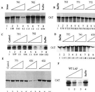
Amino acid residues critical for translation-inhibitory ac-tivity of LAP. The N terminus of all known La proteins con-tains the approximately 60-amino-acid-long La motif, which is highly conserved among various species (70). Table 1 shows a comparison of LAP amino acid sequences (amino acids 11 to 28) from various species. A close examination of this sequence revealed remarkable homology of this region from species as
diverse as human, mouse, bovine,Xenopus laevis, rat,
Caeno-rhabditis elegans, mosquito, and Drosophila melanogaster.
Al-though there was some variation within the N-terminal 11 amino acids of LAP between distant species, the C-terminal 7 amino acids of the peptide (EYYFGDF) were almost invari-ant, even between distant species, with the exception of one
C-terminal amino acid change in mosquito andDrosophilaand
two C-terminal sequence changes in the C. elegansLAP
se-quence. LAP also contains 10 hydrophobic amino acids. Due to the presence of a relatively large number of hydrophobic amino acids, LAP is not readily water soluble at high concen-trations. In addition to determining the critical amino acids for LAP’s translation-inhibitory activity, we were interested in generating an active mutant with higher hydrophilicity to im-prove solubility. For this reason, we chose to substitute the hydrophobic amino acids with the polar amino acid glutamine (Q). Table 2 lists the LAP mutants synthesized, and Fig. 8 examines the effects of these mutations on
translation-inhibi-tory activity of LAP using p2CAT template RNA in HeLa cell-free translation extracts. Substituting the first three hydro-phobic amino acids (A11, A12, and L13) with Q only partially
affected the activity of LAP; at 60M, the mutant (761)
re-tained 89% activity compared to the wt LAP (Fig. 8A and F). Substitution of two additional amino acids (A15 and I17; mu-tant 762) with Q almost completely abolished its translation-inhibitory activity (Fig. 8A). The mutant peptide (703) with all four aromatic amino acids (Y23, Y24, F25, and F28) replaced by Q was found to be totally inactive in blocking translation (Fig. 8B). Two double substitution mutants, one with both tyrosines Y23 and Y24 replaced by Q (mutant 771) and the other with both phenylalanines F25 and F28 replaced by Q (mutant 772), were found to totally inactive in blocking trans-lation (Fig. 8B). Thus, the tyrosine and phenylalanine residues within this highly conserved region of the La motif appear to be critical for translation-inhibitory activity. We then mutated the four aromatic amino acids individually. While replacing Y23 with Q (mutant 741) almost totally abolished LAP activity, mutation of Y24 (mutant 633) was as effective as the wt LAP (Fig. 8D). A similar result was obtained when F25 and F28 were mutated individually; while mutant 632 (F25Q) lost al-most all activity, mutant 631 (F28Q) still retained alal-most 75% of wt LAP activity (Fig. 8E). These results suggest that both Y23 and F25 are critical for the translation-inhibitory activity of LAP.
[image:8.603.103.484.69.214.2]We also determined the role of the charged residues on the activity of the peptide. Mutant 702 (K16Q, H19Q) had activity
FIG. 7. LAP inhibits HCV IRES-mediated translation in vivo. Huh-7 cells were preincubated with various concentrations of LAP (A) or the HIV-1 Tat peptide (B). After 2.5 h, the cells were washed free of peptides and transfected with the capped bicistronic RNA template. Duplicate samples of cells treated with FITC-LAP or FITC-Tat peptides were examined to confirm peptide cell entry. At 6 h posttransfection, the cell lysates were harvested and measured forRenillaand firefly luciferase activities. Representative data from three separate transfections are shown.
TABLE 1. LAP sequences from various species
Species (amino acids 11 to 28)Sequence
Human...AALEAKICHQIEYYFGDF
Mouse...AALEAKICHQIEYYFGDF
Bovine...AALEAKICHQIEYYFGDF
Xenopus...LDLDTKICEQIEYYFGDF
Rat ...AALEAKICHQIEYYFGDF
C. elegans...DDADQRIIKQLEYYFGNI
Mosquito ...VSKLEASTIRQEYYFGDA
Drosophila...TKQERAIIRQVEYYFGDA
on November 8, 2019 by guest
http://jvi.asm.org/
[image:8.603.301.542.624.723.2]comparable to that of the wt LAP (Fig. 8C). In contrast, mu-tant 701 (E14Q, E22Q, D27Q) had almost no activity. Thus, negatively charged but not positively charged residues appear important for the translation-inhibitory activity of LAP.
We also examined the ability of LAP mutants to transduce cells compared with that of wt LAP. Surprisingly, six out of seven LAP mutants (701, 762, 703, 771, 772, 741, and 632) that had lost their ability to inhibit translation also lost the capacity to enter cells (Table 2). Only mutant 701 could transduce cells; however, it had a much lower efficiency than wt LAP.
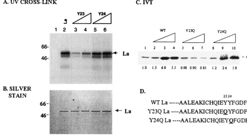
A single amino acid change within the highly conserved La motif affects both RNA binding and translation stimulation activity of La.To determine whether the two aromatic amino acids, Y23 and F25, of LAP are critical for RNA binding and translation-stimulatory activities of the full-length La protein, we used site-directed mutagenesis to make the corresponding changes in the full-length La protein. The first mutants
gener-ated were double substitutions Y23Q-Y24Q (called⌬YY) and
F25Q-F28Q (called⌬FF). After expression and purification of
[image:9.603.103.481.71.438.2]mutant polypeptides, the RNA-binding abilities of the mutants
FIG. 8. Effects of various amino acid substitutions on the translation-inhibitory activity of LAP. Various amino acid substitution mutants of LAP were synthesized chemically and purified to near homogeneity. The mutated amino acids are underlined and are shown in Table 2. The effects of 20, 40, and 60M concentrations of wt and mutant peptides were tested using uncapped p2CAT RNA template in HeLa cell-free lysates as described in Materials and Methods. The numbers at the bottom of each panel indicate relative translation as determined by quantification of the CAT polypeptide.
TABLE 2. Translation inhibition and cell entry of various LAP mutants
Peptide Sequencea Activityc Cell entryd
LAP AALEAKICHQIEYYFGDF ⫹ ⫹⫹⫹
702 AALEAQICQQIEYYFGDF ⫹ ⫹⫹⫹
701 AALQAKICHQIQYYFGQF ⫺ ⫹
761 QQQEAKICHQIEYYFGDF ⫹ ⫹
762 QQQEQKQCHQIEYYFGDF ⫺ ⫺
703 AALEAKICHQIEQQQGDQ ⫺ ⫺
771 AALEAKICHQIEYYQGDQ ⫺ ⫺
772 AALEAKICHQIEQQFGDF ⫺ ⫺
741 AALEAKICHQIEQYFGDF ⫺ ⫺
633 AALEAKICHQIEYQFGDF ⫹ ⫹⫹⫹
632 AALEAKICHQIEYYQGDF ⫺ ⫺
631 AALEAKICHQIEYYFGDQ ⫺ NDb
aMutations are indicated by underlined letters. bND, not determined.
cActivity was assayed by the ability of the peptides to block p2CAT
transla-tion.
dCell entry was determined by confocal microscopy of FITC-labeled peptides.
VOL. 78, 2004 VIRAL TRANSLATION INHIBITION BY A PEPTIDE 3771
on November 8, 2019 by guest
http://jvi.asm.org/
[image:9.603.301.540.551.685.2]were compared with that of the wt La (Fig. 9A). Briefly, the
purified proteins were UV cross-linked to uniformly 32
P-la-beled PV 5⬘-UTR RNA. After RNase treatment, the
protein-nucleotidyl complex was analyzed by SDS-PAGE. The same amount of protein used in the binding assay mixture was also concurrently loaded on a separate gel for silver staining (Fig. 9B). The purpose of the silver-stained gel was to determine the relative amounts of the protein used for cross-linking and for
densitometric analysis. Analysis of the⌬FF mutant protein by
silver staining showed an additional truncated form of La (Fig. 9B).
When Y23 and Y24 or F25 and F28 were changed to
glu-tamine, the ability to bind PV 5⬘-UTR was reduced by⬃75%
for⌬YY (Fig. 9A, lanes 6 to 8) and almost 83% for⌬FF (Fig.
9A, lanes 9 to 11) compared to that of wt La (Fig. 9A, lanes 3 to 5). Based on these results, we decided to generate single amino acid mutants of La. When Y23 was changed to Q in full-length La, the binding ability of the protein was reduced by approximately 75% (Fig. 10A and B, lanes 3 to 4) compared to that of wt La (lane 2). In contrast, changing Y24 to Q reduced
response for translation stimulation compared with wt La. These results suggest that a single tyrosine change within the La motif can significantly affect both RNA binding as well as translation stimulation from the PV IRES element.
DISCUSSION
We have shown that an 18-amino-acid-long, highly con-served sequence encompassing amino acids 11 to 28 of the human La protein is critical for its interaction with IRNA as
well as PV and HCV 5⬘-UTR sequences. Although the peptide
(LAP) itself does not appear to interact with RNA, it is capable of blocking the La-RNA interaction as well as viral IRES-mediated translation both in vitro and in vivo. In contrast, a peptide chosen randomly from a different region of La (NSP) or a totally unrelated HIV-1 Tat peptide was almost totally inactive in blocking viral IRES-mediated translation. Both UV cross-link and gel retardation studies indicated that LAP in-terferes with RNA-protein interactions presumably required for IRES-mediated translation. Rescue of LAP-mediated translation inhibition by HeLa cell proteins but not by the
5⬘-UTR RNA, as well as the lack of evidence for LAP-RNA
interaction, suggests that translation inhibition by LAP is pos-sibly mediated through components in HeLa cell lysate. Both single and multiple amino acid substitutions within LAP have identified amino acids critical for inhibition of IRES-mediated translation in vitro. We have shown that two aromatic amino acids, tyrosine Y23 and phenylalanine F25, are critical for the translation-inhibitory activity of LAP; replacement of Y23 and F25 with glutamine almost totally eliminated LAP’s transla-tion-inhibitory activity in vitro. The full-length La containing either the Y23Q or F25Q mutation was severely defective in its
interaction with the viral 5⬘-UTR (Fig. 9 and 10). Finally, we
demonstrated that the Y23Q but not the Y24Q substitution in the full-length La protein was almost totally inactive in
stimu-lating IRES-mediated translation from the PV 5⬘-UTR (Fig.
10).
One of the objectives of this study was to examine whether IRNA, apparently an IRES-specific inhibitor (17), acted by competing with PV and HCV IRES elements for the La pro-tein. Although this study did not address which La residues actually touch the RNA of interest, these results clearly dem-onstrate that the same region of La (amino acids 11 to 28) plays a critical role in binding IRNA as well as PV and HCV IRES elements. It is clear from the results presented in Fig. 2 that the LA-IRNA interaction does not require the C-terminal
half of La. In contrast, the interaction of La and HCV 5⬘-UTR
FIG. 9. Mutations in full-length La corresponding to LAP mutants 771 and 772 interfere with RNA binding. (A)32P-labeled PV 5⬘-UTR
RNA was UV cross-linked to 0.5, 1, or 1.5g of wt La (lanes 3 to 5), ⌬YY (Y23Q Y24Q; lanes 6 to 8), or⌬FF (F25Q F28Q; lanes 9 to 11). Lane 1 shows the migration of molecular weight markers. Lane 2 contains the labeled probe but no protein. (B) The lower panel is a silver-stained gel corresponding to the amount of protein used in the top panel. (C) Amino acid sequences of the wt and mutant La. The numbers at the bottom of each panel indicate relative band intensity as measured by densitometric scanning using the NIH Image J program.
on November 8, 2019 by guest
http://jvi.asm.org/
is significantly affected by C-terminal deletions, an observation consistent with previous results from other laboratories (2, 58). How does the LAP block IRES-mediated translation in trans? Gel mobility shift experiments utilizing purified La
pro-tein and 32P-labeled IRNA have shown that LAP can block
efficiently the formation of La-IRNA complex (Fig. 4). Also,
UV cross-linking of La to PV and HCV 5⬘-UTR was almost
totally blocked by LAP but not by NSP (Fig. 4 and data not shown). Thus, it appears that LAP is able to block RNA-protein interactions necessary for cap-independent translation. A simple interpretation of these results is that LAP interacts
with the viral 5⬘-UTR, thus preventing interaction of
full-length La with viral RNA. This possibility seems unlikely, how-ever, for a number of reasons. First, despite repeated attempts we have been unable to show direct interaction of LAP with
the 5⬘-UTR (or IRNA) by a variety of techniques, such as gel
shift, UV cross-link, and Northwestern analyses. Our results are consistent with previous failures of other investigators to demonstrate an interaction of the isolated La motif (amino acids 1 to 60), which contains the LAP sequence, with RNA. Also, a recent study has shown that a truncated La protein consisting of the first 100 amino acids failed to bind HCV IRES (58). Secondly, if LAP were interacting with the viral
5⬘-UTR, it would have been possible to rescue LAP-mediated
inhibition of cap-independent translation by increasing the concentration of the RNA template in the reaction mixture. We did not observe significant reversal of translation in the
presence of as much as a sixfold excess of template RNA than that normally used in the translation assay (Fig. 5). In contrast, the fact that addition of excess HeLa cell lysate could reverse LAP-mediated inhibition of p2CAT translation suggests pro-tein-protein as opposed to protein-RNA interaction as the basis for inhibition of translation by LAP. The protein-protein interaction may include the La homodimerization domain, which is required for the function of La in enhancing transla-tion of PV RNA (15). Indeed, the La protein has been shown to exist predominantly as a dimer under native conditions. Like LAP, the La dimerization domain (amino acids 226 to 348) was
found to specifically block PV 5⬘-UTR-mediated translation.
The results of Craig and coworkers (15) suggest that formation of functional La dimers requires both monomers to retain RNA-binding activity. It remains to be seen whether LAP can directly interact with La or other RNA-binding proteins that enhance IRES-mediated translation.
[image:11.603.47.528.74.340.2]The full-length La protein contains three RNA-binding do-mains: RRM1 (approximately amino acids 1 to 100), RRM2 (approximately amino acids 101 to 208), and RRM3 (approx-imately amino acids 209 to 300). Previous studies have shown that the N-terminal half of La containing RRM2 is largely responsible for interaction with PV and HCV IRES elements (58, 65). RRM3 has also been shown to interact with the HCV IRES element; however, this interaction appears to be much weaker than that with RRM2 (58). Results presented in this paper clearly demonstrate the importance of individual amino
FIG. 10. A single amino acid change in full-length La interferes with RNA binding and translation stimulation. (A) PV 5⬘-UTR was UV cross-linked to 0.5g of wt La (lane 2), 0.5 and 1g of Y23 (Y23Q; lanes 3 and 4), or 0.5 and 1g of Y24 (Y24Q; lanes 5 and 6). Lane 1 contains no protein. (B) Silver-stained gel corresponding to the amount of protein used in the UV cross-link analysis. (C) Effects of wt and mutants 741 (Y23Q) and 633 (Y24Q) on p2CAT translation in reticulocyte lysates. In vitro translation from the p2CAT RNA was performed in the absence (lane 1) or presence of 0.5 (lanes 2, 5, and 8), 1.0 (lanes 3, 6, and 9), or 1.5 (lanes 4, 7, and 10)g of wt La (lanes 2 to 4), Y23Q (lanes 5 to 7), and Y24Q (lanes 8 to 10). The numbers at the bottom of each lane indicate the fold stimulation of translation compared to the control (lane 1) without added La. (D) Sequences of the wt and mutant La. The Y23Q and Y24Q mutations were confirmed by sequencing the entire La cDNA.
VOL. 78, 2004 VIRAL TRANSLATION INHIBITION BY A PEPTIDE 3773
on November 8, 2019 by guest
http://jvi.asm.org/
sented here suggest an important role of the La motif (RRM1) in modulating RNA binding by RRMs 2 and 3. Although the RRM1 does not directly contact RNA, it may somehow regu-late the access of the RNA substrate to La RRM 2 and RRM3. For example, there may be subtle or transient interactions between RRM1 and RNA to position RRM2, resulting in a stable interaction of RRM2 with the RNA. Alternatively, in-teraction of RRM1 with another region of La (within the same molecule or between two molecules) could facilitate binding of RRM2 to the RNA. Mutations such as Y23Q and F25Q within RRM1 may interfere with such activity, leading to a lack of RNA binding. Determination of the three-dimensional struc-ture of the La-IRNA complex should shed light on the func-tions of various La RRMs.
When the carboxy-terminal tyrosines (Y23 and Y24) and phenylalanines (F25 and F28) of LAP were mutated in tandem or as a group, a significant decrease in translation activity resulted (Fig. 8). When the amino acids were individually mu-tated, Y23 and F25 appeared to be more critical for LAP activity than the Y24 and F28 residues (Fig. 8). These residues correspond to a region that is highly conserved among La
homologues (Table 1). Two mutant peptides, QQQIEYYFG
DFQQ and QQQIEYYFGDFNL, were synthesized to span the
highly conserved region to determine if a smaller sequence still
inhibited PV 5⬘-UTR-mediated translation. We found the
smaller peptides had no activity in the in vitro translation assay (data not shown). Thus, the amino-terminal residues probably contribute structurally to LAP’s activity. Protein structure-pre-dicting algorithms predict the conformation for residues 11 to
21 of LAP as an␣-helix. In mutant 761 (A1Q, A2Q, and L3Q)
the predicted␣-helix structure has been reduced to residues 11
to 18, and in mutant 762 (A1Q, A2Q, L3Q, A5Q, and I7Q) the
␣-helix has been reduced to residues 11 to 15. Perhaps it is the
shortening of the ␣-helix by 50% that eliminates activity of
mutant 762 (Fig. 9). The critical region of LAP consisting of
residues Y23, Y24, F25, and G26 is predicted to form a
-re-gion (13) and has a surface probability index of zero. This suggests that the peptide might be forming a shallow pocket for interacting with a substrate (RNA or protein). The predicted structure of mutants 703 (Y23Q, Y24Q, F25Q, and F28Q), 771 (F25Q and F28Q), and 772 (Y23Q and Y24Q) show an
ab-sence of -region and an increase in the surface probability
index, suggesting disruption of the pocket, perhaps leading to loss of activity. The computer-generated algorithms were not successful in determining structural differences between mu-tants 632 (F25Q) and 631 (F28Q) or between 741 (Y23Q) and 633 (Y24Q). Perhaps circular dichroism or nuclear magnetic
both LAP and La functions (Fig. 8 and 9 and data not shown). These results suggest that Y23 and F25 have similar roles in LAP-mediated inhibition of translation and La RNA binding and translation stimulation. However, it must be emphasized that the mechanism of action of LAP could be very different than the role these residues play in the intact La protein, particularly when it is taken out of context of full-length La. It is worth mentioning in this context that LAP-mediated inhibi-tion of p2CAT translainhibi-tion could only be partially rescued by the purified La protein (data not shown). However, there was total reversal of LAP-mediated translation inhibition by HeLa cell-free translation extracts (Fig. 5), suggesting that HeLa cell proteins other than La are likely to be involved in
LAP-in-duced inhibition of PV 5⬘-UTR-mediated translation. Our
pre-liminary experiments have shown that binding of a number of HeLa cell proteins, including La, to labeled PV and HCV
5⬘-UTRs was specifically inhibited by LAP (data not shown).
Thus, both translation rescue and RNA-binding data strongly suggest involvement of other transacting proteins in LAP-me-diated inhibition of viral translation. Future studies will exam-ine if LAP (or the LAP sequence in the full-length La protein) is involved in protein-protein interaction.
Experiments conducted with FITC-conjugated LAP clearly demonstrated that LAP entered cells (at 37°C), while FITC alone or FITC-conjugated NSP were unable to enter cells (Fig. 6). The majority of LAP was found to localize in the cell cytoplasm. At this time we do not know how LAP enters cells. We believe LAP is not endocytosed into cells, because even at 4°C LAP entered cells, although at a lower pace compared to that at 37°C (data not shown). Additional experiments will be required to fully understand the mechanism of LAP cell entry. One interesting observation from the LAP mutagenesis study was that almost all mutations that interfered with its transla-tion-inhibitory activity were also found to interfere with its ability to enter cells (Table 2). Only one mutant, LAP 701, which had lost translation-inhibitory activity, could still trans-duce cells, albeit with lower efficiency than the wt LAP. We do not know the precise reason for the requirement of the same amino acid residues for two apparently unrelated functions of this peptide. It is of interest that other peptides termed PTDs (for protein transduction domain) that can enter mammalian cells unaided have been identified. Some of the best-studied ones include basic residues 47 to 57 of the HIV-1 Tat protein, residues 267 to 300 of herpes simplex virus type 1 VP22
tran-scription factor, and the third␣-helix (residues 43 to 58) of the
Drosophilahomeotic transcription domain ANTP (encoded by
the antennapedia gene) (45). These PTDs are similar to LAP
on November 8, 2019 by guest
http://jvi.asm.org/
in that they rapidly and efficiently enter cells at 37 and 4°C. There are no sequence homologies between LAP and Tat or ANTP PTDs, but protein structure-predicting algorithms
strongly suggest that the Tat PTD can adopt an␣-helix
con-formation, and the ANTP PTD corresponds to the third␣
-he-lix of antennapedia homeodomain (19, 46). As mentioned
above, LAP has a predicted␣-helix domain, and disruption of
this domain as in mutants 761 and 762 dramatically decreases the transduction of LAP (Table 2). We have shown that LAP can transduce fluorescein into both HeLa and Huh-7 cells. It remains to be seen whether a LAP fusion polypeptide such as
LAP–-galactosidase, green fluorescent protein, or
LAP-luciferase could freely enter cells.
In summary, we have demonstrated here that an N-terminal sequence from the La motif plays a critical role in recognition
by La of HCV and PV 5⬘-UTR elements as well as IRNA.
Although this region does not interact with RNA directly, single amino acid changes within this region drastically affect both La’s RNA-binding and translation-stimulatory activities. A synthetic peptide corresponding to this important region of La is able to block RNA-protein interaction as well as
stimu-lation of viral 5⬘-UTR-mediated translation both in vitro and in
vivo. Future studies on the mechanism of action of the peptide should help to understand the role of the La protein in PV and HCV IRES-mediated translation.
ACKNOWLEDGMENTS
This work was supported by National Institutes of Health grant AI 45733 (to A.D.) and the STTR grant AI 45188 to Virasim Inc. R.E.I. was supported by a Howard Hughes graduate fellowship.
We thank Dan Keenan and J. D. Keene for the wt and mutant La constructs. We are grateful to Cathy DiMare for help with protein expression. We are grateful to the members of the Dasgupta labora-tory for helpful suggestions and to Mathew Schibler for help with the confocal microscopy.
REFERENCES
1. Ali, N., and A. Siddiqui.1997. Molecular mechanisms of translation initia-tion in eukaryotes. Proc. Natl. Acad. Sci. USA94:2249–2254.
2. Ali, N., G. J. M. Prujin, D. J. Kenan, J. D. Keene, and A. Siddiqui.2000. Human La antigen is required for the hepatitis C virus internal ribosome entry site-mediated translation. J. Biol. Chem.275:27531–27540. 3. Ali, N., and A. Siddiqui.1995. Interaction of polypyrimidine tract binding
protein with the 5⬘non-coding region of the hepatitis C virus RNA genome and its functional requirement in internal initiation of translation. J. Virol.
69:6367–6375.
4. Banerjee, R., M. Igo, R. Izumi, U. Datta, and A. Dasgupta.2000. In vitro replication of RNA viruses, p. 158–164.InA. Cann (ed.), RNA viruses: a practical approach. Oxford University Press, New York, N.Y.
5. Belsham, G. J., and N. Sonenberg.1996. RNA-protein interactions in reg-ulation of picornavirus RNA translation. Microbiol. Rev.60:499–511. 6. Belsham, G. J., and N. Sonenberg.2000. Picornavirus RNA translation: roles
for cellular proteins. Trends Microbiol.8:330–335.
7. Belsham, G. J., N. Sonenberg, and Y. V. Svitkin.1995. The role of the La autoantigen in internal initiation. Curr. Top. Microbiol. Immunol.203:85– 98.
8. Blyn, L. B., J. S. Towner, B. L. Semler, and E. Ehrenfeld.1997. Requirement of poly(rC) binding protein 2 for translation of poliovirus RNA. J. Virol.
71:6243–6246.
9. Boussadia, O., M. Niepmann, L. Creancier, A. C. Parts, F. Dautry, and H. Jacquemin-Sablon.2003. Unr is required in vivo for efficient initiation of translation from the internal ribosome entry sites of both rhinovirus and poliovirus. J. Virol.77:3353–3359.
10. Cardinali, B., C. Carissimi, P. Gravina, and P. Pierandrei-Amaldi.2003. La protein is associated with TOP mRNAs in actively translating polysomes. J. Biol. Chem.278:35145–35151.
11. Chambers, J. C., and J. D. Keene.1985. Isolation and analysis of cDNA clones expressing human lupus La antigen. Proc. Natl. Acad. Sci. USA
82:2115–2119.
12. Chang, Y. N., D. J. Kenan, J. D. Keene, A. Gatignol, and K. T. Jeang.1994.
Direct interactions between autoantigen La and human immunodeficiency virus leader RNA. J. Virol.68:7008–7020.
13. Chou, P. Y., and G. D. Fasman.1978. Empirical predictions of protein conformation. Annu. Rev. Biochem.47:251–276.
14. Coward, P., and A. Dasgupta.1992. Yeast cells are incapable of translating RNAs containing the poliovirus 5⬘untranslated region. J. Virol.66:286–295. 15. Craig, A. W., Y. V. Svitkin, H. S. Lee, G. J. Belsham, and N. Sonenberg.1997. The La autoantigen contains a dimerization domain that is essential for enhancing translation. Mol. Cell. Biol.17:163–169.
16. Das, S., M. Ott, A. Yamane, W. Tsai, M. Gromeier, F. Lasher, S. Gupta, and A. Dasgupta.1998. A small yeast RNA blocks hepatitis C virus internal ribosome entry site (IRES)-mediated translation and inhibits replication of a chimeric poliovirus under translational control of the HCV IRES element. J. Virol.72:5638–5647.
17. Das, S., D. J. Kenan, D. Bocskai, J. D. Keene, and A. Dasgupta.1996. Sequences within a small yeast RNA required for inhibition of internal initiation of translation: interaction with La and other cellular proteins influences its inhibitory activity. J. Virol.70:1624–1632.
18. Das, S., P. Coward, and A. Dasgupta.1994. A small yeast RNA selectively inhibits internal initiation of translation programmed by poliovirus RNA: specific interaction with cellular proteins that bind to the viral 5⬘ untrans-lated region. J. Virol.68:7200–7211.
19. Derossi, D., A. H. Joliot, G. Chassaing, and A. Prochiantz.1994. The third helix of the Antennapedia homeodomain translocates through biological membranes. J. Biol. Chem.269:10444–10450.
20. Ford, L. P., J. W. Sway, and W. E. Wright.2001. The La antigen associates with the human telomerase ribonucleoprotein and influences telomerase length in vivo. RNA7:1068–1075.
21. Francoeur, A. M., and B. Mathews.1982. Interaction between VA RNA and the lupus antigen La: formation of a ribonucleoprotein particle in vitro. Proc. Natl. Acad. Sci. USA79:6772–6776.
22. Fukushi, S., K. Katayama, C. Kurihara, N. Ishiyama, F. B. Hoshino, T. Ando, and A. Oya.1994. Complete 5⬘non-coding region is necessary for the efficient internal initiation of hepatitis C virus RNA. Biochem. Biophys. Res. Commun.199:425–432.
23. Gamarnick, A. V., and R. Andino.1997. Two functional complexes formed by KH domain containing proteins with the 5⬘noncoding region of poliovirus RNA. RNA3:882–892.
24. Gan, W., and R. E. Rhoads.1996. Internal initiation of translation directed by the 5⬘untranslated region of the mRNA for eIF4G, a factor involved in the picornavirus switch from cap-dependent to internal initiation. J. Biol. Chem.271:623–626.
25. Goodier, J. L., H. Fan, and R. J. Maraia.1997. A carboxy-terminal basic region controls RNA polymerase III transcription factor activity of human La protein. Mol. Cell. Biol.17:5823–5832.
26. Hellen, C. U. T., and P. Sarnow.2001. Internal ribosome entry sites in eukaryotic mRNA molecules. Genes Dev.15:1593–1612.
27. Hellen, C. U. T., G. W. Witherell, M. Schmid, S. H. Shin, T. V. Pestova, A. Gil, and E. Wimmer.1993. A cytoplasmic 57 kDa protein that is required for translation of picornavirus RNA by internal ribosome entry is identical to the nuclear pyrimidine tract binding protein. Proc. Natl. Acad. Sci. USA90:
7642–7646.
28. Hershey, J. W. B., and W. C. Merrick.2000. The pathway and mechanism of initiation of protein synthesis, p. 33–88.InN. Sonenberg (ed.), Translational control of gene expression. Cold Spring Harbor Laboratory Press, Cold Spring Harbor, N.Y.
29. Holcik, M., and R. G. Korneluk.2000. Functional characterization of the X-linked inhibitor of apoptosis (XIAP) internal ribosome entry site element: role of La autoantigen in XIAP translation. Mol. Cell. Biol.20:4648–4657. 30. Holcik, M., C. A. Lefebvre, C. Yeh, T. Chow, and R. G. Korneluk.1999. A new internal-ribosome-entry-site motif potentiates XIAP-mediated cytopro-tection. Nat. Cell Biol.1:190–192.
31. Honda, M., E. A. Brown, and S. Lemon.1996. Stability of a stem-loop involving the initiator AUG controls the efficiency of internal initiation of translation on hepatitis C virus RNA. RNA2:955–968.
32. Hunt, S. L., A. Kaminski, and R. J. Jackson.1999. unr, a cellular cytoplasmic RNA-binding protein with five cold-shock domains, is required for internal initiation of translation of human rhinovirus RNA. Genes Dev.13:437–448. 33. Iizuka, N., C. Chen, Q. Yang, G. Johannes, and P. Sarnow.1995. cap-independent translation and internal initiation of translation in eukaryotic cellular mRNA molecules. Curr. Top. Microbiol. Immunol.203:155–177. 34. Isoyama, T., N. Kamoshita, K. Yasui, A. Iwai, K. Shiroki, H. Toyoda, A.
Yamada, Y. Takasaki, and A. Nomoto.1999. Lower concentration of La protein required for internal ribosome entry on hepatitis C virus RNA than on poliovirus RNA. J. Gen. Virol.80:2319–2327.
35. Izumi, R. E., B. Valdez, R. Banerjee, M. Srivastava, and A. Dasgupta.2001. Nucleolin stimulates internal ribosome entry site-mediated translation. Virus Res.76:17–29.
36. Jacks, A., J. Babon, G. Kelly, I. Manolaridis, P. D. Cary, S. Curry, and M. R. Conte.2003. Structure of the C-terminal domain of the human La protein reveals a novel RNA recognition motif coupled to a helical nuclear retention element. Structure11:833–843.
VOL. 78, 2004 VIRAL TRANSLATION INHIBITION BY A PEPTIDE 3775
on November 8, 2019 by guest
http://jvi.asm.org/
5009–5016.
43. Kurilla, M., and J. D. Keene.1983. The leader RNA of vesicular stomatitis virus is bound by a cellular protein reactive with anti-La lupus antibodies. Cell34:837–845.
44. Liang, X. S., J. Q. Lian, Y. X. Zhou, Q. H. Nie, and C. Q. Hao.2003. A small yeast RNA inhibits HCV IRES-mediated translation and inhibits replication of poliovirus in vivo. World J. Gastroenterol.9:1008–1013.
45. Lindgren, M., A. Hallbrink, A. Prochiantz, and U. Langel.2000. Cell pen-etrating peptides. Trends Pharmacol. Sci.21:99–103.
46. Loret, E. P., E. Vives, P. S. Ho, H. Rochat, J. Van Rietschoten, and W. C. Johnson, Jr.1991. Activating region of HIV-1 Tat protein: vacuum UV circular dichroism and energy minimization. Biochemistry30:6013–6023. 47. Macezak, D. G., and P. Sarnow. 1991. Internal initiation of translation
mediated by the 5⬘leader of a cellular RNA. Nature353:90–94. 48. Madore, S. J., E. D. Wieben, and T. Pederson.1984. Eukaryotic small
ribonucleoproteins: anti-La human antibodies react with U1 RNA-protein complexes. J. Biol. Chem.259:1929–1933.
49. Maraia, R. J., D. J. Kenan, and J. D. Keene.1994. A carboxy-terminal basic region controls RNA polymerase III transcription factor activity of human La protein. Mol. Cell. Biol.14:2147–2158.
50. Meerovich, K., Y. V. Svitkin, H. S. Lee, F. Lejbkowicz, D. J. Kenan, E. K. Chan, V. I. Agol, J. D. Kenne, and N. Sonenberg.1993. La autoantigen enhances and corrects aberrant translation of poliovirus RNA in reticulocyte lysate. J. Virol.67:3798–3807.
51. Negulescu, D., L. E.-C. Leong, K. G. Chandy, B. L. Semler, and G. A. Gutman.1998. Translation initiation of a cardiac voltage-gated potassium channel by internal ribosome entry. J. Biol. Chem.273:20109–20113. 52. Oh, S. K., M. P. Scott, and P. Sarnow.1992. Homeotic gene antennapedia
mRNA contain 5⬘noncoding sequences that confer translation initiation by internal ribosome binding. Genes Dev.6:1643–1653.
53. Pardigon, N., and J. H. Strauss.1996. Mosquito homolog of the La autoan-tigen binds to Sindbis virus RNA. J. Virol.70:1173–1181.
54. Park, Y.-W., and M. G. Katze.1995. Translational control by influenza virus: identification of cis-acting sequences and transacting factors which may reg-ulate selective viral mRNA translation. J. Biol. Chem.270:28433–28439. 55. Pelletier, J., and N. Sonenberg.1988. Internal initiation of translation of
60. Rose, J. K., H. Trachsel, K. Leong, and D. Baltimore.1978. Inhibition of translation by poliovirus: inactivation of a specific initiation factor. Proc. Natl. Acad. Sci. USA75:2732–2736.
61. Schwarze, S. R., and S. F. Dowdy.2000. In vivo protein transduction: intra-cellular delivery of biologically active proteins, compounds and DNA. Trends Pharmacol. Sci.21:45–48.
62. Shimazaki, T., M. Honda, S. Kaneko, and K. Kobayashi.2002. Inhibition of internal ribosome entry site-directed translation of HCV by recombinant IFN-alpha correlates with a reduced La protein. Hepatology35:199–208. 63. Shiroki, K., T. Isoyama, S. Kuge, T. Ishii, S. Ohmi, S. Hata, K. Suzuki, Y.
Takasaki, and A. Nomoto.1999. Intracellular redistribution of truncated La protein produced by poliovirus 3Cpro-mediated cleavage. J. Virol.73:2193–
2200.
64. Svitkin, Y. V., A. Pause, and N. Sonenberg.1994. La autoantigen alleviates translational repression by the 5⬘leader sequence of the human immunode-ficiency virus type 1 mRNA. J. Virol.68:7001–7007.
65. Svitkin, Y. V., K. Meerovitch, H. S. Lee, J. N. Dholakia, D. J. Kenan, V. I. Agol., and N. Sonenberg.1994. Internal translation initiation on poliovirus mRNA: further characterization of La function in poliovirus translation in vitro. J. Virol.68:1544–1550.
66. Tsukiyama-kohara, K., N. Izuka, M. Kohara, and A. Nomoto.1992. Internal ribosome entry site within hepatitis C virus RNA. J. Virol.66:1476–1483. 67. Venkatesan, A., and A. Dasgupta.2001. Novel fluorescence-based screen to
identify small synthetic internal ribosome entry site elements. Mol. Cell. Biol.21:2826–2837.
68. Wang, C., P. Sarnow, and A. Siddiqui.1993. Translation of human hepatitis C virus RNA in cultured cells is mediated by an internal ribosome binding mechanism. J. Virol.67:3338–3344.
69. Wilson, J. E., M. J. Powell, S. E. Hoover, and P. Sarnow.2000. Naturally occurring dicistronic cricket paralysis virus RNA is regulated by two internal ribosome entry sites. Mol. Cell. Biol.20:4990–4999.
70. Wolin, S., and T. Cedervall.2002. The La protein. Annu. Rev. Biochem.
71:375–403.
71. Yang, Q., and P. Sarnow.1997. Location of the internal ribosome entry site in the 5⬘non-coding region of the immunoglobulin heavy chain binding protein (BiP) mRNA: evidence for specific RNA-protein interactions. Nu-cleic Acids Res.25:2800–2807.