A Dissertation on
“SERUM MAGNESIUM LEVELS IN ACUTE MYOCARDIAL INFARCTION ”
Dissertation submitted to
THE TAMILNADU Dr.M.G.R. MEDICAL UNIVERSITY CHENNAI – 600 032
With partial fulfillment of the regulations For the award of the degree of
M.D. GENERAL MEDICINE BRANCH – I
CERTIFICATE
This is to certify that the dissertation “SERUM MAGNESIUM LEVELS IN ACUTE MYOCARDIAL INFARCTION” is a bonafide research work done by Dr. J. ANANDARAJ Post graduate in M.D. General Medicine under my direct guidance and supervision to my satisfaction, in partial fulfillment of the requirements for the degree of M.D. General Medicine.
Date : Professor & Unit chief M-6
Date : Professor & Head Of The Department Department of General Medicine
Date : The Dean, Coimbatore medical college
DECLARATION
I hereby declare that this dissertation entitled “SERUM
MAGNESIUM LEVELS IN ACUTE MYOCARDIAL
INFARCTION” is a bonafide and genuine research work carried out by me under the guidance of Prof. Dr. Isaac Christian Moses M.D., Department of Medicine ,Coimbatore medical college, Coimbatore .
Date :
ACKNOWLEDGEMENT
I realize that dissertation being a work of cooperation and assistance, it would be far from complete without due acknowledgement of the help gratefully received.
It is my distinct honour and privilege to have under the able supervision of my teacher, Prof. Dr. Isaac Christian Moses M.D., Professor, Department of Medicine, Coimbatore medical college , Coimbatore. Indeed, I am fortunate to get the benefit of his vast experience, valuable guidance and advise at every step of my study. It is with humble gratitude, I pay respects and thanks to him for his keen interest in the study and guidance for the preparation of this dissertation.
With a deep sense of gratitude I acknowledge the guidance rendered to me by my beloved teacher Prof. Dr. Kumar Natarajan, M.D., Professor & Head of the Department of General Medicine. His proficient , incessant guidance and immutable motivation actuated me to furnish an adept study.
I thank my beloved Professors Dr.S. Chandrasekaran, M.D, Dr.M. Ravindran, M.D, Dr.N. Sundar, M.D and all my teachers in the Department of Medicine for their valuable suggestions.
the Department of Medicine for their valuable suggestions.
I also express my deep sense of gratitude to The Dean, Prof. Dr. Revwathy,M.D for the support rendered to me
I am very much thankful to my family, friends and colleagues especially for their kind cooperation.
I feel indebted to my patients and hospital, laboratory staff for their kind cooperation in the study.
Date:
LIST OF ABBREVIATIONS USED
AF ...Atrial fibrillation
CAD ...Coronary artery disease ATP ...Adenosine triphosphate VT ...Ventricular tachycardia VF ...Ventricular fibrillation ECG...Electrocardiogram IHD ...Ischemic heart disease CCF ...Congestive cardiac failure LVF ...Left ventricular failure JVP...Jugular venous pressure MgSO4...Magnesium sulphate
TABLE OF CONTENTS
S.No Title Page no.
1. INTRODUCTION 1
2. AIMS AND OBJECTIVES 3
3. REVIEW OF LITERATURE 4
4. METHODOLOGY 78
5. OBSERVATION AND RESULTS 81
6. DISCUSSION 95
7. SUMMARY 100
8. CONCLUSION 102
9. BIBLIOGRAPHY 103
10. ANNEXURES
i. Proforma 115
ii. Consent Form 121
iii.Key to Master chart 124
LIST OF TABLES
Sl.
Table
Page
No. No.
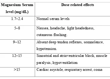
1. Manifestations of hypomagnesemia 64
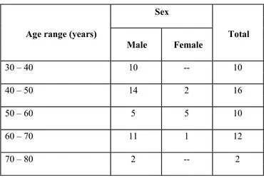
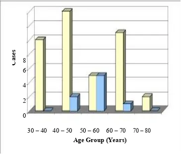
2. Age and Sex Distribution of the Study group 81
3. Religion wise Distribution of cases 83
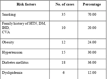
4. Risk Factors 85
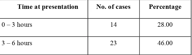
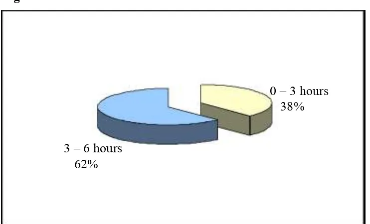
5. Time of Presentation 86
6. Serum magnesium levels in patients with arrhythmias 89
7. Serum magnesium levels in patients without arrhythmias 89
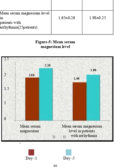
8. Mean serum magnesium level 90
9. Comparison of Serum Magnesium level in patients with 91 Arrhythmias and without Arrhythmias (Day-1)
LIST OF FIGURES
Sl. No. Table Page No
1. Age and Sex Distribution of the Study group 82 2. Religion wise Distribution of cases 83
3. Diet 84
4. Time of Presentation 87
5. Mean serum magnesium level 90
6.
Comparison of Serum Magnesium level in patients with Arrhythimas and without Arrhythmias (Day-1)
92
7.
Comparison of Serum Magnesium level in patients with Arrhythimas and without Arrhythmias (Day-5)
xiii
ABSTRACT
Background: Magnesium has been implicated in the pathogenesis of acute myocardial infarction and its complication like arrhythmia. Magnesium improves myocardial metabolism, inhibits calcium accumulation and myocardial cell death. It improves vascular tone, peripheral vascular resistance, after load and cardiac output, reduces cardiac arrhythmias and improves lipid metabolism. Magnesium also reduces vulnerability to oxygen derived free radicals, improves endothelial function and inhibits platelet function including platelet aggregation and adhesion.
Objective: To know the relationship between the serum magnesium levels and arrhythmias in patients with acute myocardial infarction.
Method: By using simple random method, 50 cases of acute myocardial infarction, admitted in Coimbatore Medical College and Hospital.
Results: There is a significant difference in the magnesium levels in patients with arrhythmias and without arrhythmias.
Conclusion: In acute myocardial infarction, patients with low magnesium levels are more prone to get arrhythmias. So magnesium treatment can be considered in patients of acute myocardial infarction with low magnesium levels.
1
INTRODUCTION
It has been known that inorganic salts are necessary for the
normal growth and functioning of all biological forms. Pasteur
(1860) proposed that yeast will grow only when inorganic
compounds were present in the culture medium. In the human
body, there is a maintenance of fluid balance, not only as a whole
but also intercompartmentally between the three compartments
such as intracellular, interstitial and intravascular fluid
compartments. Various forces such as hemodynamic, electrolyte
and other forces act together and contribute to maintain the normal
fluid balance of the body. It is now evident that not only proteins,
fats and carbohydrates, but also minerals play an essential role in
the normal homeostasis of the body. Intensive investigation is now
going on about the importance of trace elements not only of
vitamins but also of minerals.
Magnesium has been known to have an influence in the
causation of acute myocardial infarction and also its sequelae like
arrhythmias. It plays a major role in the pathogenesis of other
cardiovascular diseases as well. Magnesium ions are found to be
essential for the maintenance of the normal functional integrity of
2
Several investigations have shown that the serum
magnesium level is low in the first 48 hours following a acute
myocardial infarction and later on rose gradually to attain the
normal level in about three weeks time. Infarcted myocardium was
found to have reduced magnesium concentration. The above said
findings correlated directly with the associated complications of
acute myocardial infarction, such as arrhythmias.
In patients with sudden death because of ischemic heart
disease, magnesium concentration in the cardiac muscle was found
to be decreased2. Hypomagnesium acts a provoking factor in the
occurrence of ventricular fibrillation, which is usually the cause of
sudden death in IHD. The coronary vasospasm which occurs as a
result of hypomagnesemia has been considered as an important
factor in the causation of sudden death in IHD.
Magnesium deficiency contributes to the progression of
atheromatous plaques occuring as a result of hyperlipidemia.
Myocardial infarction is one of the common causes of death
where its prognosis depends on various factors. This study is
designed to know the contribution of magnesium levels in the
3
AIMS AND OBJECTIVES
To know the correlation between serum magnesium levels
and arrhythmias in patients with acute myocardial infarction who
4
REVIEW OF LITERATURE
Historical Review
During the time of Lavoisier (1743-1794) only 26 elements
were discovered. Followed shortly by the discovery of elements
such as sodium, potassium, calcium and magnesium. It was only
Liebig (1803-1873) who appreciated the significance of the
minerals as normal constituents of plants and animal tissues.
Eventhough the inorganic constituents of the body constitute only
small fraction of the total amount, they should not be considered as
insignificant. As a matter of fact, they are found to play a pivotal
role in the maintenance of the normal homeostasis of the body.
Greenberg and associates in 1936 demonstrated myocardial
degeneration associated with fibrosis and polyplastic infiltration in
rats fed on diets low in magnesium since birth (Burch et al, 1977)1.
From the sequence of structural abnormalities observed it was
interpreted that interference with the enzymes dependent on
magnesium which are involved in oxidative phosphorylation was
responsible for the pathogenesis of the lesion observed.
The term ‘magnesium’ derived its name from the name of
5
Humphrey Davy in 1808, investigated the alkaline earth metal and
proposed the white magnesia stone by its modern name
‘magnesium’.
Until the middle of twentieth century, magnesium
metabolism had not been given much importance, though extensive
literature about the consequences of magnesium deficiency in the
lower species was available. During this period, the progress of
the work on magnesium metabolism in man was very slow because
of the lack of uniformity and difficulty in estimating magnesium
levels and also the magnesium status of the body.
Now the availability of more accurate and uniform means of
serum magnesium estimation in the laboratory has escalated the
work on magnesium metabolism in man to greater heights.
Only 1% of total body magnesium is present in the
extra-cellular fluid and out of this, about 25% is present in the plasma,
rest is seen in the red cells. About 50% of serum magnesium is
found to be free, 32% remains bound to proteins and the rest 13%
exists as magnesium phosphate, citrate and other unidentified
complexes.
6
portion of the body involved in the homeostasis of magnesium, it is
clear that the estimation of serum magnesium levels alone does not
always indicate the actual total body magnesium stores.
As a matter of fact, magnesium deficiency can occur when
the intracellular magnesium content is normal and intracellular
magnesium deficiency may occur without any decrease in serum
magnesium levels (Vermon et al, 1978)3.
Nevertheless, compared to the complex studies on
determination of the tissue levels, measurement of serum
magnesium is the rapid, simple and most effective approach for the
assessment of magnesium deficiency states. Measurement of the
serum magnesium levels has been used widely for identifying
various clinical syndromes that have responded well to the
7
CORONARY CIRCULATION
04Anatomy of Coronary circulation
Coronary arterial system supplies blood to the heart. It is
comprised of the right coronary artery and the left coronary artery.
The origin of right coronary artery is from the anterior aortic
sinus. Soon after its origin, it courses between the right auricular
appendage and the infundibular portion of the right ventricle. It
traverses in the atrioventricular groove passing vertically
downwards. Then the artery turns posteriorly at the lower border
of the heart and courses posteriorly. It gives off several branches to
both atria and ventricles as it passes down the atrio ventricular
groove. At the inferior border of the heart, the marginal branch
traverses to the left along the right ventricle. The inferior
interventricular branch arises on the diaphragmatic surface and
courses along the groove in between the ventrices to the cardiac
apex. The terminal branches of right coronary artery anastomoses
with the terminal arterioles of the left coronary artery at the lower
aspect of left atrium.
The left coronary artery soon after its origin divides into left
8
The left anterior descending artery traverses the interventricular
groove to anastomoses with the terminal branches of the inferior
interventricular artery, which is a branch of right coronary artery, at
the apex. The left circumflex artery gives branches to the posterior
wall of the left ventricle and courses down to anastomose with the
termination of the right coronary artery, below the coronary sinus.
In 40% of the individuals it gives off a larger branch which runs
over the posterior surface of the left atrium terminates in the
auricular appendage of the right atrium at the sino-atrial node.
9
VENOUS DRAINAGE OF THE HEART
Anastomoses of Coronary Arteries
There exists an anastomoses between the terminal portions
of right and left coronary arteries in the atrrioventricular groove but
10
intercoronary anastomoses at the arteriolar level, between the
inter-ventricular arteries. If the intraventricular arteries meet at the
apex, maximum anastomoses may be provided. If the
intraventricular arteries meet slightly away from the apex above or
below, the potential anastomotic area may be diminished. In 10%
of the individuals, both the inferior as well as the anterior
interventricular artery arises out of the left coronary artery itself, in
these cases there is no anastomoses exists in this condition between
the coronaries.
There exists a potential anastomoses between the coronary
arteries and pericardial arteries which usually arises from the
pericardiophrenic, the bronchial and the internal thoracic arteries.
11
CORONARY ANASTOMOSES
Distribution of the Coronaries
Right ventricle is supplied by the right coronary artery with
the exception of the upper margin of its anterior surface which is
12
Left ventricle is supplied by the left coronary artery with the
exception of a narrow strip of the diaphragmatic surface which is
supplied by the inferior interventricular artery. The two
interventricular arteries equally supplies the interventricular
septum.
Right coronary artery supplies the anterior surface of the
right atrium. Left coronary artery supplies the posterior surface
and the auricular appendage of the left atrium.
SA Node: In 60% of cases, it is supplied by a branch of right
coronary artery and in 40% of cases, it is from left coronary artery.
The inferior interventricular artery supplies AV node and bundle
of His, which arises in 90% of cases from the right coronary artery
and in only 10% from the left coronary artery.
Dominant Arteries: Right coronary is dominant in 67% of the
cases, in 15% of cases, left coronary artery is dominant and in 18%
13
CARDIAC PHYSIOLOGY
Cardiac action potential
Physiology of Coronary Circulation
Physiologically, the right and left coronary arteries behave
like end arteries, eventhough innumerable inter coronary
anastomoses are present in most of the normal hearts.
14
Coronary blood flow in human beings during resting state is
approximately about 225 ml/ min or 0.7-0.8 ml/g of cardiac muscle
or 4.5% of the total cardiac output. This may increase upto 4 to 5
fold during exercise.
Coronary Blood Flow changes during cardiac cycle
Because of cardiac muscle contraction, blood flow to the
heart is decreased during systole and increased during diastole.
Blood supply to the left ventricle is affected more than that of the
right ventricle because of its increased thickness.
During cardiac muscle contraction, the subendocardial blood
vessels are compressed more than the epicardial vessels because of
increased myocardial pressures. The subendocardial vessels are
usually larger compared to the nutrient arteries in the middle and
external layers of the heart, which may cause a proportionate
increase in the blood flow during cardiac diastole. Hence,
subendocardial portions of the heart receive most of its blood
15
Regulation of Coronary Blood Flow:
Coronary blood flow can be regulated as follows:
1. Local myocardial metabolism
2. Nervous control
i) Local metabolism of myocardium: As the force of
contraction increases, the rate of coronary blood flow also
increases. As the activity decreases, coronary blood flow
also decreases. This is dependent on the following factors:
a) Oxygen Demand:
As oxygen extraction is near complete in resting state only,
increase in oxygen demand has to be met with by increasing the
blood flow. This is achieved probably by the following
mechanisms:
1. Vasodilator Theory: Anoxia will liberate many vasodilator
materials from myocardial cells which increase the blood flow:
i) Adenosine from the ATP
ii) Potassium ion
iii) Hydrogen ion
iv) Carbon dioxide
v) Bradykinin and possibly
16
2. Arterial Smooth Muscle Relaxation Theory: Decrease in
oxygen supply leads to anoxia of coronary arterial smooth
muscle cells, which loses their tone thus getting the artery
dilated. Factors that determine the oxygen consumption are:
a) Greater the work, the greater the oxygen consumption,
within the physiological limitations.
b) Oxygen consumption is proportionate to peak myocardial
muscle tension.
i. Increased arterial pressure, increases the work load and
hence tension.
ii. Dilatation of the heart increases the tension development in
myocardium to pump the blood according to Laplace law,
which states that tension required to generate a given
pressure increases in proportion to the diameter of the heart.
c) Other factors which increases the oxygen consumption like
stimulation of the heart by epinephrine and norepinephrine,
thyroxine, digitalis, calcium ions, increased temperature of
heart, will increase the oxygen consumption.
17
because of coronary dilatation after a brief period of
coronary occlusion.
iii) Nervous Control
a) Indirect: Sympathetics increases the heart rate and
contractility, through the local metabolic mechanisms, and
hence increases the coronary flow. Parasympathetics
decrease the heart rate and depresses the myocardium and
hence brings about coronary constriction.
b) Direct Effect:
Parasympathetics: As the vagal supply to ventricles is
negligible, except for slight dilatation which may occur,
there is no effect of its stimulation.
Sympathomimetics: Epinephrine and norepinephrine
through their receptors in coronary vessels usually bring
about vasoconstriction or no change. When alpha effect
dominates, severe constriction occurs which may bring about
18
EPIDEMIOLOGY OF CORONARY HEART DISEASE85
Coronary Artery Disease (CAD) is the major cause of
morbidity and mortality in the age group of 45 years or more all
over the world including India. Wide variations have been seen in
the prevalence rate of CAD in various geographical zones. Death
rates from CAD seems to be higher in Finland and US.
In the US, among those over 30 years of age it has been
estimated that 213 per 100,000 individuals are said to have
ischaemic heart disease.
Accurate data about the prevalence of CAD in India are not
available. Various surveys are carried out in recent years, in
various geographical locations and in small population groups
which use different protocols. It has been estimated that the
prevalence rate may be about 5% in urban population and a much
lower prevalence has been seen in the rural setting.
The pattern of CAD in India has been reported to be:
a) Males are affected more than females.
b) Hypertension and diabetes accounts for about 40% of all
cases.
19
of cases.
d) High fat & energy rich diet, sedentary life style are other
contributing factors.
It has been believed that the prevalence of CAD in India has
been increasing over the last three decades and the younger persons
are prone to the prone to develop CAD since 1970 because of the
increasing risk factors. There has been approximately 30%
reduction in mortality due to CAD.
20
PATHOPHYSIOLOGY OF ACUTE MYOCARDIAL
INFARCTION85
Myocardial infarction occurs when there is an abrupt
reduction in coronary blood flow usually occurring as a result of
thrombotic occlusion of the coronary artery already been narrowed
by the formation of atherosclerotic plaques which fissures, ruptures
or ulcerates and under favourable conditions thrombogenesis may
take place. A mural thrombus forms at the site of plaque rupture
and leads to coronary artery occlusion. An initial platelet
monolayer forms at the site of the plaque rupture, a series of
agonists (collagen, ADP, epinephrine, serotonin)released may
promote platelet activation. Following stimulation by agonists,
thromboxane A2 is produced and released. This may further
activate platelets and hence aggravating thrombogenesis.
In addition to the production of thromboxane A2, platelet
activation by agonists produce a definitive conformational change
in the glycoprotein IIb- IIIa receptors. Once it is converted to its
functional state, glycoprotein IIb- IIIa receptor develops a high
affinity for the arginine – glycine – aspartic acid sequence on the
alpha chain of the fibrinogen and also for a dodecapeptide
21
multivalent molecule and it has the ability to bind to two different
platelets simultaneously, leading to cross linking of platelets and
aggregation.
At the site of the plaque rupture, tissue factor is released
from the damaged endothelial cells, which on exposure gets
activated, followed by the activation of coagulation cascade occurs.
Through extrinsic and intrinsic pathways, Factor VII and X are
activated, ultimately leading to the conversion of prothrombin to
thrombin, Thrombin, then converts fibrinogen to fibrin. Finally,
crosslinkage of fibrin and formation of fibrin clot occurs. The
coronary artery eventually gets occluded by a thrombus made up of
platelet aggregates and fibrin strands.
Ultimately the extent of myocardial damage produced by
coronary occlusion depends upon the blood vessel affected, the
territory it supplied, whether or not the vessel becomes totally
occluded, presence of native factors which can produce early
spontaneous lysis of the occlusive thrombus, the extent of collateral
vasculature formation and the oxygen demand of the myocardium
23
Chart 2 : ACLS 2010 GUIDELINES FOR THE
24
CLINICAL FEATURES OF ACUTE MYOCARDIAL
INFARCTION85
Acute myocardial infarction usually presents itself as a
sudden catastrophic accident and its clinical picture may be
variable and unpredictable.
AMI may present with one or combination of the following:
1. Chest pain
2. Shock
3. Pulmonary edema or other evidence of LV failure.
4. Congestive cardiac failure
5. Some cases may present with combination of any of the
above.
1. Pain in the Chest:
In 80-85% of cases this is the most common presenting
complaint, characterized by a deep visceral pain usually involving
the central portion of the chest and epigastrium, felt as tightness,
heaviness or constriction in the chest. In 25% of cases, radiation of
pain to the arms especially left arm and ulnar aspect of the forearm.
It is commonly associated with weakness, forehead sweating,
25
and emotional outbursts and not relieved with rest and compels the
patient to move about to find a comfortable position.
2. Breathlessness
The next common symptom is breathlessness which may be
sudden in onset, usually grade 2 or 3, may be exertional. It is
usually seen in diabetics, elderly and those having complications
like cardiogenic shock and pulmonary edema in whom it presents
as ‘ silent myocardial infarction’.
3. Sudden loss of consciousness, a sense of profound weakness or
unexplained hypotension associated with giddiness, syncope,
confusional state and/ or convulsions may be the presenting
complaint.
4. Choking sensation felt in the neck may be the only presenting
symptom.
Very few patients present with breathlessness of gradual
onset, paroxysmal nocturnal dyspnea, abdominal pain with
oliguria and bilateral pitting pedal edema, swelling of lower
limbs, a picture characteristic of CCF.
In rare cases, endocardial thrombosis may occur where the
26
Physical Signs : Patient may arrive at the emergency room with
the hand placed over their precordium where there is maximum
intensity of pain (Levine sign).
This may often be associated with perspiration and coolness
of extremities, cyanosis may occur when the patient has severe
pulmonary edema or shock.
Pulse
Bradycardia, normal sinus rhythm, tachycardia with or
without irregularities, depends upon the presence or absence of
arrhythmias and the type of arrhythmia.
Blood Pressure
Usually there may be an increase in BP initially because of
pain, anxiety or the unfamiliarity of the environment, which
becomes normal within 3 or 4 days. Decrease in the blood pressure
may occur as a result of cardiogenic shock or due to
‘Bezold-Jarisch reflex’, which may be caused by increase vagal tone that
occurs in inferior wall infarction.
Neck veins:
Collapse of neck veins may be seen when patient is in
27
Precordium:
There is a difficulty in palpating the apical impulse. In
certain patients with anterior wall infarction, an abnormal systolic
pulsation may be observed in the periapical area during the first
few days of illness, which usually resolves later, representing a
transient, palpable systolic bulging of the infarcted ventricle.
Muffling of heart sounds, atrial (S4), ventricular (S3) gallop sounds
and paradoxical splitting of the 2nd sound. A apical systolic murmur
due to mitral regurgitation may be heard transiently secondary to
papillary muscle dysfunction in case of acute infarction. If the
infarction is transmural, pericardial friction rub may be heard.
Temperature fluctuaions in the range of 37 to 38ºC are
common during the first 3 to 4 days due because of myocardial
necrosis.
Respiratory System:
Tachypnea may be present and fine crepitations may be
heard at the base initially, then all over the lung fields depending
upon the amount of pulmonary congestion.
Gastrointestinal System:
Tender hepatomegaly may be present if there is congestive
28
Central Nervous System:
Anxiety, restlessness, stupor, coma, focal neurological
deficit may be present. When there is hypotension, fall in blood
pressure and/ or thromboembolic phenomenon.
29
MAGNESIUM HOMEOSTASIS
Magnesium (Mg) is the fourth cation to be seen in
abundance in the body. It is the second most intracellular cation
seen abundantly, next to potassium5. In adults, the normal
magnesium content of the body is about approximately 2000 milli
equivalents (meq) or 24 grams or 1000 mmol. Magnesium has an
uneven distribution, with highest concentration in tissues with the
high metabolic activity such as brain, heart and kidney.
Approximately 60% of the total body magnesium is present in the
bone. Out of which, one third has been shown to be exchangeable.
This fraction of exchangeable magnesium serves as a reservoir for
the maintenance of normal extracellular magnesium concentration.
Extracellular magnesium constitutes only about 1% of total
body magnesium content. The remaining of the body magnesium is
intracellular. The normal serum magnesium concentration is about
1.8 – 2.9 mgs/dl7approximately. On an average, 70-75% of
magnesium in plasma can be ultra filtered. The non-filterable
portion remains bound to plasma proteins, especially albumin.
The intracellular magnesium concentration in various
tissues vary widely, but is of the order of 1-3 mmols/ l8. In general,
30
content. Decrease in magnesium concentration in the serum,
usually implies magnesium deficiency. However, the serum
magnesium level may not reflect intracellular magnesium.
Intracellular magnesium depletion may be seen despite a normal
serum magnesium concentration9. Estimation of intracellular
magnesium concentration is not usually done because of the
difficulty in performing tissue and cellular assays. Hence,
determination of the serum magnesium concentration is the method
widely used in clinical practice to identify magnesium deficiency.
Myocardial Magnesium
Normal body content of magnesium is about 21 to 28 gms
or about 3 mg/kg of fat free tissue. 60% of which is present in the
bone. Magnesium is concentrated in significant amounts in cardiac
muscle which is about 17.4- 19.8 meq/l. Magnesium
concentration in ventricles are found to be higher than that in the
atria. No significant difference has been found between magnesium
concentration in the right and left ventricles or inter-ventricular
septum (Burch et al, 1977)1. Magnesium has been found to be
involved in various mechanisms essential for the contraction of
heart muscle such as ATP hydrolysis by myofibrils, sineresis and
super-precipitation of actinomycin gels, and binding and release of
31
phosphorylation in mitochondria of the cardiac muscle, and
influences sodium potassium ATPase of heart membranes, and
activates adenylyl cyclase and also phosphorylase kinase in the
heart. Magnesium also influences muscle tone and conducting
system of the heart, though sensitivity to magnesium is lesser in
myocardium compared to the nervous tissue (Wecker et al, 1968)5.
Renal handling of Magnesium
Kidneys play a significant role in the regulation of
magnesium homeostasis. Each day, about 8 meq of magnesium is
excreted into the urine. In magnesium depleted states, there is avid
retention of magnesium by the kidneys and only negligible
quantities of magnesium is lost in the urine per 24 hours period. If
the dietary magnesium intake is high or administered parenterally,
the filtered load exceeds the normal plasma concentration resulting
in rapid excretion of excess magnesium10. In humans, the renal
handling of magnesium is mainly a filtration – reabsorption
process. The proximal tubule and thick ascending limb of Henle
are found to be the important sites of magnesium retention.
In the proximal convoluted tubule, there is a passive
reabsorption of about 20 to 30 % filtered magnesium. Magnesium
32
65% of filtered magnesium is reabsorbed in the thick ascending
limb of Henle by an active transport process.
Aldosterone promotes the renal excretion of magnesium,
whereas parathormone inhibits its excretion. Parathyroid hormone
regulates both calcium and magnesium excretion and metabolism.
Parathormone reaction is reduced by increase in blood
magnesium concentration and vice versa5. In patients with either
primary hyperparathyrodism or hypoparathyrodism, the serum
magnesium concentration was found to be normal indicating that
PTH plays only a minimal role in the regulation of magnesium
homeostasis10.
Intestinal absorption of magnesium
The intestinal absorption of magnesium is inversely
proportional to the intake of magnesium11. The recommended
minimum requirement per day is 300-500 mg. The estimated
magnesium intake per day ranges from 150-350 mg/day. In
general, about 30-50% of ingested magnesium is absorbed12.
Groundnuts, cereals pulses and meat are rich in magnesium.
Absorption of magnesium takes place throughout the GIT,
the ileum and jejunum are the sites of maximum absorption.
33
environment of small intestine in chloride form13. The higher
fractional absorption at low dietary magnesium intake is because of
the existence of an unsuitable passive transport system for
magnesium absorption in addition to the hormone controlled
magnesium transport.
Hormone controlled intestinal magnesium transport
constitutes the major transport mechanism of magnesium.
Vitamin-D and its breakdown products, 25 hydroxy and 1,25 dihydroxy
compounds enhance magnesium absorption by the intestine14.
Bioavailability of magnesium may also contribute to intestinal
absorption of magnesium. The presence of certain substances such
as free fatty acids, phytates, oxalates, phosphate and fiber in excess
in the ingested food may impair absorption by binding to it 5.
Intracellular Magnesium
Magnesium is intracellularly compartmentalized and is
bound to proteins and other negatively charged molecules.
Magnesium is present in the nucleus, mitochondria and
endoplasmic reticulum as well as cytoplasm in significant
quantities15. 80% of magnesium in the cytoplasm is present as a
complex with adenosine triphosphate (ATP)00. The free ionized
magnesium (mg2+) concentration is about 0.1 mmol/l to 1 mmol/l.
34
intracellular magnesium concentration seems to be maintained
relatively constant.
Various studies about magnesium transport have suggested
that the rate at which magnesium exchange occurs in organs such
as heart, liver and kidneys far exceeds that in the skeletal muscle,
red cells and brain. There is an increase in intracellular magnesium
content in rapidly proliferating normal cells indicating a possible
relationship between the metabolic activity of a cell and relative
rates of transport of magnesium into and out of cells. Magnesium
absorbed is excreted by the kidneys and the amount excreted in
the stool is less than 1.4% of amount given.
The kidneys filter about 2.5gm of magnesium approximately
per day and retains 95% in normal conditions, excreting
approximately 100 mg/day of magnesium in the urine for
maintaining homeostasis. In magnesium depleted states, kidneys
retain magnesium and hence its excretion can be reduced to less
than 12 mg/ day.
Aldosterone promotes the renal excretion of magnesium,
whereas parathormone inhibits its excretion. Parathormone also
35
increase in the serum magnesium concentration inhibits the action
36
PHYSIOLOGICAL ROLE OF MAGNESIUM
Magnesium plays a principal role in various enzymatic
processes in the body5. It is essential for the formation of various
substrates and it has a direct role in the activation of enzymes such
as phosphofructokinase, creatine kinase, adenylate cyclase and
sodium-potassium ATPase involved in various metabolic
processes. The effect of magnesium on certain metabolic processes
such as oxidative phosphorylation, glycolysis, protein biosynthesis,
nucleotide metabolism implies the significance of magnesium in
cellular metabolism.
Magnesium activates the sodium-potassium ATPase, thereby
maintaining low extracellular and high intracellular potassium
levels against large concentration gradients. It has been shown that
hypomagnesemia may cause impairment of the ability of the cells
to maintain the potassium gradient which may lead to intracellular
potassium depletion.
The compromise in the cell membrane cation pump results
in loss of intracellular potassium associated with the accumulation
of intracellular sodium. This is similar to the effect which occurs
37
causes digitalis toxicity.
Influence of Magnesium on the tone of the blood vessels
Magnesium is known to be a naturally available calcium
antagonist41. It decreases the output of calcium out of and into the
calcium stores, and protecting the tissues against the excess
amounts of cacium occurring during states of reduced blood supply
to the heart. Magnesium decreases the resistance of both systemic
and pulmonary blood vessels resulting in drop in blood pressure
and an rise in cardiac index42. Increase in extracellular magnesium
level decreases the arteriolar tone in various arteries43 and
augments the vasodilatory action of certain substances occuring in
vivo and also administered pharmacological (isoproterenol and
nitroprusside) vasodilators43. Because of its mild reducing effect on
systolic blood pressure, magnesium may reduce the resistance of
the systemic vessels unloading the ischemic ventricles. Kugiyama
et al44 showed that in patients with variant angina, angina pectoris
stimulated by exercise may abolished by administration
magnesium parenterally, perhaps due to suppression of coronary
artery spasm thereby improving regional myocardial blood flow.
Altura and Altura45 in an experimental model using smooth musce
of the blood vessels found that hypomagnesemia could be
38
states of pregnancy. This effect may be due to potentiation of
increased intracellular calcium activity. The response to
intravenous magnesium therapy eclampsia may be due to its
calcium inhibitory effect.
39
Hypomagnesemia may be found usually alongwith hypokalemia,
sodium excess and increased excitation of the cells. Magnesium
has the following effects such as prolongation of the actual and
corrected recovery time of the sinus node, prolongation of the
function of the atrioventricular node, relative and effective periods
of refractoriness, increase in the length of the QRS complex during
pacing of the ventricle and increase in the interval between atrium
and His bundle resulting in atrioventricular nodal Wenckebach
conduction46. In 1935, Zwillinger31 first observed that magnesium
has an inhibitory effect on the initiation of arrhythmias when it was
used to convert paroxysmal tachycardia to sinus rhythm.
Thereafter, it was successfully used in the treatment of ventricular
tachycardias not responding to treatment, ventricular arrhythmias
caused by overdosage with digitalis , also in torsades de pointes,
ventricular dysrhythmia which may be prove fatal.
Magnesium may be considered to be useful in the treatment
of dysrhythmias of supraventricular origin, (e.g) atrial tachycardia
of multifocal origin. It also increases the responsiveness of
tachycardia of atrial origin to the drug therapy with digoxin.
40
considered as the drug therapy of third choice (amiodarone as the
1st choice and lidocaine as the 2nd choice ) in the ACLS protocol
for CPCR of patients with shockable peri arrest rhythms.
Infuence of Magnesium on metabolism of lipids
Magnesium has an important role in regulating the
metabolism of lipids eventhough its mechanism is not known
completely. Magnesium serves as an adjunct of two enzymes that
play an important role in the metabolism of lipids;
lecithin-cholesterol acyltransferase (LACT) and lipoprotein lipase. In an
experimental study with rabbit model, animals were fed with a
normal diet, or a diet rich in cholesterol plus supplemention of
variable quantities of magnesium. The addition of supplemental
magnesium has been found to achieve a dose-dependent drop in the
cholesterol content in large vessels such as aorta48 associated with
reduction in aortic lesions. On the other hand, in rats which are fed
with magnesium deficient diets, adverse lipid changes occured. In a
rat model, diets completely devoid of magnesium resulted in
increase in the levels of total cholesterol, LDL-cholesterol and
triglycerides in plasma, with a corresponding decrease in plasma
levels of high density lipoproteins-cholesterol (HDL-C)49.
41
for a period of 3 months and noticed that there was a 27% drop in
triglycerides and (VLDL-C) levels in plasma associated with
decrease in levels of apoprotein B and increase in HDL-C levels in
plasma.
Davis et al50 in their 4-month clinical trial reported a
remarkable increase in the ratio of HDL-C , LDL-C and VLDL-C
by the administration of magnesium 18 mmol per day
Niemela et al52 reported that in men, but not in women, the
intracellular levels of magnesium in platelets are inversely
proportional to th serum levels of total cholesterol , LDL-C and
apolipoprotein B in the serum. They also reported that decrease in
intracellular levels of magnesium in platelets may cause alterations
in the cell membrane of platelets. This, in turn may influence the
participation of platelets in thrombus and atheroma formation.
Magnesium as an anticoagulant/ antiplatelet
Greville and Lehmann53 in the year 1943, observed that
addition of little quantity of magnesium to freshly prepared human
plasma which is unclotted I nature resulted in prolongation of the
time taken for clotting . In Germany, sulphate salt of magnesium
has been used widely for skeletal muscle relaxation, and it has been
42
blood has not clotted after the administration of sulphate salt of
magnesium. Anstall et al54 in 1959 showed that magnesium can
inhibit coagulation of human blood.
Many studies have reported that magnesium by its effects as
a platelet inhibiting agent can reduce the progression of thrombi
in the coronary arteries and also complete block of the coronary
artery after recanalization which has occurred either by itself
orstimulated by the process of fibrinolysis55,56. Numerous studies
have analysed and reported the effect of magnesium in the
inhibition of aggregation of platelets in volunteers who aredevoid
of the disease55,56. Elevated levels of magnesium in plasma may
suppress clotting of blood and initiation of thrombus in the body,
decrease aggregation of platelets, decrease the production of
thromboxane A2, which acts as an agonist on platelets and suppress
the inward current of calcium, which may be stimulated by
thrombin.
Activation of platelets is the major step in involved in acute
vascular thrombosis of the vasculature, which contributes to the
causation of AMI and expected adverse effect after coronary
balloon angioplasty and stenting. Various studies have reported that
magnesium can suppress the stimulation of platelets by suppressing
43
inducing the production of factors which inhibit platelets like
prostacyclin57,58. IV infusion of magnesium in volunteers who were
healthy, suppressed the aggregation of platelets which was induced
by ADP by about 40%. The attachment of fibrinogen or
expression glycoprotein IIb-IIIa complex GMP-140 on the surface
was also inhibited by about 30%58. Hence, magnesium at
therapeutic concentrations, intensively suppresses the function of
the platelets.
Gawaz59 et al demonstrated that there was an increase in the
surface expression of P-selectin on the surface of platelets which
may be expressed on its own or induced by ADP, also a rise in the
adhesion of platelet and leucocyte in patients who were
symptomatic with coronary artery disease compared to the controls
who were healthy. However, there was a significant reduction in
both expression of P-selectin on platelet surface and adhesion of
platelet leukocyte after administration of intravenous magnesium.
Influence of magnesium on endothelial function:
In an animal model, Pearson et al60 showed that magnesium
deficiency caused selective impairment of the discharge of NO
from endothelium of the coronary vassculature. NO is a
44
aggregation of platelets . Hence, it has been considered that
magnesium deficiency may cause constriction of the vasculature
and eventually thrombosis of the coronary arteries.
Influence of Magnesium on the extent of infarction
Magnesium deficiency can cause vasoconstriction of the
coronary and systemic vasculature and may increase systemic
vascular resistance. Magnesium administration can suppress the
worsening of ischemia if started as early as possible during the
initial stages of occurrence of ischaemia. This, in turn can decrease
the occurrence of arrhythmias due to elevated levels of
catecholamines.61
In animal studies, it has been shown that low magnesium
concentrations may stimulate the causation of myocardial necrosis
induced by catecholamines61. Hypomagnesemia can also affect the
healing process, healing of the blood vessels and infarcted tissues
of the heart and can also cause incomplete angiogenesis62. These
changes can contribute to potentially lead to inadequacy in the
formation of collateral circulation and extension of the infarct.
Magnesium decreases the susceptibility of the myocardium to free
radicals released due to superoxidation, injury due to reperfusion
45
HYPOMAGNESEMIA
Magnesium has been called occasionally the “forgotten
cation”. The changing trends in the diagnosis of electrolyte
disturbances has now brought the cation into the limelight.
Hypomagnesemia is now well recognized due to increased clinical
awareness, and the greater frequency of assessment of magnesium
status. Approximately 10% of the admissions at major health care
facilities are found to be hypomagnesemic. This may escalate to as
much as 65% in severely ill patients. Hypomagnesemia
commonly occurs due to loss of magnesium from either the
gastrointestinal tract or the kidneys.
ETIOLOGY:
Due to decreased magnesium intake :
• Dependence on alcohol
• Parenteral administration of nutrition
• Starvation
Due to redistribution of magnesium
46
• diabetic ketoacidosis- treatment
• Acute pancreatitis
• alcohol withdrawal
• Hungry bone disease
Gastrointestinal magnesium loss :
• Nasogastric suction
• Diarrhea
• Vomiting
• Gastrointestinal fistulas
• Secondary to hypocalcemia
Renal magnesium loss
• Gitelman syndrome
• Familial hypomagnesemia with hypercalciuria and
nephrocalcinosis (FHHNC)
• Classic Bartter syndrome (Type III Bartter syndrome)
• ADHH
• IRH with normocalcemia
• IDH with hypocalciuria
47
Drugs :
• Antimicrobials - Amphotericin B, aminoglycosides,
pentamidine, capreomycin, viomycin, and foscarnet
• Diuretics - Loop diuretics, osmotic diuretics, and thiazides
on prolonged use
• Cisplatin
• Tacrolimus and cyclosporine
• Omeprazole, pantoprazole
Other causes :
• Ethanol
• Hypercalcemia
• Chronic metabolic acidosis
• Primary hyperaldosteronism
• Acute tubular necrosis – recovery phase
Hypomagnesemia may be seen in about 28% of patients with
acute hemorrhagic pancreatitis. The low serum magnesium
concentration may predispose to pancreatitis.
Renal wasting of magnesium
48
hypomagnesemia in many patients. Reabsorption of magnesium
from the proximal renal tubule is in parellel with the fluid flow
through the renal tubule and retention of sodium63. Hence,
intravenous fluid administration for prolonged period of time,
especially with sodium containing fluid may result in
hypomagnesemia. Similarly in diabetic mellitus, osmotic diuresis
may result in loss of magnesium in the urine.
Hypercalcemia has been known to reduce absorption of
magnesium in the proximal tubule and loop of Henle and probably
is the mechanism of renal magnesium loss or the tendency towards
hypomagnesemia in most of the conditions of hypercalcemia 64.
Gastrointestinal Disorder
The magnesium level of gastrointestinal tract is about 1
meq/l on an average. Hence, emesis and suctioning through
nasogastric tube may contribute to magnesium loss. The
magnesium lost through fistulous drainage and diarrheal fluid are
very high (up to 15 meq/l) and hence, magnesium deficiency is
much commonly seen in diarrheal diseases of acute or chronic
origin , ulcerative colitis, Crohn’s disease, fistulous lesions of the
intestinal and biliary systems 65.
non-49
tropical disease, injury due to exposure to radiotherapy given in
diseases like carcinoma related to the head of the pancreas and
cervical carcinoma can lead to magnesium depletion probably
because of the damage to the mucosa of the intestines66. Fatty
stools can cause formation of non-absorbable magnesium lipid salts
resulting in magnesium malabsorption. Small bowel surgeries used
for the treatment of certain bowel disorders may also result in
hypomagnesemia.
The commonest cause of magnesium loss is liberal use of
diuretics67. Diuretic drugs acting on the proximal renal tubules,
such as mannitol and carbonic anhydrase inhibitors,can increase
excretion of magnesium. Diuretics such as frusemide and
ethacrynic acid act at the thick ascending Henle’s loop may result
in magnesium deficiency.
Aminoglycoside therapy using capreomycin, gentamycin
and recently with tobramycin, amikacin has been shown to produce
renal magnesium wasting. Use of certain antifungal agents may
also lead to magnesium loss through the kidneys. Cisplastin, a
chemo-therapeutic drug used in the therapy for neoplasms of
50
Endocrine and Metabolic Disorders
Some endocrine and metabolic diseases are also associated
with magnesium loss, which commonly occurs through renal
magnesium loss. Diabetes mellitus is the commonest disorder of
metabolism found with hypomagnesemia68. The magnesium levels
in the serum is inversely proportional to the glucose levels in the
serum and also the extent of glycosuria. The process of magnesium
wasting in diabetes mellitus may probably be due to glycosuria
(osmotic diuresis)69. And also, insulin may cause shifting of
magnesium into the cell resulting in magnesium deficiency.
Hypomagnesemia can also be seen in various other
endocrine abnormalities. Hypophosphatemia has been shown to
produce loss of magnesium in urine resulting in hypomagnesemia.
Hence, hypophosphatemia acts as a contributing factor in the
causation of magnesium deficiency.
Other conditions such as hyperthyroidism, thyrotoxicosis
may also cause urinary magnesium wasting resulting in
hypomagnesemia. In primary hyperaldosteronism,
hypomagnesemia may be due to plasma volume expansion
51
Miscellaneous Causes
Magnesium wasting fron the entire body can occur via
uncommon modes of excretion. Abnormal sweating may result in
significant extent of magnesium losses.
Pancreatitis may also cause magnesium deficiency but the
basis of loss of magnesium seems to be unknown. It has been
postulated that abnormal deposition of magnesium and fat
complexes in the soft tissues may be one of the causes. Magnesium
redistribution into intracellular compartment or the bone is the
most common reason for reduction in magnesium levels in the
52
MAGNESIUM AND ACUTE MYOCARDIAL INFARCTION
Epidemiological studies have proposed that the incidence of
myocardial infarction and of sudden death is high in areas of soft
water intake2. Myocardial magnesium content has been found to
be decreased in patients whose death was attributed to acute
myocardial infarction16. However, it is not known whether the
decrease in magnesium content predisposes to myocardial
infarction or is result of it. Myocardial magnesium gets exchanged
rapidly with plasma magnesium. A number of clinical trials have
shown a drop in the concentration of magnesium in the serum
within the first 24 to 48 hours after acute myocardial infarction17.
Various studies have shown that there is a fall in magnesium
concentration in the infarcted myocardium but the serum
magnesium values in first 24 hours, following acute myocardial
infarction has been variable. Some studies showed no significant
change of serum magnesium. Hence, it has been proposed that
there exists a reverse relationship between serum magnesium level
in the serum and coagulability of the blood, serum cholesterol
levels, following acute myocardial infarction.
Injury to the myocardium was confirmed by histological
53
magnesium in the urine was seen during the first two hours. The
level decreased soon after that but was still maintained above the
control level. In the infarcted myocardium, magnesium content
decreased significantly.
Various authors have demonstrated a reduction in serum
magnesium level following AMI. Abraham S et al17 (1980) studied
serum magnesium levels in forty two patients with acute MI, nine
patients with coronary insufficiency and fourteen patients with
non-cardiac chest pain. In patients with acute MI and those with
acute coronary insufficiency, a reduction of magnesium levels in
the serum compared was seen, whereas no difference has been
noted in patients with non-cardiac chest pain. A marked reduction
of magnesium levels in the serum was noted during the first five
days and normal levels were recorded by the 12th day.
Singh A et al75 (1976) measured magnesium levels in the
serum of twenty patients diagnosed of having acute MI on the first
7th and 12th day of admission. Significant reduction in serum
magnesium level has been recorded in all the cases on the first day.
Babel S.Bhatnagar, HNS Bhatnagar18 (1983) studied the
prognostic significance of serum magnesium levels in patients with
acute MI. Twenty five patients diagnosed to have acute MI were
54
significantly reduced on the first day and it gradually increased and
attained the normal value by the twenty first day.
Certain studies have reported that magnesium deficiency
resulted in decrease in intracellular magnesium and potassium and
increase in intracellular calcium and sodium19. In patients with
coronary artery disease there was a decrease in the exchangeable
magnesium and in patients diagnosed of having acute myocardial
infarction, there was retention of abnormally high quantities of
magnesium during magnesium tolerance test. Skeletal muscle
magnesium content was found to be reduced, suggesting the
presence of hypomagnesemia20.
Magnesium deficiency predisposes to vascular spasms
especially coronary artery spasm and also potentiates the
contractile response to vasopressors such as angiotensin II and
norepinephrine. Magnesium deficiency may worsen angina and
may predispose to acute myocardial infarction. Magnesium
therapy in acute MI has been shown to reduce infarction size, the
55
ANTEROSEPTAL MI
Chart 1 : ACC/AHA GUIDELINES FOR THE
56
MAGNESIUM AND ARRHYTHMIAS
57
Mechanism of Tachyarrhythmias:
The mechanism by which magnesium decreases the
incidence of cardiac dysrrhythmias remains unknown. Magnesium
is essential for ATP activation, which is necessary for the
maintenance of the sodium-potassium pump. It has a significant
role in the maintenance of the resting membrane potential of
electrocardiac cells, which is dependent on the intracellular
potassium gradient. Magnesium deficiency may be associated with
decrease in intracellular potassium, an increase in intracellular
sodium concentration and an increase in excitability of the cardiac
cells. It has been hypothesized that absence of reentry of
potassium into the depolarized cell or a loss of potassium from
already depolarized cells may result in abnormal conduction,
phenomena of reentry and fibrillation 26,27 of the ventricles.
Another theory proposes magnesium to be a calcium
blocker. The increase in intracellular sodium occurring due to
magnesium depletion may be followed by a sodium-calcium
exchange, resulting in an increase in intracellular calcium. Phasic
influx of intracellular calcium may predispose to transient
58
magnesium has been shown to produce a clinical scenario similar
to that produced by the infusion of a calcium channel blocking
agent, characterized by peripheral vasodilatation, flushing,
hypotension and decreased contractile strength of the heart28.
Lysophosphatidyl choline (LPC) is an endogenous
phospholipid usually released from the cell membranes during
periods of ischemia and has significant local effects on cardiac
tissue. LPC promotes membrane depolarization by reducing
potassium conductance of the inward rectified current, thereby
inducing cardiac arrhythmias. LPC also induces intracellular
calcium accumulation in cardiac cells by the inhibition of
sodium-potassium adenosine triphosphatase (ATPase) pump29. Increase in
cytosolic calcium may prove detrimental to the cells by subsequent
activation of calcium dependent phospholipases and proteases and
by the generation of additional toxic fatty acids. Excess free
intracellular calcium may potentiate the harmful effects of free
radicals30. Magnesium serves as a critical cofactor of numerous
myocardial ion pumps and also antagonizes calcium influx.
Magnesium inhibits LPC induced increase in intracellular calcium.
59
Ventricular Tachyarrhythmias and Magnesium
Zwillinger31 in 1935, administered 15 ml bolus of a 20%
solution of magnesium sulfate (MgSO4) into the left ventricle of a
patient with refractory ventricular fibrillation resistant to other
therapy. It has been noted that the rhythm changed immediately to
sinus rhythm.
Boyd and Schesf32 in 1943 used 10-20 ml of 10% MgSO4 for
treating spontaneous dysrrhythmias..
Rasmussen et al33 administered about 1.2 g of magnesium
chloride (MgCl2) to the patients with acute MI in the first 24 hours
after AMI and about 300 mg in the second 24 hours. After
comparing the results with placebo control group, it was concluded
that those patients treated with MgCl2 had significantly lesser
incidence of dysrrhythmias (21% in control group versus 47%in
60
Digitalis toxicity induced ventricular dysrrhythmias are
highly responsive to magnesium therapy. Magnesium deficiency is
often seen during digitalis toxicity. Eventhough the serum
magnesium levels are found to be normal, the intracellular
magnesium is often decreased. Magnesium is antagonistic to the
inhibitory effects of digitalis on sodium/ potassium ATPase.
During therapy with digitalis, increase in intracellular calcium
occurs resulting in increase in cellular excitability and inotropism
34
. In a study in monkeys, low magnesium levels were found to
increase the risk and duration of digitalis toxicity.
VPCs
Holden et al35 observed a marked reduction in Mg2+ during
cardiovascular bypass surgery and also the first postoperative day.
61
partially caused by hypomagnesemia occuring as a result of
anticoagulants usage during surgery. Anticoagulants causes
hypomagnesemia by binding with Mg2+. Administration of Mg++
during the postoperative period has reduced the incidence of
dysrrhythmias.
Torsades de Pointes
Torsades de Pointes (TdP) is a life-threatening ventricular
dysrhythmia characterised repetitive polymorphic ventricular
tachycardia commonly occurs in conditions where QT prolongation
is seen. TdP is induced by type Ia antidysrhythmic drugs such as
quinidine or disopyramide. Amiodarone, which produces QT
prolongation may cause TdP. Hypokalemia and hypomagnesemia
may also induce the occurance of TdP and in rare cases can be the
cause36.
In a longitudinal study of 12 patients with TdP , a single
bolus dose of 2g of MgSO4 reverted TdP within 1 to 5 minutes in
nine patients27. In the other three patients, a second dose of MgSO4
given after 5 to 15 minutes TdP completely. No side effects were
62
63
Atrial dysrrhythmias
Treatment of atrial fibrillation (AF) becomes difficult38 in
patients with magnesium deficiency. In a study of 45 patients with
atrial fibrillation, 20% had serum magnesium levels approximately
<1.5 mEq/L. Magnesium deficient patients needed twice the
dose of IV digoxin to control AF. From the study it has been
inferred that monitoring of serum magnesium level and magnesium
replacement may prove to be useful in patients with symptomatic