STUDY ON CARDIOPROTECTIVE EFFECT OF
ENALAPRIL IN PATIENTS WITH BREAST CANCER ON
DOXORUBICIN CHEMOTHERAPY.
DISSERTATION SUBMITTED FOR THE DEGREE OF
M.D BRANCH
–
VI
PHARMACOLOGY
APRIL - 2015
THE TAMILNADU
Dr. M.G.R MEDICAL UNIVERSITY, CHENNAI.
Madurai .09.2014
CERTIFICATE
This is to certify that the dissertation entitled “STUDY ON CARDIOPROTECTIVE EFFECT OF ENALAPRIL IN PATIENTS WITH BREAST CANCER ON DOXORUBICIN CHEMOTHERAPY” is a bonafide record of work done by Dr.J.Arun kumar, under the guidance and supervision of Dr.S.Vijayalakshmi M.D., Professor, in the Institute of Pharmacology, Madurai Medical College,Madurai during the period of his postgraduate study of M.D Pharmacology from 2012-2015.
Dr. R. PARAMESWARI. M.D., Captain. Dr.B. SANTHAKUMAR,
Director & Professor, M.Sc (F.Sc), M.D (F.M),PGDMLE, DNB (F.M).,
Institute of Pharmacology, Dean,
Madurai Medical College, Madurai Medical College &
Madurai. Govt. Rajaji Hospital, Madurai.
Madurai .09.2014
CERTIFICATE
This is to certify that the dissertation entitled “STUDY ON CARDIOPROTECTIVE EFFECT OF ENALAPRIL IN PATIENTS WITH BREAST CANCER ON DOXORUBICIN CHEMOTHERAPY” is a bonafide record of work done by Dr.J.Arun kumar, under my guidance and supervision in the Institute of Pharmacology, Madurai Medical College, Madurai during the period of his postgraduate study of M.D Pharmacology from 2012-2015.
Dr. S. VIJAYALAKSHMI M.D., Professor,
DECLARATION
I, Dr.J.Arun kumar solemnly declare that the dissertation titled “STUDY ON CARDIO PROTECTIVE EFFECT OF ENALAPRIL IN PATIENTS WITH BREAST CANCER ON DOXORUBICIN CHEMOTHERAPY” has been prepared by me under the able guidance and supervision of Dr. R. Parameswari M.D, Director and Professor, Institute of Pharmacology, Madurai Medical College, Madurai, in partial fulfillment of the regulation for the award of M.D Pharmacology degree examination of the Tamilnadu Dr. MGR Medical University, Chennai to be held in April 2015.
This work has not formed the basis for the award of any degree or diploma to me, previously from any other university to anyone.
ACKNOWLEDGEMENT
I am greatly indebted to Captain.Dr.B.Santhakumar,M.Sc(F.Sc), M.D(F.M),PGDMLE, DNB(F.M)., Dean, Madurai Medical College and Govt Rajaji hospital, Madurai who initiated this interdisciplinary work with generous permission.
It is with great pleasure I record my deep respects, gratitude and indebtedness to Dr.R.Parameswari M.D., Director and Professor, Institute of Pharmacology, Madurai medical college, Madurai for her remarkable guidance, encouragement and selfless support which enabled me to pursue the work with perseverance. Her contagious enthusiasm was a source of energy to me in successfully completing my dissertation under her generous guidance.
I am extremely thankful to my guide Dr.S.Vijayalakshmi, M.D., Professor, Institute of Pharmacology, Madurai Medical College, Madurai, for her valuable suggestions and critical review at every stage for the successful completion of this study.
I am thankful to Dr.R.Sarojini M.D., Associate Professor of Pharmacology, for her valuable suggestions and support. I am thankful to Dr.K.Raadhika M.D., Associate Professor of Pharmacology, for her valuable suggestions and support. I am extremely thankful to Dr.S.Tamilalarasi, M.D., Professor of Pharmacology (Rtd), who rendered her able guidance and suggestions to complete my work.
I wish to express my sincere thanks to Dr.P.N.Rajasekaran M.D,D.M., Head of Department and Department of Medical Oncology, Government Rajaji Hospital, Madurai for the generous permission and complete co-operation to carry out the study.
I express my heartful thanks to Dr.Jebasingh M.D,D.M.,Assistant Professor, Department of Medical Oncology, Government Rajaji Hospital, Madurai for his immense help during this study.
My cordial gratitude to Dr.R.A.Janarthanan M.D,D.M, Professor& HOD, Department of Cardiology , Govt. Rajaji Hospital, Madurai and Dr.S.Ganesan MD, Professor & HOD, Department of Biochemistry, Madurai Medical College, Madurai for guiding me throughout the study.
Dr.M.S.Ahil M.D., Dr.K.Geetha M.D.,Dr.S.Sidhaarthan M.D., Dr.M.Malathi M.D., Dr.R.Navajothi M.D.
I express my heartful thanks to Dr.P.Arivarasan M.D, Director, Bose Clinical Laboratory, Madurai for his support to do investigations at affordable price for the participants of this study.
I am extremely thankful to Dr.M.Saleem M.D., Associate Professor & Dr.S.Priya M.D.,Assistant Professor , Institute of Preventive and Social Medicine, Madurai Medical College, Madurai for their valuable suggestions and support towards statistical work out.
It is my duty to express my appreciation to my colleagues Dr.K.C.SaravanaKumar, Dr.M.Mathivani, Dr. T.Gowrithilagam, Dr.N.AjayKumar, Dr.A.AbdulRahman, Dr.T.Nivethitha, Dr.B.Bhuvaneswari, Dr.M.Vijayalaksmi, Dr.S.Yesodha, Dr.S.Vasanth, Dr.G.Muthukavitha, Dr.R.Vijayarani, Dr.R.Mangaladevi, Dr.S.Kiruthika and Dr.C.Uma Maheshwari for their assistance.
I thank my family members and staff members of the Institute of Pharmacology for their kind support and encouragement throughout the study.
CONTENTS
S.No TITLE PAGE No.
1. INTRODUCTION 1
2. AIM AND OBJECTIVES 5
3. REVIEW OF LITERATURE 6 4. MATERIALS AND METHODS 82
5. RESULTS 91
6. DISCUSSION 101
7. SUMMARY & CONCLUSION 110
ANNEXURES
1. BIBLIOGRAPHY 2. PROFORMA
3. PATIENT INFORMATION SHEET 4. INFORMED CONSENT IN TAMIL 5. MASTER CHART
6. ABBREVIATION
7. ETHICAL CLEARANCE LETTER 8. ANTI PLAGIARISM CERTIFICATE
STUDY ON CARDIOPROTECTIVE EFFECT OF ENALAPRIL
IN PATIENTS WITH BREAST CANCER ON DOXORUBICIN
CHEMOTHERAPY
AIMS AND OBJECTIVES:-
To determine the Cardioprotective effect of Angiotensin Converting Enzyme
Inhibitor, Enalapril on Doxorubicin Induced Cardiotoxicity in breast cancer
patients.
METHODOLOGY:-
The present study was carried out in the inpatients of Department Medical
Oncology, Government Rajaji Hospital, Madurai after obtaining Institutional
Ethical Committee Clearance. 60 female Breast cancer patients undergoing
doxorubicin based chemotherapy were included for the study. Patients with Left
ventricular ejection fraction (LVEF) >50% were taken in to the study. Patients
were allocated into two groups 30 in each. All the 60 patients treated with FAC
Chemotherapy regimen (5-Fluorouracil 500mg/m2, Doxorubicin 50mg/m2,
Cyclophosphamide 500mg/m2) once in 3 weeks for 6 cycles. Test group received Tab. Enalapril 5 mg / once daily at bed time started after the 6th cycle of chemotherapy schedule and slowly titrated up to 10 mg once daily and continued
baseline,24 hrs after first dose of chemotherapy and at the end of the chemotherapy
schedule ( 6th cycle ). Cardiac function was also evaluated by serial measurement of Left ventricular ejection fraction (LVEF) and Fractional Shortening (FS) by
echocardiogram at baseline, 3rd cycle, 6th cycle, 6th month and 9th month of the study.
RESULTS:-
All the 60 patients were followed up to the end of the study. There was no drop
out from the study. 23.3% of patients in both the groups showed persistent
elevation of Troponin I level and those subjects were considered as High risk
groups.
The mean LVEF at 9th month in Enalapril treated and control groups were 61.90 ±2.34 & 54.57 ± 5.86 respectively. At the end of 9th month the mean LVEF was maintained in Enalapril treated group than in control group from baseline line
value which is statistically significant ( p < 0.001).
The mean FS at 9th month in Enalapril treated and control groups were 34.07 ±2.21 & 28.97 ± 3.47 respectively. When FS is compared between these two
groups Enalapril treated group showed significant improvement in FS than in
Sub clinical toxicity defined as more than 10% reduction of LVEF from its
baseline value during serial echocardiogram evalution. At the end of study 36.7%
& 3.3% of patients showed subclinical cardiotoxicity in control and Enalapril
group respectively. Cardiac events were significantly higher in control subjects
than in Enalapril treated subjects.
CONCLUSION:-Thus we conclude that the prognostic role of TnI as an early marker of
cardiotoxicity to find out the high-risk patients and Prophylactic Enalapril
administration have been showed to preserve the left ventricular function &
improved cardiac outcome. Thus early treatment with Enalapril seems to prevent
the development of late cardiotoxicity in patients undergone doxorubicin based
chemotherapy.
KEY WORDS:-
Doxorubicin, Cardiotoxicity, Troponin I, Enalapril, Left ventricular ejection
INTRODUCTION
Breast cancer is the second most common cause of cancer in females in India 1, contributing to major cause of morbidity & mortality. Breast cancer can be treated by multimodality approaches like surgery,
radiotherapy, chemotherapy & hormonal therapy. The different types of
chemotherapy for carcinoma breast vary according to the stage whether
prior surgery has been done or not. It may be adjuvant, neoadjuvant and
palliative chemotherapy 2.
The drugs commonly used to treat breast cancer include
cyclophosphamide, methotrexate, doxorubicin, 5 flurouracil, pacletaxel, docetaxel, carboplatin, trastuzumumab. Most chemotherapeutic agents are reported to cause severe adverse reactions and some of which lead to organ damage 3. But these agents cannot be avoided in the treatment of cancer though side effects cannot be abolished.
Major limitations to the clinical efficacy of chemotherapy have been toxicity to the normal tissues of the body and the development of drug resistance. In the past decade, better understandings of molecular biology and pathways/targets have led to target specific therapy. This has resulted in a paradigm shift in the management of many cancers.
anthracycline antitumor agent, plays vital role in the management of breast cancer. However, dose-dependent increased risk of heart failure and dilated cardiomyopathy has restricted its clinical use. Cardiotoxicity may compromise the efficacy of chemotherapy and affecting the quality of life & survival of the patients undergoing cancer chemotherapy. Risk factors 4 for doxorubicin induced cardiotoxicity are cumulative dose above 550 mg/m2, more than 60yrs of age, dosing schedule, mediastinal radiotherapy, previous cardiac disease, hypertension, female sex and combined chemotherapy with known cardiotoxic agents like cyclophosphamide,trastuzumab etc.
Hence patients undergoing anthracycline treatment need serial measurements of LVEF, Fractional shortening by echocardiography 6 prior to, during and after treatment to assess the left ventricular function and cardiotoxicity. Cardiac Troponin I is one of the marker for early myocardial insult has been used to monitor doxorubicin induced cardiotoxicity. An elevation in plasma troponin I level following cancer chemotherapy may be an important tool to predict the poor cardiological outcome in patients with breast cancer7.
Adjustment in doxorubicin dose is the main approach to prevent the development of cardiac dysfunction. A certain number of patients still develop severe cardiac dysfunction at doses less than 550 mg/m2. Iron chelating agent dexrazoxane and analogues of anthracycline like epirubicin, idarubicin has been used to protect patients with evidence of early cardiotoxicity at medium doses of doxorubicin. Few studies found that dexrazoxane eventhough reduce the cardiotoxicity, may also reduce the antitumor efficacy of anthracyclines 8.
useful in preventing doxorubicin induced cardiotoxicity by minimizing oxidative stress 10 and limiting left ventricular remodeling.
AIMS
&
AIM AND OBJECTIVES
To study the Cardioprotective effect of Enalapril on Doxorubicin
based chemotherapy in breast cancer patients.
To check whether Enalapril has got any protective effect on left
ventricular function in doxorubicin induced cardiotoxicity.
REVIEW
OF
REVIEW OF LITERATURE
Introduction to Breast Cancer
Cancer Chemotherapy
Doxorubicin Induced Cardiotoxicity
Cardioprotectants
ACE Inhibitors and Oxidative Stress
BREAST CANCER
Cancer is defined as a condition in which class of diseases with a group of cells display undifferentiated, uncontrolled growth, invasion via the blood, lymphatic and metastasis to the most of the organ of the body. The treatment of cancer involves surgery, radiotherapy, chemotherapy, immunotherapy and targeted therapy.
According to WHO 2012, Global burden increases to 14 million new cases and 8 million cancer related deaths in 2012. Lung cancer (13.0%) followed by breast cancer (11.9%) are the commonly diagnosed cancers across the world. More than 50% of all cancers and death related to the cancers in 2012 occurred in developing countries like India and these proportions are expected to increase further by 2025 12.Since the 2008 estimates, the incidence of breast cancer has been raised by more than 20% and mortality has been raised by 14%. The most commonly diagnosed cancer & the most common cause of cancer related death among women across the world is carcinoma breast12 , which represents one in four of all cancers in women.
ETIOLOGY AND RISK FACTORS OF BREAST CANCER 13 Etiology
Familial in 2-5% cases, BRCA1 & BRCA2 mutations in
50-70%,Li-Fraumen’s syndrome, ataxia telangiectasia, Cowden’s syndrome, p53
mutation, Hormone replacement therapy and oral contraceptive pills intake for more than 5 years.
Risk factors for breast cancer Moderate risk
Florid hyperplasia, solid duct papilloma, Obesity, alcohol, Hormone Replacement Therapy, nulliparity, age > 35 years at first birth, early menarche and late menopause.
High risk
Age more than 60 years, breast cancer in one side, proliferative benign breast diseases like lobular carcinoma in situ and atypical ductal hyperplasia14, H/O Ductal carcinoma in situ, mammographic dense breast. Very high risk
BREAST CANCER CLASSIFICATION 15 I. Non-invasive epithelial cancer
LCIS - Lobular Carcinoma In Situ
DCIS - Ductal Carcinoma In Situ – solid, papillary &
comedo
II. Invasive epithelial cancer
Invasive lobular – 10%
Invasive ductal – 70%
Medullary carcinoma – 5%
Tubular, colloid, cribriform – each 2%
Invasive papillary, metaplastic, adenoid cystic – each 1%
III. Mixed connective tissue and epithelial
Phylloides, angiosarcoma
CLINICAL PRESENTATION13,14
INVESTIGATIONS 13
Mammography:- Women between 40-49 years-studies should be done every 12 to 24 months. Annual mammogram to be done for those >50 years and women younger than 50 years of age who are in high-risk group.
Ultrasound:- Useful in young female with dense breast in whom
diagnosis is difficult to interpret. Used to localise impalpable areas of breast pathology.
CT scan :- aid in clinical staging of malignant processes.
MRI :- Best imaging modality for the breast of women with
implants.
FNAC:- done in palpable mass, mass on mammogram.
Biopsy:- Excision & Incision
Chest X-ray:- Lung metastasis, pleural effusion
Bone scan:- Identify occult osseous metastases
Liver enzymes:- SGOT,SGPT,ALP – For liver metastasis.
Hormone receptor status:- ER,PR status- to guide adjuvant therapy.
HER-2/neu receptor:- to predict prognosis and has better response to
adriamycin therapy.
Triple assessment:-Combination of physical examination,
Table – 1 BREAST CANCER - TNM STAGING SYSTEM 15 TX Primary cancer could not be assessed
T0 Absence of evidence of primary cancer
Tis Carcinoma in situ
Tis(DCIS) Ductal carcinoma insitu
Tis (LCIS) Lobular carcinoma insitu
Tis(Paget’s) Paget’s disease of the nipple without tumor T1 Tumor size 2 cm in greater dimension
T1mic Microinvasion 0.1 cm or less in greater dimension
T1a Tumor size >0.1 cm but not >0.5 cm in greater dimension
T1b Tumor size >0.5 cm but not >1 cm in greater dimension
T1c Tumor size >1 cm but not >2 cm in greater dimension
T2 Tumor size >2 cm but not >5 cm in greater dimension
T3 Tumor size >5 cm in greatest dimension
T4 Any tumor size with extension to Skin or chest wall
T4a Extents to chest wall, pectoralis muscle not involved
T4b Edema ( peau d’orange), or skin ulceration
T4c Both T4a and T4b
T4d Inflammatory carcinoma
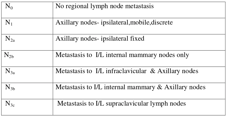
Table – 2 Regional lymph nodes—Clinical (N)
N0 No regional lymph node metastasis
N1 Axillary nodes- ipsilateral,mobile,discrete
N2a Axillary nodes- ipsilateral fixed
N2b Metastasis to I/L internal mammary nodes only
N3a Metastasis to I/L infraclavicular & Axillary nodes
N3b Metastasis to I/L internal mammary & Axillary nodes
N3c Metastasis to I/L supraclavicular lymph nodes
Table – 3 Distant metastasis (M)
MX Couldnot be assess the distant metastasis
M0 Absence of distant metastasis
[image:25.595.89.545.165.399.2]Table – 4 TNM Stage Groupings15 Stage 0 TisN0M0
Stage I T1N0M0
Stage IIa T0N1M0 T1N1M0 T2N0M0
Stage IIb T2N1M0 T3N0M0
Stage IIIa T0N2M0 T1N2M0 T2N2M0 T3N1M0 T3N2M0
Stage IIIb T4N0M0 T4N1M0 T4N2M0
Stage IIIc AnyT N3M0
Stage IV AnyT,Any N,M1
Treatment options in Carcinoma of Breast 14,16 Pimary - Surgery
Lumpectomy - Wide local excision
Simple/total mastectomy
MRM - Modified Radical Mastectomy
Radical mastectomy
Axillary lymph node dissection
Adjuvant
A. Radiotherapy
Post operative radiotherapy & Palliative radiotherapy
B. Systemic therapy
Chemotherapy
In 1960s first trails of combination chemotherapy were initiated for breast cancer management. First adjuvant chemotherapy was administered to women with positive nodes; later in 1980s the use was extended to node negative women as well.
Indications for chemotherapy 13,15
The proportional reduction in recurrences and mortality in both node positive and negative patients are similar, but given the better prognosis of node negative patients especially those node negative with small tumors(<1 cm). Younger females have proportionally greater reduction in both mortality and recurrence than older females with carcinoma breast. Combination chemotherapy has been more effective than single agent therapy.
DOSAGE AND SCHEDULE Choosing the regimen
REGIMENS 14,16
FAC:-5 Fluorouracil: 500mg/m2, Adriamycin: 50mg/m2, Cyclophosphamide: 500mg/m2, On day one & every 3 weeks for 6 cycles. AC:-Adriamycin: 60mg/m2, Cyclophosphamide: 600 mg/m2 - 4 cycles are given, once in every 3 weeks.
CMF:-Cyclophosphamide: 750 mg/m2, Methotrexate: 50 mg/m2,5-FU: 600 mg/m2- Given once in every 3 weeks, for 6 cycles.
FEC:-5 FU 500mg/m2,Epirubicin 50mg/m2,Cyclophosphamide 500mg/m2- On day one & every 3 weeks for 6 cycles.
TIMING OF TREATMENT 15,16 Adjuvant therapy:-(ACT)
Chemotherapy is given after a surgery for cure with an aim to prevent
local and systemic relapse by eradicating micrometastasis and to improve
outcome in cancers like breast, ovary, colon etc.
Neo-adjuvant therapy (NACT)
Palliative therapy
Depending on the receptor status, distant sites and those experiencing distant relapse after adjuvant treatment are treated by palliative measures either by endocrine manipulation or chemotherapy. Patients who are candidates for chemotherapy are those who fail hormonal therapy or presence of visceral metastasis.
CANCER CHEMOTHERAPY
Chemotherapy, which includes newly developed targeted treatments, is the
principle tool to treat most cancers. The development of effective
combination chemotherapy for Hodgkin’s lymphoma, childhood leukemia
and lymphomas in the 1960s provided curative therapeutic strategies for
patients with advanced malignancies of all types.
Historical perspective
Paul Ehrlich coined the term chemotherapy. Alkylating agents represent
the first class of chemotherapeutic drugs to be used in the clinical setting.
First clinical use of nitrogen mustard in a patient with non–Hodgkin’s
Clinical application of chemotherapy 17
Chemotherapy is used in four main clinical settings:-
(a) Primary induction treatment for advanced cancers for which there are no
other effective treatment. (b) As the primary or neoadjuvant treatment for
patients with localized disease for which local forms of therapy, such as
surgery, radiation, or both, are ineffective by themselves. (c) Adjuvant
treatment for early-stage disease following local modes of treatment like
radiotherapy, surgery or both. (d) Directly instilled into sites of specific
regions of the body directly affected by the cancer.
Primary Chemotherapy:
Cancers for which chemotherapy is a primary treatment modalityin cancers like Acute leukemia, Non-Hodgkin lymphoma, Myeloma, Hodgkin lymphoma, Germ cell cancer, Lymphoma, Ovarian cancer, Small cell lung cancer, Wilms tumor and Embryonal rhabdomyosarcoma.
Neoadjuvant Chemotherapy:
Cancers for which neoadjuvant chemotherapy is indicated for locally advanced diseases like Non–small cell lung cancer, Head and neck cancer,
Bladder cancer, Ovarian cancer, Breast cancer.
lung cancer, Osteogenic sarcoma, Colorectal cancer, Gastric cancer and Anaplastic astrocytoma.
Principles of cancer cell kinetics 2,17
The antineoplastic agents follow the logarithmic cell-kill kinetics to
exert their cytotoxic effects. Constant fraction of cells not numbers are
killed by these drugs. If a anticancer agent leads to a 4 log kill of neoplastic
cells and reduces the tumor burden from 1012 to 108 cells, the same dose is
used at a tumor burden of 107 cells reduces the tumor mass to 103. Cell kill
is therefore proportional, regardless of tumor burden.
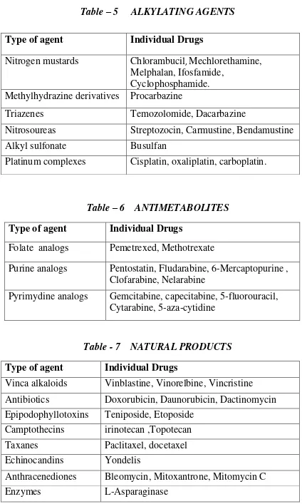
CLASSIFICATION OF ANTI NEOPLASTIC AGENTS 18
Table – 5 ALKYLATING AGENTS
Type of agent Individual Drugs
Nitrogen mustards Chlorambucil, Mechlorethamine,
Melphalan, Ifosfamide, Cyclophosphamide. Methylhydrazine derivatives Procarbazine
Triazenes Temozolomide, Dacarbazine
Nitrosoureas Streptozocin, Carmustine, Bendamustine Alkyl sulfonate Busulfan
Platinum complexes Cisplatin, oxaliplatin, carboplatin.
Table – 6 ANTIMETABOLITES Type of agent Individual Drugs
Folate analogs Pemetrexed, Methotrexate
Purine analogs Pentostatin, Fludarabine, 6-Mercaptopurine , Clofarabine, Nelarabine
Pyrimydine analogs Gemcitabine, capecitabine, 5-fluorouracil, Cytarabine, 5-aza-cytidine
Table - 7 NATURAL PRODUCTS
Type of agent Individual Drugs
Vinca alkaloids Vinblastine, Vinorelbine, Vincristine Antibiotics Doxorubicin, Daunorubicin, Dactinomycin Epipodophyllotoxins Teniposide, Etoposide
Camptothecins irinotecan ,Topotecan Taxanes Paclitaxel, docetaxel Echinocandins Yondelis
Anthracenediones Bleomycin, Mitoxantrone, Mitomycin C
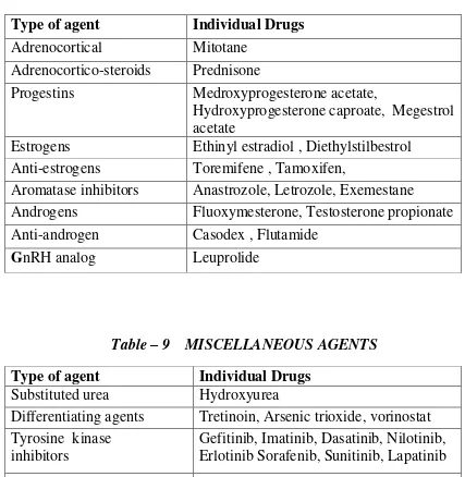
[image:32.595.94.518.64.771.2]Table - 8 HORMONES AND ANTAGONISTS
Type of agent Individual Drugs
Adrenocortical Mitotane Adrenocortico-steroids Prednisone
Progestins Medroxyprogesterone acetate,
Hydroxyprogesterone caproate, Megestrol acetate
Estrogens Ethinyl estradiol , Diethylstilbestrol Anti-estrogens Toremifene , Tamoxifen,
Aromatase inhibitors Anastrozole, Letrozole, Exemestane
Androgens Fluoxymesterone, Testosterone propionate Anti-androgen Casodex , Flutamide
GnRH analog Leuprolide
Table – 9 MISCELLANEOUS AGENTS
Type of agent Individual Drugs
Substituted urea Hydroxyurea
Differentiating agents Tretinoin, Arsenic trioxide, vorinostat Tyrosine kinase
inhibitors
Gefitinib, Imatinib, Dasatinib, Nilotinib, Erlotinib Sorafenib, Sunitinib, Lapatinib Proteasome inhibitor Bortezomib
Biological response modifiers
[image:33.595.91.518.121.559.2]
THE CELL CYCLE 19
Many cytotoxic agents act by damaging DNA. Their toxicity is greatest during the S, or DNA synthetic phase of the cell cycle. Others, vinca alkaloids and taxanes, block the formation of a functional mitotic spindle in the M phase. These agents are most effective on cells entering mitosis, the most vulnerable phase of the cell cycle. All cells display a similar pattern of cell cycle progression.
Phase that precedes DNA synthesis (G1) DNA synthetic phase (S)
An interval which is followed by the termination of DNA synthesis (G2)
The mitotic phase (M) in which the cell, containing a double complement of DNA, divides into two daughter G1 cells a probability
of moving into a quiescent state (G0) and failing to move forward for
long periods of time.
Figure – 1 Cell cycle18
DOXORUBICIN
Doxorubicin otherwise known as Adriamycin was the first anthracycline isolated from Streptomyces peucetius 21 in the year 1963 by both Italian and French group of scientists. Italian group isolated natural product doxorubicin from Streptomyces peucetius var. caesius. The French group produced semi synthetic derivatives. Epirubicin and Idarubicin are analogs of daunorubicin and doxorubicin respectively, only slightly differ in their chemical structures. Doxorubicin exerts broad-spectrum activity against solid human cancers. These drugs have the ability to produce free radicals and cause an irreversible, unusual cardiomyopathy which is related to exposure of the total dose of the drug.
CHEMISTRY
Figure – 2 Chemical Structure of Doxorubicin21
Absorption, fate, and excretion20
Doxorubicin is very poorly absorbed and is therefore administered parenterally. It is relatively rapidly distributed throughout the body including breast milk and bound to plasma proteins moderately. There is no evidence that it crosses the placenta but it may cause harm to the fetus.
Doxorubicin is cleared by complex hepatic metabolism and excrete via bile. The plasma disappearance curve is triphasic, with the first phase t1/2
10-20 mts due to distribution, the second phase 1.5 - 10 hrs largely to
Doxorubicin is converted into an alcohol intermediate 22 which plays a different role in its therapeutic activity. The drug rapidly enters the lungs, heart, liver, spleen and kidneys. It does not cross the blood-brain barrier. Doxorubicin is eliminated by its metabolic conversion into aglycones and few inactive products. Idarubicin is mainly metabolized into idarubicinol that accumulates in plasma and which is responsible of its activity. In the presence of hepatic failure the clearance of anthracyclines and their active alcohol metabolites are delayed. 50% of initial dose reduction should be considered in patients if serum bilirubin level is elevated.
FDA approved indications of doxorubicin18
Mechanism of action 18,20
Anthracyclines are directly affecting the transcription and replication of the neoplastic cells by intercalating with DNA. Their important action is mediated by their ability to form a tripartite complex with DNA and topoisomerase II. Topoisomerase II is an ATP-dependent enzyme that binds to DNA. It causes double-strand nicks at the 3'-phosphate backbone and allowing strand passage and uncoiling of super-coiled DNA. Then topoisomerase II religates the DNA strands. This enzymatic function is important for DNA replication and repair. The tripartite complex formation with anthracyclines or with etoposide inhibits the re-ligation of the broken DNA strands, which leads to apoptosis. Any defect in DNA double-strand break repair sensitizes neoplastic cells to damage by these drugs. But over expression of transcription-linked DNA repair may lead to drug resistance.
Anthracyclines generates free radicals in solution and in both normal and malignant tissues because of their quinone moieties. Anthracyclines can form semiquinone radical intermediates 22 that can react with O2 to produce
superoxide anion radicals which is responsible of their antitumor activity and also its cardiotoxicity.
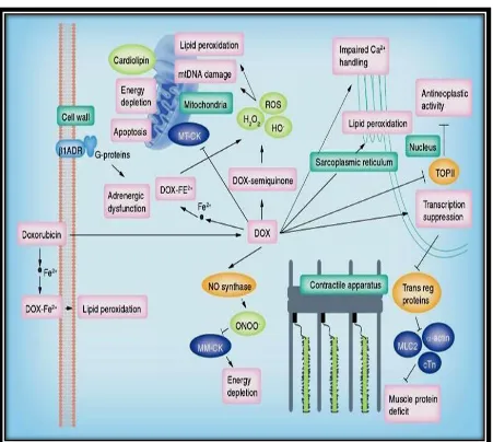
Figure-3 Mechanism of action of doxorubicin
cardiotoxicity. Anthracyclines exposure to the myocardial cells to leads to apoptosis. This process is mediated by p53 – a DNA-damage sensor and proteases, activated caspases, ceramide, and the Fas receptor-ligand system also have been implicated.
Dosage and administration 22
Doxorubicin is usually given as a single intravenous infusion dose of 60-75 mg/m2 slowly over 4 to 5 minutes that is repeated after 3 weeks. Since it is a vesicant, care must be taken to avoid extravasations, tissue necrosis. Drug can be given as a continuous infusion over 2-4 days through central venous access line. Dose reduction is needed in patients with liver failure. Reduce 50% of dose if serum bilirubin between 1.2 – 3.0 mg/dl 22. Infuse only 25% of dose if sr.bilirubin level greater than 3 mg/dl. Weekly therapy may be more cytotoxic to cancer cells than comparable doses of monthly bolus schedules36.
Drug interactions21
Doxorubicin is a potent radiosensitizing agent and radiation recall
reactions are potentially dangerous.
Radiation may increase the risk of cardio toxicity of doxorubicin.
Cyclosporine increases the toxicity of doxorubicin by inhibiting
P-glycoprotein
Vitamin D 3 enhances the doxorubicin induced oxidative damage in
breast cancer cells
Pacletaxel modifies the pharmacokinetic profile of doxorubicin.
CLINICAL TOXICITIES 18,22,23
Myelosuppression is the most common toxicity
Stomatitis, mucositis, occurs in nearly 10% of patients.
Total or near total alopecia 21 occurs in nearly every patient.
Extravasations
Severe nausea and vomiting are common
Anorexia and diarrhoea may occur in less than 10% of patients.
Facial flushing, conjunctivitis, and lacrimation.
Cardiotoxicity:-18,22 Cardiac toxicity is a rare unusual but peculiar
CYCLOPHOSPHAMIDE
Cyclophosphamide is an alkylating agent and chemical derivative of mechlorethamine which was first synthesized in Germany in 1958 26.It is used as a single agent to treat burkitt´s lymphoma. As combination chemotherapy 17 in carcinoma breast, ovary, lung and multiple myeloma. It is an immunosuppressant – used in nephritic syndrome, psoriasis.
Metabolism
Causes Microsomal hydroxylation
It is hydrolysis to Phosphoramide mustard (active) and acrolein.
Excretion as inactive oxidation products.
Mechanism of action
Produce DNA alkylation via the formation of reactive intermediates
that attack nucleophilic sites.
Cell cycle non specific 20
Dose schedule
Intravenous – 400 to 2000 mg/m2
Oral – 100 mg/m2 or 1 to 25 mg/kg/day in divided doses.
Figure – 4 Metabolism of Cyclophosphamide18
PHARMACOKINETICS
Bioavailability:- Oral >75 % ; Protein bound > 60 %
Primary elimination t ½:-Parent drug:- 3 – 10 hrs, Aldophosphamide:- 1.6 hrs, Phosphoramide mustard:- 8.7 hrs
Toxicity 18,19
nausea, vomiting, pulmonary fibrosis, transient myopia, cataract, Haemorrhagic cystitis and SIADH.
Teratogenesis – category D in FDA Cardiotoxicity 22,29,
Dose limiting cardiac toxicity occurs when using 7 fold increased normal dose, ≥180 mg/kg for 4 or more days or > 1.55 g/m2/day, mainly with transplantation doses. Mechanisms underlying the toxicity are believed to be injury of both endothelial cells and myocytes, and a picture of hemorrhagic myocardial necrosis can emerge. It causes low grade delayed cardiotoxicity which is not related with cumulative dose toxicity, more with patients older than 50 yrs of age. Maximum tolerated dose - 7000 mg/m2 Precautions- Use MESNA ( 2- Mercaptoethane sulfonate ) with high dose therapy.
Drug interactions20
Increased cytotoxicity with radiation sensitizers and glutathione
depletion.
Inhibit pseudo cholinesterase – risk of apnoea with succinyl choline.
Risk of cardiomyopathy when combine with anthracyclines.
Cimitidine enhances myelosuppression of cyclophosphamide.
5 – FLUOROURACIL ( 5 – FU )
5-Fluorouracil is an antimetabolite and structural analogue of DNA precursor of thymine 22. It is developed in 1957 by Heidelberger and Ansfield. It is used to treat carcinoma breast, gastrointestinal tract and many cancers.
Metabolism
5-FU is a prodrug that enters cells and phosphorylated to series of metabolites. It is converted enzymatically to active nucleotide forms intracellularly. DPD (Dihydropyrimidine dehydrogenase) catalyzes the initial, rate limiting step in 5 FU catabolism.
Mechanism of action 18,22
Fluorouridine triphosphate incorporated into RNA which interferes with RNA synthesis and its function. Thymidylate synthase inhibition is mediated by fluorodeoxyuridylate (FdUMP) which leads to thymidine 5´ monophosphate and thymidine 5´ triphosphate depletion. It also causes accumulation of deoxyuridine monophosphate and deoxyuridine triphosphate. Incorporation of fluorodeoxyuridine triphosphate and deoxyuridine triphosphate into DNA may affect DNA stability. Genotoxic stress triggers programmed cell death pathways.
Figure – 5 Mechanism of action of 5- Fluorouracil
Pharmacokinetics
Half life is 8 – 14 mts after iv bolus infusion.
Nonlinear pharmacokinetics due to saturable catabolism
Total body clearance decreases with increasing doses.
Elimination
90% eliminated by metabolism; less than 10% excreted as unchanged
in urine. 5 FU and its catabolites undergo biliary excretion.
Drug interactions
Dipyridamole increases 5 –FU clearance .
Sequential Methotrexate (Mtx) use Mtx increases 5- FU toxicity
and increases fluorouridine triphosphate (FUTP) incorporation into RNA; may antagonize DNA directed toxicity of 5 –FU.
Inhibitors of de novo pyrimydine synthesis increase 5 – FU anabolism
to the ribonucleotide level and 5 FU – RNA incorporation.
Toxicity
Gastrointestinal epithelial ulceration, Myelosuppression, skin rashes
Neurotoxicity – cerebellar ataxia, cognition dysfunction
Cardiac toxicity
It causes coronary spasm 22 & myocardial ischemia ( 1.6%) 29.
More common with high continuous infusion than iv bolus
administration. Fluorouracil can cause acute ischemic syndromes ranging from angina to MI and this can occur in patients without CAD (approximately 1% of patients), although it is more common in patients with pre-existing disease (4% to 5%).
Vasospasm is believed to be the mechanism triggering
ischemia, although thromboembolic events are also increased.
CHEMOTHERAPY INDUCED CARDIOTOXICITY 24
There are 2 types of drug induced cardiac toxicities commonly observed. Type I cardiotoxicity is more serious and causes permanent damage to the myocardium and type II which is usually reversible.
[image:49.595.103.536.395.730.2]Type I cardiotoxicity found in Anthracyclines, type II cardiotoxicity found in traztuzumab 23. Anthracyclines induced cardiotoxicity is a cumulative dose dependent toxicity which is characterized by each administration constitutes additive or sequential damage to the heart.
Figure – 6 Chemotherapy induced cardiotoxicity24
ANTHRACYCLINE INDUCED CARDIO TOXICITY 23 - 30
Acute and sub acute cardiotoxicity
Delayed onset- irreversible dilated cardiomyopathy
ACUTE TOXICITY 25
Acute toxicity manifest during or soon after administration of the drug. It is generally minor and reversible and independent of anthracycline dose. Manifestations are asymptomatic ECG changes, Myocarditis, Pericarditis, transient heart failure may develop. Incidence - approximately 11 %
ECG changes 27,29 may be manifests are ST changes, Low voltage QRS complex, Poor R wave progression, Prolongation of QT interval, Atrial / ventricular ectopics, T wave inversion, supra ventricular tachycardia, ventricular arrhythmia. Conduction block is more common in paediatric age group.
An acute reversible reduction in ejection fraction is observed in some patients in the 24 hours after a single dose, and plasma troponin I, a cardiac enzyme released with myocardial damage, may increase in a minority of patients in the first few days following drug administration.
SUB ACUTE TOXICITY
effusion, Arrhythmias – VT , SVT. Insult to the myocardium can be early detected by monitoring serum Troponin I level 27 which may be used to predict the future development of ventricular dysfunction.
DELAYED TOXICITY
Chronic or delayed cardiotoxicity may manifests months or years after anthracycline therapy3,27. Commonest presentations:-Congestive heart failure, Dilated cardiomyopathy. Incidence – up to 20%.These produces progressive, irreversible myocardial damage which are fatal and results in cardiac death. Dilated cardiomyopathy is the most important long-term toxicity of doxorubicin 25.
CUMULATIVE DOSE RELATED CARDIOTOXICITY
Cardiotoxicity is mainly due to dose administered during each cycle and on total cumulative dose28,. The risk of anthracycline cardiomyopathy depends on cumulative dose. A 5% risk is seen at 400 to 450 mg/m2 for doxorubicin 27, 900 mg/m2 for daunorubicin, 800 to 935 mg/m2 for epirubicin29, and 223 mg/m2 for idarubicin.
RISK FACTORS FOR CARDIOTOXICITY 26,
Cumulative dose - most significant risk factor
Age – elderly more than 60 yrs and children up to 12 yrs
Sex - females are more vulnerable 26
Length of infusion – risk more with rapid bolus infusion 26
Patients with abnormal cardiac function
Mediastinal irradiation – prior or concomitant irradiation 30
Trisomy 21- higher risk of early clinical toxicity 32
Combination chemotherapy24,25 with high dose cyclophosphamide,
bleomycin,vincristine, mitoxantrone , pacletaxel etc.
Obesity – more drug need because of more surface area
Diseases – Hypertension, Diabetes mellitus, Coronary artery disease,
PROPOSED MECHANISMS OF DOXORUBICIN INDUCED CARDIAC TOXICITY 33,34
Oxidative stress 30
Mitochondrial dependant ROS
NOS dependant ROS
NADPH dependant ROS
Fe – Doxorubicin complex
Intracellular calcium dysregulation
Changes in high energy phosphate pool
Endothelin-1upregulation
Extracellular matrix remodelling
RAS Activation
Multiple mechanisms may involve in the formation anthracycline induced cardiomyopathy. Oxidative stress is the well accepted mechanism of the above all.
MITOCHONDRIAL DEPENDANT ROS33
Mitochondria produce more than 90% of ATP needed for cardiomyocyte. Doxorubicin is a cationic drug which forms an irreversible complex with cardiolipin and the complex sequestered in the inner mitochondria. Doxorubicin alters cardiolipin protein interface within electron transport chain leads to more superoxide formation27,30. Protein responsible for carnitine transfer within mitochondria also disrupted, which leads to mitochondrial dysfunction. Pathological changes found are mitochondrial swelling, Myelin figure formation.
Figure – 7 Mechanism of semiquinone formation
In an animal model of anthracycline induced cardiomyopathy showed that defect in long chain fatty acid oxidation, which coupled with excessive glucose metabolism within cardiac mitochondria. Development of congestive heart failure was mainly due to shift of aerobic metabolism into anaerobic one.
mitochondria play a vital role in the development of doxorubicin cardiomyopathy. So preservation of mitochondrial function will lead to better cardiac outcomes.
NOS DEPENDANT ROS
Nitric oxide synthase is an enzyme with three isoforms. These are
eNOS – Endothelial
iNOS – Inducible
nNOS – Neuronal
NOS has two domain
Oxygenase
Reductase
NADPH dependant ROS38
[image:56.595.103.535.251.530.2]Doxorubicin combines with NADPH and forms oxygen free radicals. Acute cardiotoxicity is associated with SNP in the p22phox and Rac2 subunits. Chronic toxicity is mediated by NADPH oxidase subunit NCF4.
Figure-8 Doxorubicin induced apoptotic cardiac cell death
Fe – Doxorubicin Complex28,34
Doxorubicinol forms complex with thiol group of cytoplasmic aconitase or iron regulatory protein (IRP) and enhances stability of transferring mRNA & prevents its translation. Further decrease in IRP increases free iron level which promotes free radical generation. These free radicals interfere with iron sequestration and causes doxorubicin cardiomyopathy. ROS interferes with the function of G protein through lipid peroxidation. ROS alter the tertiary structure of the proteins. ROS also induces calcium release in myocardium.
APOPTOSIS30,32
Reactive oxygen species induce the formation of free radicals which stimulate proapoptotic genes and induce apoptosis via both intrinsic and extrinsic pathways, causes death of cardiac myocytes. In an apoptotic model, heat shock protein 1(HSP1) is activated by oxidative stress which promotes more HSP 25 protein production. By stabilizing p53 gene, HSP 25 promotes proapoptotic pathway. HSP acts as molecular chaperones which stabilize the proteins involved in antiapoptotic pathway by inhibiting their dephophorylation, ubiquination and its degradation33. By these mechanisms HSP 10,27,60 and Bcl-2 are maintains mitochondrial functions.
Figure – 9 Role of Heat Shock Proteins in Cardiotoxicity
INTRACELLULAR CALCIUM DYSREGULATION
It may be due to ROS production. Reactive oxygen species and hydrogen peroxide alter normal calcium homeostasis through disruption of sarcoplasmic reticulum in cardiac myocytes. This is mainly mediated by decreased SERCA2 mRNA expression which leads to poor calcium handling. It may also due to ryanodine receptor activation.
Figure – 10 Intracellular calcium dysregulation
Doxorubicin induces release of calcium from sarcoplasmic reticulum and opens the calcium channels. Doxorubicin increases L Type of calcium channel activity and inhibit sodium calcium exchanger channel in sarcoplasmic reticulum39.
MODIFICATIONS IN HIGH ENERGY PHOSPHATE POOL
Mitochondrial damage impairs the ability to generate adenosine tri phosphate (ATP). ATP depletion due to doxorubicin reduces the affinity of HSP 90 to ErbB2 which is a cardio protective protein. Because of ATP depletion, ErbB2 level also decreased. So HSP 90 unable to maintain its chaperone role. ATP depletion may be due to apoptosis and calcium dependent proteases which also consumes ATP.
Tokarska – schlattner et al40. demonstrated in an animal model of doxorubicin may impair energy signaling through AMP activated protein kinase (AMPK) which is involved in the inhibition of oxidation of fatty acid and to reduce mitochondrial function.
ENDOTHELIN-1 UPREGULATION33
EXTRACELLULAR MATRIX REMODELING33
Anthracyclines inhibits matrix metalloproteinase 1(MMP-1) and its transcription in tumor cells. But in heart it has opposite effect by increasing MMP-2 and MMP-9 levels. This leads to weakening of collgenous matrix and further progress to pathological remodeling which is depend on NADPH level.
DOXORUBICIN INDUCED UPS ACTIVITY34
UPS is a proteolytic system which enhances the degradation and post translational modification of proteins. Anthracyclines activates UPS mediated proteolysis by act on proteosomes. Doxorubicin potentiates MAPKS, P38 and JNK which induces cardiomyocyte apoptosis by reduce the expression of antiapoptotic proteins like Bcl-2 and increases proapoptotic agents like Bax,caspase-3,caspase-9 etc.
ACTIVATION OF RENIN ANGIOTENSIN SYSTEM (RAS) & ACE ACTIVITY:-41-43
ACE inhibition significantly improved cardiac function and survival rate in hamsters. Lisinopril, treated hamsters improved mortality, cardiac remodeling and cardiac dysfunction in doxorubicin-induced cardiomyopathy suggest that ACE, plays a crucial role in the production of cardiomyopathy following the doxorubicin.
Toko et al42. found that AT1-mediated Ang II signaling pathway plays vital role in DOX-induced cardiac impairment. AT1 antagonist can be used to prevent DOX-induced cardiomyopathy. These results suggest that inhibition of the RAS in the heart may attenuate DOX-induced cardiac damage via a mechanism independent of blood pressure.Wen-na zong et al59. in their animal model of doxorubicin induced heart failure in rats explored that the changes in plasma level of angiotensin-(1–7)[Ang-(1–7)] and myocardial expression of angiotensin II type 1/2 receptors (AT1R / AT2R) and Mas receptor.
Cardiac toxicity of anthracyclines may result from low levels of catalase in the heart combined with excessive mitochondria and myoglobin of the heart, which enhances the drug activation, as well as the sensitivity of cardiac glutathione peroxidase to free radicals attack. This destroys the activity of glutathione peroxides at the same time anthracycline administration stimulates cardiac hydrogen peroxide formation.
[image:63.595.103.548.330.727.2]
In contrast anthracyclines can be easily activated to its intermediates by hepatic enzymes; the liver has more active free radical defense systems and is able to actively efflux anthracyclines and its metabolites. It may result from doxorubicin itself or its major metabolite, doxorubicinol. The leading hypothesis for doxorubicin induced cardiotoxicity involves oxidative stress induced by free radicals formation.
MONITORING
Treatment with anthracyclines may necessitate lifelong cardiac monitoring. General cardiac evaluation should be done to all the patients who are going to undergo doxorubicin therapy. Patients with cardiovascular risk factors need close and frequent monitor and keeping track on cumulative dose of doxorubicin44.
Electrocardiogram28
Sinus tachycardia in previously normal heart rate is the earliest sign34 of cardiotoxicity. Failure to return baseline heart rate, resting tachycardia, loss of respiratory variations to the heart rate are the important predictors of future cardiotoxicity.
Echocardiogram44,46
LVEF44:-
Baseline and serial measurement of LVEF by echocardiogram is standard protocol in patients on anthracycline treatment44. It is the most commonly used non invasive parameter to detect cardiotoxicity earliest. LVEF = EDV-ESV X 100
EDV EDV – End Diastolic Volume
ESV - End Systolic Volume
Normal LVEF is between 50% - 70%. LVEF is calculated by 2D echocardiogram and Modified Simpson’s formula. To decrease the inter-observer variability, echo done by same physician for each patient at different time points. Mild – Ejection fraction less than 45%, Intermediate – EF between 45 – 50% , severe toxicity EF < 45%.Stop doxorubicin therapy if LVEF decrease 15% from baseline value.
FRACTIONAL SHORTENING 36,46,:-Calcuted from the following formula
FS = LVIDd – LVIDs X 100
LVIDd
Both LVEF and FS decrease in left ventricular systolic dysfunction44.Reduction in LVEF and FS precedes development of CHF, if we stop further infusion of doxorubicin we can decrease further progression of heart failure.
All patients should have a baseline measure of LVEF. For doxorubicin, poor-risk patients should have a repeat study at 200 mg/m2, and all patients should have a follow-up study at 300 to 400 mg/m2 and every 50 to 100 mg/m2 thereafter28.
Radionuclide ventrculography46
Well established method to determine LVEF35. It can be used to determine regional wall motion and diastolic function by serial monitoring. It is an invasive procedure and exposure to radiation is limits its use routinely.
Cardiac biomarkers46
Troponins28,47
Quantification of cardiac contractile proteins such as Troponin is a highly sensitive method of detecting myocardial cell injury and anthracyclines induced cardiotoxicity as well. Troponin I levels may be elevate in doxorubicin treated patients before deterioration in LVEF. Elevation of troponin I level after exposure to anthracyclines, an indicator of early cell death which may be beginning of ongoing process. Elevation of troponin I which correlate with subclinical LV Dysfunction.
Natriuretic peptide27
These are released from the atria (ANP), left ventricle (BNP) in response to circulating volume and intra cardiac pressures. BNP level correlates more with diastolic dysfunction than systolic dysfunction. Both troponin and BNP are highly specific and sensitive tool for early detection of myocardial defect.
MRI:-Valuable tool to assess myocardial function and damage
Computed tomography:-High radiation exposure, Low temporal resolution
Endo Myocardial Biopsy (EMB)28,30
An invasive procedure, biopsy taken from endocardium of right ventricle of the heart. Gold standard investigation for anthracycline cardiomyopathy and provides histological evidence of cardiotoxicity. Total doses of doxorubicin as low as 250 mg/m2 can cause pathological changes in the myocardium, as demonstrated by subendocardial biopsies. Histological changes may be found are mitochondrial swelling, Myofibrillar drop out, stellate scars, Vesicular dilatation, Adria cells, Myocytes hypertrophy, Myocytes degeneration and Loss of cross striations. Disadvantages are High cost, Need experts to do biopsy and to interpret its histology, only small sample of myocardium is tested.
STRATEGIES TO REDUCE DOXORUBICIN CARDIOTOXICITY
With awareness of the cardiac risk, anthracycline cardiotoxicity may be avoided by recognizing risk factors, early detection, limiting total cumulative dose, and more recently, using cardio protective agents or modified infusion drug regimens.
The prevention of doxorubicin induced cardiotoxicity depends on three approaches:
Using analogues of anthracycline with less cardiotoxicity
Modified administration of doxorubicin30.
Doxorubicin toxicity can be minimized by:
Changes in doxorubicin administration to continuous infusion.
Minimizing the total cumulative dose to<400 mg/m2
Using liposomal - encapsulated doxorubicin28.
Using dexrazoxane to decrease the free iron formation. But it should
be used only after exposure of doxorubicin dose of 300mg/m2 because of the possibility of its ability to diminish the effect of anticancer agents.
Using antioxidant to reduce the production of ROS with N-acetyl
cysteine, coenzyme Q10.
Treatment of early cardiac events needed, to minimize the evolution
of doxorubicin-induced cardiotoxicity.
ACE Inhibitors and β-blockers are routinely used to treat the late
RISK FACTORS SCREENING AND PREVENTION OF CARDIAC EVENTS
All patients should be screened for preexisting cardiac risk factors like hypertension, diabetes, obesity and smoking before commencing therapy with doxorubicin. ACE inhibitors with or without beta blocker administration during the early course of the toxicity shows increased efficacy in the prevention of delayed cardiac events. Pretreatment with these agents can prevent the fall in ejection fraction seen with patient’s undergone high-dose chemotherapy.
Dose limitation
The risk of cardiotoxicity can be reduced by keeping the total cumulative dose of doxorubicin well below550mg/m2. It is recommended that the lifetime dose should be <450-550 mg/m2 for doxorubicin. It is better to limit the doxorubicin cumulative dose less than 350 mg/m2 in patients with cardiovascular risk factors which decrease the need of careful monitoring.
Schedule Modification32
cardiotoxic than bolus injections. An important strategy to be followed to reduce the risk of cardiotoxicity is infusion over several hours. Weekly regimen schedule allowed more drug up to 200 mg/m2 doxorubicin which may be tolerated by the individuals compared to 21 day routine cycle and also improve biopsy score. Continuous infusion (24-96 hrs) are safe by minimize the plasma drug concentration while maintaining antitumor effect which is confirmed by endomyocardial biopsy44. These measures reduce the cardiotoxicity of anthracyclines but did not prevent it. Rapid bolus infusion saline increased risk of extravasations / tissue necrosis. Using central line for prolonged >24 hrs infusion, would reduce the risk of extravasations. Drawback of central line infusion
Increase the risk of infections, more time consuming to set up and flush the line weekly, line position checked by chest x ray, more than 96 hrs continuous infusions may leads to more chance of hand foot syndrome and mucositis, need of portal infusion pump and indwelling catheters.
ALTERNATIVE STRATEGIES Liposomal Anthracyclines28,44
Formulations of liposomal anthracyclines18
Pegylated liposomal doxorubicin
Non pegylated liposomal doxorubicin
Other unique delivery systems :- Starch microspheres, Albumic
microspheres, Lipeodal Doxorubicin
FDA recently approved liposomal formulations of doxorubicin for Kaposi sarcoma, carcinoma ovary, breast and Multiple Myeloma. These formulations have same efficacy with minimal cardiac toxicity when compared with free doxorubicin. Less chance of developing cardiotoxicity with pegylated liposomal doxorubicin than with normal one in all subgroups analyzed ( age>65 yrs, prior anthracyclines therapy). However they may cause hand–foot syndrome and Stomatitis.
Non-pegylated liposomal doxorubicin is less cardiotoxic than conventional doxorubicin. It has comparable antitumor activity with less diarrhea and nausea/vomiting, neutopenia. Findings from a meta-analysis showed a lower rate of clinical and subclinical heart failure with nonpegylated liposomal doxorubicin.
Anthracycline analogues27
Analogue development holds the promise of good antitumor activity with less cardiotoxicity. Epirubicin produce heart failure only in 10% of breast cancer patients took cumulative dose of more than 600mg/m2.
Idarubicin
The dose of idarubicin is 12 mg/m2/day for 3 days by intravenous injection. Slow injection over 10-15 minutes is followed to avoid extravasations. It has less cardiotoxicity than doxorubicin.
Daunorubicin
Daunorubicin (rubidomycin, daunomycin) is infused intravenously. The dose is 25-45 mg/m2/day for 3 days. Total doses of >1000 mg/m2 are associated with risk of cardiotoxicity.
Epirubicin
It is indicated in adjunctive therapy for treatment of breast cancer. It is administered in doses of 100-120 mg/m2 intravenously every 3-4 weeks. Total doses >900 mg/m2 sharply increase the risk of cardiotoxicity. Its toxicity profile is the same as that of doxorubicin.
Valrubicin
bladder once a week for 6 weeks. Less than 10% of instilled drug is absorbed systemically.
REGIMENS WITHOUT ANTHRACYCLINES44:-
Another strategy to reduce cardiotoxicity is simply not to use doxorubicin. Although taxanes also have a potential cardiovascular risk, in a comparative study found that treatment with docetaxel conferred no apparent cardiotoxicity benefit than doxorubicin. Thus, the choice of agent than doxorubicin should be balanced against reduction in antitumor efficacy and against the overall toxicity profile of the two agents. Cardiac events occur during doxorubicin schedule or in the first year after its completion is often manifesting as arrhythmias, such as atrial fibrillation or pericarditis. These should be treated according to guidelines, which may heart rate or rhythm modifying drugs and antithrombotic agents. Patients with symptomatic CHF should receive ACE inhibitors and beta blockers.
Blocking agents44
Differences in the biochemical mechanisms of antitumor and cardiotoxic activity provide a potential avenue of selectively inhibiting or preventing the adverse effect.
Targets for the approach
Agents that prevent free radicals generation
FREE RADICALS50
Oxidative Free radicals are constantly formed in the human system and has been implicated in several human diseases. These free radicals are scavenged or removed by enzymatic and non enzymatic antioxidant defense mechanism. When these defense mechanisms are inadequate, oxidative stress can damage proteins, carbohydrates, lipids and nucleic acids.
A free radical contains one or more unpaired electrons in their orbitals. When two radicals meet, they can combine their unpaired electrons and join to form a covalent bond
AO + AO --- A ---A
Types of free radicals
Hydroxyl radicals (*OH)
It is one of the most reactive free radical, important role in tissue damage by radiation.*OH radical causes DNA fragmentation and extensive alterations in the purine and pyrimidine bases. Hydroxyl radicals (*OH) are also known to induce lipid peroxidation of fatty acid side chains of membrane phospholipids. Accumulation of lipid peroxides in a biological membrane disrupts its integrity and function.
Superoxide radicals
O 2 + e
-Superoxide radicals (O2 .
-) is an oxygen molecule deficient of one electron, involved in the regulation of fibroblast proliferation, vasodilatation and phagocytosis.H2O2 is not a free radical but is a powerful oxidizing agent and
is removed by the enzymes catalase and glutathione peroxidase. Nitric oxide
NO has one unpaired electron and can be regarded as free radical. Combination of NO with superoxide radicals results in the formation of peroxynitenite (OONO) which is highly toxic.
Transition metal ions and free radicals
Iron and copper ions are excellent promoters of free radical formation. These can convert H2O2 in to highly reactive *OH , leads to accumulation of cytotoxic end products of lipid peroxides.
Antioxidant defensive mechanisms
Antioxidant or free radical scavenger is a substance at low concentrations can prevent or delay the oxidation of an oxidizable substrate.
Major intracellular Antioxidants50
SOD, Catalase, Glutathione peroxidase, α Tocopherol, Ascorbic acid Major Extracellular Antioxidants
OXIDATIVE STRESS AND DISEASE50
Disease involving several organs / systems
Rheumatoid arthritis, Myocardial infarction, Malignancy and Aging Disease involving specific organs / systems
Nervous system:- parkinsonism, Alzheimer’s disease; Renal system :-heavy metal, aminoglycoside & NSAIDs toxicity; Respiratory system :-emphysema, oxygen toxicity; Endocrine :- diabetes mellitus; Drug toxicity:- Anthracyclines, halothane, chloroform, Lead poisoning, Antimalarials – Haemolysis.
ANTIOXIDANTS OF NATURAL ORIGIN
Superoxide dismutase (SOD)
The enzyme superoxide dismutase (SOD), found in all cells and is necessary for scavenging the Superoxide radical and prevent its accumulation and production of oxidative stress to the living system. SOD catalyzes a dismutation reaction, where by one O2
. –
is oxidized to O2 and
the other reduced to H2O2.
O2 + O2 . –
+ 2H --- H2O2 + O2
It protect against ischemic or reperfusion injury seen after MI, cerebral stroke or organ transplants. The large inflow of oxygen during reperfusion results in generation of O2
. –
Alpha Tocopherol
Severe neurological damage, hemolytic syndrome, atherosclerosis, retrolental fibroplasias.
Ascorbic acid
Vitamin c is an essential component of human diet and functions as a cofactor of several hydrolyze enzymes. Useful in atherosclerosis and cancers.
Physiological antioxidants50
Lactoferrin – iron binding protein, Nicotanamide, Glutathione – organ transplantation, cystic fibrosis, AIDS- p.carnii pneumonia, Acetylcysteine – paracetamol poisoning, Beta carotene – anti cancer effect, Melatonin.
Synthetic antioxidants:- Allopurinol , Desferrioxamine, Probucol.
Drugs with additional antioxidant property50
ACE Inhibitors, Calcium channel blockers, NSAIDs, Selegiline.
Free radical scavengers50
↑itamin E (α Tocopherol),N-Acetylcysteine, Coenzyme Q10,
DEXRAZOXANE8,49
Dexrazoxane is a prodrug of bisdioxopiperazine derivative. It is an iron chelating agent. Highly effective in reducing doxorubicin induced cardiotoxicity and extravasations injury. It is the only FDA approved drug to prevent doxorubicin induced cardiotoxicity. Intracellularly it is hydrolyzed to form bidentate chelator. Structurally it is similar to EDTA also a strong inhibitor of topoisomerase II enzyme. It quickly binds to intra cellular iron and strips Fe 2+ from iron – doxorubicin complex by which it decrease the generation of free radicals8. Although its benefits against late cardiotoxicity not proved, but it is known to decrease the acute cardiotoxicity of doxorubicin. Dexrazoxane also have decrease antitumor response rate to the anthracycline treatment. Route of administration – intravenous. Biphasic elimination – distribution T ½ - 0.8 hrs, elimination T
½ -9 hrs. Though dexrazoxane may be useful to reduce cardiotoxicity; its use
should be weighed against the risk of a lower response rate49 and the additional costs of treatment.