“A STUDY ON THE SURGICAL MANAGEMENT OF ACUTE INTESTINAL
OBSTRUCTION IN ADULTS
THE TAMILNADU DR. M.G.R. MEDICAL UNIVERSITY
In partial fulfillment of the regulations for the award of the degree of
M.S. IN GENE
THANJAVUR MEDICAL COLLEGE,
THANJAVUR
THE TAMILNADU DR. M.G.R. MEDICAL UNIVERSITY
DISSERTATION ON
A STUDY ON THE SURGICAL MANAGEMENT OF ACUTE INTESTINAL
OBSTRUCTION IN ADULTS”
Dissertation submitted to
THE TAMILNADU DR. M.G.R. MEDICAL UNIVERSITY
In partial fulfillment of the regulations for the award of the degree of
M.S. IN GENERAL SURGERY
BRANCH
–
I
THANJAVUR MEDICAL COLLEGE,
THANJAVUR
-
613 004
THE TAMILNADU DR. M.G.R. MEDICAL UNIVERSITY
CHENNAI
-
600 032
APRIL
-
2014
Page | 0
A STUDY ON THE SURGICAL MANAGEMENT OF ACUTE INTESTINAL
THE TAMILNADU DR. M.G.R. MEDICAL UNIVERSITY
Page | 1
CERTIFICATE
This is to certify that this dissertation entitled “A STUDY ON THE SURGICAL MANAGEMENT OF ACUTE INTESTINAL OBSTRUCTION IN ADULTS ” is the bonafide work of Dr.AQUINAS. B in partial fulfilment of the requirements for M.S Branch -I (General Surgery) Examination of the Tamilnadu Dr. M.G.R. Medical University to be held in APRIL - 2014 under my guidance and supervision during the academic year march- 2012 to december - 2013.
Prof.Dr.V. BALAKRISHNAN, M.S.,
Head of the Department, Department of General surgery, Thanjavur Medical College, Thanjavur - 613 004.
Prof. Dr. K.MAHADEVAN M.S.,
DEAN,
Thanjavur Medical College, Thanjavur - 613 004. Prof. Dr. R. YEGANATHAN, M.S. D.A.,
Unit Chief S-VI,
Page | 2
DECLARATION
I, Dr.AQUINAS. B, solemnly declare that the dissertation titled “A
STUDY ON THE SURGICAL MANAGEMENT OF ACUTE INTESTINAL OBSTRUCTION
ADULTS” is a bonafide work done by me at Thanjavur Medical College, Thanjavur
during March - 2012 to december - 2013 under the guidance and supervision of
Prof. Dr. R. YEGANATHAN M.S. D.A., Unit Chief S-VI, Thanjavur Medical College,
Thanjavur.
This dissertation is submitted to Tamilnadu Dr. M.G.R Medical University
towards partial fulfilment of requirement for the award of M.S. degree (Branch -I)
in General Surgery.
Place: Thanjavur.
Page | 3
ACKNOWLEDGEMENT
I gratefully acknowledge my sincere thanks to Prof. Dr. K.Mahadevan M.S., Dean, Thanjavur Medical College, Thanjavur, for allowing me to do this dissertation and utilize the institutional facilities.
I am extremely grateful to Prof. Dr. V. Balakrishnan M.S., Head of the Department, Department of General surgery, Thanjavur Medical College, for his full-fledged support throughout my study and for his valuable suggestions and guidance during my study and my post graduation period. I am greatly indebted to Prof. Dr. R. Yeganathan M.S. D.A., my Professor and Unit Chief, who is my guide in this study, for his timely suggestions, constant encouragement and scholarly guidance in my study and in my post graduation period.
I profoundly thank my respected professors, Prof. Dr. Maragadhamani M.S., Prof. Dr. M.Elangovan, M.S., Prof .Dr. Shanthini, M.S., Prof. Dr. Karunaharan M.S, M.D., and Prof.Dr. P.
Rajagopal, M.S., for their advice and valuable criticism which enabled me to do this work effectively.
My sincere thanks to assistant professors, Dr. K. Sathyabama, M.S, and Dr. P. Vanathi M.S., for their motivation, encouragement and support.
A special mention of thanks to all the patients who participated in this study for their kind cooperation.
Page | 4
CONTENTS PAGE NO.
1. INTRODUCTION--- 5
2. OBJECTIVES--- 6
3. REVIEW OF LITERATURE--- 7
4. METHODOLOGY--- 77
5. RESULTS--- 83
6. DISCUSSION--- 93
7. CONCLUSION--- 101
8.BIBLIOGRAPHY--- 102
9. ANNEXURES (i) PROFORMA--- 107
(ii) KEY TO MASTER CHART--- 113
ABSTRACT
BACKGROUND AND OBJECTIVES
Intestinal obstruction remains as one of the most common intra-abdominal
pathologies encountered by surgeons whether it is caused by hernia, neoplasm,
adhesions or any biochemical disturbances. Intestinal obstruction of the small or
large bowel continues to be an important cause of morbidity and mortality. The
objective of this study is to analyse the clinical features, treatment and outcome of
patients with acute intestinal obstruction along with the cause of obstruction and
causes of bowel ischaemia, necrosis and perforation. The following are the
objectives of the study:
(1)to study various patterns of presentation, various causes, significance of early
recognition, diagnosis and treatment.
(2) to study various influencing factors like age, sex, diet and socio-economic
status in the pathogenesis of acute bowel obstruction.
(3)to study morbidity and mortality rates in acute intestinal obstruction.
METHODS
The materials for the clinical study of bowel obstruction were collected from
intestinal obstruction have been studied. Patients were belonging to the age groups
from 12 years to 85 years, paediatric age group (<12 yrs) is excluded from this
study. The case selection criteria was based on history, physical findings,
radiological findings and haematological parameters. The study has been divided
into Clinical study, Investigations and Treatment. Postoperative Follow-up was
done in majority of the patients upto six months after the discharge of patients.
The results have been tabulated stressing on following points: age, sex, symptoms,
clinical findings, investigations, abnormalities, possible causative factors, operative
findings and operative procedure done and complications if any occurred.
RESULTS
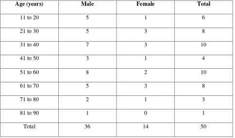
The study group had 50 cases of acute bowel obstruction in the adult age group
from 12 years to 85 years. The prevalent age groups are 31-40 and 51-60 age group
with around 20% each in the total study. The most common cause of acute
intestinal obstruction in the adults in this study series has been post-operative
Adhesions (40%) and the next being obstructed Hernia (30%). The clinical features
of abdominal pain, vomiting, constipation have been the main symptoms in this
study. Abdominal Tenderness, guarding, rigidity, rebound tenderness and shock
The commonest type of obstruction has been due to adhesions or band arising from
the previous surgeries. This has constituted to about 40% of the cases of the study
group. The second most common type of bowel obstruction is due to
obstructed/strangulated external hernia. Salient features had been pain in the groin
swelling, acute onset of swelling which is tender, not reducible and without cough
impulse. Obstructed hernia constituted about 30% of the total cases studied.
Volvulus of the sigmoid colon was 4% in this series. Conservative measures
included insertion of ryle’s tube and iv fluids but all the cases underwent
laparotomy due to failure of the recovery of symptoms. Derotation of the volvulus
and sigmoidopexy was done in one case and in one case with vascular
compromise, resection and anastamosis was done. Malignancy of the colon was
seen in 7 patients constituting 14% of cases. 65% of the malignancies were in the
age group of 35-75 years. Of these 2 patients have been managed with Hartman’s
procedure. One case has been managed with loop transverse colostomy and
remaining patients were managed with resection and anastamosis. Most of the
deaths occurred those with malignancy.
Although pulmonary tuberculosis is more prevalent in India, due to the use of
antitubercular drugs, abdominal tuberculosis is becoming less prevalent. In this
study incidence of ileocaecal tuberculosis was 4% and both patients were managed
obstruction was 6%. One case was managed with simple reduction and the
remaining two patients underwent resection and anastamosis. One case of
mesenteric ischaemia was recorded is our study. This patient was managed with
resection and anastamosis but patient expired due to septicemia. The complication
rate in this study was 18%. Overall mortality of this study was 14%. The results
obtained from this study was comparable to various other studies. Malignancy and
mesenteric ischemia had more mortality than simple obstruction due to
postoperative adhesions. The poor prognosis of the disease was because of late
presentation to the hospital with a high incidence of bowel damage with associated
faecal peritonitis. The morality in the postoperative period was mostly due to
faecal peritonitis, pneumonia and respiratory tract infection.
INTERPRETATION AND CONCLUSION
Acute intestinal obstruction remains to be an important surgical emergency in the
surgical field. Success in the treatment of acute bowel obstruction depends mainly
on the early diagnosis and efficient management and treating the pathological
effects of the obstruction as much as the treatment of the cause itself. Erect
abdomen X-ray is a valuable tool in the diagnosis of acute intestinal obstruction.
Postoperative adhesions have been the most common cause to produce bowel
diagnose the intestinal obstruction. Mortality is still significantly high in acute
intestinal obstruction in adults.
KEY WORDS: Blood pressure; Computed tomography; Central venous
pressure; Superior mesenteric artery; Acute intestinal obstruction; Extra cellular
Page | 5
INTRODUCTION
Bowel obstruction remains one of the most common intra-abdominal problems
faced by general surgeons in their practice whether caused by hernia, neoplasm,
adhesions or related to biochemical disturbances intestinal obstruction of either the
small or large bowel continues to be a major cause of morbidity and mortality.1
They account for 12% to 16% of surgical admissions for acute abdominal
complaints. Manifestations of acute intestinal obstruction can range from a fairly
good appearance with only slight abdominal discomfort and distension to a state of
hypovolemic or septic shock (or both) requiring an emergency operation.
The death due to acute intestinal obstruction is decreasing with better
understanding of pathophysiology. Improvement in diagnostic techniques, fluid
and electrolytes correction, much potent anti-microbials and knowledge of
intensive care. Most of the mortalities occur in elderly individuals who seek late
treatment and who are having associated pre-existing diseases like, diabetes
mellitus, cardiac diseases or respiratory disease.
Early diagnosis of obstruction skillful operative management, proper technique
Page | 6
OBJECTIVES
1. To study the various ways of presentation, various etiologies, importance of
early recognition, diagnosis and management.
2. To study the various influencing factors like age, sex, diet and socio-economic
status in the pathogenesis of acute intestinal obstruction.
Page | 7
REVIEW OF LITERATURE
HISTORICAL REVIEW
The attempts to treat acute intestinal obstruction goes back centuries. In 6th
century, Sushrutha wrote oldest known descriptions of intestinal surgery. Forms of
intestinal obstruction like strangulated hernia, intussusception had been known to
the ancient Egyptians. Intestinal obstruction had been observed by Hippocrates
(460-370 BC). The earliest surgery was performed by Proxogorous (350 BC), who
made an enterocutaneous fistula to relieve obstruction.
• Fabricus d’Aquopendente in 12th century AD described a procedure of
bowel repair involving end-to-end anastamosis.
• Sanctus in 16th century treated intestinal obstruction by administering
metallic mercury to patients.
• John Arderence (1306-1390) was the first person who wrote the book on
“Passio Iliaca” (Appendicitis or intestinal obstruction).
• Ambrose Pare (1510-1590) was the first to recognize intestinal obstruction
as a pathological entity. For severe patients he used mercury in water, lead
bullets smeared with mercury.3
Page | 8
• Kerckring in 1670 described the jejunal valvulae conniventes.
• Johnston in 1938, Harris in 1945, Cantor in 1946 and Gafton Smith in 1952
described many other tubes for gastrointestinal drainage.
• Bishop and Allock (1960) researched the bacteriology of gut above the
obstruction.
• Bilgutay (1963) has encouraged the use of faradic current for intestinal
paresis following operations.
• The experimental use of Doppler ultrasonography to determine the viability
of ischaemic intestine was first described by Wright in 1975.
• Marfuggi and Greenspan in the year 1981 reported 93% accuracy of a
fluoroscopic dye injection technique to determine the viability of ischaemic
bowel.
• Bookstein in 1982, used angiography for diagnosing and treating small
bowel bleeding.
• In 1996, Akgun gained attention in mesosigmoplasty as a definitive
operation sigmoid volvulus.5
• In 1997, Yaco et al. evaluated the diagnostic procedure for diverticular
Page | 9
EMBRYOLOGY OF SMALL INTESTINE
During the early stage of embryogenesis, the primitive bowel is in free
communication with rest of the yolk sac. In the cephalic and caudal parts of the
human embryo, the primitive gut creates a blind ending tube the foregut and the
hindgut and the middle part, the midgut remains temporarily connected to the yolk
sac.
In the 5th week embryo, there will be a rapid elongation of the gut and its
mesentery resulting in formation of a primary intestinal loop. At its apex, the loop
continues to be in open connection with the yolk sac by way of the
vitello-intestinal duct. The cephalic part of the loop develops into the distal part of the
duodenum, jejunum and part of the ileum. The caudal limb develops into the lower
portion of the ileum, the caecum, the appendix, the ascending colon and the
proximal two-thirds of the transverse colon. The hindgut develops into distal
one-third of transverse colon, the sigmoid, the rectum and also part of anal canal.
Fixation of the gut
Small and large bowel are suspended from the posterior abdominal wall by Means
of a mesentery. After the complete rotation of the gut, the duodenum, the
ascending colon, the descending colon and the rectum come to lie in the
Page | 10
Page | 11 There are three errors in the stages of rotation.
1. Non-rotation
2. Reversed rotation
3. Malrotation
Accessory bands of peritoneum
Can cause (i) Intestinal obstruction (ii) Kinking (iii) Angulation of bowel. Failure
of part of the original membrane to disappear or minor alterations in the
development of secondary mesentery may result in accessory peritoneal bands.
These are:
• Lane’s ileal band: This is a thickened peritoneal band extending from the
right iliac fossa to the 5 cm of ileum which on continuous contraction causes
kinking of the small bowel and resulting in obstruction.
• Mesosigmoid membrane (Lane’s first and last band): This is formed by the
thickening of peritoneum which extends from the pelvic brim of the left iliac
region to the junction of descending and sigmoid colon.
• Genitomesenteric fold of Douglas: causes kinking of the appendix causing
obstructive appendicitis as it extends from the back of the terminal
Page | 12
ANATOMY
The small intestine is the longest part of the gastrointestinal tract and it extends
from the pyloric orifice of the stomach to the ileocaecal fold. This forms a hollow
tube, which is approximately 6-7 m long with narrowing diameter from the
beginning to end, it consists of the duodenum, the jejunum and the ileum.7
The adult duodenum is 20-25 cm length and the name is coined as duodenum
because length is as long as width of 2 fingers. It is shortest, widest and the most
fixed part. It does not have any mesentery and is partially covered by peritoneum.
Its course presents a remarkable curve somewhat like a horseshoe type, the
convexity is being directed towards the right and concavity directed to the left
embracing the head of the pancreas. It is divided into four portions. First part
(superior portion), Second part (descending portion), third part (horizontal portion)
and fourth part (ascending portion).
Blood supply and nerve supply
Arteries supplying the duodenum are found to arise from the right gastric artery,
supraduodenal, right gastroepiploic, the superior and the inferior
pancreaticoduodenal arteries.
Page | 13
Nerves: They come from the coeliac plexus.
Lymph nodes: along inferior and posterior pancreatico duodenal artery.
Jejunum and ileum
In small intestine excluding duodenum, upper 2/5 are formed by jejunum and
lower 3/5 are ileum. The rest of the small intestine extends from the
duodeno-jejunal flexure to the ileo-caecal valve, ending at the junction of the caecum and
ascending colon. It is totally covered by the peritoneum and it is arranged in a
series of coils attached to the posterior abdominal wall by the mesentery. The
jejunal loops are characteristically situated in the upper abdomen to the left of the
midline, whereas the ileal loops tends to lie in the lower right part of the abdomen
and pelvis. This distribution can be reversed during paralytic ileus or small bowel
obstruction due to rotation around the mesentry attachment following bowel
distension.
The wall of jejunum and ileum is composed of serosa of visceral peritoneum,
muscular of longitudinal and circular smooth muscle fibres and a mucosa of
Page | 15
Blood supply
Blood supply is by the superior mesenteric artery which is a branch of abdominal
aorta, the branches of which, reaching the mesenteric border, extend between the
serosal and muscular layers. After this, numerous branches traverse the muscle,
supplying it and thus forming an intricate submucosal plexus from which many
minute vessels pass to glands and villi. The superior mesenteric veins
correspondingly follow the arteries.
Nerve supply
Nerve supply is by vagi and thoracic splanchnic nerves through the celiac ganglia
and the superior mesenteric plexus.
Large intestine
It is about 150 cm in length, it extends from the terminal ileum to the anus. Its
function is mainly absorption of fluids and solutes and it differs in its structure,
size and arrangement from the small bowel in the following ways:
• It is for the most part fixed in position.
• Its longitudinal muscle, though being a complete layer, is concentrated into
Page | 16
• The colonic wall is puckered into sacculations (haustrations) and appendices
epiploicae by the taeniae.
• It has a great diameter.
The divisions are the caecum, colon proper and the rectum.
Caecum
The caecum is like a blind pouch that lies in the right iliac fossa, with its average
axial length being about 6 cm and its breadth is about 7.5 cm. this continues
proximally with the distal ileum and distally it continues with the ascending colon
and it is related posteriorly to iliopsoas muscle and the femoral nerve, anteriorly
related to the abdominal wall, greater omentum and the loops of ileum. Almost the
entire posterior part of the caecum is attached to the abdominal wall, but in some
cases it is wholly unattached.
Ileocaecal valve
The ileum opens onto the posteromedial aspects of the caeco-colic junction and
two flaps which form a projection into the lumen of the colon. The valve is
actually closed by sympathetic tone. It is mechanically closed during the
distensions of caecum and this prevents the reflux of contents of the caecum into
Page | 17
Colon
The colon is conveniently sub-divided in four parts: (1) Ascending, (2) Transverse,
(3) Descending and (4) Sigmoid colon.
Ascending colon
It is usually fused with the posterior body wall and is covered by peritoneum
anteriorly. It constitutes about 15 cm in length and is narrower than the caecum. It
ascends onto the inferior surface of the right lobe of the liver, on which it causes a
shallow depression. Here, it turns abruptly forwards and to the left forming right
colic flexure.
Hepatic flexure
Anteriorly it is covered by the peritoneum, but posteriorly it is not covered by
peritoneum and It lies in direct contact with renal fascia. It is related posteriorly to
the infero-lateral part of the anterior surface of the right kidney above, and
anterolaterally lies the right lobe of the liver, anteromedially are the descending
part of the duodenum and the fundus of the gallbladder.
Transverse colon
It extends from the hepatic flexure to the left colic flexure measuring 50 cm. The
Page | 18 that had secondarily fused with posterior wall of the omental bursa. The transverse
colon hangs in either U or V shaped curve. Above the transverse colon lies the
liver and gallbladder, the greater gastric curvature and the lateral end of spleen,
below lies the small intestine, in front are the posterior layers of the greater
omentum and behind are the descending part of the duodenum, the pancreatic
head, the upper end of the mesentery, duodenojejunal flexure and the loops of the
jejunum and ileum. The transverse colon sometimes may be interpositioned
between liver and diaphragm (Chilaiditi syndrome).
Splenic flexure
This is the junction of the transverse and the descending colon in the left
hypochondriac region. It is closely related to the lower part of the spleen and
pancreatic tail above and medially with the anterior of the left kidney. It is attached
to diaphragm by means of phrenico-colic ligament, which lies below the
antero-lateral pole of the spleen. It lies more superiorly and more posteriorly than the
hepatic flexure at the level of 10th and 11th ribs.
Descending colon
It is about 25 cm in length and it extends from the splenic flexure to pelvic brim,
and in the whole of its course, it is plastered to the posterior abdominal wall by
Page | 19 caliber more deeply placed and more frequently covered posteriorly by
peritoneum. The descending colon lies on the lumbar and iliac fascia. It ends at the
pelvic brim about 5 cm above the inguinal ligament.
Sigmoid colon
It is about 40 cm in length. Sigmoid colon extends from the descending colon at
the pelvic brim to the commencement of rectum in front of the third piece of the
sacrum. The sigmoid mesocolon has an inverted ‘V’ attachment to the posterior
abdominal wall.
Blood supply
Blood supply is by the branches from the superior mesenteric artery and inferior
mesenteric artery. Superior mesenteric artery supplies upto the junction of middle
1/3rd of transverse colon and colon beyond this is supplied by the inferior
mesenteric artery.
Nerve supply
Sympathetic nerve supply to the midgut from coeliac ganglion (T1-L1).
Page | 20 Hindgut portion receives sympathetic nerve supply from the lumbar sympathetic
chain from the L1-L2 and parasympathetic supply from the pelvic splanchnic
nerves.
Rectum
The rectum is about 12 cm long and it is continuous with the sigmoid colon at S3.
The human rectum follows the posterior concavity of sacrum and shows three
lateral curves or flexures that are most prominent when the viscus is distended,
upper and lower curves are convex to the right and a middle curve is convex to the
left, the lowest part is slightly dilated as the rectal ampulla. It ends 2-3 cm in front
and below the tip of the coccyx, turning abruptly downwards and backwards
through the levator ani muscle to become the anal canal 4 cm from the anal verge.
Blood supply
Blood supply is mainly from the superior rectal artery, with contributions from the
middle, inferior rectal and median sacral vessels. Veins correspond to the arteries,
but they anastamose freely with one another, forming an internal rectal plexus in
Page | 21
Nerve supply
The sympathetic nerve supply is derived by branches from the hypogastric plexus.
The parasympathetic supply is from S2 and S3 by the pelvic splanchnic nerves.
Lymphatic drainage of colon
Lymph from the colon passes through 4 sets of lymph nodes: (a) Epicolic lymph
nodes, lying on the wall of the colon, (b) the Paracolic nodes lying on the medial
side of ascending, descending and mesocolic border of transverse and sigmoid
colon, (c) Intermediate nodes along the main branches of the vessels, (d) Terminal
nodes at the origin of SMA and IMA, finally drains into para-aortic nodes.
PHYSIOLOGY
The gastrointestinal system consists of the gastrointestinal tract and the associated
glandular organs that produce the gastro-intestinal secretions. The major
physiological functions of the gastrointestinal system are to digest food stuffs and
to absorb nutrient molecules into the blood stream. Mainly the small intestine and
large intestine carry out these functions by motility, secretion, digestion and
Page | 22 Motility of the bowel refers to the movements that mix and circulate the
gastrointestinal contents and propel them along the length of the gatrointestinal
tract. The contents are usually propelled in the antegrade (forward) direction.
Secretion – refers to the processes by which glands in the GIT associated with the
small intestine and large intestine release water and other substances into the
lumen.
Digestion – defined as the processes by which the food and large molecules are
chemically degraded to produce smaller molecules that can be easily absorbed
along the wall of the bowel.
Absorption refers to the processes by which nutrients are absorbed by cells that
live in the intestine and enter the circulation.
Properties of succus entericus
Volume – 180 ml/day
Reaction – Alkaline
Page | 23
Functions of Succus Entericus9
1. Digestive function – The enzymes of succus entericus act on the partially
digested food and convert them into the final digestive products.
2. Protective function – The mucus present in the succus entericus helps in
protecting the intestinal wall from the acid chime, which enters into the
intestine from stomach paneth cells secrete defensins which are the
antimicrobial peptides.
3. Activator function – The enzyme enterokinase present in intestinal juice
activates trypsinogen into trypsin.
4. Haemopoietic function – Intrinsic factor of castle present in the intestine
plays important role in erythropoiesis.
5. Hydrolytic process – Intestinal juice helps in all the enzymatic reactions of
digestion.
Functions of small intestine
1. Mechanical function
2. Secretory function
3. Hormonal function
4. Digestive function
Page | 24 6. Hemopoietic function
7. Hydrolytic function
8. Absorptive function
Large Intestine
Secretions
Large intestine juice
Water 99.5%
Solids 0.5%
Organic substances
1. Albumin
2. Globulin
Functions of large intestine
1. Absorptive function – absorption of various substances such as water,
electrolytes, organic substances like glucose, alcohol, drugs like anaesthetic
Page | 25 1. Excretory function
2. Secretory function
3. Synthetic function – synthesis of folic acid, vitamin B12 and vitamin K
Movements of small intestine
The movements of small intestine is essential for the mixing of chime with
digestive juices, propulsion of food and for its absorption.
Four stages of movements take place in the small intestine.
1. Mixing movements
a. Segmentation movements
b. Pendular movements
2. Propulsive movements
a. Peristaltic movements
b. Peristaltic rush
3. Peristalsis in fasting – Migrating motor complex
4. Movements of the villi
Movements of large intestine
• Segmental contractions
Page | 26
Intestinal bacteria
The bacteria in the gastrointestinal tract can be divided into three types.
1. Some are pathogens that cause disease.
2. Others are symbionts that benefit the host and vice versa, and most of them
are commensals.
PATHOPHYSIOLOGY OF BOWEL OBSTRUCTION
Management of acute bowel obstruction depends largely on early
recognition, skillful management and appreciation of the importance of treatment
of the pathological effects of the obstruction just as much as the etiology itself.
If detected early, the prognosis will be excellent after relief of obstruction
but in late cases, where there will be vascular compromise due to obstruction,
where relief of obstruction is not enough, rather it calls for many other surgical
procedures like resection, anastamosis, etc.
.Pathophysiological changes in acute bowel obstruction
1. Intestinal distension
Although being a constant feature of bowel obstruction, the mechanism
underlying the intestinal distension has not been described completely. Most of the
Page | 27 from swallowed air. Other sources include: fermentation, production of carbon
dioxide by interaction of acid from stomach and bicarbonates in pancreatic and
biliary secretions, and the diffusion of oxygen and carbon dioxide from the blood.
Following dilatation and inflammation, activated neutrophils and macrophages
accumulate in the bowel wall because increased blood flow to the gut. These
release reactive proteolytic enzymes, cytokines, and other locally active substances
which either inhibit or damage the secretory and the motor processes of the gut.
The nitric oxide produced during the process causes smooth muscle relaxation,
further aggravating the bowel distension and inhibiting gut contractility. The
normal intraluminal pressure of 2 to 4 cm of water increases to 8 to ‘to cm of water
in case of intestinal obstruction, which may reach upto 30 to 60 cm of water in
close loop obstruction. The reactive oxygen radicals produced during these
changes not only affect gut motility but also gut permeability.
During the first 12 hours of obstruction, water and electrolytes accumulate
within the lumen secondary to a decrease in absorption. By 24 hours, accumulation
occurs more rapidly because of a further decrease in absorption and in addition to
the increase in intestinal secretion secondary to mucosal injury and increased
permeability. Although the role of neural or systemic humoral/hormonal
mechanisms in aggravating the distension remains likely, it has been poorly
Page | 28 increase in intraluminal secretion leads to excessive fluid losses which can further
lead to dehydration. Although the intestinal wall distal to the obstruction maintains
its normal function, the inability of the intra-luminal content to reach the
unobstructed gut further compounds the dehydration.
2. Intestinal Motility
Intestinal motility, absorption and secretion are also altered after intestinal
obstruction but the point at which the normal bacterial barrier function of the
viable gut fails after intestinal obstruction is still unclear.10
In the early phase of intestinal obstruction, intestinal contractility increases
in an attempt to propel intraluminal contents past the site of obstruction. Later, it
diminishes secondary to intestinal wall hypoxia and exaggerated intramural
inflammation; However, the exact mechanisms behind this have not been
completely elucidated. Some investigators have suggested that the alterations in
bowel motility are secondary to a disruption of the normal autonomic,
parasympathetic and sympathetic & splanchnic innervation.
3. Circulatory Changes
Ischaemia of the intestinal wall occurs by several different mechanisms.
Extrinsic compression of the mesentery by the presence of adhesions, fibrosis,
Page | 29 (e.g., a fibrous band), or progressive distension in the setting of a closed-loop
obstruction can all cause vascular compromise or strangulation. The consequences
of the vascular compromise are more disastrous in large intestinal obstruction, as in
nearly a third of people ileocaecal valve is competent,11a which can functionally
lead to a closed-loop obstruction between the competent ileocaecal valve and the
site of obstruction in the large intestine.
Progressive distension of the bowel lumen with a concomitant rise in
intraluminal pressure leads to increased transmural pressure on capillary blood
flow within the bowel wall. In the simple (non-closed loop) obstruction, this occurs
rarely, as the obstructed distended intestine can decompress proximally. Severe
intestinal distention, however, is self- perpectuating and progressive, intensifying
the peristaltic and secretory abnormalities and increasing the risks of dehydration
and progression to strangulating obstruction. Strangulation is obstruction with
compromised blood supply; it occurs in nearly 25% of patients with small bowel
obstructions. It is usually associated with obstructed hernia, volvulus, and
intussusception. Strangulating obstruction may progress to infarction and gangrene
of the bowel in as little as 6 hours. Venous obstruction usually occurs first,
followed by arterial occlusion, resulting in rapid ischemia of the bowel wall. The
ischemic bowel becomes edematous and infarcted, leading to gangrene and
Page | 30 luminal diameter is greatest and (by Laplace’s law) the wall tension (and
ischemia.) is also maximum. This makes large bowel obstruction a more surgical
emergency than small bowel obstruction. With strangulation, there also can be
blood loss into the infarcted bowel, which together with the preexistent fluid loss
can lead to further hemodynamic instability, exacerbating the already
compromised blood flow to the intestinal wall.
4. Microbiological changes and Bacterial Translocation
The upper small bowel contains gram-positive facultative organisms in small
concentrations, usually <1000000 colonies/mL. More distally, the bacterial count
rises in concentration to about 108 colonies/mL in the distal ileum, with flora
changing mainly to coliforms and anaerobes. In the presence of obstruction,
bacteria can proliferate rapidly proximal to the obstruction in direct proportion to
the duration of intestinal obstruction, reaching a plateau of 109-1010 colonies/mL
after 12-48 hours of obstruction. The bowel distal to the obstruction tends to retain
its usual bacterial flora until paralytic ileus sets in, following which there is
generalized bacterial proliferation. Toxins produced by these bacteria disrupt the
mechanical integrity of the intestinal mucosa. Once the gut mucosal barrier is lost,
translocation of bacteria occurs as the luminal bacteria invade the submucosa and
Page | 31 Due to these alterations in resident microbial flora, the risk of infective
complications in bowel obstruction has increased markedly, especially if bowel
resection is required or if any inadvertent enterotomy is made with intraperitoneal
spillage of "obstructed" intestinal contents. With strangulation, there is systemic
entry of bacterial products, the activation of immunocompetent cells, release of
cytokines, and increased formation of reactive oxygen intermediates, this
eventually leads to systemic inflammatory response syndrome and multiple organ
dysfunction syndrome.
PATHOLOGY
Distension occurs proximal to the obstruction and it starts immediately after
the obstruction begins. Causes of bowel distension are: fluid, gas and intestinal
toxins.
(a) Fluid: made up by the various digestive juices about 8 litres/day.
In obstructive pathology, there will be increased enteric pressure leading to
edema, shortening and clubbing of the intestinal villi leading to disturbed
absorption. Also there will be a depletion of water and electrolytes due to
vomiting, defective absorption, sequestration within the lumen of bowel.
(b) Gas: Consists of swallowed atmospheric air, diffused air from blood into the
Page | 32 activity. When the O2 and CO2 has been absorbed into the blood stream, the
resulting mixture is made up of N2 (90%) and H2S.
(c) Intestinal toxins: In unrelieved strangulation, toxic substances tend to appear
in the peritoneal cavity only when the viability of the intestinal wall is affected.
When the obstruction is relieved, these toxic products may pass on to the bowel
where absorption occurs. It is probable that the substances involved are mainly
endotoxins of Gram negative bacilli.
The first effect of strangulation is compression of the veins so as to cause
ischaemia. When the venous return is totally occluded, the colour of the intestine
turns from purple colour to black. Due to raised edema at the point of obstruction,
there will be rupture of capillaries with haemorrhagic infiltration. Thrombosis in
intramural and mesenteric veins augments the ischaemia. Mucosal necrosis first
appear and spread towards the serosa causing wet gangrene of the bowel.
Increasing venous and arterial occlusion causes extravasation of blood under
the serosa and effusion into the lumen of the intestine. In the case of strangulated
external hernia , only a small segment of the bowel is involved, the blood that is
sequestrated is minimal but when a large coil of the gut becomes strangulated, the
blood loss is quite significant to make the patient hypovolemic and in severe cases
Page | 33 In case of obstruction due to closed loop as seen in the case of
carcinomatous stricture of the colon. Proximally the competent ileocaecal valve
prevents the regurgitation of contents whereas distally the colon is obstructed by
the neoplasm. This leads to high pressure inside the caecum. If obstruction is not
relieved, due to compression of the blood vessel in wall, ulceration, gangrene and
eventually perforation of the caecum will occur.
Adynamic obstruction
Adynamic obstruction is a condition when there is failure of neuro-muscular
mechanism i.e. the Auerbach’s and Meissener’s plexuses resulting in atony of the
intestine, causing loss of peristalsis and accompanied by abdominal distension.
The factors that leads to paralytic ileus are:
Humoral
Neural
Drugs
Postoperative
Page | 34
CLASSIFICATION OF INTESTINAL OBSTRUCTION
Acute intestinal obstruction is most commonly a disorder of small intestine
and accounts for approximately 20% of all the surgical admissions.
Intestinal obstruction can be classified into two types.
1. Dynamic obstruction
2. Adynamic obstruction
DYNAMIC OBSTRUCTION: Where peristalsis is working against a mechanical
obstruction.
Irrespective of the aetiology or acuteness of the onset, in dynamic
obstruction the proximal intestine dilates and develops an altered motility. Below
the obstruction the intestine exhibits normal peristalsis and absorption until it
becomes emptied at which point it tends to contract and becomes immobile.11
The causes of intestinal obstruction can be :12
Intraluminal
• Intussusception
• Bezoar
• Foreign bodies
Page | 35
• Mucosal tumours
Intramural
• Stricture
• Malignancy: Carcinoid, Lymphoma, Leiomyosarcoma
• Inflammation: Crohn’s disease, Tuberculosis
• Haematoma
• Endometriosis
Extramural
• Bands/adhesions
• Hernia: External – Inguinal, Femoral, Incisional, Obturator hernias
Internal – Paraduodenal, Epiploic foramen, Diaphragmatic,
Transmesenteric hernias.
• Tumours: Peritoneal metastasis, Desmoid tumour.
• Abscess: Diverticulitis, Pelvis inflammatory disease, Crohn’s disease
ADYNAMIC OBSTRUCTION: This can occur in two forms:
a. Peristalsis can be absent e.g.: Paralytic ileus
b. Peristalsis may be present but in a non-propulsive form e.g.: (1) Mesenteric
Page | 36
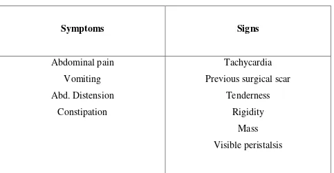
CLINICAL FEATURES
Cardinal features of acute intestinal obstruction are:
1. Abdominal pain.
2. Vomiting
3. Abdominal distension.
4. Constipation
1. Pain Abdomen.
Abdominal pain is the usually first symptom. The onset may be insidious or
abrupt in simple obstruction, but with strangulation the onset is sudden and severe.
The pain will diffuse, poorly localized and is felt across the upper abdomen in case
of high obstruction, at the level of the umbilicus in low ileal obstruction, in the
lower abdomen in colonic obstruction and in the perineum in rectosigmoid
obstruction.
2. Vomiting
Vomiting is the next most common symptom. Being a constant symptom,
the early vomiting is reflex in nature followed by quiescent period, before real
vomiting due to obstruction begins. This quiescent period will be of shorter
duration in high-level obstruction and longer in lower small intestinal obstruction.
Page | 37 Initially it contains partly digested food, followed by bilious vomiting. Finally it
becomes faeculent.
3. Distension
In the early cases of obstruction of the small intestine, abdominal distension
is often slight or even may be absent. When the proximal jejunum is obstructed,
the stomach gets distended with gas and the accumulated secretions, so that the
epigastric region may, in later stages become more prominent and tense. When the
ileum is involved, the central part of the abdomen is moderately blown out and
when the distal colon is blocked, there will be considerable universal distension of
abdomen, with well-marked bulging in the region of flanks. Visible peristalsis can
be present.
4. Constipation
In complete obstruction, after the contents of the intestine below the
obstruction have been evacuated, there will be constipation and usually neither the
faeces nor flatus is passed i.e. absolute constipation.
The rule that constipation is present in intestinal obstruction does not apply
in cases with Richter’s hernia, gall stone obstruction, mesenteric vascular
Page | 38
History and physical examination
A detailed history and physical examination usually helps in the diagnosis
and management of intestinal obstruction. In simple mechanical obstruction, there
will be very few abdominal signs. Whereas with strangulated obstruction, patient
will be toxic, tachycardiac and hypotension will be there. Any past history of
abdominal surgery, acute cholecystitis, appendicitis or any other intra-abdominal
infections, suggests adhesion to be the cause of obstruction. Hernia of long
duration gives rise to strangulation. There may be the following history:
• Alternate diarrhoea and constipation with weight loss suggests tuberculosis
and malignancies.
• Recent onset constipation suggests malignancy in elderly people.
Physical examination
Skin turgor: can be lost due to dehydration, may be cold and calmy.
Tongue: which may become dry and coated due to dehydration.
Nail and sclera: Anaemia or jaundice may be evident.
Rapid low volume pulse, low blood pressure, cold extremities, anxious look
Page | 39
Examination of abdomen
• Inspection: On inspection previous surgical scar indicates adhesions or
cancer. In early stage visible peristalsis can be seen. All hernial orifices has
to be examined.
• Type of abdominal distension:
• Type of peristalsis:
Palpation
• Abdomen must be examined for presence of any palpable mass, localized
tenderness, rebound tenderness which may be suggestive of strangulation. In
peritonitis there will be a generalized rigidity and tenderness. During pain
heaped up coils of intestine or prominent distended coils can be seen.
• Auscultation: In simple mechanical obstruction, sounds will become loud,
high pitched and metallic. In late stages bowel sounds will be absent due to
paralysis of bowel musculature. Bowel sounds can be absent in strangulation
and ileus or low-pitched tingling sounds will be heard due to movements of
fluid from one coil to another.
• Rectal examination: must be performed in all cases of obstruction, may
reveal faecal impaction, mass, red currant bleeding in intussusception. A
Page | 40 may be present. Ballooning of the rectum which usually occurs in the
intestinal obstruction may be due to obstruction to nerves which causes
sympathetic paralysis.
CLINICAL FINDINGS IN SMALL BOWEL OBSTRUCTION
Features Proximal/High
obstruction
Distal/low obstruction
Onset of symptoms Sudden Gradual
Pain Epigastric, intense,
colicky usually relieved
by vomiting
Periumbilical, colicky
Vomiting Early, bilious,
voluminous, frequent
Later, infrequent feculent
Tenderness Epigastric / periumbilical
mild unless strangulated
Diffuse and progressive
Distension Absent Diffuse and progressive
Obstipation Absent or mild Mild to moderate
Radiologic findings Distended proximal small
bowel loops or gasless
Diffusely distended small
bowel loops, air fluid
Page | 41
LABORATORY INVESTIGATIONS
(i) Haematological tests
A complete blood count, packed cell volume, serum electrolyte
determination and blood urea level estimation should be done. Leucocytosis is
suggestive of strangulation, extremely high counts are mainly suggestive of
mesenteric thrombosis.
(ii) Urine examination
Specific gravity of the urine will give a rough idea of the degree of
dehydration in case of intestinal obstruction.
(iii) Diagnostic aspiration
It is very important in the distinction between simple and strangulated
obstruction. Aspiration of peritoneal cavity with a fine needle and withdrawal of
blood stained fluid is diagnostic of strangulation.
X-ray diagnosis
The finding in erect abdominal X-ray for small intestinal obstruction is the
triad of dilated small bowel loops (>3 cm in diameter), air fluid levels on upright
Page | 42
MULTIPLE AIR-FLUID LEVELS
Page | 43
Gas shadows: When the jejunum, ileum or the colon are distended with gas, each
structure has significant radiological images. Jejunum is characterised by ‘valvulae
conniventes’ that pass from the anti-mesenteric to the mesenteric border in the
regular fashion. Ileum radiography is described by Wangensteen as being
‘characterless’. Large intestine reveals haustral markings which unlike the valvulae
conniventes are spaced irregularly and they do not completely traverse the
circumference of the bowel and gas shadows of large intestine will be located
peripherally.
Fluid levels: In adults, two inconstant fluid levels are regarded as physiological,
one at the duodenal cap and the other in the terminal ileum. In obstruction, fluid
levels tend to appear later than the gas shadows; the number of fluid level is
proportional to the degree of obstruction and to its location in the small intestine.
In upper small intestinal obstruction, fluid levels will be in left upper
quadrant and they will be few in number, multiple fluid level seen all over the
abdomen in case of lower small bowel obstruction. The presence of gas within the
wall of the bowel is highly significant sign of bowel necrosis, which was
demonstrated by Schorr in 1963.
Volvulus of sigmoid colon shows greatly distended sigmoid loop filling the
Page | 44 and Righler pointed out that the “coffee bean” sign in pathognomonic sign of
caecal volvulus.
Barium enema: In intussusception, barium is seen like a ‘claw’ around a negative
shadow of intussusceptions, whereas in sigmoid volvulus, barium column ends at
the level of the distal sigmoid in a characteristic Twisted Bird’s Beak deformity.
Computerized tomography (CT)
CT demonstrates the cause of intestinal obstruction.
Computerized tomography is very much useful in establishing the site, level
and the cause of obstruction and in displaying signs of threatened intestinal
viability. CT scan is most valuable when there are systemic signs indicative of
infarction, an associated palpable abdominal mass. In these cases CT scan may
confirm the presumptive diagnosis or reveal other causes mimicking obstruction
such as appendicitis or diverticulitis. In strangulated obstruction, target sign or
pneumatosis intestinalis and haemorrhage in the mesentery can be seen.
TREATMENT OF ACUTE INTESTINAL OBSTRUCTION
With few exceptions, an urgent intervention is needed in a case of intestinal
obstruction. Eventhough it is difficult to differentiate a simple and strangulated
Page | 45 clinical examination. Investigations are done to find out whether the obstruction is
mechanical or adynamic and to make out the level of obstruction.
The treatment must be planned according to the above assessment which
includes both supportive management and surgical management. There are four
important measures in the management of obstruction.
Conservative management
Cases with a partial intestinal obstruction can be treated conservatively with
resuscitation and tube decompression alone. Regression of the symptoms and
discharge without the need of surgery have been reported in 60-85% of patients
with a partial obstruction.16 Simple obstruction caused by post-operative early
adhesions or kinking may resolve spontaneously with the institution of
conservative management and it is indicated in following situations.
• Post-operative early adhesions
• Paralytic ileus of non-paralytic origin
• Inflammatory condition which cause obstruction
• Obstruction because of worm impaction
The initial conservative management is used in the above said conditions. This
facilitates spontaneous relief of obstruction and avoids more adhesion formation
Page | 46 clinician involved and general status of the patient. The conservative management
includes.
• GI decompression
• Fluid and electrolyte therapy.
• Antibiotics.
Surgical Management
With regard to the timing of the operation, all patients should be operated on
promptly after volume resuscitation if there is any evidence or suspicion that bowel
is ischaemic.17 Early surgery indicated in (1) obstructed and strangulated hernia,
(2) internal intestinal strangulated obstruction (3) acute obstruction, The classical
clinical saying that the sun should not set and rise in a case of unrelieved intestinal
obstruction is sound and it should be followed.
Laparotomy
In patients with small intestinal obstruction, who have not had any previous
abdominal surgery or in those with clinical evidence of ischaemia, a laparotomy is
essential.18
When the cause of intestinal obstruction lies within the abdomen and but its
Page | 47 obstruction is defined left mid or lower paramedian incision may be preferred,
abdominal cavity is inspected which reveals the underlying pathology.
Haemorrhagic fluid suggests strangulation; clear straw-coloured fluid denotes
simple obstruction. The operative assessment is directed at:
• Site of obstruction
• Viability of the bowel
• The nature of obstruction
The type of surgical procedure required will depend on the nature of the
cause, following relief of obstruction the viability of the involved segment of
bowel should be carefully assessed. In case of a viable bowel, peritoneum will be
shiny, mesentery bleeds on prick whereas with a nonviable bowel, peritoneum is
lusterless, mesentry does not bleed on prick.
Difference between viable and non-viable bowel
Intestine Viable Non-viable
Circulation Dark colour becomes
lighter, mesentery bleed
on prick
Dark colour remains
mesentery does not bleed
Page | 48
Peritoneum Shiny Dull and lusterless
Intestinal musculature Firm, Pressure rings may
or may not disappear,
peristaltic movements
may be observed
Flabby, thin and friable
pressure rings persist, no
peristaltic movements are
observed
Doppler ultra-sonography can also help to establish the circulation in the
mesenteric vasculature (a most accurate modality of testing viability). If viability
of the bowel is in doubt, it should be placed in warm moist pads for ten minutes
along with administration of 100% oxygen in the anaesthetic gas. Then after ten
minutes it has to be re-examined, in doubtful cases resection has to be done.
Principles of large bowel obstruction
As most of the large intestinal obstruction are due to malignancy, volvulus
or secondary to adhesive bands, which more commonly occur, in elderly patient.
When the lesion is operable and found in the caecum, ascending colon or
proximal transverse colon, a right hemicolectomy should be performed; if lesion is
fixed, a proximal stoma (colostomy. or ileostomy if ileocaecal valve is
Page | 49 obstructing lesions of the splenic flexure (malignant) should be treated by an
extended right hemicolectomy. If one stage resection and anastomosis is not
feasible a covering colostomy to protect the site of anastomosis is safer, where the
distal segment could not be brought to the surface a proximal stoma and the distal
end can be closed and returned to abdomen (Hartman’s procedure) or both the ends
may be brought outside, proximal as stoma and distal as mucus fistula, followed by
second stage colorectal anastomosis which can be planned when the patient is fit.
In very old or cachectic patients when an obstructing carcinoma of rectum which is
fixed, left iliac colostomy is the best site for the placement of a permanent artificial
anus. In rare circumstances, or if caecal perforation is strongly suspected, we
should wait for some time for improvement of the patient’s condition and later
relief of obstruction can be done by doing an emergency caecostomy through a
small incision in the right iliac fossa.
INTESTINAL OBSTRUCTION BY ADHESIONS AND BAND
It is the most common cause of intestinal obstruction in the developing
countries. The pathology lies with peritoneal irritation resulting in local outpouring
of fibrin which produces adhesions between the opposed peritoneal surfaces. These
fibrinous adhesions may become vascularised and become a mature fibrous tissue,
infection being an important cause. Also foreign materials like the silk thread,
Page | 50 These commonly occurs following laparotomies. Once adhesions have
developed, progression to obstruction is very much inevitable in a significant
proportion.19 Ileum is the most common segment to be obstructed due to adhesions.
After abdominal surgeries, about 5% of the patients subsequently develop
obstruction due to adhesions, whereas operation in the colon carry a high incidence
of obstruction, about 1/5th of cases. About 20% of the obstruction occurs within
the first year after laparotomy and most of these occur during the first few weeks
after surgery and are termed as early postoperative obstructions and most get
resolved by conservative treatment.20
Treatment
The treatment for adhesions is the same as the general principles of
management often is curative in early type of adhesions, but conservative
treatment should not be prolonged beyond 48-72 hours and should not be
continued if symptoms and signs are progressive even after initial resuscitation.
When laparotomy is done multiple adhesiolysis should be done.
Recurrent intestinal obstruction due to adhesion
Adhesions are a major cause of late morbidity. Approximately 10% of all
Page | 51 operation at a later date for the same issue. A further 10% may require a third
operation for adhesive obstruction.
There are often chances of recurrent obstruction after the first adhesiolysis.
The following procedures may be considered for recurrent adhesive obstruction
• Intestinal intubation
• Charles- Phillips transmesenteric plication
• Noble plication
• Repeat adhesiolysis (enterolysis)
Internal hernias
When a segment of the small intestine becomes herniated into one of the
retroperitoneal Fossae, it is termed as internal hernias. This can occur in following
sites:
• Supra-vesical hernia
• Foramen of Winslow
• Diaphragmatic hernia: Acquired/Congenital.
• Caecal or appendiceal: Retroperitoneal fossae superior or retrocaecal
• A hole in the mesentery or mesocolon and defects present in the broad
Page | 52
• Paraduodenal fossae: Right/left paraduodenal fossae.
Internal herniation in the absence of adhesions is quite uncommon and a
preoperative diagnosis is unusual. The standard treatment for a hernia is to release
the constricting agent by divison.11
Internal herniations are treated by laparotomy and the release of constriction
ring. The distended loop first needs to be decompressed and then reduced.
Unviable segment of the bowel has to be resected and anastomosed.
INTUSSUSCEPTION
When one portion of the bowel invaginates into the immediately adjacent
loop, the condition is called as an intussusception. Most of the time, it is proximal
segment of bowel that invaginates into the distal segment. It is one of the
commonest cause in paediatric age group. It can also occur in adults. In adults, our
2/3rd rule may be applied. Two-thirds of cases of adult intussusceptions are from
known causes. Of these thirds are due to neoplasms. Of these neoplasms,
two-thirds will be of malignant pathology.24 An intussusception constitutes following
parts:
• Intussusceptum
• Intussuscepiens
Page | 53
• Neck
The types of intussusception are as follows:
Ileocolic (77%)
Ileo-ileocolic (12%)
Ileo-ileal (5%)
Colo-colic (2%)
Multiple (1%)
Retrograde (0.2%)
Others (2.8%)
In the paediatric age group, intussusception can occur in any age but more
commonly in the period between 3 months to 9 months when the weaning from the
breast milk is started. The incidence is higher in the first-born child. It is more
Page | 54
Etiology
Most of the cases of intussusception occur in the paediatric age group.
Majority of the patients are classified as idiopathic and there may be association
with acute gastroenteritis or URI , when the baby gets weaned from breast milk, it
is believed to be due to hyperplasia of Peyer’s patches in the terminal ileum,
polyps, Meckel’s diverticulum, duplication. H.S. Purpura or appendix as the
common lead points. Most patients with adult intussusception are benign and
enteric origin and most sensitive diagnostic modality is abdominal CT scanning.25
Large bowel intussusception is more common in adults. Half of the cases are
secondary because of neoplasia either benign or malignant. Among benign
conditions, fibroma, leiomyoma, polyps and submucous lipoma, Peutz Jeghers
syndrome are the commonest one.
Clinical features: The typical presentation is with intermittent abdominal pain.
Usually it is of sudden onset. During the attacks of pain the child cries with pallor
over the face and becomes quiet after few minutes. Vomiting and blood stained
stools may be present. On examination, during pain free period a sausage shaped
lump may be palpable with concavity towards umbilicus, which becomes hard on
palpation. Right iliac fossa on palpation may be peculiarly empty (sign-de-dance).
Page | 55
Radiography
Plain X-ray shows multiple air fluid levels. Barium enema in ileo-colic type
shows the typical ‘pincer shaped’ or ‘coiled spring’ deformity or ‘pinch fork’ sign.
Treatment
Intravenous fluid administration should be begun, decompression of small
bowel through nasogastric suction is achieved. The reduction of the
intussusception can be done by any of the following ways which may be
non-operative reductions: Hydrostatic or pneumatic.
Operative management
Laparotomy is done by making right lower paramedian incision.
Intussusception mass is identified and a trial of manual reduction is attempted.
The reduction is effected by squeezing the apex of the intususception retrogradely,
but at no time the segments are pulled. Open method of inserting the little fingers
into the neck and separating the adhesions between the intussusception in difficult
cases can be tried (Copes method).
Indications for resection
When there is presence of a polyp, tumour or Meckel’s diverticulum or if the
Page | 56 gangrenous bowel is there, intestine should be resected without attempting for
reduction.
The whole intussusception is usually resected with primary ileocolostomy.
The ileum is resected proximal to the intussusception and the proximal end is
anastomosed end to side with the transverse colon which is distal to the
intussusception.
VOLVULUS
Volvulus refers to the axial rotation of a portion of alimentary tract on its
mesentery. It may be either primary or secondary. Primary volvulus can occur due
to malrotation of the gut, abnormal attachment of the mesentry or congenital
bands. E.g. Caecal volvulus, volvulus neonatorum and sigmoid volvulus.
A secondary volvulus is due to the actual rotation of a segment of bowel
around an acquired adhesion or a stoma. When it is completed, it forms a close
loop of obstruction with ischaemia of the involved segment.
PRIMARY VOLVULUS
Volvulus neonatorum
This condition is predisposed by arrested rotation of the gut causing the