FLOATIN MATRIX TABLETS OF RITONAVIR”
Dissertation submitted to
THE TAMILNADU Dr.M.G.R. MEDICAL UNIVERSITY,
CHENNAI – 32.
In partial fulfillment of the requirements for the award of the degree of
M
M
A
A
S
S
T
T
E
E
R
R
O
O
F
F
P
P
H
H
A
A
R
R
M
M
A
A
C
C
Y
Y
I
I
N
N
P
P
H
H
A
A
R
R
M
M
A
A
C
C
E
E
U
U
T
T
I
I
C
C
S
S
Submitted byR
R
e
e
g
g
.
.
N
N
o
o
.
.
2
2
6
6
1
1
2
2
1
1
0
0
8
8
0
0
6
6
Under the Guidance ofMr.K.G.PARTHIBAN M.Pharm, (Ph.D).
,
Professor
DEPARTMENT OF PHARMACEUTICS
J.K.K.MUNIRAJAH MEDICAL RESEARCH FOUNDATION
COLLEGE OF PHARMACY, KOMARAPALAYAM-638183.
J.K.K.Munirajah Medical Research Foundation College of Pharmacy,
Komarapalayam-638183.
C
C
E
E
R
R
T
T
I
I
F
F
I
I
C
C
A
A
T
T
E
E
This is to certify that the work embodied in this dissertation entitled
“FORMULATION AND IN-VITRO EVALUATION STUDIES ON ORAL FLOATING MATRIX TABLETS OF RITONAVIR ” submitted in the partial
fulfillment for the degree of MASTER OF PHARMACY in Pharmaceutics, The
Tamil Nadu Dr. M.G.R. Medical university, Chennai, is a bonafide work, which was
carried out by Mr. KANNEDHARA GOPALA RAO (Reg.No.261210806) under
my guidance and supervision during the academic year 2013-2014.
Place:komarapalayam Mr. K.G. PARTHIBAN M.Pharm., (Ph.D).,
Date: Professor,Guide and Head ,
Department of Pharmaceutics
J.K.K.Munirajah Medical Research Foundation College of Pharmacy,
Komarapalayam-638183.
CERTIFICATE
This is to certify that the work embodied in this dissertation entitled
“FORMULATION AND IN VITRO EVALUATION STUDIES ON ORAL FLOATING MATRIX TABLETS OF RITONAVIR” submitted to The Tamil Nadu
Dr. M.G.R. Medical University, Chennai, was carried out by Mr. KANNEDHARA
GOPALA RAO (Reg.No.261210806), for the partial fulfillment for the degree of
MASTER OF PHARMACY in Pharmaceutics under the guidance of
Mr.K.G.Parthiban, M.Pharm., (Ph.D)., Professor, Department of Pharmaceutics,
J.K.K.Munirajah Medical Research Foundation College of Pharmacy, Komarapalyam,
during the academic year 2013-2014.
Place: Komarapalayam. Dr.
N.SENTHIL KUMAR,
M.Pharm., Ph.D.,Date: Principal,
J.K.K.Munirajah Medical Research Foundation College of Pharmacy,
Komarapalayam-638183.
D
D
E
E
C
C
L
L
A
A
R
R
A
A
T
T
I
I
O
O
N
N
The work presented in this dissertation entitled “FORMULATION AND IN
VITRO EVALUATION STUDIES ON ORAL FLOATIN MATRIX TABLETS OF
RITONAVIR” was carried out by me under the guidance of,
Mr.K.G.PARTHIBAN,M.Pharm.,(Ph.D).,Professor in Department of
Pharmaceutics, J.K.K.Munirajah Medical Research Foundation College of Pharmacy,
and Komarapalayam.This work is original and has not been submitted in part or full
for the award of any other degree or diploma of any other university.
Place: Komarapalayam. Mr. KANNEDHARA GOPALA RAO
First of all, I thank the ALMIGHTY who is constantly showering his blessing on me.
It gives me immense pleasure to profusely thank my esteemed Academic guide K.G.PARTHIBAN., M.Pharm, (PhD).,Professor, Department of Pharmaceutics, JKKMMRF College of pharmacy, for the unflagging zeal with which they guided me
in carrying out this research work.
I would like to express my gratitude to Dr.N.Senthil Kumar, Principal, JKKMMRF College of pharmacy for his support and gratitude.
My heartfelt thanks to Mr.Ramesh M.Pharm Asst.Professor, Dept. of Pharmaceutical Analysis, JKKMMRF College of pharmacy for their moral support
and help throughout the course.
We express our sincere thanks to our respected Correspondent,Dr. J.K.K. MUNIRAJAH, M.Tech (Bolton) for providing a good infrastructure and facilities
during the entire course of B. Pharmacy.
I express my profound indulge to all my staff members, Lab assistants and Librarian for their kind co-ordination during this work.
S. No
CONTENTS
PAGE. No
1
Introduction
1
2
Literature Review
35
3
Aim and Plan of work
50
4
Materials and methods
53
5
Results and discussion
98
6
Summary and conclusion
120
Chapte
r No. Chapter Page No.
1 Introduction 1-37
2 Literature review 37-53
3 Aim and Plan of work 54-56
4 Materials and Methods 57-103
5 Results and Discussion 104-124
6 Summary and Conclusion 125-126
7 Bibliography -
LIST OF ABBREVIATIONS USE
% - Percentage
°C - Temperature on Celsius scale
API - Active pharmaceutical ingredient
Conc. - Concentration
CP - Centi-poise
CRDDS - Controlled release drug delivery system
DDS - Drug delivery system
FT-IR - Fourier Transform Infra Red
G/ml - Grams per milliliter
GIT - Gastro intestinal tract
Gm - Gram
HPMC - Hydroxy propyl methyl cellulose
L - Liter
m Pa S - milli Pascals seconds
M - Molarity
Min - Minutes
N - Normality
No. - Number
pH - Negative log of Hydrogen ion
concentration
RH - Relative Humidity
rpm - Rotation per minute
Rt - Retention time
SD - Standard deviation
Sem - Scanning electron microscopy
SEM - Standard error mean
SS - Stock solution
TK - Tyrosine kinase
UV - Ultra Violet
v/v - volume by volume
Vs - Verses
W/v - weight by volume
W/w - weight by weight
λmax - Absorption maxima
µg/ ml - Microgram per milliliter
µl - Microliter
1. INTRODUCTION
The oral drug administration has been the predominant route for drug
delivery due to the ease of administration, patient convenience and flexibility in
formulations. However, it is a well accepted fact today that drug absorption
throughout the GI tract is not uniform. Using currently utilized release technology,
oral drug delivery for 12 or even 24 hours is possible for many drugs that are
absorbed uniformly from GI tract. Nevertheless this approach is not suitable for a
variety of important drugs characterized by narrow absorption window in the upper
part of GI tract i.e. stomach and small intestine.
The design of oral controlled drug delivery systems (DDS) should be
primarily aimed to achieve the more predictability and reproducibility to control the
drug release, drug concentration in the target tissue and optimization of the
therapeutic effect of a drug by controlling its release in the body with lower and less
frequent dose.
The controlled release systems for oral use are mostly solid and based on
dissolution or diffusion or a combination of both the mechanisms in the control of
release rate of drug. Depending upon the manner of drug release they are classified as
follows
1.1. CLASSIFICATIONOFCONTROLLED DRUG DELIVERY SYSTEM [4, 27,
37]
A. Continuous release system: These systems release the drug for a prolonged
period of time along the entire length of GIT with normal transit of the dosage form.
The various systems under this category are:
I. Dissolution controlled release systems
III. Dissolution and diffusion controlled release systems
IV. Ion-Exchange resins – drug complexes
V. slow dissolving salts and complexes
VI. pH–dependent formulations
VII. Osmotic pressure controlled systems
VIII. Hydrodynamic pressure controlled systems
B. Delayed transit and continuous release system: These systems are designed to
prolong their residence in the GIT along with their release. Often, the dosage is
fabricated to retain in the stomach and hence the drug present therein should be stable
at gastric pH. Systems included in this category are:
I. Altered density systems
II. Mucoadhesive systems
III. Size-based systems
C. Delayed release systems: The design of such systems involve release of drug only
at a specific site in the GIT.
The drugs contained in such system have following category:
Destroyed in the stomach or by intestinal enzymes
Known to cause gastric distress
Absorbed from a specific intestinal site, or
Meant to exert local effect at a specific GI site.
The two types of delayed release systems are:
I. Intestinal release systems
II. Colonic release systems
Oral controlled release dosage forms have been developed for the past three
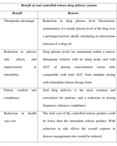
Table No.1: Advantages
Benefit of oral controlled-release drug delivery systems
Benefit Reason
Therapeutic advantage Reduction in drug plasma level fluctuations;
maintenance of a steady plasma level of the drug over
a prolonged period, ideally simulating an intravenous
infusion of a drug ref
Reduction in adverse
side effects and
improvement in
tolerability
Drug plasma levels are maintained within a narrow
therapeutic window with no sharp peaks and with
AUC of plasma concentration versus time
comparable with total AUC from multiple dosing
with immediate release dosage form.
Patient comfort and
compliance
Oral drug delivery is the most common and
convenient for patients, and a reduction in dosing
frequency enhances compliance.
Reduction in health
care cost
The total cost of the controlled release product could
be lower than the immediate release product. With
reduction in side effects the overall expense in
disease management also would be reduced.
Despite several advantages associated with controlled drug delivery system,
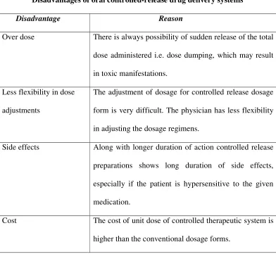
Table No.2: Disadvantages
Disadvantages of oral controlled-release drug delivery systems
Disadvantage Reason
Over dose There is always possibility of sudden release of the total
dose administered i.e. dose dumping, which may result
in toxic manifestations.
Less flexibility in dose
adjustments
The adjustment of dosage for controlled release dosage
form is very difficult. The physician has less flexibility
in adjusting the dosage regimens.
Side effects Along with longer duration of action controlled release
preparations shows long duration of side effects,
especially if the patient is hypersensitive to the given
medication.
Cost The cost of unit dose of controlled therapeutic system is
higher than the conventional dosage forms.
To overcome these problems and improve the efficacy of oral administration,
some recent studies have reported that controlled oral drug delivery system with
prolonged gastric residence time, such as floating dosage system have been proved to
be advantages.
A gastrointestinal drug delivery system can be made to float in the stomach by
a gelling process of hydrocolloid materials or by incorporating a floatation chamber
with vacuum or gas. In this way bulk density less than that of gastric fluid is
produced. However, most of the devices generating gas or gelling need time to be
form from transiting into the small intestine along with food before floating in
stomach.
1.2. GASTRO-RETENTIVE DOSAGE FORMS (GRDF):[1,5,12,14,15,50]
These are primarily controlled release drug delivery systems, which gets
retained in the stomach for longer periods of time, thus helping in absorption of drug
for the intended duration of time. Gastric retentive drug delivery devices can be useful
for the spatial and temporal delivery of many drugs.
The gastric emptying time mainly depends upon the design of the dosage form
and physiological state of the subject, which last from a few minutes to 12 hrs. The
average gastric emptying time in human is 2-3 hrs through major absorption zone
(stomach and upper part of the intestine), which leads to incomplete drug release from
the DDS leading to diminished efficacy of the administered dose. So drugs which
have stability problem, GRDF plays an important role. These considerations have led
to the development of oral controlled release dosage forms possessing gastric
retention capabilities.
GRDF will also greatly improve the pharmacotherapy of the stomach itself
through local drug release leading to high drug concentrations at the gastric mucosa,
which are sustained over a long period of time.
1.2.1. Anatomy and Physiology of Stomach: [44]
1.2.1. a) Anatomy:
The stomach is j- shaped organ located in the upper left hand portion of the
abdomen just below the diaphragm. It occupies a portion of the epigastric and left
hypochondriac region. The main function of the stomach is to store the food
temporarily, grind it and then release it slowly into the duodenum. Due to its small
Figure No. 1: Anatomy of the stomach.
The stomach has four main regions:
1. Cardia 3. Body and
2. Fundus 4. Pylorus
The main function of the fundus and body is storage, whereas that of cardia
is mixing or grinding. The fundus adjusts the increased volume during eating by
relaxation of the fundus muscle fibers. The fundus also exerts a steady pressure on the
gastric contents pressing them towards the distal region, to pass through the pyloric
sphincter into the small intestine.
1.2.1. b) Physiology:
Various factors like absorption ability, presystemic clearance, gastric
motility; gastrointestinal transit time and gastrointestinal emptying time will have an
influence on the bioavailability of drug from the dosage form.
Absorption ability:
The absorption capability of various segments of gastrointestinal tract differs
from each other. i.e. most of the absorption takes place in small intestine and lesser
extent in colon and stomach. Unless drugs are absorbed equally in both the colon and
major limiting factor for sustained release and controlled release drug delivery
systems.
Presystemic clearance:
Even if the drugs that can be absorbed equally well throughout the
gastrointestinal tract, bioavailability is significantly reduced by the site-specific
changes in presystemic clearance. Degradation of the drug is also carried out by
hydrolysis in the stomach, enzymatic digestion, and metabolism in the brush border of
the gut wall and by the microorganisms.
Such degradation may leads to high variation in plasma drug concentration
and poor absorption of drug in to the systemic circulation.
Gastric motility:
Gastric emptying occurs during fasting as well as fed states. During the fasting
state an interdigestive series of electrical events take place, which cycles through
stomach and intestine every 2 to 3 hours. This is called the interdigestive myloelectric
cycle or migrating myloelectric cycle (MMC), which is further divided into 4 phases
as described by Wilson and Washington.
After the ingestion of a mixed meal, the pattern of contractions changes from
fasted to that of fed state. This is also known as digestive motility pattern and
comprises continuous contractions as in phase II of fasted state. These contractions
result in reducing the size of food particles (to less than 1 mm), which are propelled
toward the pylorus in a suspension form. During the fed state onset of MMC is
delayed resulting in slowdown of gastric emptying rate. Scintifc graphic studies
release dosage forms are subjected to basically two complication, that of short gastric
residence time and unpredictable gastric emptying rate.
1. Phase I (basal phase) lasts from 40 to 60 minutes with rare
contractions.
2. Phase II (preburst phase) lasts for 40 to 60 minutes with intermittent
action and potential contractions. As the phase progresses the intensity
and frequency also increases gradually.
3. Phase III (burst phase) lasts for 4 to 6 minutes. It includes intense and
regular contractions for short period. It is due to this wave that all the
undigested material is swept out of the stomach down to the small
intestine. It is also known as the housekeeper wave.
4. Phase IV lasts for 0 to 5 minutes and occurs between phases III and I
of 2 consecutive cycles.
Gastrointestinal Transit Time:
Food content remains in each segment of the gastrointestinal tract for
different periods of time. The resident time for both liquid and solid foods in each
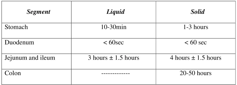
Table No.3: Transit time of food in each segment of the gastrointestinal tract
Segment Liquid Solid
Stomach 10-30min 1-3 hours
Duodenum < 60sec < 60 sec
Jejunum and ileum 3 hours ± 1.5 hours 4 hours ± 1.5 hours
Colon --- 20-50 hours
Since most of the drugs are absorbed from the upper part of intestine, the total
effective time for the drug absorption is 3-8 hours. So one has to take most of the
drugs 1-2 times a day.
1.2.1.c) Factors affecting the gastric emptying time:[32]
I. State of the stomach: gastric emptying time depends upon the fed state of the
stomach, which increases the gastric emptying time as compared to unfed
state.
II. Circadian rhythms: which are increased in day time and less during night, also
affects the gastric retention time (GRT).
III. Size of the Meal: greater the energy content of the meal (carbohydrate and
high fat content), longer the duration of emptying.
IV. Density of the oral dosage form: The density of the gastric fluid is reported to
be 1.2g/cm. The density of the dosage form should be less than this for the
buoyancy so that it is retained in the stomach for longer period of time.
V. Diseased state: State of the stomach also affects the environment for the
dosage form as in case of ulcers, flatulence and spasms.
VI. Drug therapy: Plays an important role in gastric emptying e.g. prokinetic drugs
VII. Age: Increase in age decreases the gastric motility thereby increasing the
gastric emptying time.
VIII. Posture: It was seen that the supine posture on the right side showed better
results than on the left side.
1.2.1. d) Criteria for selection of drug candidate for GRDF: [32]
The gastric retentive drug delivery systems are suitable for following types of
drug therapy:
I. Absorption from upper part of GIT: Drugs have a particular site for maximum
absorption e.g. ciprofloxacin, whose maximum absorption is in the stomach
only. The absorption of metformin hydrochloride is confirmed to small
intestine only and the conventional sustained release dosage forms may be
poorly bioavailable since absorption appears to diminish when the dosage
form pass in to large intestine.
II. Drugs having low pKa, which remains unionized in stomach for better
absorption.
III. Drugs having reduced solubility at higher pH e.g.captopril and
chlordiazepoxideand the bioavailability of drugs that get degraded in alkaline
pH can be increased by formulating gastro-retentive dosage forms. e.g.
doxifluridine, which degrades in small intestine.
IV. Local action as it is seen in the treatment of H.Pylori by amoxicillin and
misoprostolfor ulcers.
V. To minimize gastric irritation this may be caused by sudden increase of drug
VI. Improve effectiveness of particular drugs. E.g. antibiotics in the colon tend to
disturb the microflora causing overgrowth of microorganisms like Clostridium
difficile causing colitis.
1.3. GASTRO RETENTIVE DRUG DELIVERY SYSTEM: [55(b), 32, 45]
Oral drug delivery is the most desirable and preferred method of
administering therapeutic agents for their systemic effects. Oral medication is
generally considered as the first avenue investigated in the development of
pharmaceutical formulations because of patient acceptance, convenience in
administration and cost effective manufacturing processes. Oral route offers an
attractive approach of drug targeting at the specific site within GI tract for certain
types of drug.
1.3.1 Approaches to Gastric Retention:
A number of approaches have been used to increase the GRT of a dosage form
in stomach by employing a variety of concepts. These include:
a) Floating Systems: [23]
Floating Drug Delivery Systems (FDDS) have a bulk density lower than
gastric fluids and thus remain buoyant in the stomach for a prolonged period of time,
without affecting the gastric emptying rate. While the system is floating on the gastric
contents, the drug is released slowly at a desired rate from the system. After the
release of the drug, the residual system is emptied from the stomach. This results in an
increase in the GRT and a better control of fluctuations in the plasma drug
concentrations.
Floating systems can be classified into two distinct categories,
(A) effervescent
b) Bio/Muco-adhesive Systems: [27]
Bio/Muco-adhesive systems are those which bind to the gastric epithelial
surface or mucin and serve as a potential means of extending the GRT of drug
delivery system (DDS) in the stomach.
The surface epithelial adhesive properties of mucin have been well recognized
and applied to the development of GRDS based on bio/muco-adhesive polymers. The
ability to provide adhesion of a drug (or a delivery system) to the GI wall provides a
longer residence time in a particular organ site, thereby producing an improved effect
in terms of local action or systemic effect.
Binding of polymers to the mucin/epithelial surface can be divided into three broad
categories:
1. Hydration-mediated adhesion.
2. Bonding-mediated adhesion.
3. Receptor-mediated adhesion.
c) Swelling and Expanding Systems:
These are the dosage forms, which after swallowing; swell to an extent that
prevents their exit from the pylorus. As a result, the dosage form is retained in the
stomach for a longer. These systems may be named as “plug type system” since they
exhibit the tendency to remain logged at the pyloric sphincter if that exceed a
diameter of approximately 12-18 mm in their expanded state. Such polymeric
matrices remain in the gastric cavity for several hours even in the fed state.
A balance between the extent and duration of swelling is maintained by the
degree of cross-linking between the polymeric chains. A high degree of cross-linking
d) High Density Systems
These systems with a density of about 3 g/cm3 are retained in the rugae of the
stomach and are capable of withstanding its peristaltic movements. A density of
2.6-2.8 g/cm3 acts as a threshold value after which systems can be retained in the lower
part of the stomach. High-density formulations include coated pellets. Coating is done
by heavy inert materials such as barium sulphate, zinc oxide, titanium dioxide, and
iron powder.
e) Incorporation of Passage Delaying Food Agents:
Food excipients like fatty acids e.g. salts of myristic acid change and modify
the pattern of the stomach to a fed state, thereby decreasing gastric emptying rate and
permitting considerable prolongation of release. The delay in the gastric emptying
after meals rich in fats is largely caused by saturated fatty acids with chain length of
C10-C14.
f) Ion Exchange Resins: [33]
A coated ion exchange resin bead formulation has been shown to have gastric
retentive properties, which was loaded with bicarbonates. Ion exchange resins are
loaded with bicarbonate and a negatively charged drug is bound to the resin. The
resultant beads were then encapsulated in a semi-permeable membrane to overcome
the rapid loss of carbon dioxide. Upon arrival in the acidic environment of the
stomach, an exchange of chloride and bicarbonate ions take place, as a result of this
reaction carbon dioxide was released and trapped in the membrane thereby carrying
beads towards the top of gastric content and producing a floating layer of resin beads
g) Osmotic Regulated Systems:
It is comprised of an osmotic pressure controlled drug delivery device and an
inflatable floating support in a bioerodible capsule. In the stomach the capsule quickly
disintegrates to release the intragastric osmotically controlled drug delivery device.
The inflatable support inside forms a deformable hollow polymeric bag that contains a
liquid that gasifies at body temperature to inflate the bag. The osmotic controlled drug
delivery device consists of two components, drug reservoir compartment and
osmotically active compartment.
1.3.2 Types of Floating Drug Delivery Systems (FDDS): [16, 42]
Based on the mechanism of buoyancy, two distinctly different technologies
have been utilized in the development of FDDS, which are:
A. Effervescent System
I. Gas generating system
1. Gastric bilayered floating tablets
2. Multiple unit type floating pills
3. Multi-unit type oral floating stages of floating mechanisam dosage
system
II. Volatile liquid/vacuum containing system
1. Intragastric floating gastrointestinal drug delivery system
2. Inflatable gastrointestinal delivery system
3. Intragastric osmotically controlled drug delivery system
B. Non- Effervescent System
2. Alginate beads
3. Hollow microspheres
A. Effervescent System: [2]
Effervescent systems include use of gas generating agents, carbonates (sodium
bicarbonate) and other organic acid (citric acid and tartaric acid) to produce carbon
dioxide (CO2) gas, thus reducing the density of the system and making it to float on
the gastric fluid. These effervescent systems further classified into two types.
I. Gas Generating Systems:
Intra Gastric Single Layer Floating Tablet or Hydro dynamically Balanced System
(HBS)
Figure No. 2: hydro dynamically balanced system
These are formulated by mixing the CO2 generating agents and the drug within
the matrix tablet (Fig. 2). These have a bulk density lower than gastric fluids and
therefore remain floating in the stomach unflattering the gastric emptying rate for a
system and after the complete release the residual system is expelled f
stomach. This leads to an increase in the GRT and a better control over fluctuations in
plasma drug concentration.
1. Intra Gastric Bilayered Floating Tablets:
These are also compressed tablet and contain
i) Immediate release layer
Figure No. 3: Intra Gastric Bilayer Floating Tablet.
2. Multiple Unit type floating pills:
These systems consist of sustained release pills as ‘seeds’ surrounded by
double layers. The inner
of swellable membrane layer. When the system is immersed in dissolution medium at
body temperature it sinks at once and then forms swollen pill like balloon and float as
the density decreases (Fig. 4&5).
system and after the complete release the residual system is expelled f
stomach. This leads to an increase in the GRT and a better control over fluctuations in
plasma drug concentration.
. Intra Gastric Bilayered Floating Tablets:
These are also compressed tablet and contain two layers for:
Immediate release layer and ii) Sustained release layer (Fig. 3).
Figure No. 3: Intra Gastric Bilayer Floating Tablet.
Multiple Unit type floating pills:
These systems consist of sustained release pills as ‘seeds’ surrounded by
double layers. The inner layer consists of effervescent agents while the outer layer is
of swellable membrane layer. When the system is immersed in dissolution medium at
body temperature it sinks at once and then forms swollen pill like balloon and float as
Fig. 4&5).
system and after the complete release the residual system is expelled from the
stomach. This leads to an increase in the GRT and a better control over fluctuations in
and ii) Sustained release layer (Fig. 3).
These systems consist of sustained release pills as ‘seeds’ surrounded by
layer consists of effervescent agents while the outer layer is
of swellable membrane layer. When the system is immersed in dissolution medium at
3. Multi-unit type oral floating Stages of floating mechanism dosage system
A Penetration of water
B Generation of CO2 and floating Dissolution of drug
C Release of drug from the resorvoir
II. Volatile Liquid / Vacuum Containing Systems:
1. Intragastric Floating Gastrointestinal Drug Delivery System: [26]
This system can be made to float in the stomach because of flotation chamber,
which may be a vacuum or filled with air or a harmless gas, while drug reservoir is
[image:25.612.188.497.299.439.2]encapsulated inside a microporous compartment (Fig. 6).
Figure No. 6: Intra Gastric Floating Gastrointestinal Drug Delivery Device
2. Inflatable Gastrointestinal Delivery Systems:
In these systems an inflatable chamber is incorporated, which contains liquid
that gasifies at body temperature to cause the chamber to inflate in the stomach. These
systems are fabricated by loading the inflatable chamber with a drug reservoir, which
can be a drug impregnated polymeric matrix, then encapsulated in a gelatin capsule.
After oral administration the capsule dissolves to release the drug reservoir together
with the inflatable chamber. The inflatable chamber automatically inflates and retains
the drug reservoir compartment in floating position. The drug continuously released
Figure No. 7: Inflatable Gastrointestinal Delivery System
3. Intragastric Osmotically Controlled Drug Delivery System:
It is comprised of an osmotic pressure controlled drug delivery device and an
inflatable floating support in a biodegradable capsule. In the stomach capsule quickly
disintegrates to release the intragastric osmotically controlled drug delivery device.
The inflatable support inside forms a deformable hollow polymeric bag that contains a
liquid that gasifies at body temperature to inflate the bag. The osmotic pressure
controlled drug delivery device consists of two components; drug reservoir
compartment and an osmotically active compartment.
The drug reservoir compartment is enclosed by a pressure responsive
collapsible bag, which is impermeable to vapour and liquid and has a drug delivery
orifice. The osmotically active compartment contains an osmotically active salt and is
enclosed within a semi permeable housing. In the stomach, the water in the GI fluid is
continuously absorbed through the semi permeable membrane into osmotically active
compartment to dissolve the osmotically active salt. An osmotic pressure is thus
created which acts on the collapsible bag and turns in forces the drug reservoir
compartment to reduce its volume and activate the drug reservoir compartment to
reduce its volume and activate the drug release in solution form through the delivery
The floating support is also made to contain a bioerodible plug that erodes
after a predetermined time to deflate the support. The deflated drug delivery system is
then emptied from the stomach (Fig. 8).
Figure No. 8: Intragastric Osmotically Controlled Drug Delivery System
B. Non Effervescent Systems:
The Non effervescent FDDS is based on mechanism of swelling of polymer or
bioadhesion to mucosal layer in GI tract. The most commonly used excipients in
non-effervescent FDDS are gel forming or highly swellable cellulose type hydrocolloids,
polysaccharides and matrix forming materials such as polycarbonates, polyacrylates,
polymethacrylates, polystyrenes and bioadhesive polymer such as chitosan and
carbopol.
The various types of these systems are:
1. Single Layer Floating Tablets: [58, 49]
They are formulated by intimate mixing of drug with a gel-forming
hydrocolloid, which swells in contact with gastric fluid and maintain bulk density of
less than unity. The air trapped by the swollen polymer confers buoyancy to these
2. Alginate Beads: 47
Multi unit floating dosage forms were developed from freeze-dried calcium
alginate. Spherical beads of approximately 2.5 mm diameter can be prepared by
dropping a sodium alginate solution into aqueous solution of calcium chloride,
causing precipitation of calcium alginate leading to formation of porous system,
which can maintain a floating force for over 12 hours. These floating beads gave a
prolonged residence time of more than 5.5 hour.
3. Hollow Microspheres:
Multiple-unit hollow microspheres by emulsion solvent diffusion technique
were prepared with Drug and acrylic polymer. These were dissolved in an
ethanol-dichloromethane mixture, and poured into an aqueous solution of PVA with stirring to
form emulsion droplets. The rate of drug release in micro balloons was controlled by
changing the polymer to drug ratio. Micro balloons were floatable in-vitro for 12
hours when immersed in aqueous media. Radio graphical studies proved that micro
balloons orally administered to humans were dispersed in the upper part of stomach
and retained there for 3 hours against peristaltic movements.
1.4.3 Application of FDDS: [32]
1. For treating local inflammation and stomach ulcers.
2. For treating H. Pylori associated ulcers.
3. In chronic diseases associated with frequent medication and prolonged medication,
FDDS can be promising drug delivery system.
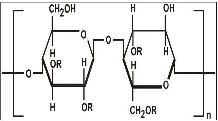
1.5 MATRIX SYSTEMS:[13,30,39,55]
A matrix is a uniform mixture of drug and excipients. e.g. polymer that is
The drug substance, which has a solubility S gm /cm3 in the dissolution medium, is dispersed in the matrix which is insoluble in the dissolution medium, is
dispersed in the matrix which is insoluble in the dissolution medium. The
concentration of drug in the matrix is ‘A’ gm / cm3. The matrix is porous, with a
porosity of ‘Є’ and diffusion coefficient of ‘Dm’. The drug release from such system
can be described by dQ/dt = 2SDmAt. Liquid will intrude from the bulk liquid. The
rate and extent of intrusion will follow the following equation:
L q L Qr dt dL − = − =
η
8 2Where, L is the length of the intrusion at time t, r is the average radius of the
pores, ŋ is the viscosity of the liquid and Q is a constant21.
Figure No. 9: Dissolution of drug from a solid matrix
1.5.1. HYDROPHILLIC MATRIX SYSTEM:
A hydrophilic matrix controlled release system is a dynamic system composed
of polymer wetting, polymer hydration and polymer dissolution. At the same time
other soluble excipients or drug will also wet, dissolve and diffuse out of the matrix
while insoluble materials will be hold in place until the surrounding polymer/
excipients / drug complex erodes or dissolves away.
The main principle is that a water-soluble binder, present throughout the
tablet, partially hydrates on the outer tablet “sink” to form a gel layer. Throughout the
X = L X = h C = S
No solid present
Liquid enters
C = 0 X = 0
life of ingested tablet the rate of drug diffusion (if soluble) out of the wet gel and the
rate of tablet erosion control the overall dissolution rate and drug availability.
Figure No. 10 : Matrix System
.1.5.1.(a). ADVANTAGES OF HYDROPHILIC MATRIX SYSTEM: [21]
-A hydrophilic matrix system essentially consists of a drug dispersed in a water
swelling viscous polymer. These systems offer a number of advantages over other
sustained release technologies namely.
1. Simplicity of formulation.
2. High drug loading as high as 80 % is possible in many cases.
3. The system is usually inexpensive as the rate-controlling agent is usually a
GRAS (generally accepted as safe) food polysaccharides.
4. Number of matrix former is available allowing development of formulations
that meet special needs and avoid patent infringement.
5. The systems are eroded as they pass the GIT thus there are no accumulation of
6. As system depends on both diffusion and erosion for drug release, release is
not totally dependent on GI motility.
7. No specialized equipment is required which substantially reduces
manufacturing costs.
8. Offer easy scalability and process validation due to simple manufacturing
processes.
9. The above listed advantages overshadow the undesirable property of reducing
release rates with time.
1.5.2. DISADVANTAGES OF HYDROPHILIC MATRIX SYSTEM
1. Poor patient compliance, increased chances of missing the dose of a drug with short half-life for which frequent administration is necessary.
2. The unavoidable fluctuations of drug concentration may lead to under medication or over medication.
3. A typical peak-valley plasma concentration-time profile is obtained which makes attainment of steady-state condition difficult.
4. The fluctuations in drug levels may lead to precipitation of adverse effects especially of a drug with small Therapeutic Index (TI) whenever over medication occur.
1.5.3. MATRIX TYPE: [22]
The most common controlled delivery system has been the matrix type such as
tablets and granules, where the drug is uniformly dissolved or dispersed throughout
the polymer, because of its effectiveness, low cost, ease of manufacturing and
prolonged delivery time period.
Hydrophilic polymers are becoming more popular in formulating oral
controlled release tablets, it is well documented that the dissolution curve of drug
release from a hydrophilic matrix shows a typical time dependent profile. The release
release rate, due to the dissolution of the drug present at the surface of the matrix
followed by a rapidly declining drug release rate. The enhanced release rate observed
at the beginning for the short time of release process is known as “burst effect” and is
many a time undesirable since it may, have negative therapeutic consequences. After
this burst effect, hydration and consequent swelling and/or erosion of related polymer
occur. These phenomenons control the release process but with time, the diffusion
path length increases and saturation effect is attained, resulting in a progressively
slow release rate during the end of dissolution span.
Figure No. 11: Schematic showing the burst effect in a zero-order
Drug delivery system:
In many controlled release formulations immediately upon placement in
release medium, an initial large bolus of drug is release medium; an initial large bolus
of drug is released before the release rate reaches a stable profile. This phenomenon is
referred to as burst release.
1.5.4. Cause of burst release
1) Processing conditions
2) Surface characteristics of host material
5) Morphology and porous structure of dry material.
1.5.5. Prevention of burst release:
Several advanced technologies to avoid burst include
a) Surface extraction of active agent
Approaches have been taken to reduce the initial burst, such as extracting the
drug formulation for a short period of time in vitro before using them in-vivo
application. Burst effect is reduced because drug is removed from the outer layers of
controlled release devices. E.g. Lee showed the effectiveness of surface extraction in
reducing burst release of Oxprenolol HCl from P-HEMA hydrogen.
b) Coated surface
It is another method which prevents burst release is surface modification by
additional coating steps to provide an outer layer with no drug. Colombo and
co-workers have done extensive work in understanding the influence of exposed surface
area on drug release. They defined a dimensionless parameter so, the swelling area
number as
dt
dA
D
1
=
S
d.
--- (2)Where,
dA/dt = rate of releasing area charge
D = drug diffusion coefficient
c) Drug loading distribution
Non uniform drug loading, i.e. the increasing concentrations away from
surface overcome the growing rubbery gel layer. This gel layer typically leads to
d) Polymer morphology and composition
The polymer microstructure and hydrophilic/hydrophobic interaction also play
an important role in determining drug distribution profiles and release characteristics.
e) Surface modification
To prevent burst release from porous polymer structures caused by solvent
evaporation during processing many methods have been attempted which are based
on changing the surface characteristics of the devices.
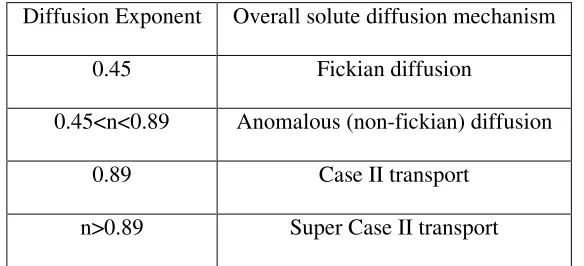
1.6. SWELLING CHARACTERISTICS OF POLYMER: [27]
The Peppa’s plot is useful to determine whether the drug release from the
matrix is controlled by swelling of the polymer or not. The Peppa’s equation is
log Q = log (kt½) ---(3)
log Q = log k + 0.5 log t ---(4)
Where,
Q = amount of drug release in time t per unit area
k = release constant.
If the slope of a plot of log Q Vs log t is exactly 0.5, then the drug release
occurs by perfect diffusion obeying Higuchi’s and Fick’s law. If it is in the range of
0.5 – 1, then the mechanism of release is diffusion and rate of diffusion is controlled
by swelling of polymer. If it is bellow 0.5, then there is no swelling of matrix occur.
1.7. MECHANISM OF DRUG RELEASE FROM MATRIX SYSTEM: [46, 21]
When a hydrophilic matrix system containing a swellable glassy polymer
comes in contact with an aqueous medium, the fall in glass transition temperature
leads to an abrupt change from a glassy to a rubbery state, causing swelling of the
this gel diffusional barrier and/or by surface erosion of the gel. Surface leaching of
the drug can lead to an initial burst, especially with highly soluble drugs.
Hydration of individual polymer chains leads to expansion in their end to end
distance and radius of gyration to a new solvated state due to lowering of the polymer
transition temperature, a sharp distinction between glassy and rubbery region is
observed and the matrix increases in volume because of swelling.
When the system is hydrated to the core, the drug concentration falls below its
solubility value and the release rate of the drug begins to decline. A concurrent
increase in the thickness of the barrier layer with time increases the diffusion path
length, further reducing the release rate. Drug release kinetic associated with this gel
layer dynamics, range initially from Fickian to anomalous (Non-Fickian) and
subsequently from quasi-constant (near zero order) to constant. Matrices of highly
molecular weight polymers rarely shows all three regimens (Fickian, Non-Fickian and
quasi-constant) of drug release because of a low chain disentanglement rate and
insufficient external polymeric mass transfer.
Soluble drugs are primarily released by diffusion through aqueous filled
porous network formed in the inert matrix former due to dissolution and erosion of
the polymer from the surface. For poorly soluble drugs dispersed in inert polymer
systems erosion is the primarily release mechanisms.
There are two major processes that control the drug release from swelling
controlled matrix systems, these include:
1. Ingress of aqueous medium into the matrix followed by a hydration,
gelation or swelling and
Simultaneous occurrence of these processes leads to the formation of two
fronts within the hydrating matrix, this are a swelling front, at the junction of the
unhydrated glassy matrix and the hydrated matrix and an eroding front where the
polymer is completely hydrated. Thickness of the diffusion layer, i.e. the distance
between the two fronts, depends on the relative rates at which the swelling and
erosion occurs.
If the polymer gels slowly, solvent can penetrate deep into the glassy matrix,
thus dissolving the drug; therefore, gel layer thickness and its stability are crucial in
controlling drug release. Numbers of techniques have been used to study the swelling
of matrix tablets and to characterize the gel layer and front movement such as, optical
imaging,1H- NMR, pulsed filled gradient spin echo NMR, confocal laser scanning
microscopy, cryogenic scanning electron microscopy and texture analysis. The gel
layer thickness is determined by the relative position of the swelling and erosion front.
1.8. DRUG RELEASE MECHANISM FROM MATRICES: [29]
From time to time, various authors have proposed different types of drug
release mechanisms from matrices. It has been proposed that drug release from the
matrices usually implies water penetration in the matrix, hydration, swelling,
diffusion of the dissolved drug (polymer hydro fusion), and / or the erosion of the
gelatinous layer. However, it is worth mention that the release mechanism of a drug
would depend on the dosage form selected, pH, and nature of the drug and of course,
the polymer used.11
a) Zero order kinetics:
Drug dissolution from pharmaceutical dosage from that do not disaggregate
and release the drug slowly (assuming that area does not change and no equilibrium
W0 –Wt = Kt --- (5) Where,
W0 = initial amount of drug in the pharmaceutical dosage form,
Wt = amount of drug in the pharmaceutical dosage form,
t = time,
K = Proportionality constant.
The pharmaceutical dosage form following this profile release the same
amount of drug by unit of time and in this model can be explained by following
equation:
Qt = Q0 + K0t --- (6) Where,
Qt = Drug dissolved in time t,
Q0 = Initial amount of drug in solution,
K0 = Zero order rate constant.
OR,
W = K.t (Xu and Sunada, 1995) --- (7)
Where,
W is percentage drug release at time t,
K is the release rate constant
b) First order kinetics:
The application of this model to drug dissolution studies was first proposed by
Gibaldi and Feldman (1967) and later by Wagner. The dissolution phenomena implies
a surface action, as can be seen by Noyes – Whitney equation,
) (C C K
dt dc
s −
= --- (8)
CS = solubility in equilibrium at experience temperature. k =First order proportionality constant
Hixson and Crowell adapted the above equation as
) (C C KS
dt Dw
s −
= --- (9)
Where, w = amount of solute in solution at time t,
S = Solid area accessible to dissolution.
log Qt = log Q0 + K1.t / 2.303 --- (10)
Where, Qt = amount of drug release in time t,
Q0 = initial amount of drug in solution,
K1 = First order release constant.
Above equation also represents this model.
The pharmaceutical dosage form following this dissolution profile, such as
those containing water soluble drugs in porous matrices release drug in a way that is
proportional to amount of drug remaining in its interior in such a way that amount of
drug released by unit of time diminish.
OR,
In (100 – W) = In 100 – k t (Singla and Medrata, 1988; and Sunada 1995)-- (11)
1.9. Definition of HIV:
Human immunodeficiency virus infection / acquired immunodeficiency
syndrome (HIV/AIDS) is a disease of the human immune system caused by infection
with human immunodeficiency virus (HIV).[1] During the initial infection, a person
may experience a brief period of influenza-like illness. This is typically followed by a
including opportunistic infections and tumors that do not usually affect people who
have working immune systems.
HIV is transmitted primarily via unprotected sexual intercourse (including
anal and even oral sex), contaminated blood transfusions, hypodermic needles,
and from mother to child during pregnancy, delivery, or breastfeeding.[2] Some bodily
fluids, such as saliva and tears, do not transmit HIV.[3] Prevention of HIV infection,
primarily through safe sex and needle-exchange programs, is a key strategy to control
the spread of the disease. There is no cure or vaccine; however, antiretroviral
treatment can slow the course of the disease and may lead to a near-normal life
expectancy. While antiretroviral treatment reduces the risk of death and complications
from the disease, these medications are expensive and may be associated with side
effects.
Acquired immunodeficiency syndrome (AIDS) is defined in terms of either a
CD4+ T cell count below 200 cells per µL or the occurrence of specific diseases in
association with an HIV infection. In the absence of specific treatment, around half of
people infected with HIV develop AIDS within ten years. The most common initial
conditions that alert to the presence of AIDS are pneumocystis
pneumonia (40%),cachexia in the form of HIV wasting syndrome (20%)
and esophageal candidiasis. Other common signs include recurring respiratory tract
infections.
Opportunistic infections may be caused by bacteria, viruses, fungi and parasites that
are normally controlled by the immune system. Which infections occur partly depends
on what organisms are common in the person's environment. These infections may
People with AIDS have an increased risk of developing various viral induced
cancers including Kaposi's sarcoma, Burkitt's lymphoma, primary central nervous
system lymphoma, and cervical cancer. Kaposi's sarcoma is the most common cancer
occurring in 10 to 20% of people with HIV. The second most common cancer is
lymphoma which is the cause of death of nearly 16% of people with AIDS and is the
initial sign of AIDS in 3 to 4%. Both these cancers are associated with human herpes
virus. Cervical cancer occurs more frequently in those with AIDS due to its
association with human papillomavirus (HPV).
Additionally, people with AIDS frequently have systemic symptoms such as
prolonged fevers, sweats (particularly at night), swollen lymph nodes, chills,
weakness, and weight loss. Diarrhea is another common symptom present in about
90% of people with AIDS. They can also be affected by diverse psychiatric and
neurological symptoms independent of opportunistic infections and cancers
Classification of Anti Viral drugs:
Agents to treat Herpes Simplex Virus (HSV) & Varicella Zoseter Virus (VZV)
infections
Acyclovir, Valcyclovir, Famciclovir, Penciclovir, Trifluridine.
Agents to treat Cytomegalovirus (CMV) infections
Ganciclovir, Valganciclovir, Cidofovir, Foscarnet, Fomivirsen.
Antiretroviralagents:
Nucloside Reverse Transcriptase Inhibitors (NRTIs)
Zidovudine, Didanosine, Lamivudine, Zalcitabine, Stavudine, Abacavir.
Antiretroviralagents:
Nucleutide inhibitors
Antiretroviralagents:
Non Nucleoside Reverse Transcriptase Inhibitors (NNRTIs)
Nevirapine, Delaviridine, Efavirenz.
Antiretroviralagents:
Protease Inhibitors
Saquinavir, Ritonavir, Lopinavir, Indinavir, Nelfinavir, Ampernavir.
Fusion Inhibitors
Enfuvirtide (HIV), Docosanol (HSV).
Anti-Hepatitis agents
Lamivudine, Ribavirin, Pegylated interferon alpha, Interferon alpha, Adefovir.
Anti-Influenza Agents
Amantadine, Rimantadine, Zanamivir, Oseltamivir.
Other Antiviral agents
2. LITERATURE REVIEW
Syed Ershad et al., (2013) prepared Floating microspheres of Ritonavir by ionic
gelation method with an aim of increasing the gastric residence time and for
controlled release. Sodium alginate, polymeric mixture of Sodium alginate and
xantham gum were used as polymers. Sodium bicarbonate was used as the
gas-forming gent. The prepared floating microspheres were evaluated with respect to
particle size distribution, floating behavior, drug content, entrapment efficiency,
morphology and in vitro release study. These results indicated that the release rate
was found to decrease with increase in concentration of coating material applied. The
wall thickness of microspheres was found to be increased with the increase in
concentration of coating material applied. The floating microspheres followed zero
order kinetics and the mechanism of drug release was governed by peppas model.
K.P.R Chowdhary et al., (2011) prepared, characterized and evaluated starch
phosphate, a new modified starch as a carrier in solid dispersions for enhancing the
dissolution rate of ritonavir. The feasibility of formulating solid dispersions of
ritonavir in starch phosphate into compressed tablets with enhanced dissolution rate
was also investigated. Starch phosphate was prepared by reacting starch with
di-sodium hydrogen orthophosphate anhydrous at elevated temperatures. It was insoluble
in water and has good swelling (400%) property without pasting or gelling when
heated in water. Solid dispersions of ritonavir in starch phosphate were prepared by
solvent evaporation method employing various weight ratios of drug: starch
phosphate such as 2:1(SD-1), 1:1(SD-2), 1:2(SD-3), 1:3(SD-4) and 1:9(SD-5) and
were evaluated for dissolution rate and efficiency. All the solid dispersions prepared
and 94.41 fold increase in the dissolution rate (K1) of ritonavir was observed with
solid dispersions SD-4 and SD-5 respectively. The DE30 was also increased from
6.80% in the case of ritonavir pure drug to 76.25% and 84.05% in the case of these
solid dispersions. Ritonavir (50 mg) tablets were prepared employingritonavir alone
and its solid dispersions SD-3 and SD-4 by wet granulation method and were
evaluated. Ritonavir tablets formulated employing its solid dispersions in starch
phosphate gave rapid and higher dissolution rate and DE30 when compared to plain
and commercial tablets. A 9.95 and 28.14 fold increase in the dissolution rate (K1)
was observed with tablet formulations containing solid dispersions SD-3 and SD-4
respectively when compared to plain tablets.
M.S. Hasoliya et al., (2012) prepared Floating microspheres of Ritonavir by simple dripping method with an aim of increasing the gastric residence time and for
controlled release. A polymeric mixture of Sodium alginate and hydroxy propyl
methyl cellulose was used. Sodium bicarbonate was used as the gas-forming gent. The
solution was dropped to 1% calcium chloride solution containing 10 % acetic acid for
carbon dioxide release and gel formation. The prepared floating microspheres were
evaluated with respect to particle size distribution, floating behavior, drug content,
entrapped, morphology and in-vitro release study. Effect of sodium bicarbonate on the
above mentioned parameters were evaluated and it was found that the sodium
bicarbonate had a pronounced effect on various parameters. The enhanced buoyancy
and controlled release properties of sodium bicarbonate containing microspheres
made them an excellent candidate for floating dosage form.
Kanagala Vijayasri et a l., (2013) prepared and evaluated montmorillonite (natural
dispersions of ritonavir in montmorillonite into compressed tablets. Solid dispersions
of ritonavir in montmorillonite were prepared by solvent evaporation method
employing various weight ratios of drug: montmorillonite such as 1:1(SD-1),
1:2(SD-2), 1:4(SD-3) and 1:7 (SD-4). The tablets were evaluated. Prior to compression, the
pre-compression parameters showed satisfactory flow properties. Post-compression
parameters showed that all tablet formulations had acceptable mechanical properties.
The compatibility of the drug in the formulation was confirmed by IR and DSC
studies. Ritonavir tablets formulated employing its solid dispersion in montmorillonite
gave rapid and higher dissolution rate and DE30 when compared to plain tablets. A
14, 12 and 8 fold increase in the dissolution rate (k1) was observed with tablet
formulations containing solid dispersions SD-1, SD-2 and SD-3 respectively when
compared to plain tablets
K.P.R. Chowdhary et al., (2012) comparatively evaluated three commercially
available DCVs namely Lubritose AN, Lubritose SD, Lubritose MCC and one
laboratory made DCV namely starch phosphate, a new modified starch in the
formulation development of three antiretroviral drugs by direct compression method.
Tablets of (i) Efavirenz (100 mg) (ii) Ritonavir (100 mg) and (iii) Stavudine (30 mg)
were formulated employing the four directly compressible vehicles and the tablets
were evaluated for various physical properties and dissolution rate. All the tablets
gave rapid dissolution of the contained drug. The dissolution was complete (100%)
within 15 – 30 min with all the drugs and the dissolution was much higher than the
official requirement in each case. Stavudine tablets exhibited faster dissolution than
those of efavirenz and ritonavir with all the four DCVs. Hence these DCVs are
recommended for the preparation of tablets of antiretroviral drugs by direct
Rabhi Narayan parhi et al., (2013) developed a floating drug delivery system of
ritonavir (RN) in order to prolong the gastric residence time and increase its
bioavailability. The floating tablets of RN were prepared by direct compression
technique, using polymers such as different grades of hydroxypropyl methylcellulose
(HPMC, Methocel E15LV, E50LV, K100LV and K4M) and polyvinyl pyrrolidone
(PVP K30). Sodium bicarbonate was used as gas releasing agent. The formulations
were optimized on the basis of matrix integrity, duration of floating, swelling
behavior and in vitro drug release. Except series FA, where floating time was 10 hr,
other series such as FB, FC and FD were showing more than 12 hr of floating time.
The mechanism of RN release from the floating tablets for FA, FB and FC series is
anomalous diffusion transport and follows zero order kinetics, but FD series indicated
Higuchi kinetics with release rate exponent (n) of 0.44. Further, the scanning electron
microscopy showed porous structured formed on the tablet surface at different times
(0, 3, 6, 9 and 12 hr) of dissolution for the selected batch FC3. Finally, FC3 batch
showed no significant change in above parameters after storage at room temperature
(28-32°C), 40°C and 50°C for one month.
Raju B et al., (2012) studied and developed ritonavir is an antiretroviral drug with activity against Human Immunodeficiency Virus (HIV) type 1. In the present work
an attempt is being made to provide for parenteral drug delivery with having
improved therapeutic index for Ritonavir and anintention to develop a stable and
effective parenteral formulation, containing the drug Ritonavir. Ritonavir is
practically insoluble in water and unstable at higher temperature. The effects of
various co solvents in the solubility ofRitonavir have been evaluated. Ritonavir was
tried with co solvents such as Sodium-p-hydroxy benzoate, Sodium glycinate and
as a SVP. Various batches of Ritonavir injection formulation were prepared in order
to assess the influence of heat, light, atmospheric oxygen and antioxidant on the
stability of the drug and the formulations were also subjected to accelerated stability
test. Out of all trials, formulation containing Sodium thiocyanate was found to be
more soluble, stable and passed all tests satisfactorily.
Chandira Margret R et al., (2010) developed floating tablets of Itopride hydro
chloride, a novel pro kinetic drug, which after oral administration are designed to
prolong the gastric residence time and thereby increase drug bioavailability and drug
release rate. This would help in promoting gastro intestinal transit and speed up
gastric motility and thereby it will relieve the symptoms associated with it. Floating
tablets were fabricated using direct compression method containing itopride
hydrochloride, polymers HPMC K100M, HPMC K15M and carbopol 934P along
with gas generating agent sodium bicarbonate and citric acid. The addition of
carbopol aided in the reduction of drug dissolution due to their hydrophobic nature.
The concentration of these agents was also optimized to get desired controlled release
of drug. The floating tablet formulations were evaluated for physical characterization,
assay, swelling index, in-vitro drug release, hardness, friability and weight variation.
The results indicated that gas powered floating tablets of Itopride hydrochloride
containing 125mg HPMC K 100M, 40 mg HPMC K 15M and 40 mg carbopol
provides a better option for 24 hrs release action and improved bioavailability
mechanism.
Margret Chandira et al., (2009) Prepared Diltiazem Hydrochloride undergoes an
extensive biotransformation, mainly through cytochrome P-450 CYP3A, which
results in less than 4% of its oral dose being excreted unchanged in urine. Suffers
It has an elimination half-life of 3.5 hrs and an absorption zone from the upper
intestinal tract. Thus the present work is aimed to formulate floating tablets of
Diltiazem Hydrochloride using an effervescent approach for gastro retentive drug
delivery system. Floating tablets were prepared using direct compression technique
using Hydrophilic polymer like HPMC K4M, HPMC K15M and hydrophobic
polymer like Ethyl cellulose as matrix materials in various quantities (%w/w), sodium
bicarbonate, citric acid, magnesium stearate, talc and lactose in varying ratio to
formulate the floating tablets. Observations of all formulations for physical
characterization had shown that, all of them comply with the specification of official
pharmacopoeias and/or standard reference. It was observed that tablets of batch F6
followed the results obtained, it was concluded that the formulation F6 is the best
formulations as the extent of drug release was found to be around 99.81 % at the
desired time 12 hrs.
Desai.S et al., (1993) studied A novel floating controlled-release drug delivery was
formulated in an effort increase the gastric retention time of the dosage form and to
control drug release. The buoyancy was attributed to air and oil entrapped in the agar
gel network. A floating controlled-release 300mg theophylline tablet having a density
of 0.67 was prepared and compared in-vitro and in vivo to Theo-dur. The in-vitro
release rate of the floating tablet was slower. In-vivo scintigraphic studies for a
floating and a heavy nonfloating tablet, under fasting and nonfasting conditions,
showed that the presence of food significantly increa