A STUDY ON PREVALENCE OF LEFT VENTRICULAR DYSFUNCTION AND ITS CORRELATION WITH ESTIMATED GLOMERULAR FILTRATION RATE (eGFR) IN CKD PATIENTS
DISSERTATION SUBMITTED FOR
DOCTOR OF MEDICINE
BRANCH - I (GENERAL MEDICINE)
APRIL 2015
THE TAMILNADU
BONAFIDE CERTIFICATE
This is to certify that the dissertation entitled “A STUDY ON
PREVALENCE OF LEFT VENTRICULAR DYSFUNCTION AND ITS CORRELATION WITH ESTIMATED GLOMERULAR FILTRATION RATE (eGFR) IN CKD PATIENTS”submitted by DR. TINA ANN
ANTONY to the Tamil Nadu Dr. M.G.R. Medical University, Chennai in partial
fulfillment of the requirement for the award of M.D Degree Branch I (General
Medicine) is a bonafide research work carried out by her under my direct
supervision & guidance.
Captain Dr. B. Santhakumar
M.Sc (F.Sc), M.D(F.M), PGDMLE, Dip.N.B (F.M),
The Dean,
Madurai Medical College,
Government Rajaji Hospital,
BONAFIDE CERTIFICATE
This is to certify that the dissertation entitled “A STUDY ON
PREVALENCE OF LEFT VENTRICULAR DYSFUNCTION AND ITS CORRELATION WITH ESTIMATED GLOMERULAR FILTRATION RATE (eGFR) IN CKD PATIENTS”submitted by DR. TINA ANN
ANTONY to the Tamil Nadu Dr. M.G.R. Medical University, Chennai in partial
fulfilment of the requirement for the award of M.D Degree Branch I (General
Medicine) is a bonafide research work carried out by her under my direct
supervision & guidance.
Dr. S.Vadivel murugan M.D, Dr.V.T. PREMKUMAR M.D.,
Professor and HOD, Professor of Medicine Dept. of General Medicine, Dept. of General Medicine,
Government Rajaji Hospital, Government Rajaji Hospital,
Madurai Medical College, Madurai Medical College,
DECLARATION
I, Dr. TINA ANN ANTONY declare that, I carried out this work on, “A STUDY ON PREVALENCE OF LEFT VENTRICULAR DYSFUNCTION AND ITS CORRELATION WITH ESTIMATED GLOMERULAR FILTRATION RATE (eGFR) IN CKD PATIENTS” at the Department of
Medicine, Govt. Rajaji Hospital during the period of July 2013 to August 2014.
I also declare that this bonafide work or a part of this work was not submitted by
me or any others for any award, degree, diploma to any other University, Board
either in India or abroad.
This is submitted to The Tamilnadu Dr.M.G.R.Medical University,
Chennai in partial fulfilment of the rules and regulations for the M.D degree
examination in General Medicine.
Place :Madurai Dr. TINA ANN ANTONY
ACKNOWLEDGEMENTS
At the outset, I wish to thank our Dean, Captain Dr. B. Santhakumar,
M.Sc (F.Sc), M.D(F.M), PGDMLE, Dip.N.B (F.M), for permitting me to use the
facilities of Madurai Medical College and Government Rajaji Hospital to conduct
this study.
My beloved Head of the Department of Medicine,
Prof. Dr.S.Vadivel Murugan M.D., has always guided me with his valuable
words of advice and has encouraged innovative thinking and original research
work done by post graduates.
I shall remain eternally grateful to my unit chief
Prof. Dr. V.T.Premkumar M.D, who has given me his moral support and
encouragement through the conduct of the study.
I also sincerely thank our beloved professors Dr.R.Balajinathan M.D,
Dr.M.Natarajan M.D, Dr.Bagialakshmi M.D, Dr.J.Sangumani. M.D,
Dr.C.Dharmaraj M.D., and Dr.R.Prabhakaran M.D, for their par excellence
I am extremely grateful to the Department of Nephrology and Prof.
Dr.Shanmuga Perumal M.D D.M for their constant support, guidance,
cooperation and encouragement.
I would also like to express my deep felt gratitude to Department of
Cardiology and Dr.A.S.Arul M.D D.M and retired professor and H.O.D of
Cardiology Dr.R.A. Janarthanan M.D D.M for their support, encouragement
and guidance.
I offer my heartfelt thanks to my unit Assistant Professors Dr.K.S.
Maniappan M.D., Dr. M.Sooryakumar, M.D, and Dr. P.Manimegalai M.D,
for their constant encouragement, timely help and critical suggestions
throughout the study.
I would also like to sincerely thank the Department of Biochemistry for
helping me out with the laboratory investigations.
My patients, who form the most integral part of the work, were always
kind and cooperative. I pray to God to give them courage and strength to endure
their illness.
I thank my friends and family who have stood by me during my times of
need. Their help and support have always been invaluable to me. And last but
not the least I would like thank the Lord Almighty for His grace and blessings
ABBREVIATIONS
CKD Chronic Kidney Disease
GFR Glomerular Filtration Rate
LV Left Ventricular
LVH Left Ventricular Hypertrophy
CAD Coronary Artery Disease
eGFR estimated Glomerular Filtration Rate
NKF National Kidney Foundation
KDOQI Kidney Disease Outcome Quality Initiative
SEEK Screening and Early Evaluation of Kidney disease
TGF β Transforming Growth Factor β
FGF - 23 Fibroblast Growth Factor - 23
PTH Parathormone
CRP C Reactive Protein
ECF Extracellular Fluid
ACE Angiotensin Converting Enzyme
ARB Angiotensin Receptor Blocker
HIV Human Immunodeficiency Virus
NHANES National Health and Nutrition Evaluation Survey
MRFIT Multiple Risk Factor Intervention Trial
ADMA Asymmetric Dimethyl Arginine
ECHO Echocardiography
BP Blood Pressure
MDRD Modified Diet in Renal Disease
BMI Body Mass Index
PICP Peptide of collagen type 1 Protein
CT -1 Cardiotrophin -1
DD Diastolic dysfunction
CONTENTS
TOPIC PAGE NO.
1. INTRODUCTION
2. AIMS AND OBJECTIVES
3. REVIEW OF LITERATURE
4. METHODS AND MATERIALS
5. OBSERVATION AND RESULTS
6. DISCUSSION
7. SUMMARY
8. CONCLUSION
9. ANNEXURE
•
Bibliography
•
Proforma
•
Master chart
•
Ethical clearance
•
Turnitin Certificate
1
ABSTRACT
BACKGROUND AND OBJECTIVE:
In Chronic Kidney Disease there is a higher incidence cardiovascular events.
Most of the patients with CKD succumb to cardiovascular disease even before they
reach the end stage of renal disease. Hence all efforts should be given in earlier
stages of CKD to prevent the development of cardiovascular complications.
LV diastolic dysfunction is found to antedate LVH and systolic dysfunction.
It is not just associated with hemodynamic factors like anaemia and hypertension,
but also with uraemia related non hemodynamic factors like secondary
hyperparathyroidism, altered mineral metabolism, cardiotrophysin etc.
ECHO provides a simple non-invasive method to assess the left ventricular
structure and function, which helps us in identifying those prone for cardiovascular
complications at an earlier stage of CKD.
This study was done to find out the prevalence of LV dysfunction and its
correlation with eGFR in CKD patients.
METHODS:
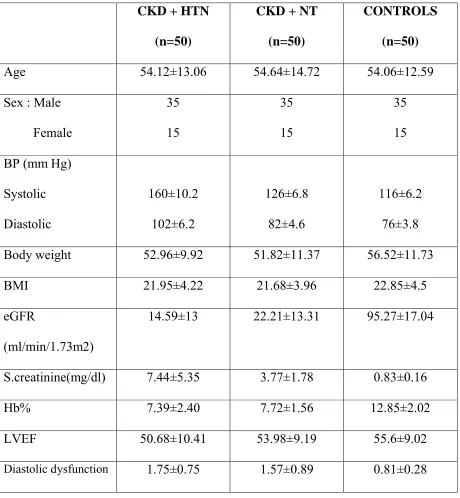
50 hypertensive CKD patients and 50 normotensive CKD patients admitted
for the presence of LV dysfunction and LVH and the results were compared with
that of 50 age and sex matched individuals.
Patients with acute kidney injury, prior coronary artery disease, valvular
heart disease, cardiomyopathy, diabetic individuals and CKD patients on renal
replacement therapy or transplant patients were excluded from the study.
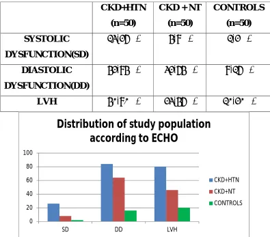
The CKD patients were divided into various stages of CKD based on their
eGFR (calculated according to MDRD formula) and they were evaluated by
ECHO. Those with an ejection fraction < 50% were considered to have systolic
dysfuction. Diastolic dysfunction was calculated based on the E/A ratio. E/A ratio
<0.8 was grade I, 0.8 – 1.5 was grade II and >2 was grade III diastolic dysfunction.
This was compared with the ECHO findings of the controls. The collected data
was analysed using various statistical methods.
RESULTS:
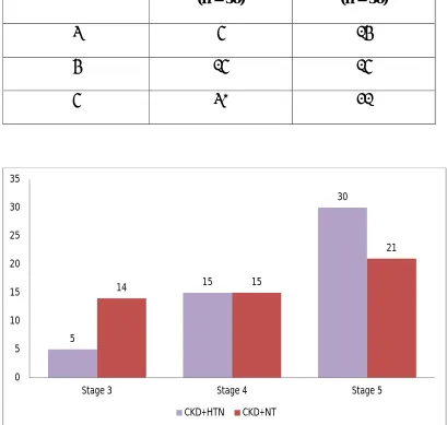
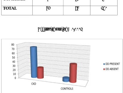
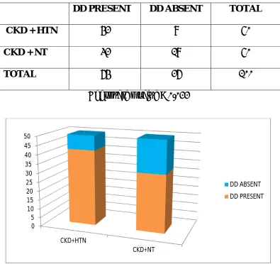
74% of CKD patients had LV diastolic dysfunction (p <0.0001). Diastolic
dysfunction was found to occur in 84% of the hypertensive CKD and in 64% of the
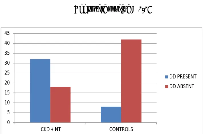
Comparing the CKD normotensives with the control group, 64% among the
normotensive CKD had diastolic dysfunction whereas only16% of controls had
diastolic dysfunction (p<0.05).
There is a negative correlation between the eGFR and diastolic
dysfunction.So as the eGFR falls, the diastolic dysfunction increases.
INTERPRETATION AND CONCLUSION:
Even in the absence of hypertension, LV diastolic dysfunction can occur in
CKD. This emphasizes the need for correction of not just hemodynamic factors but
also the uraemia related factors.
KEY WORDS:
INTRODUCTION
Chronic kidney disease (CKD) is a spectrum of pathophysiological
process that is associated with dysfunction of kidney and a progressive
decrease in Glomerular Filtration Rate (GFR). (1)
It refers to functional or structural abnormalities of kidneys for
more than three months, irrespective of cause.
It is a global health concern. CKD has been the 12th leading cause
of death, and the 17th leading cause of disability. CKD has a number of
co-morbidities and hence is a disease with high mortality.(2)
An analysis of risk factors for development and progression of
CKD is necessary in clinical practice.
The result of CKD is loss of kidney function which in the long run
leads to kidney failure, decreased kidney function and its complications,
development of cardiovascular disease and death.(3) Improving outcomes
in CKD requires prevention, detection, evaluation, and management of
other chronic diseases, such as hypertension, hypercholesterolemia ,
The number of CKD patients is on the rise. The prevalence rate of
CKD in India is not available due to lack of adequate data recording. In
community based studies, the prevalence rate of CKD is from 0.79% to
1.4%. The studies were done to detect stage 3 CKD or worse. The exact
prevalence is higher than the one reported. The end stage renal disease
(ESRD) incidence is reported as 160 to 232 per million population and
the projected ESRD prevalence rate was 785 to 870 per million
population.(4)
Screening and Early Evaluation of Kidney disease (SEEK); a
community based study which was done recently reported a very high
prevalence rate of CKD which is about 17.4%.(5) The Indian CKD registry
was formed in the year 2005 with a target to serve as a comprehensive
nationwide data warehouse for studying the different aspects of CKD.
The data house has enrolled about 63,538 patients and 74% of the cases
have CKD stage 4 and stage 5.
The prevalence of End Stage Renal Disease (4) (ESRD) and the
patients who are on renal replacement therapy has increased since last 2
decades. Only 20% of ESRD patients are on Renal Replacement Therapy
(RRT). It is estimated that 1,00,000 new patients of ESRD undergo renal
The most common cause for CKD in India is Diabetes. It accounts
for about 30 to 40% of patients. With the increasing number of diabetic
patients there is a parallel increase in the incidence of CKD. In India the
number of patients with diabetes is now about 41 million and is on the
rise. Most of the patients develop diabetic nephropathy and land up in
ESRD.
In Chronic Kidney Disease there is a higher incidence of
cardiovascular events. Cardiovascular disease is one of the prime causes
of morbidity and mortality in all the stage of CKD. Most of the patients
with CKD develop cardiovascular disease before reaching ESRD. The
CKD registry (6) has shown that patients with CKD stage 4 have a high
incidence of cardiovascular complications. Most of the patients die before
they have ESRD. Patients in early stages of CKD are also found to have a
high cardiovascular morbidity. Hence we should look into the prevention
of cardiovascular complications.
A large cohort study of patients with CKD stages 2 to 4 showed
that death was seen more often than progression to kidney failure in all
stages of CKD. There was a high baseline prevalence of cardiovascular
disease in the patients who died, compared to those who survived, which
the deaths. Hence, most patients in the earlier stages of CKD do not
progress to kidney failure because of the mortality caused due to
cardiovascular disease. Cardiovascular disease has been seen as a major
“competing outcome or risk” of kidney failure. (3)
The cardiovascular manifestation in CKD includes: (13)
Ischemic heart disease – myocardial infarction and angina
Left ventricular hypertrophy
Heart failure : LV diastolic and systolic dysfunction and
Dilated cardiomyopathy.
Heart and kidney are linked to each other in the hemodynamic and
regulatory functions. There are multiple communications among the two
organs like Sympathetic Nervous System, Anti Diuretic Hormone, Renin
Angiotensin Aldosterone System and natriuretic peptides.(8) National
Kidney Foundation formed a Task Force which considered Coronary
Artery Disease (CAD) and LVH as the target conditions, recommended
for decreasing cardiovascular mortality in ESRD.(9)
LVH is associated with both systolic and diastolic
dysfunction. LVH associated with systolic dysfunction is expressed by
fraction. These findings show us that mild reduction in the renal perfusion
which is caused by the slightly impaired LV systolic function, is
associated with pathological, highly pulsatile perfusion in the
microvasculature of the kidney. This might be the mechanism through
which a progressive reduction of renal function takes place in patients
with pre-existing renal damage. This renal insufficiency produced,
adversely affects the function of the heart, which produces a vicious
cycle where the renal failure further impairs cardiac performance.
Diastolic dysfunction is associated with higher incidence of
episodes of intradialytic hypotension and higher peri-operative death
from pulmonary oedema at the time of renal transplantation. It is
significant to recognize and correct the cardiovascular complications and
risk factors before the commencement of dialysis.
Echocardiography will provide us with a very simple and
non-invasive assessment of the structure and function of the heart. It also
helps us to identify the people who are at greater risk. Strategies to
prevent the development and progression of LV dysfunction at an early
AIMS AND OBJECTIVES:
1. To assess the prevalence of Left Ventricular (systolic and/ or
diastolic) dysfunction in patients with CKD.
2. To evaluate the correlation between Left Ventricular
dysfunction and eGFR in CKD patients
3. To compare the Left Ventricular dysfunction in hypertensive
REVIEW OF LITERATURE
NORMAL ANATOMY AND PHYSIOLOGY OF KIDNEY
EMBRYOLOGY OF KIDNEY:
The kidney is one of the most highly differentiated organs in the
body (10) They develop from the intermediate mesoderm under the timed
and sequential control of genes. The excretory tubules (nephrons) develop
from the metanephric blastema. The collecting part arises from the
ureteric bud, which arises from the lower part of mesonephric duct. The
ureteric bud grows cranially towards the metanephric blastema, and
becomes dilated to form an ampulla. Ampulla undergoes division to form
the major calyces, then minor calyces and finally the collecting tubules.
(11)
GROSS ANATOMY:
Kidneys are a pair of excretory retroperitoneal organs, situated on
each side of the vertebral column. It consists of two poles, extending from
T12 to L3. The left kidney is at a lower level than the right. The
approximate size of each kidney is 11 cm long, 5 cm wide and 3 cm in
thickness. The weight of a single kidney is about 125 to 170 grams.
Renal vessels, renal pelvis, lymphatics and nerve plexus enter through the
hilum. Kidney consists of an outer cortex and an inner medulla. Cortex
contains all the glomeruli and portions of the tubules, whereas the
medulla is made of renal pyramids and renal columns. Minor calyx arises
from the pyramids and joins to form the major calyces which form the
renal pelvis. (10)
THE NEPHRON:
Nephrons are the functional units of kidney. Kidney contains
approximately about 1.2 million nephrons. The nephron consists of
malphigian or renal corpuscle (Bowman’s capsule and the glomerulus),
the site at which blood is filtered and a renal tubule (the proximal tubule,
thin and thick ascending limb, distal tubule and collecting ducts) from
which solutes are reabsorbed. (10)
Glomerulus (Renal corpuscle):
It is a network of capillaries lined by endothelial cells, central
mesangial cells, visceral epithelial cells and its basement
membrane, parietal layer of Bowman’s capsule with its basement
Visceral epithelium becomes continuous with the parietal
epithelium at the vascular pole. Here the afferent arteriole enters
and efferent arteriole exits. (10)
It also helps in the production of an ultrafiltrate of plasma
Tubules:
Continues from Bowman space and consists of proximal loop of
Henle and distal tubule.
The proximal convoluted tubule is lined by cuboidal cells which
aids in the absorption of solutes.
Loop of Henle is made of a thin descending limb and a thick
ascending limb, extending into cortex. The thick end of ascending
limb reaches the glomerulus of the nephron and nestles between its
afferent and efferent arterioles. Specialised cells at the end form the
macula densa, particularly close to the afferent arteriole.(12)
The macula densa, the neighboring lacis cells, and the
renin-secreting juxtaglomerular cells which are present in the afferent
Distal convoluted tubules coalesce to form the collecting ducts.
The epithelium of collecting duct has principal (P cells) and
intercalated (I cells). P cells are involved in Na+ reabsorption and
vasopressin stimulated water reabsorption. The I cells are
concerned with acid secretion and HCO3- secretion.(12)
VASCULAR SUPPLY OF KIDNEY:
Kidney is supplied by a single renal artery, a direct branch from
abdominal aorta. Renal artery enters the hilum and divides into an
anterior and a posterior branch.
Lobar arteries: these are 3 segmental arteries arising from the
anterior branch and supply upper, middle and lower third of the
anterior surface of the kidney. Posterior branch supplies posterior
half. These are end arteries and no collateral circulation has been
demonstrated.(10)
Inter lobar arteries: arises from the lobar arteries runs between the
renal columns and pyramids.
Arcuate arteries: arises from the interlobar arteries.
Interlobular arteries: arises from the arcuate arteries and forms the
FUNCTIONS OF THE KIDNEY: (11)
A. Maintain solute and water homeostasis.
B. Serves as an endocrine organ by producing erythropoietin, active
form of vitamin D and renin.
C. Excretion of metabolic products.
D. Regulation of blood pressure and intraglomerular hemodynamics.
E. Regulation of acid bases.
F. Elimination of toxic substances from the body.
G. Involved in catabolism of small peptide hormones.
H. Regulation of extra cellular volume and osmolarity.
AGE RELATED CHANGES OF KIDNEY:
The kidney attains its full anatomical and functional maturity by the end
of 3rddecade of life. From then on, involutive changes start. Up to 6th
decade these changes are slow and after this a rapid progression occurs
due to reduced renal perfusion. Despite this, under normal circumstances
KIDNEY IN YOUNG ADULTS:
Anatomical characteristics:
During this period, there is full maturation of all renal structures.
The cortico-medullary index (cortex: medulla) increases from 1.64:1 in
the new born to 2.59:1 in adults. The kidneys reach their maximum size
during this period. (10)
Functional characteristics:
The kidneys are fully functional in adults due to its complete
maturation. The normal Glomerular Filtration Rate (GFR) at this period is
120 to 130 ml/min/1.73m2. GFR decreases as age advances.(10)
KIDNEY IN THE ELDERLY:
Anatomical characteristics:
Age produces changes that are similar to those in chronic kidney
diseases. There is a progressive loss of kidney mass. The weight of both
the kidneys decreases to 110 – 120 gram as the age increases. The lost
kidney mass is mainly in the cortex, characterised by a reduction in the
number of functional nephrons, whereas medulla is relatively spared.
There is an enlargement of the mesangial matrix, thickening of the
the renal corpuscles. With ageing, renal vasculature undergoes changes
regardless of hypertension or other diseases. The renal arteries undergo
sclerosis resulting in ischemic nephropathy.
Functional characteristics:
In normal healthy persons the age related changes develop slowly.
Increase in vascular resistance in the afferent and efferent arterioles
decreases renal blood flow. There is a progressive decrease in the
intensity of glomerular filtration. The creatinine clearance decreases at an
average of 0.83ml/min/m2 per year. However serum creatinine does not
change as there is a progressive decline in the muscle mass. The sodium
preserving capacity is lowered due to its insufficient intake and there is
reduced reabsorption of sodium by the distal tubules due to interstitial
fibrosis. The renin-angiotensin-aldosterone system activity is reduced.
The concentrating and diluting capacity of kidney is also reduced.
ADAPTATION OF KIDNEYS TO NEPHRON LOSS
The kidney’s ability of maintaining constancy of the extracellular
fluid volume and composition is usually well preserved till late in the
course of CKD. When nephrons are lost through disease, the rest of the
hyperfunction, which compensates for the loss. Studies in patients with
CKD showed that; if the GFR falls below a critical level, the disease
leads to end stage; even when initial disease activity has been controlled.
This mechanism for the disease progression of CKD has been based on
the Bricker’s intact nephron hypothesis, which states that:
“As CKD progresses; the kidney function is maintained by a decreasing
pool of hyperfunctioning nephrons rather than a relatively constant
number of nephrons with decreasing function”.
RENAL PROGRESSION: (13)
Renal progression is a term used to define primary nephron
loss which produces a maladaptive deterioration in the remaining
nephrons. Irrespective of injury in the glomeruli or tubulointerstitium, it
follows a common pathway. The mechanisms involved are:
Glomerular hypertension reduces the single nephron GFR and the
associated proteinuria.
Proteinuria produces accumulation of interstitial mononuclear
cells.
Activation of nephritogenic T lymphocytes produces interstitial
Epithelial – mesenchymal transition forming new interstitial
fibroblasts.
Fibrosis of adjacent capillaries and tubular nephrons results in
acellular scar.
ROLE OF ANGIOTENSIN II IN RENAL PROGRESSION:
Angiotensin hastens renal progression in CKD patients. Aldosterone
increases the renal vascular resistance and glomerular capillary pressure
which complements the detrimental effects of Angiotensin. It causes renal
damage by:
Efferent arteriolar vasoconstriction which increases the
intraglomerular capillary pressure.
By selectively altering the glomerular size it induces protein
ultrafiltration.
Podocyte function is altered by increasing intracellular calcium.
TUBULAR FUNCTION IN CHRONIC RENAL FAILURE:
The loss of functioning nephrons in chronic renal failure produces
a persistent intraglomerular hypertension.
Glomerulotubular balance is maintained; inspite of loss of
functioning nephrons, by which the remaining nephrons increases the
single nephron glomerular filtration. To maintain the solute homeostasis
the tubular function is altered in CKD. (14)
1. SODIUM:
The transport of sodium and its ability to maintain extracellular
volume is well preserved till late in CKD. Sodium excretion is increased
by decreasing its reabsorption in the Henle’s loop and distal nephrons.
There is increased excretion of organic and inorganic anions, and
increased expression of atrial natriuretic peptide . As the disease
progresses, compensatory changes are lost resulting in sodium retention,
expansion of intravascular volume, oedema and worsening hypertension.
(14)
2. Potassium:
Hyperkalemia can occur when the compensatory functions of the
1. Urinary dilution and concentration:
CKD patients lose their ability to concentrate or dilute the urine. So the
urine specific gravity is fixed and the patients are prone for nocturia.
2. Acid base regulation:
The solute load of the remaining nephrons is increased which produces
impaired total body H+ excretion. The hydrogen ion pumps are lost with
reduction in ammoniagenesis resulting in non-delta acidosis. In advanced
stages, the ammoniagenesis is further decreased by elevated potassium
resulting in type IV renal tubular acidosis.
3. Calcium and phosphate:
Kidneys and intestines are the key regulators of calcium phosphate
metabolism. In chronic renal failure the excretion of phosphate is reduced
resulting in hyperphosphatemia. Calcium level is reduced by the
following ways:
1. Decreased absorption in the gut
2. Decreased calcitriol formation
3. Increased serum phosphate
CHRONIC KIDNEY DISEASE
Chronic kidney disease (CKD) includes a spectrum of different
pathophysiologic processes associated with abnormal kidney function and
a progressive decline in glomerular filtration rate (GFR).(2)
Chronic Renal Failure is a term used for the continuing significant
irreversible reduction in nephron number and corresponds to CKD stages
3 – 5.(12)
End-Stage Renal Disease (ESRD) is defined as a stage of CKD
where toxins, electrolytes and fluids which are normally excreted by
kidney are accumulated and it is responsible for uremic syndrome. Which
ultimately leads to death of the patient unless a renal replacement therapy
using dialysis or renal transplantation is done where, the toxins are
removed. (12)
The kidney functions as biosynthetic, excretory and metabolic
organs, necessary for maintaining normal physiology. Although daily
dialysis can replace some kidney functions, it cannot replicate the normal
DEFINITION OF CHRONIC KIDNEY DISEASE:
The National Kidney Foundation’s (NKF) “Kidney Disease
Outcomes Quality Initiative (KDOQI)” has proposed a definition and
classification scheme of CKD. The NKF guidelines define CKD on the
basis of kidney damage and or reduced renal function. (2)
Glomerular filtration rate is calculated from serum creatinine and
equations with the help of age, sex, race, and BMI. CKD can be classified
based on the level of GFR . (1) Stages 1 and 2 are kidney damage, stages 3
and 4 are decreased kidney function and stage 5 is kidney failure. (3)
CRITERIA: (2, 1, 13)
1. Kidney damage ≥ 3 months, either functional or structural
abnormalities, with or without decreased GFR, are seen by the
following manifested
Pathological abnormalities or
Markers of kidney damage
ii. Imaging abnormalities. (Bilaterally
shrunken kidneys)
iii. Blood abnormalities.
2. GFR <60 ml/min/1.73m2 for 3 months or longer and with or without
CLASSIFICATION OF CKD:
STAGE GFR / ml / 1.73 m2 ACTION
0 >90 a Screening: CKD risk reduction
1 >90 b Diagnosis and treatment
Slowing the progression of
the disease
Treatment of comorbidities
CVD risk reduction
2 60 -89 Estimating progression
3 30 – 59 Treating complications
4 15 – 29 Prepare for kidney replacement
5 <15 Kidney replacement
a - risk factors for CKD
b - demonstrated kidney damage – persistent proteinuria,
abnormal urine sediment,
abnormal blood and urine chemistry,
abnormal imaging studies (13)
Staging of CKD was based on the estimated glomerular filtration rate
(eGFR) and not on serum creatinine level. In stage 1 and stage 2 the GFR
structural or functional defect (eg, proteinuria, or abnormal imaging
studies) to classify under it.
RISK FACTORS FOR CKD: (14)
Hypertension
Diabetes mellitus
Autoimmune disease
Older age
African ancestry – genetic – APOL 1 gene
Family history of renal disease
Previous episode of acute kidney injury
Proteinuria
Abnormal urine sediment
Structural abnormalities of the urinary tract
Nephrotoxins
Dyslipidemia
Smoking
PATHOPHYSIOLOGY OF CHRONIC KIDNEY DISEASE:
It’s divided into two causes
1) Initiating mechanism
Initiating mechanisms – this is caused due to a specific
etiology. For example genetically determined abnormalities in
the renal integrity or its development, immune complex
deposition and inflammation in certain types of
glomerulonephritis, toxin exposure in interstitial nephritis etc
Progressive mechanisms –this is caused due to the
hyperfiltration and hypertrophy of the remaining viable
nephrons due to decreased renal mass, irrespective of
etiology. These responses were mediated by the cytokines and
growth factors, vasoactive hormones, and transforming
growth factor (TGFß). (13)
The increased intra-renal renin activity contributes to both initial
inciting event and also to the progressive mechanisms, finally leading on
Risk Factors for Chronic kidney disease and its outcome (14)
Risk Factors Definition Examples
Susceptibility
factors Increase susceptibility to kidney damage Older ageFamily history of Chronic reduction in kidney mass low birth weight
kidney disease
Initiation factors Directly initiate kidney
damage
Diabetes
high blood pressure autoimmune diseases urinary stones
lower urinary tract obstruction, systemic infections
urinary tract infections drug toxicity
Progression factors
Cause worsening kidney damage and faster decline in kidney function after initiation
of kidney damage
poor glycemic control in diabetes Higher level of proteinuria
smoking
systolic blood pressure
End-stage factors
Increase morbidity and mortality in kidney
failure
Lower dialysis dose low serum albumin level
Examples of Epidemiologic Studies Involving Patients with Kidney Disease
Study
Design Examples Prospective
cohorts Population Based Cohorts Atherosclerosis Risk in Communities Study
(ARIC)
Framingham Heart Study
Cardiovascular Health Study (CHS) Multi-Ethnic Study of Atherosclerosis (MESA)
Health, Aging and Body Composition (Health ABC)
Coronary Artery Risk Development in Young Adults (CARDIA)
Cohorts of Patients with Kidney Disease
Chronic Renal Insufficiency Cohort (CRIC) Cardiovascular Risk in Birmingham (CRIB) United States Renal Data System (USRDS) Choices for Healthy Outcomes in Caring for End Stage Renal Disease (CHOICE)
Interventio
n trials Trials in Patients with Kidney Disease Modification of Diet in Renal Disease Study
(MDRD)
African American Study of Kidney Disease (AASK)
Study
Design Examples
Deutsche Diabetes DialyseStudie (4D) Reduction of Endpoints in NIDDM with the Angiotensin II Antagonist Losartan (RENAAL) Irbesartan in Diabetic Nephropathy Trial
(IDNT)
Hemodialysis Study (HEMO)
The Ramipril Efficacy In Nephropathy (REIN)
Post Hoc Analysis of Trials in Populations with CVD or CVD Risk Factors
Heart Outcomes Prevention Evaluation Trial (HOPE)
CAUSES OF CHRONIC KIDNEY DISEASE:
Diabetic glomerulosclerosis
Hypertensive nephrosclerosis
Glomerular Diseases
Systemic lupus erythematosus.
Amyloidosis, light chain disease.
Glomerulonephritis
Wegener’s granulomatosis
Tubulointerstitial Diseases
Obstructive nephropathy (stones, benign
prostatic hypertrophy).
Analgesic nephropathy.
Myeloma kidney.
Reflux nephropathy.
Vascular
Diseases Atheroembolic renal disease
Vasculitis.
Scleroderma.
Reno vascular renal failure
Cystic
Diseases Medullary cystic kidney disease
CLINICAL PRESENATION:
Usually the patients are asymptomatic in the early stages of CKD.
Hypertension and proteinuria are the most common features of CKD and
are present in all stages of the disease. Uraemia leads to disruption in the
function of all organ systems. Most common complications include
anaemia, decreased appetite, and abnormalities in potassium, sodium,
water and acid-base homeostasis, abnormalities in phosphorus, calcium
and mineral regulating hormone.(13)
PATHOPHYSIOLOGY AND BIOCHEMISTRY OF UREMIA:
1. Consequent to accumulation of toxins that are normally excreted:
Even though urea and creatinine measures the excretory capacity of
kidney, it is not the accumulation of these toxins that are responsible for
the uremic symptoms. The toxins that are implicated in uremic syndrome
include: hydrophobic, protein bound, water soluble, charged and
uncharged molecules. Additional nitrogenous excretory products include
urates, hippurates, guanidino compounds, polyamines, products of
nucleic acid metabolism, myoinositol, benzoates, indoles and phenols.
Compounds called middle molecules (molecular mass of 500 – 1500 Da)
2. Consequent to loss of other renal functions:
Suppression of metabolic and endocrine functions results in anemia,
malnutrition, abnormal metabolism of protein, carbohydrates and fats.
FGF 23, PTH, insulin, glucagon, Vitamin D, sex hormones all change
with renal failure. It occurs due to abnormal retention, decreased
degradation or abnormal regulation. (13)
3. Progressive systemic inflammation and its vascular and nutritional consequences:
Elevated CRP and acute phase reactants
Negative acute phase reactants- albumin and fetuin decline.
So, the inflammation associated with CKD is important in
malnutrition inflammation-atherosclerosis/calcification syndrome.
The clinical presentation depends on the organ system involved, the
1. Fluid, electrolyte and acid-base disorders:
Sodium and water homeostasis:
In the early stages of CKD, the total body content of sodium and water is
increased. Dietary intake of sodium exceeds the urinary excretion
resulting in sodium retention and extra cellular (ECF) volume expansion.
Thus resulting in hypertension. Hyponatremia is not commonly seen in
CKD patients, if it is present, it will respond to water restriction. Some
patients may have impaired renal conservation of sodium and water. (13)
Potassium homeostasis:
Hyperkalemia in CKD may be due to the following mechanisms:
A. Increased dietary potassium intake.
B. Protein catabolism.
C. Haemolysis, haemorrhage
D. Transfusion of stored blood and
E. Metabolic acidosis.
F. Drugs – ACE inhibitors, ARBs, spironolactone, potassium
Hyporeninemic hypoaldosteronism, obstructive uropathy and
sickle cell nephropathy may cause disruption of potassium secretory
mechanism out of proportion to decline in GFR.
Hypokalemia is not common in CKD. It may occur in reduced
dietary intake, excessive diuretic therapy, gastrointestinal losses, primary
potassium wasting conditions – Fanconis syndrome, Renal Tubular
Acidosis
Metabolic acidosis:
Commonly seen in patients with advanced CKD. The combination
of hyperkalemia and hyperchloremic metabolic acidosis is often present.
Non anion gap acidosis occurs in the early stages of CKD but in later
stages as the disease progresses anion gap acidosis ensues. Treatment of
hyperkalemia increases the renal ammonia production; improve the renal
production of bicarbonate thereby improving the metabolic acidosis. (13)
Disorders of calcium and phosphate metabolism:
2. Bone manifestations of CKD:
Due to high bone turn over - secondary hyperparathyroidism produces
adyanamic bone disease and osteomalacia. Reduced GFR (< 60 ml/min)
leads to reduced excretion of phosphate - retained phosphate stimulates
PTH- secondary hyperparathyroidism. PTH is by itself a uremic toxin.(13)
Fibroblast growth factor 23(FGF-23) is a phosphatonin that promotes the
renal phosphate excretion. Studies have shown that high levels of FGF-23
are an independent risk factor for left ventricular hypertrophy. Adynamic
bone disease is most common in diabetics and elderly patients. It is also
associated with cardiac and vascular calcification. Osteomalacia is caused
by vitamin D deficiency, metabolic acidosis. Renal osteodystrophy results
from secondary hyperparathyroidism. (13)
Hyperphosphatemia and hypercalcemia causes increased vascular
calcification of the media of coronary arteries and heart valves and
increased cardiovascular mortality. (13)
Calciphylaxis (calcific uremic arteriolopathy) due to vascular and soft
3 Neuromuscular abnormalities:
It results in peripheral neuropathy, autonomic neuropathy and
myopathy. These abnormalities are due to the nitrogenous metabolites
and middle molecules. In the early stages patients present with
disturbances in the memory, concentration and sleep. Neuromuscular
irritability, including hiccups, cramps, and fasciculations or twitching of
muscles are seen in later stages. Asterixis, myoclonus, seizures and coma
may occur in advanced untreated cases. (13)
Peripheral neuropathy occurs from stage 4 of CKD and its
evidence without any other cause is a firm indication for renal
replacement therapy. Sensory nerves, lower extremities, distal parts are
more involved than the motor, upper extremities and proximal part.
‘Restless leg syndrome’ is a common manifestation.
4. Gastrointestinal and nutritional manifestations:
Uremic fetor can lead to dysgeusia. Other manifestations include
gastritis, peptic ulcer disease, and mucosal ulcerations resulting in
abdominal pain, nausea, vomiting and GI bleeding. Constipation is
Protein energy malnutrition due to low protein and calorie intake is
common in advanced stages. Metabolic acidosis and activation of
inflammatory cytokines promotes protein catabolism. All the patients
should be assessed for malnutrition from stage 3 of CKD. (13)
5. Endocrine – metabolic manifestations:
Glucose metabolism is often impaired in CKD. Plasma insulin
levels are elevated in most of the uremic patients as the kidneys fail to
remove it. Due to the diminished degradation of insulin, insulin and
hypoglycaemic agents require dose reduction.
In women with CKD, oestrogen levels are low. Menstrual
abnormalities and infertility are common. Pregnancy may hasten the
progression of the disease. Spontaneous abortions and miscarriages are
common. Males have reduced plasma testosterone levels, sexual
dysfunction and oligospermia. (13)
6. Dermatologic manifestations:
Pruritus is the most common and devastating complication of
CKD. Hyper pigmentation of the skin due to the deposition of pigmented
metabolites (urochromes) can occur. Nephrogenic fibrosing dermopathy
condition is characterised by progressive subcutaneous induration of arms
and legs. (13)
7. Hematologic abnormalities: Abnormal haemostasis:
Prolonged bleeding time
Decreased activity of platelet factor 3
Impaired prothrombin consumption
Abnormal platelet adhesion and aggregation
Thrombophilia occurs due to nephrotic range proteinuria, which
ANAEMIA AND CHRONIC KIDNEY DISEASE
Anaemia is most common in CKD patients and the cause for
anaemia is multifactorial. Even though considered as normocytic
normochromic due to erythropoietin deficiency, other factors like iron
deficiency contributes a major proportion and this is worsened in patients
on dialysis.
Anaemia develops earlier in CKD patients with diabetes and it is
more severe than with non-diabetic patients. The degree of anaemia
reflects the severity of disease.
Based on KDOQI guidelines, anaemia in CKD is defined as
haemoglobin less than 12 gm/dl for men & postmenopausal women and
11 gm/dl in premenopausal women.
In general anaemia becomes more frequent as renal function
declines, becoming almost universal in ESRD. Studies have demonstrated
correlation of anaemia with progression of renal failure. A randomized
cohort study RENAAL (Reduction of Endpoints in NIDDM with the
Angiotensin II Antagonist Losartan) reports that for every 1g/dl decrease
ETIOLOGY OF ANAEMIA IN CKD:
1) Relative erythropoietin deficiency.
2) Iron deficiency (absolute/functional).
3) Decreased RBC survival.
4) Reduced dietary intake and absorption.
5) Bleeding diathesis.
6) Urinary loss of transferrin as a part of proteinuria leading to
CONTRIBUTORY FACTORS:
Uremic toxins.
Immunosuppressive drugs.
Aluminium toxicity.
Secondary hyperparathyroidism and bone marrow fibrosis.
Folate and vitamin B12 deficiency.
Associated HIV/HCV infections.
Chronic inflammation and release of inflammatory cytokines.
Haemoglobinopathy.
Co-morbid conditions like hypo/hyperthyroidism, pregnancy,
MANIFESTATIONS OF ANEMIA:
Decreased quality of life, fatigue and diminished exercise tolerance
Decreased mental acuity and cognitive functions.
Decreased tissue oxygen delivery and utilization, increased cardiac
output.
Left ventricular dilatation and hypertrophy – Heart failure
Angina/myocardial infarction
Impaired immune response.
Growth retardation in children
An association between modest decline in hemoglobin level and
progressive left ventricular growth has been shown in the patients who
have early renal insufficiency (15). There is decrease in left ventricular
dimensions and an improvement in exercise capacity and cognitive
HYPERTENSION AND CHRONIC KIDNEY DISEASE
Hypertension is a universal disease in all CKD patients and it is
often manifested as an early sign of the disease. Hypertension is seen
early in the course of CKD and is associated with left ventricular
hypertrophy and accelerated progression of CKD to end stage renal
disease. Hypertension can lead to the early development of cardiovascular
disease in CKD. Cardiovascular disease is one of the main cause of
increased morbidity and mortality. Thus we have to identify them at an
earlier stage.
Hypertension in CKD patients is due to an: (8)
Expanded extracellular volume and a diminished sodium excretion.
Activation of renin –angiotensin– aldosterone system (RAAS) and
the activation of sympathetic nervous system.
Endothelial dysfunction
Large artery stiffening
Patients with hypertension frequently have elevated serum uric acid
levels and this may result in vascular damage contributing further to the
Several prospective trials, like Multiple Risk Factor Intervention
Trial (MRFIT)(16) and the systolic hypertension in elderly program, have
shown a strong association between hypertension and rate of decline in
kidney function and development of kidney failure. Analysis of National
Health and Nutrition Evaluation Survey (NHANES) III data suggests that
adequate BP control is achieved in only 11% of patients of CKD. More
recent analysis of NHANES IV indicates that only 37% of hypertensive
patients with CKD have BP controlled to a level of less than 130/80 mm
Hg.
Risk factors for uncontrolled hypertension in CKD:
Age more than 60
Presence of albuminuria
Associated other comorbid illness like diabetes, coronary artery
disease and cerebrovascular accident.
Documentation of BP is important in the assessment of CKD because
this is strongly associated with kidney disease progression and
cardiovascular mortality. Treatment of hypertension in CKD should
include specification of target blood pressure levels, non pharmacological
Antihypertensive drugs reduce the albuminuria even in
normotensive diabetic patients. Degree of blood pressure control appears
to be an important factor in controlling the rate of progression of CKD.
The absence of hypertension in CKD may be due to: (13)
• Salt losing nephropathy – reflux nephropathy
o interstitial nephropathies
o post obstructive uropathy
o medullary cystic disease
o acute phase of acute tubular necrosis
• Anti-hypertensive therapy
• Volume depletion
• Poor left ventricular function
Low blood pressure actually carries a worse prognosis. This along with
reduced body mass index and hypolipidemia shows advanced
CARDIOVASCULAR DISEASE AND CKD:
Cardiovascular disease is the most common cause of mortality in
all stages of CKD. Age adjusted cardiovascular mortality is about 30
times higher in ESRD than in normal populations and more in dialysis
patients. The risk is more in younger individuals with CKD. Most of the
patients with CKD succumb to the cardiovascular disease even before
they reach the end stage of renal disease. Hence all efforts of care should
be given in the earlier stages of CKD to prevent the development of
cardiovascular complications. (17)
Potential Mechanisms for Increased Cardiovascular Disease Risk in Chronic kidney disease (17)
1. Chronic kidney disease is associated with an increase in
prevalence of cardiovascular disease risk factors and these
cardiovascular disease risk factors promote development and
progression of CKD.
2. Cardiovascular disease is a risk factor for CKD.
3. Chronic kidney disease is an independent risk factor for
The clinical consequences of cardiac disease in CKD are
cardiomyopathy, ischemic vascular disease, left ventricular hypertrophy,
myocardial ischemia and cardiac failure, valvular lesions (due to dystro
CARDIOVASCULAR DISEASE AND STAGE OF CKD:
Presentation with cardiovascular disease is influenced by the duration,
severity and type of renal disease. LVH is already present in earlier stages
of CKD. As the disease progresses; geometry of heart changes from
concentric hypertrophy with normal LV volume; to eccentric hypertrophy
with LV dilatation. Eventually LV growth and a decrease in myocardial
contractility contribute to elevated LV pressures for a given volume, thus
Manifestations of Cardiovascular Disease in CKD and Risk Factors
Pathology
Traditional Risk
Factors Non-traditional Risk Factors
Cardiomyopathy Older age Albuminuria
Hypertension Reduced glomerular filtration
rate
Valvular disease Anemia
Dyslipidemia Inflammation
Smoking Arteriosclerosis
Diabetes Extracellular fluid volume
overload
Abnormal calcium/phosphate metabolism
Atherosclerosis Older age Albuminuria
Male gender Reduced glomerular filtration
rate
Hypertension Anemia
Diabetes Inflammation
Dyslipidemia Oxidative stress
Smoking Endothelial dysfunction
Physical inactivity Homocysteine
LVH Lipoprotein (a)
Malnutrition
Thrombogenic factors Sympathetic activity
Insulin resistance/metabolic syndrome
Arteriosclerosis Older age Albuminuria
Male gender Reduced glomerular filtration
Pathology
Traditional Risk
Factors Non-traditional Risk Factors
Smoking Endothelial dysfunction
Hypertension Abnormal calcium/phosphate
metabolism
Diabetes Metabolic syndrome
PATHOPHYSIOLOGY OF CARDIOVASCULAR DISEASE IN CKD
Heart failure and Atherosclerosis are more common in CKD
patients thereby increasing the cardiovascular related mortality.
Cardiovascular disease accelerates the progression of CKD and vice
versa. In addition, CKD patients usually present with atypical symptoms
making the diagnosis difficult.
Cardiomyopathy may present as an enlarged, dilated left ventricle
with or without systolic dysfunction, or as a hypertrophic ventricle with
normal left ventricular volume and diastolic dysfunction. Left ventricular
disease is more common in CKD patients and is already present in about
85% patients of the patient starting on dialysis. Microvascular disease is
also more common in CKD. Intramyocardial arterial wall thickening is
the consistent finding independent of hypertension. It interferes with the
perfusion reserve of the myocardium. There is also reduction in the
capillary density interfering with myocardial blood and oxygen supply.
Besides the anticipated accelerated atherosclerotic changes in the aorta,
peripheral arteries and veins, calcification of the arterial media especially
in the aorta leading to aortic stiffness, is an independent cardiovascular
The inflammatory state associated with CKD causes elevation of
acute phase reactants due to the release of inflammatory cytokines like
C-reactive protein. There is an accelerated vascular occlusive disease in the
presence of inflammation and the associated low levels of Feutin with
hyperphosphatemia permits more rapid vascular calcification. (13)
Thus, evaluation of a CKD patient should be properly investigated
ISCHEMIC HEART DISEASE
Ischemic heart disease most results from atherosclerosis of the
coronary arteries. Factors like hypertension, diabetes, and dyslipidemia
accelerates atherosclerosis in CKD patients. Uremic state provides the
hemodynamic, humoral, and metabolic abnormalities that induces
endothelial cell activation and injury.
Coronary reserve is reduced in CKD caused because of marked left
ventricular hypertrophy and microvascular disease. The episodes of
recurrent hypotension and hypovolemia in haemodialysis patients may
further aggravate coronary ischemia. Nitric oxide is reduced because of
the elevated Asymmetric Di Methyl-Arginine (ADMA) augmenting the
coronary ischemia. ADMA is the competitive inhibitor of nitric oxide
synthase and is emerging as the strongest predictor of cardiovascular risk
LEFT VENTRICULAR HYPERTROPHY AND CKD (23)
It is an adaptive remodelling process, which compensates for the
increased work load with the aim of minimising ventricular wall stress.
Pressure overload caused by increased afterload as in hypertension,
arteriosclerosis, or aortic stenosis creates greater intracavitary
pressure during ventricular contraction. This is achieved by
arraying contractile proteins in parallel. As a result there is an
increase in the wall thickness of interventricular septum and LV
free (or “posterior” wall) and cavity volume remains normal. So
that relative to the LV end diastolic diameter, wall thickness is
increased. This is called as concentric hypertrophy. This leads to
fall in diastolic compliance and there is a great risk of myocardial
ischemia, even when there is no coronary artery disease. (22)
In diseases like anemia, extracellular volume overload, and
arteriovenous fistulas which causes volume overload, there is
lengthening of contractile units this can lead into increase in the
systolic stroke volume as described by Starling’s law. Left
ventricular dilatation produces increased wall tension, increased
oxygen requirements and myocyte damage. Therefore decrease
dilatation and ventricular hypertrophy as secondary adaptations.
This produces eccentric hypertrophy. (22)
The underlying molecular mechanisms are hypertrophy, apoptosis,
and fibrosis that are influenced by growth factors, hormones, genetic
factors and cytokines such as insulin like growth factor, angiotensin II,
endothelin 1 and tumor necrosis factor alpha. Certain humoral factors like
cardiac natriuretic peptide, troponin, homocysteine; asymmetric dimethyl
Left ventricular (LV) pressure overload, LV volume overload and
Risk factors for left ventricular hypertrophy in patients with chronic renal insufficiency(47)
Not easily reversible Easily Reversible
Older age Diabetes
mellitus Abnormally stiff
large Arteries
Arteriovenous connections
Anaemia
Hypertension
Extracellular fluid volume
expansion
Uremic internal milieu
Abnormalities of calcium
phosphate homeostasis
Eventually LV hypertrophy becomes maladaptive; there is a
resultant energy deficit. The electrophysiological abnormalities and
maintenance of systolic efficiency occurs at the expense of impaired
diastolic filling. Arrhythmias can occur due to conduction abnormalities
secondary to fibrosis and due to prolongation of action potential from
slower reuptake of calcium by sarcoplasmic reticulum. This along with
CONGESTIVE HEART FAILURE AND CKD:
The prevalence of heart failure increases with declining kidney
function. It is the leading cause of mortality in CKD patients with higher
mortality for diastolic than systolic failure.
Pathophysiology:
The mechanisms that causes LV failure in CKD
pressure overload (due to hypertension and vascular
stiffness),
volume overload,
CKD associated non hemodynamic factors
Catalytic iron dependent oxidative stress
Inappropriate activation of the renin-angiotensin system,
Inflammation
Stimulation of prohypertrophic and profibrogenic factors
(transforming growth factor B , cardiotrophin-1, fibroblast growth
Pathophysiology of cardiomyopathy and ischemic heart disease in
Imbalance between increased collagen synthesis and
decreased collagen degradation causes myocardial fibrosis which is
responsible for LV stiffness, disturbances in diastolic filling and
increased LV filling pressure which leads on to diastolic
dysfunction.
The syndrome of heart failure is characterised by fatigue, effort
intolerance and oedema. LVH, LV systolic and diastolic dysfunction can
be diagnosed by Tissue Doppler Echocardiography imaging.
The current Kidney Disease Outcome Quality Initiative (KDOQI)
guidelines recommend ECHO for all patients with stage 5 CKD, 1- 3
months after renal replacement therapy and 3 years thereafter, regardless
of symptoms. However earlier screening and closer follow up may
increase the prognostic value.
Prevention and treatment:
Prevention plays an important role in the progression of CKD. BP
control and modification of the risk factors which causes CHF can help us
to stop the further progression of the disease. Dietary salt restriction
forms the mainstay of counselling. Intensive diuretic therapy is needed to
treat the CHF as compared to CHF in a patient with normal kidney
mineralocorticoid receptor blockers are required for management of CHF
in CKD. Recent studies suggest that these of carvedilol and bisoprolol in
CKD with CHF. (48)
Other management strategies include
- Correction of anaemia (Hb> 10g/dl) reduces LVH. Intravenous
iron and erythropoiesis stimulating agents shows us that it
improves exercise tolerance but they have shown that there is no
survival benefit.
- Minimising vascular calcification by control of calcium and
phosphorus concentration – using non calcium containing
phosphate binders , avoiding excessive low or high parathyroid
hormone concentrations and adequate vitamin D status
Premature cardiovascular disease plays a significant role in the
morbidity and mortality in CKD. ECHO is an excellent non-invasive
method which can help us with the wall dimensions, details of anatomy
SYSTOLIC DYSFUNCTION
LVH is associated with both systolic and diastolic dysfunction.
LVH is associated with systolic dysfunction expressed by decreased mid
wall systolic fractional shortening and decreased ejection fraction. When
there is a mild reduction in renal perfusion induced by slightly impaired
LV systolic dysfunction, which is accompanied with pathological, highly
pulsatile perfusion in kidney microvasculature might be mechanisms
through which a progressive reduction of renal function takes place in
patients with pre-existing renal damage. This renal insufficiency
produced, affects cardiac function, producing a vicious cycle were the
DIASTOLIC DYSFUNCTION
Diastolic dysfunction refers to a mechanical failure of the heart’s
chambers to fill properly with blood during the diastole phase of the
cardiac cycle. This is caused by inadequate relaxation of the ventricles
during diastole. Here the systolic function or the mechanical contraction
of the heart is normal. Impairment of the diastolic phase can lead to
inadequate filling of the left ventricle leads to a decrease in the amount of
blood that is pumped out of the heart to oxygenate. In addition, if the
ventricle does not fill with blood adequately, blood is drawn back into the
atrium and eventually into the lungs, raising the pressure gradient of
blood in the pulmonary vessels. This mismatched pressure gradient
causes fluid or transudate to flow from these vessels into the lung alveoli,
thereby causing pulmonary oedema.
The LV diastolic dysfunction appears to be the initial left
ventricular dysfunction and might even preceed left ventricular
hypertrophy. Diastolic heart function is influenced by numerous factors
such as myocardial relaxation and compliance, transvalvular pressure
gradient, heart rate, atrial contraction, preload, respiratory variant, the
restraint of pericardium and thoracic wall, as well as arrhythmias and
valve incompetence. The abnormal ventricular filling in uraemia results
delayed relaxation. By virtue of an increase in LV stiffness, small
changes in LV volume results in large changes in LV pressures, thus
predisposing to symptomatic pulmonary oedema. Sometimes volume
depletion can result in a large fall in LV pressure with symptomatic
hypotension and hemodynamic instability.
Hypertension, anaemia and alteration in fluid and electrolyte
balance are identified as major determinants of LVH in CKD. However
beyond hemodynamic factors, inappropriate activation of RAAS,
inflammation, oxidative stress, collagen and muscle growth plays a role.
Impairment of diastolic dysfunction can occur early, even in the absence
of LVH. So early detection of LV dysfunction in CKD could lead to an
improvement in CV outcomes in CKD. (23)
Although patients with CKD have high traditional risk factors, this
isn’t fully responsible for CV mortality (27). Other factors such as:
secondary hyperparathyroidism ,
changes in mineral metabolism
Impaired clearance of some growth factors. The carboxy
cardiotrophin- 1 (CT-1) were elevated in CKD patients than
those with normal function.
osteoblasts and osteophytes secretes FGF 23, which maintains
serum phosphate concentrations in CKD by stimulating
phosphorus excretion and decreasing dietary phosphorus
absorption through inhibition of 1, 25OH vitamin D
synthesis. (39, 40, 41, 42). This increased concentrations of FGF
23 in CKD patients induces fibrosis. This increased
myocardial fibrosis has a central role in diastolic
dysfunction.
There is also a role for inflammation in diastolic dysfunction
as demonstrated by increased CRP. (50)
Grading of diastolic dysfunction:
Grade I –Inversion of ratio of peak early to peak atrial velocity –
curve, with reduced early ventricular filling, due to reduced
ventricular relaxation
This is the mildest form of diastolic heart failure. Patients are
with normal filling pressure. Grade 1a = impaired relaxation pattern with
increased filling pressure
Grade II –Pseudonormalisation of E/A flow pattern, due to
increased atrial and ventricular filling pressures and decreased
ventricular relaxation.
Grade III – This is a severe form of diastolic dysfunction
characterized by restrictive filling of the heart that leads to
symptoms of advanced heart failure. When the patient is asked to
perform the Valsalva maneuver during echocardiography, the
diastolic abnormalities seem to reverse. This grade III dysfunction
is therefore also called reversible restrictive diastolic dysfunction.
Grade IV – This is also a severe form diastolic dysfunction
characterized by restrictive filling. However, at this stage, the
abnormalities are not reversible and grade IV diastolic dysfunction
Approach to treatment of left ventricular disorders and cardiac failure in patients
VALVULAR CALCIFICATION(13)
Valvular lesions in CKD are acquired and are due to dystrophic
calcification. Aortic and mitral valves (annulus and leaflets) are
commonly involved. Aortic valve stenosis evolves from valve
sclerosis and is associated with increased cardiovascular mortality.
Risk factors associated are age, duration of dialysis,
hyperphosphatemia, and elevated calcium phosphate product.(13)
PERICARDIAL DISEASE:
Pericarditis is very common. It is an absolute indication for dialysis.
GLOMERULAR FILTRATION RATE (7)
GFR is the primary metric for kidney function and its direct
measurement involves administration of inulin or iothalamate a
radioactive isotope that can be filtered at the glomerulus but neither
secreted no