ROLE OF DIAGNOSTIC LAPAROSCOPY IN
DIAGNOSING CHRONIC ABDOMINAL PAIN
DISSERTATION SUBMITTED TO
THE TAMIL NADU DR.M.G.R.MEDICAL UNIVERSITY
IN PARTIAL FULFILLMENT OF THE REGULATIONS FOR THE AWARD OF THE DEGREE OF
M.S (GENERAL SURGERY) BRANCH-1 GOVT.RAJAJI MEDICAL COLLEGE & HOSPITAL
MADURAI
THE TAMIL NADU DR.M.G.R MEDICAL UNIVERSITY
CHENNAI,TAMIL NADU
Submission author: Assignment title: Submission title: File name: File size: Page count: Word count: Character count: Submission date: Submission ID:
Digital Receipt
This receipt acknowledges that Turnitin received your paper. Below you will find the receipt information regarding your submission.
The first page of your submissions is displayed below.
221211121.ms General Surgery VAI… TNMGRMU EXAMINATIONS
ROLE OF DIAGNOSTIC LAPARASC… thesis_final_backup.doc
2.7M 140 18,827 104,483
DECLARATION
I ,solemnly declare that the dissertation titled “ROLE OF DIAGNOSTIC
LAPAROSCOPY IN DIAGNOSING CHRONIC ABDOMINAL PAIN” was
done by me at Govt.Rajaji Medical College and Hospital during 2013-2014
under the guidance and supervision of DR.A.M.Syed Ibrahim M.S , Professor
of Surgery
This dissertation is submitted to the Tamil Nadu Dr.M.G.R
Medical University towards the partial fulfillment of requirements
for the award of M.S DEGREE(BRANCH- I) in General Surgery
.
Place: Madurai Dr.Vaitheeswaran
ACKNOWLEDGEMENT
I am greatly indebted to our Dean, Prof.Capt. Dr. SANTHAKUMARM.D
,Government Rajaji Hospital, Madurai for his kind permission to allow me to
utilize the clinical material from the hospital.
I am extremely thankful to Head of the department of General
Surgery Prof .Dr. M. SHANKARA MAHALINGAM M.S for his excellent
guidance in doing this study and for the inspiration and encouragement .
I am thankful to my unit chief Professor. Dr. A.M. SYED IBRAHIM M.S., for
his guidance and support he rendered in every stage of this study and my
Assistant Professors. Dr. J. AMUTHAN M.S.,, DLO , Dr. M. LAKSHMI
I am also extremely grateful to Prof . Dr.S.MEENAKSHI SUNDARAM M.S
,Prof.Dr.S.R.DHAMOTHARAN M.S and Prof.Dr.P.AMUTHA M.S for their
outstanding support and guidance throughout my study that has enabled me
to complete my study with success
I whole heartedly thank all the patients who willingly co-operated
and rendered themselves for the study without whom this study could not have
LIST OF ABBREVIATIONS USED
(in alphabetical order)
CO2 Carbon dioxide
CT Computed Tomography
D-Lap Diagnostic laparascopy
5- HT 5 Hydroxy Tryptamine – Serotonin
HPE Histo Pathological Examination
IBD Inflammatory Bowel Diseases
NSAID Non Steroidal Anti Inflammatory Drug
T Thoracic Segment of spinal cord
UGI Upper Gastro Intestinal endoscopy
TABLE OF CONTENTS
Page No.
1. Introduction
2. Aims and Objective
3. Review of Literature
4. Methodology
5. Results
6. Discussion
7. Conclusion
8. Summary
9. Bibliography
10. Annexure
a. Proforma
b. Master Chart
c. Key to Master Chart
111 103 87 4 3 1
INTRODUCTION
Patients with chronic abdominal pain are amongst the most difficult
to manage. Potentially it can be unrewarding for both the patient and the treating
physician.1 It can affect a patient both physically and mentally. Chronic abdominal
pain is associated with poor quality of life.2
Most patients in this group would have already undergone many
diagnostic procedures. No results can be reached in > 40% patients even after
complete evaluation with imaging studies . These searches for pathology often
include such procedures as upper and lower gastrointestinal endoscopies,
computerized tomography and screening for undetected carcinoma.
When the limits of reasonable noninvasive testing are reached in an
individual patient’s illness, which is likely to occur without the extensive testing
practiced today, the surgeon is often consulted. A high chance of a non therapeutic
abdominal exploration naturally results. Clearly diagnostic laparoscopy is an
important intermediate option between refusing to explore a patient’s abdomen and
Diagnostic laparoscopy can be done under direct vision with simple
equipment as it does not require a video camera or the electronic gadgetry
associated with laparoscopic surgery
.
With advances in optics, laparoscopy allows perfect visual examination of the peritoneal cavity and further makes possible,histological diagnosis of target biopsy under vision. Laparoscopy is as much a
surgical procedure as an exploratory laparotomy, often just as informative, and to
the trained surgeon affords a better view of the entire peritoneal cavity than the
usual exploratory laparotomy
.
To achieve a high rate of positive diagnosis from laparoscopy requires much more than correct technique, it requires thoroughbackground of surgery, sound clinical acumen as also knowledge and awareness of
abdominal pathology
In many cases it prevents unnecessary negative laparotomy. The early
recovery and return to daily activities are a source of gratification for the operating
AIMS OF THE STUDY
To study the efficacy of diagnostic laparoscopy in identifying the etiology of undiagnosed chronic abdominal pain.
OBJECTIVES OF THE STUDY
1)
Toestablish that laparascopy can also be offered as effective therapeutic option for patients with chronic abdominal pain
2)
To evaluate causes for chronic abdominal pain using laparascopy in orderREVIEW OF LITERATURE
History of Laparoscopy
We are witnessing the dawn of a new era, in which closed body cavity
procedures are more and more often being performed through minimal access by
endoscopic visualization.10 Through the effort of many individuals, increasingly
more procedures once thought to be impossible through endoscopic access are now
widely performed. The wide spread acceptance of this technique has been largely
propelled by public awareness that minimally invasive surgery is associated with
less pain ,quicker return to normal activity and better cosmetic results
Ever since the dawn of civilization, advances in technology and
advances in surgery have been inseparably linked. A glance at ancient textbooks
of surgery reveals examples of the ingenuity of surgeons who devised instruments
and operations to deal with deformity and disease.11 To begin with, their
application was limited by the problems of pain, shock and infection. The turn of
the nineteenth century saw a revolution in the way surgery was practiced which
was brought on by advances in anaesthesia, asepsis and anti-septics and the role of
blood transfusion
At the turn of the twenty first century we are at the brink of another
optics, three chip cameras and 3 dimensional and high definition displays, the
quality of imaging has improved leaps and bounds.
Greek and Roman physicians made the earliest attempts to view the
interior of body cavities.11 Records from 400BC show the physicians of
Hippocrates’ school using rectal and vaginal speculae. Similar instruments have
been described from Pompeii (destroyed in AD70) and from Babylon (AD500)11.
It is assumed that the earliest endoscopes used daylight or a naked flame as
illumination.
By the end of eighteenth century Phillip Bozzini came with idea of Lichtleitr
,an instrument that employs mirrors,speculum and candle to visualize
proctum,bladder and also has a role as colposcopy
Antonin Desormeaux in 1853 invented an universal endoscope that
used alcohol as source of light and an array of system of lens that intensified
visual field illumination . It was further enhanced by Bruck ,a Polish dentist to
a internal speculum with platinum tip mounted light. It had disadvantages of
flare and thermal injury that prompted changing of angle of the lens from
Maximilian Nietze and Reinecke by 1879 came u;p with an idea of three lens system that led to the development of first endoscopes and cystoscopes
.Leiterr and Newman made significant contributions to this development.
Nietze ‘ s design incorporated many operating ports and crystalline lens
that allowed for excellent viewing that paved way for natural orifice/open
laparascopy.
Diagnostic Laparoscopy And Thoracoscopy12
Laparoscopy (extracted from Greecian term (lapro -flank, and
skopein- examine) was initially done by George Kelleng of Germany in 1901 in a
dog by Nietze ‘. He described the use of a cystoscope to examine the effects of
increased intra-abdominal pressure on the small bowel.13
Hans Christinian Jacobeus of Sweden, performed the first
laparoscopy and thoracoscopy ever in human beings, describing the abdominal
TB, malignancy and malignancy through the diagnosis of endoscopy.
Bertram Berneheim from U.S.A first did laparoscopy at Johns Hopkins
Roger Korbesch , a German was the first to publish a comprehensive txt
book on laparascopy and thoracoscopy .Additional indications are covered in
this book
Heinze Kalik , a German hepatologist came forward with an upgraded
version of oblique forward-viewing scope(45 degree), that increased operator
orientation favourably and reduced blind spot of operator to nearly 10 degrees.
Richard Zollikoefer ,a Swiss surgeon invented the idea of inert Co2 for
creation of pneumoperitoneum
Janeos Veress, a Hungarian by 1938 was the first to use Veress needle for
insuffulation
Therapeutic Laparoscopy And Thoracoscopy
12By early 1900s role of endoscope in thoracic surgery was grasped and
employed with success. To create artificial pneumothorax in patients with
pleural adhesions ,thorascope is employed for adhesiolysis
.
of dual trocar for liver biopsy. Carl Ferevers ,a German surgeon first performed
adhesiolysis for interloop adhesions causing acute intestinal obstruction. Han
Frangenheim, a German surgeon described first, the CO2 insuffulator
By late nineties , two vital developments were brought essential for
proclaimed use of laparoscopy.
The first was the introduction by Harold Hopkins from England,a rod lens
system that raises the transmission of light by former scopes by 80-times.
The second was fibreoptic cables with cold light transmission described
by Hirschoweitz , a gastro surgeon from Michigan
German Gynaecologist Karl Semm first described automatic insuffulator .
He also introduced many devices and techniques, like endo-loop with
pretied suture, the laparoscopic clip applier, the saline irrigator and many
laparascopic instruments.The credit of World‘s first laparascopic
Laparoscopy in General Surgery
12The development of silicon chip ,a charge coupled device with
solid state image sensor and mini camera linked main stream surgery with
laparascopy .It allows visualization for all operating members simultaneously in
comparison to single eye piece of past
The first reported laparoscopic cholecystectomy in man is performed by the
surgeon Philip Mouret of France.
Karl Miller,14 in 1996, reported that laparoscopy provided diagnosis in 89.8% of patients
.
Klingensmith et al,15 in 1996, suggested that laparoscopy can also be used as a diagnostic device and has resulted in favourable outcomes in several noted
cases
Salky BA, Edye MB,16 in 1998, concluded laparoscopy as a means of
diagnosis for abdominal conditions presenting with chronic and acute pain
Lavonius M et al,17 in 1999, suggested that laparoscopy is a indispensable
technique for identifying and treating abdominal conditions presenting with
chronic pain .
Raymond P,18 in 2003, concluded that laparoscopy has a vital role as a
DJ Swank et al,19 in 2003, established role of laparascopic lysis of
adhesions in patients whom they are constant source of pain .
Arya PK and Gaur KJBS,20 in 2004, concluded D-Lap as a high reliable
diagnostic tool for cases of unrevealed abdominal pain by other modalities
Paajanen,4 in 2005, concluded that by careful selection, for patients with
chronic abdominal pain laparoscopy alleviates the symptoms in more than 70% of
patients and it should be considered if other diagnostic tests are negative.
Krishnan P et al,21 in 2008, concluded laparascopy can offer histological
diagnosis in pts with evidence of GI Tuberculosis with low level of
complication rate ( 8% )
Nafeh MA et al,22 in 1992, performed laparoscopy in 200 patients with
undiagnosed ascites and concluded that laparoscopy was an effective tool in
diagnosing tuberculous peritonitis.
S Rai et al23 recorded a 92 % diagnostic rate in their study to diagnose
patients with abdominal tuberculosis.
Embryology And Anatomy of Abdominal Organs
24The endodermal gut tube created by body folding during the 4th week
consists of a blind ended cranial foregut, a blind-ended caudal hindgut, and a
By the fifth week, the abdominal portion of the foregut is visibly divided
into the esophagus, stomach, and proximal duodenum. The stomach is initially
fusiform, and differential growth of its dorsal and ventral walls produces the
greater and lesser curvatures. Meanwhile, hepatic, cystic, and dorsal and ventral
pancreatic diverticula bud from the proximal duodenum into the mesogastrium and
give rise, respectively, to the liver, gallbladder, cystic duct, and pancreas. In
addition, the spleen condenses from mesenchyme in the dorsal mesogastrium.
During the sixth and seventh weeks , the stomach rotates around
longitudinal and dorsoventral axes so that the greater curvature is finally directed
to the left and slightly caudally. This rotation shifts the liver to the right in the
abdominal cavity and brings the duodenum and pancreas into contact with the
posterior body wall, where they become fixed (i.e., secondarily retroperitoneal.)
This event converts the space dorsal to the rotated stomach and dorsal
mesogastrium into a recess called the lesser sac of the peritoneum. The pouch of
dorsal mesogastrium forming the left lateral boundary of the lesser sac
subsequently undergoes voluminous expansion, giving rise to the curtain- like
The midgut differentiates into the distal duodenum, jejunum, ileum,
cecum, ascending colon, and proximal two thirds of the transverse colon. The
future ileum elongates more rapidly than can be accommodated by the early
peritoneal cavity, so that by the fifth week the midgut is thrown into an
anteroposterior hairpin fold, the primary intestinal loop, which herniates into the
umbilicus during the sixth week. As the primary intestinal loop herniates, it rotates
around its long axis by 90 degrees counterclockwise (as viewed from the ventral
side) so that the future ileum lies in the right abdomen and the future large
intestine lies in the left abdomen. Meanwhile, the cecum and appendix
differentiate, and the jejunum and ileum continue to elongate. During the 10th
through 12th weeks, the intestinal loop is retracted into the abdominal cavity and
rotates through an additional 180 degrees counterclockwise to produce the
definitive configuration of the small and large intestines
.
The hindgut gives rise to the distal one third of the transverse colon ,rectum , the sigmoid and descending colon, . Just superior to the cloacal membrane, the
primitive gut tube forms an expansion called the cloaca. During the 4th to 6th
weeks, a coronal urorectal septum partitions the cloaca into the urogenital sinus,
which will give rise to urogenital structures and a dorsal anorectal canal. The distal
anal pit. Between the 6th and 8th weeks, the lumen of the gut tube becomes
solidly filled by epithelium, and then is gradually recanalized. During
recanalization, mesodermal extensions project into the lumen and together with
the overlying epithelium form the villi of the intestines. Cytodifferentiation of the
gut epithelium depends on interactions with the underlying mesoderm and is
regionally specified based on the cranial – caudal axis and radial axis (lumen to
outer tunic ) of the gut. Migrating neural crest cells form the enteric nervous
system
.
Abdominal Planes And Regions
25For descriptive purposes, the abdomen can be divided by a number of
imaginary horizontal and vertical lines drawn using the skeletal landmarks of the
thorax and abdomen . Projection of these lines into the sagittal or transverse planes
can then be used to define certain abdominal 'planes'. Apart from dividing the
abdomen into different
regions for descriptive purposes, these planes are also of value in defining
approximate vertebral levels and the positions of some relatively fixed
Vertical Planes
25In addition to the midline, which passes through the xiphisternal process and
the pubic symphysis, there are two paramedian planes which are projected from
the midclavicular line (also sometimes called the lateral or the mammary line).
This line passes through the midpoint of the clavicle, crosses the costal margin just
lateral to the tip of the ninth costal cartilage, and passes through a point mid way
between the pubic symphysis and ASIS. It approximates to, but does not exactly
correspond to, the lateral border of rectus abdominis
.
Horizontal Planes
25Several horizontal planes have been defined, but only the subcostal,
transpyloric and transtubercular planes are in common clinical use.
The transpyloric plane lies midway between the Xiphisternum and the umbilicus. It
usually lies at the level of the body of L1 near its lower border and meets the
costal margins at the tips of the ninth costal cartilages, where a distinct 'step'
maybe felt at the costal margin. The linea semilunaris crosses the costal margin on
the transpyloric plane. The hilum of both kidneys, the origin of the superior
mesenteric artery, the termination of the spinal cord, the neck, adjacent body and
head of the pancreas, and the confluence of the superior mesenteric and splenic
veins as they form the portal vein may all lie in this plane. The pylorus may be
The transtubercular plane joins the tubercles of the iliac crests and usually lies at the level of the body of the L5 near its upper border. It indicates, or is just
above, the confluence of the common iliac veins and marks the origin of the
inferior vena cava
.
Abdominal Regions25
The abdomen can be divided into nine arbitrary regions by the transpyloric
and transtubercular planes and the two midclavicular planes projected onto the
surface of the body. These regions are used in practice for descriptive localization
of the position of a mass or the localization of a patient's pain. They may also be
used in the description of the location of the abdominal viscera. The nine regions
thus formed are: epigastrium (2); right and left hypochondrium (1 ,3); central or
umbilical (5); right and left lumbar (4, 6); hypogastrium or suprapubic (8); right
and left iliac fossa (7, 9). The general visceral cavity of body is formed by
abdominal and pelvic cavity ,continuous with each other . They confer protection
to urinary ,digestive and reproductive systems and support through the fascial and
ligamentary attachments. Neuro vascular structure supplying viscera also pass
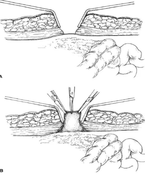
Fig 1 :Regions of the abdomen
Orientatation of viscera on entering abdominal cavity –Liver is
located along the right side beneath the ribs ,stretching along the midline till the
point inferior to Xiphoid process . Stomach lies to the left of liver ,the greater
omentum arises from the greater curve of stomach . , The coils of the small
intestine are generally seen beneath it, while the cecum and the left colon lies in
the RIF and LIF respectively and are p exposed partly. The appendix is attached
sub caecal, retro caecal, para caecal or sub hepatic in position. The appendix can
be identified by tracing the taenia coli. The base of the appendix lies where the
three taenia coli meet. It is attached to the caecum about 2cm below the ileo caecal
valve. The rectum is confined to the sacral concavity being overshadowed by
coils of intestine ,the bladder lies in the anterior pelvis but projects above pubic
symphysis on distension.Sigmoid colon lies between bladder and rectum 18
Stomach continues as duodenum, the pyloric sphincter marks point of
continuity and it appears like a thick ring .
The duodenum lies beneath the inferior surface of the liver and
disappears from the view . Retracting the omentum from the view will reveal
passage of duodenum across the left and it’s transformation into jejunum and
then ileum . The length is about 6 metres , and if the ileum is traced it will be
seen entering into the caecum in the RIF . From the cecum the large intestine
takes an arched course, passing at first upward on the right side, then across the
midline and downward on the left side and forming respectively the ascending,
transverse and descending parts of the colon. In the pelvis it assumes the form of a
loop, the sigmoid colon, and ends in the rectum
By retraction of the stomach towards the right ,the spleen is
A small sac like outpouching is seen on the undersurface of the right lobe of the
liver- the gall bladder. It might overhang and project as far below to the right iliac
fossa too when grossly distended. The pancreas is usually not visualized easily
since it is a retroperitoneal structure. If the lesser sac is entered, it is seen extending
from the “c loop” of the duodenum obliquely to the left crossing the midline
towards the splenic hilum.
The kidneys too being retroperitoneal are not visualized unless specifically
looked for. They lie in the lumbar region with their concavities facing the midline.
The Peritoneum covering the inner surface of abdomen wall and viscera
gives it a shining appearance.26
General Arrangement of the Peritoneal Cavity
25The peritoneum forms the largest serous membrane of the body, and its
arrangements are complex. In males it forms a closed sac, but in females it is open
at the lateral ends of the uterine tubes. It consists of a single layer of flat
mesothelial cells lying on a layer of loose connective tissue . The peritoneal cavity
is a large continuous space lying between the parietal peritoneum covering the
inner surface of abdominal wall and the visceral peritoneum enveloping the
viscera. The main region of peritoneal cavity is the the greater sac, which is
equivalent to the main abdominal cavity surrounding the majority of the abdominal
and pelvic viscera . The lesser sac, or omental bursa, is a small diverticulum lined
with peritoneum, which is situated behind the stomach and lesser omentum and in
front of the pancreas and retroperitoneum. These lesser and greater sac
communicates via the Winslow’s epiploic foramen.
For clinical purposes, the peritoneal cavity can be divided into several
spaces because pathological processes are often contained within these spaces and
their anatomy may influence diagnosis and treatment . Functionally the peritoneal
cavity is divided into into two main compartments , supramesocolic and
inframesocolic, which are partially divided by the transverse colon and Gastrocolic
Supramesocolic Compartment
The supramesocolic space lies above the transverse mesocolon between
the diaphragm and the transverse colon. It can be functionally divided into Rt and
Lt supramesocolic spaces. These regions can be further subdivided into a number
of subspaces, which communicates with each other under normal conditions, but
are frequently subdivided by inflammatory adhesions in disease. The right
supramesocolic space can be divided into three subspaces; the Rt subphrenic
space, the right subhepatic space, and the lesser sac. The left supramesocolic space
can be divided into two subspaces; the left subphrenic space and the left
perihepatic space.
Lesser sac (omental bursa)
The lesser sac is a cavity lined with peritoneum and connected to the larger
general peritoneal cavity (greater sac) by the epiploic foramen. It is considered part
of the right supramesocolic space because embryologically the liver grows into the
right peritoneal space and stretches the dorsal mesentery to form the lesser sac
behind the stomach . The sac varies in size according to the size of the viscera
Fig 3 : Recesses of the Peritoneal Cavity
.
The posterior peritoneal layer of the lesser omentum, the peritoneum over
the posterior wall of the stomach and first part of the duodenum, and the uppermost
part of the anterior layer of the greater omentum forms the anterior wall of the
omental bursa
The posterior wall is formed mainly by the peritoneum covering the
posterior abdominal wall in this area. The superior border of the lesser sac is
narrow and lies between the right side of the oesophagus and the upper end of the
posterior wall of the lesser sac is reflected anteriorly from the diaphragm to join
the posterior layer of the lesser omentum.
The inferior border of the lesser sac runs along the line of the fusion between
the layers of the greater omentum. This runs from the gastrosplenic ligament to the
peritoneal fold behind the first part of the duodenum. In cases where the layers are
not completely adherent to each other , the lesser sac may extend as far as the
bottom of the two lamina of the greater omentum
.
The right border of the lesser sac is formed by the reflection of the peritoneum from the pancreatic neck andhead onto the inferior aspect of the first part of the duodenum . Above the epiploic
foramen the right border is formed by the reflection of peritoneum from the
diaphragm to the right margin of the caudate lobe of the liver and along the left
side of the inferior vena cava, enclosing the hepatic recess.
The left border of the lesser sac runs from the left end of the root of the
transverse mesocolon and is mostly formed by the inner layer of peritoneum of the
splenorenal and gastrosplenic ligaments. The part of the lesser sac lying between
the splenorenal and gastrosplenic ligaments is referred to as the splenic recess.
Above the level of the spleen, the two ligaments are merged as the short
lesser sac. The two layers of the gastrophrenic ligament diverge near the abdominal
oesophagus, leaving part of the posterior gastric surface devoid of peritoneum. The
left gastric artery runs forwards here into the lesser omentum.
Epiploic foramen (of Winslow)
The epiploic foramen (foramen of Winslow, aditus to the lesser sac), is a short ,
vertical slit , usually 3 cm in height in adults , in the upper part of the right border
of the lesser sac . It leads into the greater sac. The anterior border contains the
common bile duct (on the right ), portal vein (posteriorly) and hepatic artery (on
the left) between its two layers. Superiorly the peritoneum of the posterior layer
of the hepatoduodenal ligament runs over the caudate lobe of the liver which forms
the roof of the foramen Winslow . To the right, the rim of the foramen is
continuous with the peritoneum of the greater sac. The roof is continuous with the
peritoneum on the inferior surface of the right hepatic lobe. The anterior and
posterior walls of the foramen are normally apposed.
Inframesocolic Compartment
The inframesocolic compartment lies below the transverse mesocolon and
transverse colon are far as the true pelvis. It is divided by the root of the mesentery
gutter and left paracolic gutters lying lateral to the ascending and descending
colon. As a consequence of the mobility of the transverse mesocolon and
mesentery of the small intestine, disease processes are rarely well contained within
these spaces, and fluid within the infracolic space tends to descend into the pelvis
or the paracolic gutters
.
Lateral to the descending and ascending colon lies a shallow recess –
Rt and Lt paracolic gutter . The right (lateral) paracolic gutter communicates from
the right subhepatic space , down to the space in and around the caecum. It
descends over the pelvic brim into the pelvis. Superiorly, it is continuous with the
the lesser sac through the epiploic foramen,. Thus Bile, pus or blood released
from viscera may run along the gutter and collect in locations quite far from the
suspected organ of origin. Any collections from the RIF may collect in the lesser
sac by coursing along rt paracolic gutter communicating with foramen of
Winslow. In cases of perforation of stomach in erect posture the gastric content
may track to the RIF mimicking appendicitis . The right subhepatic collections
are more common than left because of wider space offered by the right
Pathophysiology of chronic abdominal pain
27One of the most common reasons an individual seeks the advice of a
physician is because he or she is in pain. Pain was called by Sherrington, “the
physical adjunct of an imperative protective reflex.” Painful stimuli generally
initiate potent withdrawal and avoidance responses . It turns out to be immensely
complex because when pain is prolonged and tissue is damaged, central nociceptor
pathways are sensitized and reorganized.
A 2009 report in “Scientific American” indicated that 10–20% of the US
and European populations experienced chronic pain ;59% of these individuals were
women. Nearly 20% of adults with chronic pain indicated that they have visited an
alternative medicine therapist.
Classification of Pain
Definition of pain by the International Association for the Study of Pain (IASP)
is as, “an unpleasant sensory and emotional experience associated with actual or
potential tissue damage, or described in terms of such damage.”
Pain is frequently classified as physiologic or acute pain and pathologic or
typically has a sudden onset and recedes during the healing process. Acute pain
can be considered as “good pain” as it has role in protective mechanism. The
withdrawal reflex can be considered as an example in this aspect
.
Chronic pain can be considered “bad pain” because it persists long after
recovery from an injury and is often refractory to common analgesic agents,
including nonsteroidal anti-inflammatory drugs (NSAIDs) and opiates. Chronic
pain can result from nerve injury (neuropathic pain) including diabetic neuropathy,
toxin-induced nerve damage, and ischemia.
Pain and temperature sensations arise from unmyelinated dendrites of
sensory neurons located around hair follicles throughout the glabrous and hairy
skin as well as deep tissue. Impulses from nociceptors (pain) are transmitted via
two fiber types. One system has thin fibred myelinated A δ fibers (2–5 μm in
diameter) with property of rapid conduction. The other one is non myelinated C
fibers(0.4–1.2μm in diameter) which has low velocity of conduction.28
Visceral Pain 28
Viscera has mostly pain receptors in majority than other . Also, visceral pain
differs from surface pain in several important aspects. The main contrasting
is sharp in nature . Conversely, diffuse stimulation of pain nerve endings
throughout a viscus as caused by termination of the blood supply to a wide area
of gut activates several diffuse pain fibers at the same time and can result in
unbearable pain.
Morever, pain originating from viscera are poorly localized,with nausea,
and frequently accompanied with autonomic disturbances like sweating and
changes in blood pressure. In addition to that of being poorly localized, radiation
of visceral pain is common and is referred to remote areas. The Autonomic
system , similar to the somatic has afferent components, higher integrating
stations and effector pathways.
Causes Of True Visceral Pain
:28Any stimulus that stimulates wider range of receptors in large areas of the viscera results in visceral pain. Such stimuli include ischemia from occlusion
of vessels, chemical damage to the viscera, spasm from repeated contraction of
the smooth muscle of a hollow viscus due to obstruction or distension and
excess strain on the connective tissue surrounding or lying within the viscus. It s
through the small Type C fibres visceral pain are transmitted
Ischemia:
Bradykinin , lactate and other acidic end products of anaerobic metabolism resulting from ischaemia of viscera can excite the pain receptors and cause
visceral pain
Chemical Stimuli:
Following perforation of the gastrointestinal tract damaging substances like proteolytic acidic gastric juice gets seeped into peritoneal cavity that causes
wide spread digestion of visceral peritoneum stimulating wider areas of nerve
stimulation resulting in severe excruciating pain.
Spasm Of A Hollow Viscus:
Following Spasm of a part of the hollow viscus like the gut, the, bile duct, a ureter and the gall bladder can result in pain, by mechanical excitation of the
free nerve endings and pain receptors or the spasm might be severe enough to
cause decreased blood to the musculature along the wall of hollow viscus,
compounded with the increased requirement for nutrients causing severe pain.
Usually pain originating from a spastic viscus is in the form of cramps, with the
increasing severity of pain reaching peak and then subsiding. This process
cycles occur due to intervals of contraction of smooth muscle. For example,
every time a peristaltic wave passes along an stmulated spastic gut, a cramp
results. This cramping type of pain commonly occurs in appendicitis,
gastroenteritis, menstrual bleeding, at the time of delivery, gallbladder disease and
ureteral obstruction.
Overdistention Of A Hollow Viscus :
Extreme loading of a hollow viscus also can cause pain, probably due to
overstretching of the tissues themselves. Overdistention can result in compression
of the blood vessels along the wall of hollow viscus , thus accentuating
ischemic pain.
Insensitive Viscera:
A few visceral surfaces are almost nonresponsive to any type of noxious stimuli. Few instances are the liver parenchyma and the pulmonary alveoli. Even
then the capsule of liver is extremely stimulatory to reflexes from direct injury
and stretch, and the bile ducts are also increasingly sensitive to noxious stimuli .
Apart from the alveoli that are nonsensitive in lungs ,the bronchi as well as the
“Parietal Pain” Caused By Visceral Disease
When a disease affects a viscus, the disease process often spreads to the parietal peritoneum, pleura, or pericardium. These parietal surfaces, like the skin,
are supplied with extensive pain innervations from the peripheral spinal nerves.
Therefore, pain from the parietal wall overlying a viscus is frequently sharp
.
Localization Of Visceral Pain— “Visceral” And The “Parietal”
Pain
Transmission Pathways
Pain arising from viscera cannot be localized, due to variety of
reasons. First, the patient’s brain is not aware of the existence of variety of
internal organs ; hence any pain that arises internally will be localized
externally in a superficial fashion . Second, sensory impulses from the abdominal
viscera are conducted via two pathways to the central nervous system—the
original visceral and referred parietal pathway. Real visceral pain is conducted
by noxious sensory fibers within the nerve bundles of autonomic system and
also are referred to superficial surface of the body often remote from the excited
spinal nerves from th e parietal peritoneum, pleura, or pericardium, and these pain
impulses are generally felt directly over the inciting area
.
Referred Pain
27Following irritation of an intra abdominal organ it usually produces pain felt remote from the site in a somatic structure that may be far away. Such type
of pain referred to the parietal structure is referred pain .
Localization Of Referred Pain Transmitted Via Visceral Pathways
28When visceral pain is referred to the body surface, the person usually
regionalises it to the dermatome that gave rise to visceral organ in the embryo,
not strictly the site the viscera now lies. For example , the neck and upper
thorax are the sites of origin of heart, so in a way the cardiac visceral fibers for
pain runs superiorly along the sympathetic nerves for sensation and terminate in
the segments C-3 and T-5 of spinal cord. Hence, cardiac pain is referred along
lateral neck, left side of shoulder, adjoining pectoral muscles, down the arm to the
tip of finger and into the upper part of the chest. These are the sites of the
parietal region that gives their own somatic sensory nerve fibers into the C-3 to
T-5 cord segments. The origin of stomach is roughly from the T7 to T9
segments of the embryo. Hence , gastric pain is referred to the upper
innervated by T7 to T9 segments.
The basis for referred pain may be convergence of both somatic and
visceral pain fibers on the identical II order neurons in the dorsal horn that
project to the thalamus and then to the somatosensory cortex. This is called the
convergence– projection theory. Somatic and visceral neurons converge in the
ipsilateral dorsal horn. The somatic nociceptive fibers normally do not excite the
II order neurons, but following prolongation of the visceral stimulus, facilitation
of the somatic fiber endings occurs. They now stimulate the second-order neurons,
and of course the brain cannot decide if the stimulus arose from the viscera or
from the area of referral.
Fig 5: Areas of referred pain
Parietal Pathway For Transmission Of Abdominal And Thoracic
Pain.
Pain arising in the viscera is usually localized to two areas of the body surface concomitantly due to the dual pathway of pain transmission one via
the referred visceral pathway and other via direct parietal pathway. The figure
below shows dual pathway of pain transmission due to appendicitis .Impulses
initially pass from the appendix via visceral pain fibers situated inside
sympathetic nerve bundles, and then terminate in the spinal cord at the level of
T-10 or T-11 segments ; It is referred to peri umbilical region . And also pain
impulses originating from the parietal peritoneum following contact of
abdominal wall with the inflamed appendix is transmitted directly over the
Fig 7: Parietal Pathway Of Pain In Appendicitis
Biochemical Mechanisms
27Serotonin is mainly secreted from the GIT . 5-HT mediates numerous
functions such as bowel contraction or relaxation, intestinal secretion and the
sensation of pain and nausea. Depending on its subtype and location 5-HT is
actively released from enterochromaffin cells as a result of mechanical or chemical
stimuli and increases peristalsis, bowel wall tone and sensory perception. At the
spinal level, repeatedly stimulated afferent fibers increase second order neuronal
responsiveness, possibly through the release of stimulatory neuropeptides (eg.
Substance P, neurokinin and calcitonin gene related peptide among others) and
excitatory amino acids (eg. Glutamate). These substances increases membrane
excitability and activate post synaptic receptors ( primarily N-methyl D- aspartate,
release and influx of intracellular Ca which in turn activates phospholipase C,
protein kinase C, nitric oxide and other second messengers which increase
neuronal excitability and pre-synaptic transmitter release thereby permitting more
Ca influx and creating a positive feedback loop. These substances may also
increase the expression of proto-oncogenes such as c-Fos that act as third
messengers in the transcriptional control of genes that encode neuropeptides such
as dynorphin. Increased dynorphin gene expression can enhance neuronal
excitability for days to weeks.
Chronic pain is often refractory to most conventional therapies such as
NSAIDs and even opioids . In new efforts to treat chronic pain, some therapies
focus on synaptic transmission in nociceptive pathways and peripheral sensory
transduction. TRPV1, a capsaicin receptor, is activated by noxious stimuli such as
heat, protons and products of inflammation. Capsaicin transdermal patches or
creams reduce pain by exhausting the supply of substance P in nerves. Nav1.8 (a
tetrodotoxin-resistant voltage-gated sodium channel) is uniquely associated with
nociceptive neurons in dorsal root ganglia. Drugs like Lidocaine and mexiletine
relieves chronic pain by this channel blockade
.
Ziconotide, a voltage-gated N-type Ca 2+ channel blocker, has been approved for intrathecal analgesia in patientsPhysical Examination
As is done with any other patient, general physical examination starts with a primary general survey of the patient before proceeding to a more specific
and detailed systemic examination.
The overall appearance of the patient is noted along with a simultaneous
assessment of his built and nourishment status. Starting from head downwards the
following are looked for
Pallor
Icterus
Signs of vitamin deficiency
Cyanosis
Clubbing
Koilonychia
Peripheral limb edema
Generalized lymphadenopathy with special attention to Virchows and Irish node.
Systemic examination begins with examination of the system in question,
abdomen, in cases of chronic pain abdomen. It begins with proper exposure of the
parts in question. Abdominal examination by palpation is done after inspection for
any obvious masses or dilated veins or previous scars which might give a valuable
superficial palpation of all abdominal regions after noting for any local rise in
temperature and tenderness. A more detailed deep palpation is then carried out in
all regions of the abdomen to look for the possible cause of abdominal pain. The
knowledge of anatomy is vital to arrive at a proper anatomical source of diagnosis.
Renal angles and hernia orifices including the external genitalia also form a very
important part of the examination and should not be missed. The rest of the
systems should also be examined in detail.
During the physical examination, patients with functional abdominal pain
syndrome, may not exhibit autonomic arousal. In fact, the presence of tachycardia,
diaphoresis, and blood pressure changes suggests a more acute peripheral source of
pain.
The “closed eyes sign” may be noted. When the abdomen is palpated,
patients with functional abdominal pain syndrome may wince with eyes closed,
whereas those with abdominal pain from organic causes keep their eyes open in
anticipation of fear from the examination. “Stethoscope Sign”, that is, gentle
pressure at the painful site of the abdomen with the diaphragm of the
stethoscope, elicits no or only a minimal response and thereby affords a more
The need to review the prior investigations cannot be over emphasized here. It
provides a valuable clue to the pattern of pain and helps in deducing the probable
etiology and source of pain abdomen in these patients
.
Causes of Chronic Abdominal Pain
29Peptic ulcer disease
Gall stones
Chronic pancreatitis
Recurrent /chronic appendicitis
Abdominal neoplasms
Inflammatory bowel diseases
Mesenteric ischemia
Intestinal adhesions
Intestinal obstruction
Intestinal tuberculosis
Tuberculous peritonitis
Abdominal neoplasms:
Gastrointestinal lymphomas
Gastrointestinal carcinoid tumour and the carcinoid syndrome
Endocrine tumours of the pancreas and the gastrointestinal tract
Tumours of the stomach
Pancreatic cancer, cystic pancreatic neoplasms and other non- endocrine
pancreatic tumours
Tumours of the gall bladder, bile duct and ampulla
Hepatic tumours and cysts
Small intestinal neoplasms
Malignant neoplasms of the large intestine
Investigations
The following are a battery of investigations that are carried out in patients presenting with chronic pain abdomen. Needless to say, not all investigations are
done for all patients. The choice of investigations are the sole discretion of the
treating surgeon and the clinical picture that the patients presents with.
Hematological: Hemoglobin%, Total Leucocyte Count, Differential Count,
Erythrocyte Sedimentation Rate, Pro Thrombin time- International Normalised
Ratio( PT-INR)
Test, Serological markers
Urine: albumin, sugar, microscopy
Stool: for occult blood
Ultra Sonography of the abdomen
X-Ray: chest, erect abdomen
Upper GI endoscopy for APD and Gastrointestinal tumors
Colonoscopy for IBD and large intestinal tumors
Sigmoidoscopy for IBD and large intestinal tumors
Barium studies
USG / CT guided FNAC
Diagnostic Laparoscopy as an Investigative Tool
Diagnostic laparoscopy is rapidly becoming a popular procedure among the
general surgeons in increasing numbers and is used in patients where other routine
investigative modalities have not thrown any light on the possible cause for
chronic pain abdomen. The indications include abdominal pain (acute and
chronic), focal liver diseases, ascites , pre operative evaluation of malignant
diseases etc. the overall diagnostic rate is 99% for abdominal conditions with
acute pain, 70% for abdominal conditions with chronic pain , 95% for focal
hepatic disorders, 95% for abdominal masses, 95% for ascites and 80% for retro
frequency when a tissue diagnosis is needed.
Indications For Diagnostic Laparoscopy
3Elective
Emergency
Detailed evaluation of chronic pain
syndrome
Assessing cause for acute abdominal
pain and peritonitis
Detailed study of focal hepatic disease Blunt and penetrating trauma
Assessing the cause for ascites Evaluation of ICU patients
Staging laparascopy for malignancy
Workup for fever of unknown origin
As a second-look procedure
Evaluation of small insignificant
inguinal or ventral hernia
Contraindications To Diagnostic Laparoscopy:
7Severe and unstable cardiopulmonary disorder
Uncorrected coagulopathy
Infections of the parietal wall
Laparoscopic guided needle biopsy is far superior to blind biopsy via
percutaneous route in the diagnosis of cirrhosis and is usually performed under
LA . Chances of blind biopsy causing injury to the liver and missing small
inconspicuous lesions such as small metastases are higher. Laparoscopy is
indicated for assessing the cause for ascites following failure of imaging
modalities to reveal the cause . Diagnostic laparoscopy will also help in ruling
out a nonoperable cause for abdominal pain . By laparoscopy the rate of negative
laparotomy can be greatly minimised .Especially laparoscopy is fruitful in young
women experiencing gynecological problems to rule out appendicitis. In
younger individuals with ill-defined abdominal pain of diagnostic difficulty D-
Lap can be used to reveal diagnosis wher other imaging modalities are of little
help. In the intensive care unit (ICU), laparoscopy plays a major role in
preventing non therapeutic celiotomy in patients with a suspected abdominal
catastrophe.
It can be done under local or general anaesthesia in a standard operating
room set-up, with proper port placement and normal laparoscopic equipment.
GA allows handling of inflamed tissues without pain, biopsy of peritoneal and
omental lesions and safe conversion to an open procedure. The following
laparascopic devices may be required for aiding in D-Lap and should be
Angled laparoscope (30-degree)
Scissors
Grasping devices
Liver retractor
Blunt probe
Babcock clamp
Hook cautery
Cupped forceps
Biopsy forceps
Uterine retractor
Hollow suction/ irrigation probe
Pre-Operative Evaluation And Patient Preparation
A well taken history and meticulous physical examination are the deciding
factors of patient selection for surgery. Any comorbid conditions that expose
patients to both anaesthetic and surgical complications should be recognised. Any
severe cardiac or lung disorder should be searched for based on symptoms and
The patient must be aware of the potential advantages ,proposed risks and other alternatives to the planned laparoscopic procedure and must know about
the possibility of requirement for conversion to an open procedure . Preoperative
antibiotics is administered before skin incision is performed in any procedure
where bacterial infection of the wound is possible (i.e., clean contaminated
cases). Also, antibiotics need to be administered following placement of
prosthetic mesh during the surgery. For immune deficient patients antibiotics
to counteract skin flora should also be given. From the probable risk of DVT
following laparoscopic surgeries , several surgeons recommend administering a
single dose of subcutaneous heparin several hours before surgery. Mechanical
bowel preparation is not absolutely required prior to procedure
Equipments Used In Laparoscopy
10Equipment falls into two broad categories: those major pieces of equipment
that enable the surgeon to perform laparoscopy and those instruments related
to the performance of specific tasks or procedures .
I. Imaging System
A. Laparoscopes
Laparoscopes can be of rigid or fibre optics variety . Frequently employed ones are rigid scopes, like 0°, 30°, 3mm, 5mm and 10mm. The 30° angled
scopes are amenable for rotation and used to look down , see up the anterior
abdominal wall and can be viewed side ways.
B. Light Source
The new light source such as 250 watt halogen lamp has been provided with
a cooloing condenser system, but Xenon lamp (cold light source)can be too used
that gives better visual clarity. The intensity of light can be adjusted dither
manually or automatically. Xenon lamp with superior intensity gives far better
visual and photographic clarity
.
C. Light Cable
Light carrier can be a fluid or either a glass fibre light cable .The plasic sheath surmounting the light cable should be devoid of cracks and bends . The
cables come in several diameters and lengths. The diameter of the fibre bundle
should be chosen in such a way it is mildly larger than the contained lens system
D. Cameras
Currently small and low weight cameras are present, which is handy
providing optimally sharp pictures of increased resolution and brilliant colour
reproduction .The resolution of one chip camera is of point 450- 600;While that
of 3 chip cameras with greater than 750 horizontal lines give brilliant visual
clarity. However one chip camera is suffice for routine laparoscopic surgeries
but if the need for recording surgery into a larger film or video production
arises, three chip camera is preferred . Currently an enhanced version of 3 digital
chip cameras integrating image processing modules are available .
E.Monitors
Following the S-VHS connection an ideal video monitor must produce
images with higher resolution. Video screen of 20 inches and above and without
flickering is preferred
.
II. Gas For Pneumoperitoneum
Initially pneumoperitoneum was created using air , but it was abandoned
following the risk of air embolism.
The ideal insufflating agent should have following features:
and should not explode in the presence of electrocautery or laser
coagulation.
It should have high blood solubility .
The insufflating gas should be easily available, of low cost and
nontoxic.
1.Carbon Dioxide
Carbon dioxide is an odorless, colorless gas. It is a easily obtained, stable at
room temp, naturally produced in the tissues and subsequently removed from
body in the lungs. Due to these features, Carbon dioxide is the most frequently
used gas for insufflations during any laparoscopic procedure.
Advantages
It has very low risk of venous gas embolism
It is non combustible
Disadvantages
Hypercarbia and acidosis
The direct effects of carbon dioxide and acidosis can lead to depressed
cardiac contractility, pulmonary hypertension and systemic vasodilation
2. Nitrous Oxide
Nitrogen is non reactive , colorless, gaseous element stable at room
conditions and easily available . Nitrous oxide has been advocated for the
techniques performed under local anaesthesia, or for patients undergoing
procedures for longer duration with poor pulmonary reserve .
Advantages
Negligible changes in acid-base balance.
Significantly reduced pain
Disadvantages
Aids combustion in the along with hydrogen or methane gas.
3. Helium (He)
Helium is a gas with no color, no odour, without taste and it is obtained from natural source. It is inert gas and neither is combustible , nor aids
combustion. Helium is comparatively less soluble in water than carbon dioxide
and hence more chance of air embolism
Advantage
The main benefit is the negligible effect on acid- base balance.
Disadvantages
Owing to it s poorly solublity in water , it is associated with
increased risk of subcutaneous emphysema .
water compared to carbon dioxide.
It is readily diffusible due of its reduced density.
4. Argon
Argon gas has no color, no odour, is non combustible and chemically inert
.
Advantage
The stable acid base status of the gas is it’ s main advantage.
Disadvantage
The most worrisome disadvantage of the gas is it s propensity to
cause cardiac depression.
III. Laproflattor
The electronic carbondioxide Laproflattor is an all purpose insufflations unit used routinely in theatres . Insufflations of the peritoneal cavity under
controlled pressure achieves the essential working space for surgery by
causing distension of the peritoneal cavity and collapsing the hollow organs.
Automatic insufflators enables the surgeon to predetermine the pressure and
provides gas till the required level is reached. Further fall in insuffulation
pressure due to leakage of gas from ports is checked by automatic activation of
the insuffulator . It is possible to alter Insufflation pressure from 0 to 30 mm Hg
as and when needed ; total gas flow volumes can be set to any value in the range
pressure, flow rate and amount of gas used.
IV. Suction Irrigation Machine
Suction And Irrigation Hand Apparatus
Irrigation and suction are most vital at the time of laparoscopic surgeries specially to sustain clear visual field and confirm established hemostasis. It ‘ s
available in 5mm and 10mm sizes and can be reused. The suction tip is helpful
for occasional suction and can be also used as a blunt instrument for dissection
like a finger in conventional surgeries. The suction irrigation apparatus is most
commonly during surgery to maintain a clear field . Normal saline or ringer
lactate is used for irrigation purposes. Sometimes, heparinized saline is
employed to dissolute clots to enhance proper suction from increased
bleeding.
V. Operative Hand Instruments
Both reusable and one use instruments are widely available. One use
instruments offer superior performance and increased safety on single use.
Reusable instruments are cost effective for the long run, however, they require
A. Insuffalation Cannulas
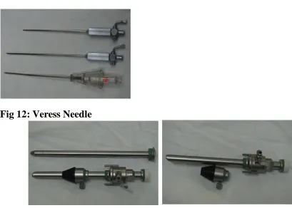
1. Veress Needle
Veress needle was introduced by a thoracic physician for aspirating fluid in pleural effusion hoping that its spring mechanism and blunt tip will not cause
injury to lung tissues. Veress needle has an bevelled edged outer cannula for
cutting through tissues .Within the cannula lies an inner stylet, that is loaded
with a spring which springs in forward direction in response to the rapid fall in
pressure upon moving beyond the abdominal wall and reaching the peritoneal
cavity. The hole on the lateral aspect of the stylet allows for CO2 gas to be
delivered intra-abdominally. In the beginning Veress needle based creation of
pneumoperitoneum is practiced so the trocar can enter abdominal wall safely
without injuring the abdominal viscera
Veress needle technique is the commonly practiced way for creation of
initial pneumoperitoneum . It is mandatory to verify Veress needle every single
time before employing it, for its proper functioning . The size of Veress needle
can be 80mm, 100mm, 120mm. In thick abdomen walled patients 120mm is
required and in very frail patients with thin abdomen 80mm Veress needle is the
needle of choice . Veress needle must be used and held like a dart during
insertion.
2. Hassan Cannula
It is not commonly used like that of a Veress needle . There is less chance of risk of injury to vascular structures and hollow visceral . It is the preferred
instrument to access the abdomen, needful to say in patient who has underwent
intra-abdominal procedures in the past . It is made of three pieces: a blunt tipped
obturator ,a conical sleeve, a metal or plastic sheath with either a trumpet or flap
valve. Over the sheath are 2 struts for binding 2 fascial sutures. These sutures
are then tightly rolled around the struts, effectively seating the conical sleeve
into the laparoscopic port. This generates an efficient seal for maintaining
pneumoperitoneum.
B. Trocars
Though the term “trocar” refers to the entire assembly but actually it is a stylet that is passed through the cannula. Various trocars are there with
various tip types . The cutting tips may be in the appearance of a 3 edged
pyramid or a flat 2 edged blade. Conical tipped trocars are believed to be
atraumatic to the body tissues. It can penetrate via abdominal wall without
damage to the tissues and a reduced risk of herniation or bleeding is reported.
Cannulas are made from either plastic or metal. Plastic devices though they can
be transparent or opaque, should be designed in so as to minimize the glare
blade. These are highly efficient in penetrating the parietal wall by dividing the
tissue as they pass through. Many of the single use plastic trocar has associated-
spring mechanism that sharply withdraws the pointed tip immediately once it
Passes through the parietal wall to lower the risk of injury to viscera. Trocar
and cannula are of several sizes and diameter varying upon the device for which it
is designed . The diameter of outer cannula varies from 3 mm to 30 mm; the
most common size being 5mm and 10 mm . Some recent single use trocar
incorporate special design features as direct serial incision of the tissue guided
under visual control [Excel trocar]. Each and every cannulas come with a valve
mechanism at the upper part . Always ensure that in trocar all the valves move
freely and the insufflation valve remain closed (to avoid escape of air from
created pneumoperitoneum).The valves within cannula offer internal air seals,
that enable devices to move within cannula without loss of created
pneumoperitoneum. They can be either oblique, transverse, or in piston
arrangement. The valves are designed in such way they can be manually or
automatically retracted during passage of device .It is imperative to remember
that sharp trocars are far way better than blunt ones, as they require less amount
of force to be introduced into the abdominal cavity obviating risk of injury to
viscera . The cannula ‘ s end can be either straight or oblique. An cannula with
C. Reducing Sleeve
It is employed to minimize the port size from 10mm to 5mm or 5mm to 3
mm, in such a way the pneumoperitoneum will be maintained even when
surgeon opts for a instrument from larger diameter to smaller diameter .
D. Needle Holder
Laparoscopic needle holder comes with either a straight or curved tip. It
requires 2 needle holders for performing a swift endo-suturing, although it is
possible for satisfactory endo- suturing with a single needle holder and a grasper
.
E. Port Closure Instrument
These are excellent hand instruments for closure of the laparoscopic ports,
especially 10mm or larger ports.
VI. Other Hand Instruments
Disposable or Reusable Instruments
Reusable instruments are expensive during initial buy but they are cost effective on a long term usage . In developing nations, single use instruments are
less frequently used owing to low labor cost . However in Europe and USA,
Insulation of single use instrument are amenable to damages easily that can
lead to electrosurgical injuries. Usually the lap instruments differ in diameter
from 1.8 to 12mm though greater part of instruments were made to pass
through 5 to 10mm of cannula. They also vary differently in length based on the
manufacturer but they are easier to work with if they uniformly are os same
length of about 36 cm in adult and 28 cm in younger age group. For pediatric
surgery instruments varying in size from 18 to 25 cm are convenient . However
they can also be used in adults where the operating space is narrow . 45
centimeter devices are used in obese individuals .Suited for better handling half
of the instruments should lie within the abdomen and half should lie outside. This
makes the instrument to act like class I lever thus stabilizing the port nicely and
making surgery more convenient.
F. Instruments For Sharp Dissection
Scissors
Electro surgery hook
HF Electro surgery spatula (Berci)
HF Electro surgery knife
Scissors
It uses two blades which when glided with one another causes fine cutting tissues
Types of Laparoscopic Scissors
(1) Straight Scissors
(2) Curved Scissors
(3) Serrated Scissors
(4) Hook Scissors
(5) Micro-tip Scissors
Spatula, Hook And Harmonic Scalpel
Spatula with flat tip is ideal for dissecting the gall bladder from the gall bladder fossa . It is extremely safer when compared to hook. Hook has a L shaped
tip. Frequently it is used to dissect out the gallbladder from the Gall bladder fossa
. Some surgeons also use this device for enterotomy. In modern era of
laparoscopic surgery ultrasonic scalpel (Harmonic scalpel) is used for advanced
procedures.
Clip Applicator
They come in both single use and reusable forms . Reusables forms occur
artery & duct according to their size. Disposable clip applicator is available with
preloaded 20 clips per unit similar to the Protack that comes with 30 clips per
unit.
Electrocautery / LASER
Electrocautery or LASER energy is used to dissect tissue. Either energy
modality will also adequately achieve hemostasis of small blood vessels.
Electrocautery uses microwave wavelength en