ACKNOWLEDGEMENT
My sincere thanks and deep sense of gratitude to
Dr. Capt. S.
Gokulanathan,
B.Sc,
M.D.S., Dean and Dr. N. Balan, M.D.S.,
Principal, Vivekanandha Dental College for Women, for permitting me to
pursue this dissertation.
I owe an immense debt of gratitude to my guide and mentor
Dr. V. Mahesh Mathian, M.D.S., Professor and Head,
Department of
Pedodontics and Preventive Dentistry, Vivekanandha Dental College for
Women for his constant guidance and support which was instrumental in
driving me towards achieving completion of this dissertation. This work
would not have seen the light of the day without his dedicated efforts
which was the backbone behind my progress.
I thank Dr. R. Rajkumar, M.D.S., Senior Lecturer without whom
this work would not have seen the light of the day without his affectionate
and compassionate counseling, which reposed by confidence in myself to
complete this work.
Pedodontics and Preventive Dentistry, Vivekanandha Dental College for
Women, for their valuable guidance that enabled me to comprehend this
dissertation and reach its successful culmination.
I sincerely acknowledge my seniors
Kamatchi.M,
Gaytry.S.S and
Kiruthiga Revathy.A,
my batch mates Ramyalakshmi.I.K and
Niranjana.A
and my juniors
Anjugam.P, Dhivya.S, Menaka.E.K
for
their support and encouragement.
My lists of acknowledgements would go meaningless without
dedicating all my efforts of my parents, my brother and my sister in law.
Words cannot express what they have done for me.
Above all, I bow my head to
God Almighty, for showering his
blessings and making me what I am today.
Dr. S. Preethi Archana
Post Graduate Student
Contents
CONTENTS
S.NO
CONTENTS
PAGE NO
1
Introduction
1
2
Aim and objectives of the study
5
3
Review of literature
7
4
Materials and methodology
19
6
Results
42
7
Discussion
48
8
Conclusion
56
9
References
57
Contents
LIST OF TABLES
S.NO
TABLES
PAGE NO
Table 1
Composition of RMGIC
23
Table 2
Composition of biodentine
24
Table 3
Composition of Adper single bond
27
Table 4
Composition of Universal bond
28
Table 5
Shear bond strength values for all groups
42
Table 6
Mean shear bond strength of Biodentine and
RMGIC groups
43
Table 7
Comparison of mean shear bond strength of
Biodentine and RMGIC groups
44
Table 8
Microleakage values for all groups
45
Table 9
Mean microleakage of Biodentine and
RMGIC of the study groups
46
Table 10
Comparison of mean microleakage of
Contents
LIST OF FIGURES
S.NO
FIGURES
PAGE NO
Figure 1
Samples for shear bond strength
20
Figure 2
Samples for microleakage
21
Figure 3
Materials used in this study
29
Figure4 a,b
Shear bond samples after cavity
preparation
30
Figure 5 a,b,c
Shearbond samples after restoration of
cavity with biodentine and RMGIC
31
Figure 6
Restoration with composite resin
31
Figure 7
Universal testing machine
32
Figure 8 a,b,c,d
Samples under shear stress
33
Figure 9 a,b
Microleakage samples after cavity
preparation
34
Figure 10 a,b,c
Microleakage samples after cavity base
restoration
35
Figure 11
Restoration with composite resin
36
Figure 12
Samples in thermocycle unit
37
Figure
13a,b,c,d
Microleakage samples after coated with
Contents
Figure 14
Microleakage samples immersed in 5%
methylene blue dye
38
Figure 15
Microleakage samples after immersed in
5% methylene blue dye
39
Figure 16
Teeth samples after sectioning
39
Figure 17
Stereomicroscope
40
Figure
Contents
LIST OF GRAPHS
S.NO
GRAPHS
PAGE NO
Graph 1
Distribution of scores across various
groups and samples
43
Contents
LIST OF ABBREVATIONS
ABBREVIATION
EXPLANATION
RMGIC
Resin-modified glass ionomer cement
GIC
Glass ionomer cement
CEJ
Cemento enamel junction
HEMA
Hydroxyethyl methacrylate
MTA
Mineral trioxide aggregate
SBS
Shear bond strengths
DBA
Dentin bonding agents
CR
Composite resin
LC
Light cure
PVC
Poly Vinyl Chloride
p – value
Significant value
BD ASB
Biodentine with Adper single bond
BD UB
Biodentine with Universal bond
RM ASB
Resin-modified glass ionomer cement
with Adper single bond
RM UB
Resin-modified glass ionomer cement
with Universal bond
MPa
Megapascal
Abstract
ABSTRACT
BACKGROUND & OBJECTIVES
It is important to have good marginal seal and better bond strength for the
longevity of restorative material, thereby reducing the marginal leakage which is the
precursor of secondary caries, staining of restoration, tooth discoloration, marginal
deterioration, postoperative sensitivity and pulpal pathology. Several factors
contribute to the high incidence of recurrent caries in the gingival area. These include
improper restorative technique by the clinician, plaque accumulation due to patient
difficulty in cleaning and lack of patient compliance with proper oral hygiene. It is
therefore critical to achieve a seal on the gingival margin of class II sandwich
restorations.
The objective is to evaluate and compare the shear bond strength and
microleakage in premolars using Biodentine – tricalcium silicate–based bioactive
restorative material and resin modified glass ionomer cement with Adper single bond
(etch and rinse adhesive system) and Universal bond – 3M (1 step self etch adhesive
system) adhesive systems.
MATERIALS AND METHODOLOGY
80 premolars extracted for orthodontic purpose were collected. The samples
were divided into two groups namely, Group A (Biodentin group) and Group B (resin
modified glass ionomer cement group respectively. Each group is then further
subdivided into subgroup1 (Adper single bond) and subgroup 2 (Universal Bond).
The occlusal surfaces of the tooth were ground flat and PVC molds were stabilized
over flat dentin surface. It is then filled according to the groups ascertained which is
Abstract
Universal Testing Machine (INSTRON) using a steel knife edge (1mm thickness) at a
cross head speed of 0.5 mm/ minute.
Standardized class II cavities were prepared in premolars and restored with
tricalcium silicate-based restorative material (Biodentine) and resin modified glass
ionomer cement and was applied with adhesives, according to groups ascertained.
After the application of adhesives, composite resin material is restored, using open
sandwich technique. The samples were then subjected to 1000 thermocycles followed
by methylene blue dye penetration. The extent of microleakage was examined using
stereomicroscope at 40X magnification.
RESULTS
The result showed that the shear bond strength of Biodentine with Adper
single bond was better with the mean value of 12.4 and there was a statistical
significance when compared to other groups and RMGIC with Adper single bond
shows less microleakage with the mean value of 2.7 when compared to other groups
but there was no statistical significance.
CONCLUSION
Biodentine with Adper single bond shows better shearbond strength which is
followed by RMGIC with Universal bond (self etch system) and RMGIC with Adper
single bond and then Biodentine with Universal bond.
RMGIC with Adper single bond shows less microleakage when compared to
RMGIC with Universal bond which is then followed by Biodentine with Adper single
bond and then the Biodentine with Universal bond.
KEYWORDS
Shearbond strength, Microleakage, Biodentine, Resin modified glass ionomer
Introduction
1
INTRODUCTION
Restorative dentistry has seen a standard change from the invasive surgical
approach which is given by G.V. Black “extension for prevention” to a minimally
invasive approach with improvement in diagnostic system and revolution in adhesion
technology [1].
The physiological integrity of tooth structure is hindered majorly due to the
loss of dentin. The lost coronal or radicular dentin should be replaced with a
alternative material to restore this physiological integrity. There has been a continuous
enhancement in the restorative approach to repair and replace lost tooth structures.
Dentin substitutes are substances, which are used to replace dentin in the crown and
root region as these materials possesses the mechanical properties which are similar to
dentin [2].
An ideal dentin substitute should have the properties like an adhesive, tooth
colored, wear resistance, good biocompatibility, long-term impermeability,
antibacterial properties, ability to induce hard tissue regeneration, good stability,
lower solubility rate, non-absorbability, non toxic and ease of handling. Thus
numerous materials have been developed with these properties over the years for the
replacement of dentin [3].
One such material is Glass ionomer cement introduced by Wilson and Kent in
1972, which has been used extensively in many ways. To replace enamel a technique
called the sandwich technique was developed as a restorative technique using glass
ionomer cement as a dentin substitute and composite to restore it. But the limitation of
Introduction
2 Thus to overcome the limitations resin modified glass ionomer cements were
introduced as dentine replacement materials [2].
Resin-modified glass ionomer cement (RMGIC) was introduced as both a
restorative and base material because it has improved mechanical and physical
properties over conventional GICs. Resin modified Glass ionomer cements have
subjected to remarkable development potential and have been capable to conquer the
majority of the disadvantages of other restorative materials, as it possess high
strength, wear resistance, a chemical adhesion to tooth structure, fluoride release and
radio opacity. In addition, it is stated to be less technique sensitive to saliva and
highly stable with superior physical properties [5].
It is difficult to control the moisture and blood contamination in the deep
carious lesions which extends below CEJ. In this situation a material with pulp
healing potentiality is required [5].
Polymerizable monomers and photo initiators are the constituents of RMGICs.
RMGICs were essentially formed by adding components of methacrylate to the
polyacrylic acid, which are polymerizable by light-curing which complementing the
basic acid-base reaction. However its clinical use is limited bacuase of their
mechanical properties and esthetic appearance. Thus, the alleged sandwich restoration
or “composite-laminated GIC” technique has been used by dentist to protect and
conserve the fluoride release mechanism and the chemical bond to tooth structure
which is provided by the GIC and RMGIC, and to increase the esthetic and
Introduction
3 Another new bioactive cement named Biodentine was introduced which is also
termed as smart dentin replacement, which has the similar indications and mode of
action as calcium hydroxide, but does not acquire its drawbacks [7].
Thus new tricalcium silicate-based bioactive cement named was introduced by
Septodont’s research group. Biodentine is the rudimentary all-in-one bioactive and
biocompatible dentine substitute developed from distinctive Active Biosilicate
TechnologyTM and proposed to treat dentinal damage. Bioactivity can be defined as
‘materials that elicit a specific biological response at the interface between tissues and
the material, which results in the formation of a bond’ [8]
.
Biodentine material can be placed directly in the cavity with bulk amount
without requiring any specific conditioning of the dentin surface as it does not require
photo activation. Its setting time is concise enough to complete the total procedure in
a single appointment. This material demonstrates the similar outstanding biological
properties as that of MTA and it can be placed in direct contact with dental pulp.
Thus, Biodentine can be used as both a dentin substitute base and cement for
preserving the vitality of pulp and accelerating the formation of hard tissue, (i.e.)
formation of both reactionary and reparative dentin [9].
For the longevity of restorative material it is important to have good marginal
seal and better bond strength. Numerous factors contribute to the high incidence of
recurrent caries in the gingival area. These include inappropriate restorative technique
by the dentist, accumulation of plaque due to difficulty in cleaning by the patient and
Introduction
4 It is therefore critical to achieve a seal on the gingival margin of class II
sandwich restorations thus causing marginal leakage which is the precursor of
secondary caries, staining of restoration, tooth discoloration, marginal deterioration,
postoperative sensitivity and pulpal pathology. Thus for the success of all restorations
the ideal requirement is an production of a material with stable long term bond to the
tooth substance [11].
Following the success of adhesion to enamel through acid etching technique
by Buonocore in 1952, dentin adhesion was the major regard as most of the materials
used such as silicate cements, unfilled resins are deficient in this property. Ideal
requirement of an adhesion is that the adhesion should permit the placement of a more
conservative restoration, reduces microleakage and dentin sensitivity [12].
Acid etching is chemically treating to modify the surface characteristics of
enamel to allow adhesion of acrylic resins to the tooth enamel surface. Enamel
etching leads to total etch techniques, in which both the enamel and dentin surfaces
are acid conditioned to allow for resin adherence. Dentin adhesives are at presently
available in different systems such as three-step, two-step and single-step systems,
depending upon the three cardinal steps of etching, priming and bonding to tooth
substrate are accomplished [13].
In the past, various studies have been performed to evaluate the microleakage
and shear bond strength of RMGIC and biodentine when restored with composites.
This study was aimed at comparing RMGIC and biodentine to find out the best
dentinal substitute when used either with Adper single bond or Universal bond
Aim and Objectives
5 AIM AND OBJECTIVES
Aim of the study
The aim of the study was to evaluate and compare the shear bond strength and
microleakage of Biodentine – tricalcium silicate-based bioactive material (Septodent)
and resin modified glass ionomer cement (Fuji II LC GIC) with two adhesive systems
in premolars.
Objectives of the study
1. To evaluate the shear bond strength of Biodentine – tricalcium silicate–based
bioactive material (Septodent) with Adper single Bond (etch and rinse
adhesive system – two step) and Universal bond – 3M (1 step self etch
adhesive system) adhesive systems in premolars.
2. To evaluate the microleakage of Biodentine – tricalcium silicate–based
bioactive material (Septodent) with Adper single Bond (etch and rinse
adhesive system – two step) and Universal bond – 3M (1 step self etch
adhesive system) adhesive systems in premolars.
3. To evaluate the shear bond strength of resin modified glass ionomer cement
(Fuji II LC GIC) with Adper single Bond (etch and rinse adhesive system –
two step ) Universal bond – 3M (1 step self etch adhesive system) adhesive
systems in premolars.
4. To evaluate the microleakage of resin modified glass ionomer cement (Fuji II
LC GIC) with Adper single Bond (etch and rinse adhesive system – two step )
Universal bond – 3M (1 step self etch adhesive system) adhesive systems in
Aim and Objectives
6 5. To compare the shear bond strength of Biodentine – tricalcium silicate–based
bioactive material (Septodent) and resin modified glass ionomer cement (Fuji
II LC GIC).
6. To compare the microleakage of Biodentine – tricalcium silicate–based
bioactive material (Septodent) and resin modified glass ionomer cement (Fuji
Review of Literature
7
REVIEW OF LITERATURE
Hallett & Garcia-Godoy (1993) compared the marginal sealing ability of two
resin-modified GIC restorative materials with two conventional GIC materials using a
standardized Class V cavity preparation and to evaluate the nature of the tooth
restorative interface using SEM analysis and concluded that the clinical marginal seal
between the resin modified GIC restorations examined and tooth structure is unable to
completely prevent the leakage of fluid, as shown by the microleakage results [20].
Edward. J Swift et al (1998) evaluated bond strengths obtained by several
one-bottle bonding agents and one conventional unfilled resin as a control and
concluded that one-bottle bonding agents, with the exception of the Syntac material,
provide enamel bond strengths at least equal to that of a conventional unfilled
resin [21].
Juliana Godoy-Bezerraa et al (2006) conducted an in vitro study to evaluate
three different enamel pretreatments such as etching with 10% polyacrylic acid,
etching with 37% phosphoric acid and no acid etching on the shear bond strength of
commercially available RMGIC in a saliva-contaminated environment and concluded
that in a saliva-moistened environment RMGIC achieved higher shear bond strength
when 37% phosphoric acid is used [22].
AE Souza-Gabriel et al (2006) evaluated the in vitro effect of Er:YAG laser
(erbium-doped yttrium aluminium garnet laser, erbium YAG laser) irradiation of
enamel and dentin on the shear bond strength of resin-modified glass ionomer
cements (RMGIC) and concluded that bond strength values obtained for enamel were
higher than those recorded in dentin and the tested restorative systems (Fuji II LC and
Review of Literature
8 Rebeca Di Nicolo et al (2007) evaluated the shear bond strength of a RMGIC
to primary dentin, after cutting with diamond or carbide rotary instruments with or
without phosphoric acid conditioning and concluded that early shear bond strength of
RM-GIC to primary dentin was affected by the acid conditioning, but the different bur
types used in this study had no influence [24].
Taher NM, Ateyah NZ (2007) conducted a in vitro study to determine the
shear bond strength and the type of bond failure when resin-modified glass ionomer
cement (RMGIC) (FL) was bonded with different tooth-colored restorative materials
such as composite resin Point-4 (P4), Compomer Dyract AP (DY), and Ormocere
Admira (AD) with and without the use of etchant and concluded that chemical
bonding did exist between FL and esthetic tooth-colored restorative materials and
etching the surface of RMGIC (FL) did not improve the bond. AD showed the highest
shear bond strength to RMGIC [10].
Mauro S J et al (2009) evaluated the bond strength values of RMGIC (Fuji II
LC) to dentin, employing polyacrylic and phosphoric acid as dentin surface
pre-treatments and concluded that the treatment of dentin with 20% polyacrylic acid
showed a non-significant increase in bond strength values when compared to 37%
phosphoric acid with dry dentin [25].
Yaseen SM et al (2009) conducted a study to compare and evaluate shear
bond strength of two self-etching adhesives (sixth and seventh generation) on dentin
of primary and permanent teeth and concluded that for all treatment groups, Clearfil
S3 (seventh generation) showed higher shear bond strength than Contax (sixth
generation) dentin bonding systems. Permanent teeth showed higher shear bond
Review of Literature
9 Ana Carolina Maito Villela-Rosa et al (2011) evaluated the dentin shear
bond strength of four adhesive systems such as Adper Single Bond 2, Adper Prompt
L-Pop, Magic Bond DE and Self Etch Bond) in regards to buccal and lingual surfaces
and dentin depth and concluded that the shear bond strength of dentin is dependent on
material (adhesive system), substrate depth and adhesive/depth interaction [26].
Shaila Masih et al (2011) compared the marginal sealing ability of the two
glass ionomer cements, GC Fuji II LC (Improved) and GC Fuji IX GP and also to find
out which material will result in the least microleakage in primary molars, and to
ascertain which of the two is a better restorative material for the primary dentition and
concluded that both the materials, GC Fuji II LC (Improved) and GC Fuji IX GP were
similar in performance and can be considered to be safest materials for Pedodontics
usage and thus decreasing bacterial penetration [6].
Sabah A et al (2012) compared the shear bond strength of chemically cured
(Conventional) glass ionomer cement and light cured (Resin modified) glass ionomer
cement to resin composite and evaluated the effect of acid etching of the glass
ionomer cements on the shear bond strength and concluded that shear bond strength
(SBS) of RMGIC to resin composite was significantly higher than that of
conventional GIC. Acid etching the GIC surface did not improve the SBS of GICs to
resin composite [11].
S. Koubi et al (2012) compared the in vitro marginal integrity of
open-sandwich restorations based on aged calcium silicate cement versus resin-modified
glass ionomer cement and concluded that resin modified glass ionomer cement and
the calcium silicate cement allowed similar glucose diffusion at the interface between
Review of Literature
10 Vishnu Rekha, et al (2012) evaluated the tensile bond strength and
microleakage of a conventional glass ionomer (Fuji IX GP, GC products), resin
modified glass ionomer (Fuji II LC, GC products) and a compomer, vivadent products
(Compoglass) in primary molars and also compared the bond strengths with the
degree of microleakage exhibited by the same materials and concluded that
- Tensile bond strength of compoglass is significantly greater than Fuji IX GP and
Fuji II LC.
- Fuji IX GP and compoglass showed moderate to severe leakage in contrast to
minimal leakage with Fuji II LC [28].
Khoroushi et al (2012) evaluated the effect of etch-and-rinse and self-etch
bonding systems v/s cavity conditioner, and in comparison to similar composite resin
restorations on maintaining the marginal sealing of RMGI restorations and concluded
that the use of the dental adhesive systems improved the marginal integrity of RMGI
cervical restorations. Bonding of RMGIC to both enamel and dentin with a two-step
self-etching adhesive rather than the use of conventional cavity conditioner
(polyacrylic acid) improves marginal sealing of cervical restorations [29].
Anne Raskin et al (2012) evaluated the marginal sealing efficacy of
Biodentine at the cervical margins of approximal cavities placed in molars and also
the use of Biodentine in combination with resin-based adhesives and a resin
composite, compared with a resin-modified glass-ionomer cement (Fuji II LC) and
concluded that Biodentine when used as dentin substitute in cervical lining or as a
restorative material in a proximal cavities when the cervical extent is below the CEJ
seems to carry out well without any conditioning treatment. However, the operating
Review of Literature
11 Mesut Enes Odabas, Mehmet Bani and Resmiye Ebru Tirali (2013)
conducted a study to measure the shear bond strength of different adhesive systems to
Biodentine with different time intervals and the least value was obtained for group 1
(etch-and-rinse adhesive) at a 12-minute time period, and the highest was obtained for
group 2 (two-step self-etch adhesive) at a 24-hour time period. The placement of
composite resin used with self-etch adhesive systems over Biodentine showed better
shear bond strength [31].
Gilles Koubi et al (2013) conducted a multicentric randomized, 3-year
prospective study to determine for how long the Biodentine which is a new
biocompatible dentine substitute, can remain as a posterior restoration and concluded
that Biodentine is capable to restore posterior teeth for up to six months. When
subsequently covered with Z100, it is a convenient, efficient and well tolerated
dentine substitute [32].
MR Meharry et al (2013) compared shear bond strengths (SBSs) to dentin
and enamel of nine dentin bonding agents (DBAs) from three generations, after
simulated aging with thermocycling.and concluded that fourth- and sixth generation
DBAs generally showed stronger SBS values than the seventh-generation all in one
DBAs [33].
Vignesh Guptha Raju et al (2014) studied the shear bond strength and
microleakage of tricalcium silicate-based restorative material and glass ionomer
cement (Fuji IX GP) in primary and permanent teeth and concluded that glass
ionomer cement (Fuji IX GP) has better shear bond strength and exhibits more
microleakage when compared with tricalcium silicate-based restorative material
Review of Literature
12 Raji Viola Solomon et al (2014) studied the sealing ability of a new calcium
silicate based material as a dentin substitute in class II sandwich restorations and
found calcium silicate based material used in class II open-sandwich restorations
scores better than the resin-modified GIC [35].
Timothy F. Watson et al (2014) conducted Biophotonics based interfacial
analyses on present and future of glass-ionomers and calcium-silicate cements as
bioactive materials in dentistry and reviewed the underlying chemistry and the
interactions between glass ionomer cements and calcium silicate cements with tooth
tissue, concentrating on the dentin–restoration interface and concluded that the local
bioactivity of the calcium-silicate based materials has been shown to yield
mineralization within the underlying dentin substrate, extending deep within the
tissues. This suggests that the local ion-rich alkaline environment may be more
favorable to mineral repair and re-construction, compared with the acidic environs of
comparable glass ionomer based materials [12].
Shafiei and Akbarian (2014) investigated the marginal sealing of nano filled
resin modified glass ionomer cement and silorane-based or methacrylate-based
composite open sandwich technique with simultaneous bonding application compared
with that of the conventional technique in deep Class II restorations and concluded
that the simultaneous application of bonding agent in the modified sandwich
restorations (with SE Bond or Silorane Adhesive) resulted in a considerable reduction
in the cervical microleakage compared with that of the conventional bonding.
However, microleakage of the modified technique was similar to that of the total
Review of Literature
13 Boutsiouki et al (2014) evaluated the recently introduced base materials such
as biodentine and ever X posterior directly placed under composite resin in
comparison to traditional materials, such as flowable resin and RMGI cement and
concluded that Biodentine and Ever X Posterior exhibit comparable results.
Biodentine provides a satisfying seal, even when composite resin is directly placed,
and thus it is suggested to finish the combined restoration in a single patient visit [37].
Cantek and AVC (2014) evaluated bond strength of methacrylatebased (MB)
composites, silorane-based (SB) composites, and glass ionomer cement (GIC) to
Biodentine® and mineral trioxide aggregate (MTA) and concluded that the new pure
tricalcium based pulp capping, repair, and endodontic material showed higher shear
bond scores compared to MTA when used with the MB composite [38].
Kaup et al (2015) conducted a study to compare the shear bond strength of
Biodentine, ProRoot MTA (MTA), glass ionomer cement (GIC) and composite resin
(CR) on dentine and concluded that after 7days Biodentine showed comparable shear
bond values than GIC, whereas the shear bond values for MTA were significantly
lower even after 14days. The adhesion of Biodentine to dentine surface seems to be
superior compared to that of MTA [39].
Mostafa Sadeghi1 et al (2015) conducted a study to compare the shear bond
strength (SBS) of resin composite to RMGIC utilizing three different generations
resin adhesives versus a GIC-based adhesive (GC Fuji bond LC) and concluded that
there was no significant difference in SBS of resin composite to RMGIC utilizing
different generations of resin adhesives and Fuji Bond LC, therefore they suggest that
Review of Literature
14 cure technique for sandwich restorations instead of employing the resin adhesive
systems [40].
In their study, Fereshteh Shafiei et al (2015) compared the effect of delayed
light activation (DLA) on marginal sealing of a RMGI when different types of surface
pretreatments were used including cavity conditioner, acid etching, Vitremer primer
and CPP-ACP and concluded that a 3-min delay in light activation of the RMGI might
yield different outcomes on enamel and dentinal marginal sealing depending on
surface conditioning and structural characteristics of the treated surfaces [41].
Aggarwal et al (2015) evaluated the marginal adaptation of Biodentine, MTA
Plus, and resin-modified glass ionomer cement (kept as control group) when used in
an open sandwich technique under Class II composite resin restorations with two
different bonding strategies and were subjected to alternate ageing in phosphate
buffered saline (PBS) and cyclic loading. He concluded that Biodentine and
resin-modified glass ionomer cement (RMGIC), when used as a dentin substitute under
composite restorations in open sandwich technique, gave satisfactory marginal
adaptation values [42].
Faika et al (2016) conducted an in vitro study to assess and compare the SBS
of four dentin substitute/ replacement materials such as Multicore Flow, Fuji II LC,
Biodentine and SDR (Smart Dentin Replacement) to caries affected dentin of primary
teeth and concluded that the highest SBS value is for SDR followed by Multicore
Flow then Fuji II LC and the lowest was for Biodentine [43].
Fahad I and Zeeshan H (2016) conducted an in vitrostudy to determine and
Review of Literature
15 restorative with other bulk-fill restorative materials such as surefil (SDR), Biodentine,
ever X posterior and concluded that flowable and fiber-reinforced composites have
better shear bond strength and microleakage properties when compared to biodentine
which shows least values [44].
Velagala L Deepa et al (2016) compared and evaluated the bonding ability of
resin composite (RC) to three different liners such as TheraCal LCTM (TLC), a novel
modified (RM) calcium silicate cement, BiodentineTM (BD), and
resin-modified glass ionomer cement (RMGIC) using an universal silane – containing
adhesive and characterizing their failure modes and concluded that the bond strength
of composite resin to TLC and RMGIC was similar and significantly higher than that
of BD following application of universal adhesive [45].
Niranjan et al (2016) investigated microleakage of three different bases such
as GIC, MTA, biodentine under composite resin in sandwich technique using dye
penetration and dentin surface interface using scanning electron microscope (SEM)
and concluded that Biodentine exhibits superior marginal sealing ability as well as
marginal adaptation under composite resin as compared to MTA and GIC [46].
Hayes et al (2016) conducted a randomized controlled clinical trial to
compare a calcium silicate cement (Biodentine), a high-viscosity glass ionomer
cement (Fuji IX GP Extra), and a resin-modified glass ionomer cement (Fuji II LC)
and concluded that Biodentine cannot be recommended for the operative
management of root caries. Fuji IX GP Extra and Fuji II LC displayed similar success
rates, and highviscosity glass ionomer cement and resin-modified glass ionomer
Review of Literature
16 Namith Rai et al (2017) evaluated the effects of three different conditioning
agents on the shear bond strength of resin‑modified glass ionomers to human dentin
and concluded that surface conditioning of dentin resulted significantly higher bond
strength than unconditioned dentin surfaces [48].
SB Abraham et al (2017) investigated the cavity adaptation of mineral
trioxide (ProRoot MTA/MT), tricalcium silicate (Biodentine/BD), and glass ionomer
(Equia Fil/EF) cements used as liners and the interfacial integrity between those liners
and a composite resin placed as the main restorative material by computerized X-ray
microtomography and concluded that when used as a composite liner, ProRoot MTA
showed inferior cavity adaptation at dentin/liner interface when compared to
Biodentine and Equia Fil [49].
Meraji and Camilleri (2017) conducted a study to characterize and evaluate
the adequacy of different bonding systems (Total etch and bond and self etch
adhesive) to layer composite or glass ionomer cement over 3 dentin replacement
materials (Biodentine, Theracal LC, and Fuji IX glass ionomer [GC, Tokyo, Japan])
using dynamic aging and concluded that dynamic aging is necessary to have clinically
valid data. Bonding composite resin to water-based dentin replacement materials is
still challenging, and further alternatives for restoration of teeth using such materials
need to be developed [50].
Sedef Aksoy and Murat Unal (2017) compared the shear bond strength
(SBS) of various adhesive systems to a bioactive dentin substitute Biodentine with
different time intervals (12 min, 24 h, 48 h, 72 h, 96 h) and concluded that before
applying compomer material on Biodentine, a waiting period of at least 24 h can be
Review of Literature
17 performance may also be obtained without an acid etching procedure, as universal
adhesive systems applied on Biodentine show similar bond values in SE and ER
modes. In this manner, ease of use is provided for predominantly uncooperative
pediatric patients by reducing the number of procedure steps, and also thereby
reducing the risk of contamination with saliva [51].
Pradeep PS et al (2018) evaluated the shear bond strength of biodentine and
MTA and concluded that Biodentine had better bond strength when compared to
MTA and also suggest that in cases of perforation seal, root end filling or as a base
material biodentine can be recommonded over MTA [52].
Buldur B et al (2018) compared the bond strength of Biodentine and Imicryl
MTA to a compomer material, and examined the effect of the setting time (12
minutes, 24 hours, 48 hours, 72 hours, and 96 hours) on the bond strength and
concluded that the use of Biodentine in pediatric dental practice can be recommended
due to its advantages such as short cure time, ease of manipulation and no color
change as Biodentine had higher SBS values to compomer than Imicrly MTA in all
setting time groups [53].
Jewel Darsan et al (2018) evaluated and compared gingival microleakage at
tooth restoration interface in deep Class II composite closed sandwich restorations
(mesio-occlusal) using different liners like Resin Modified Glass Ionomer Cement
(RMGIC), Biodentine, Theracal LC and concluded that use of a liner beneath deep
Class II composite restoration reduced microleakage and Theracal LC performed
similar to Biodentine and better than RMGIC, when used as a liner in deep Class II
Review of Literature
18 MH Nekoofar et al (2018) measured the micro-shear bond strength of a resin
composite (RC) using several adhesive systems such as no adhesive, etch and rinse,
two-step self-etch and universal adhesive in self-etch mode and a resin-modified glass
ionomer cement (RMGIC) to different aged Biodentine specimens and concluded that
there is a difference in micro-shear bond strength of various adhesives bonded to
different aged Biodentine blocks. Universal bonding agent in self-etch mode applied
over Biodentine specimens at 12-min setting time exhibited significantly higher mean
Materials and Methodology
19
MATERIALS AND METHODOLOGY
STUDY POPULATION:
80 extracted human premolars which was extracted due to orthodontic reasons
were collected and it was divided into two groups – group A and group B. The study
was conducted in the Department of Pedodontics and Preventive Dentistry,
Vivekanandha Dental College for Women, Tiruchengode, in collaboration with
Department of Oral Pathology, Vivekanandha Dental College for Women,
Tiruchengode and Department of Plastic Engineering, Central Institute of Plastics
Engineering & Technology (CIPET), Chennai.
SELECTION CRITERIA OF THE TOOTH:
1. Premolars extracted for orthodontic purposes.
2. Teeth with sound structure.
3. Tooth without caries, cracks, restorations and fracture defects.
SAMPLE COLLECTION AND PROCEDURE:
80 premolars extracted for orthodontic purpose were taken for this study. The
samples are then divided into two groups namely, Group A (Biodentine group) and
Group B (resin modified glass ionomer cement) respectively. Each group is then
further subdivided into subgroup1 (Adper single Bond - 3M) and subgroup 2
(Universal Bond - 3M).
Extracted premolars are selected, cleaned of debris and stored in distilled
Materials and Methodology
20 STUDY DESIGN:
TOTAL SAMPLE SIZE (80)
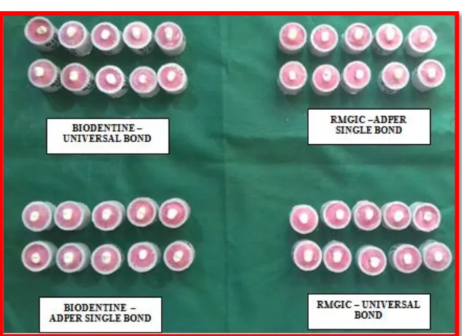
[image:40.595.149.479.419.659.2]FIGURE 1: SAMPLES FOR SHEAR BOND STRENGTH Group A (40)
(Biodentine) (BD) Subgroup A1 (20) (Adper single bond) (ASB) Subgroup A2 (20) (Universal Bond) ( UB)
Materials and Methodology
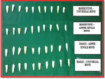
[image:41.595.142.485.83.342.2]21 FIGURE 2: SAMPLES FOR MICROLEAKAGE
ARMAMENTARIUM:
1. 80 extracted premolars.
2. Distilled water.
3. Acrylic mold.
4. Cold cure resin.
5. Airoter burs
6. Restorative instruments.
7. Composite finishing burs
8. Biodentine (Septodent).
9. Resin modified GIC (Fuji II LC GIC).
10.Prime and Bond NT (3M ESPE).
11.Universal Bond (3M ESPE) (self etch).
Materials and Methodology
22 13.Diamond disc.
14.5% Methylene blue dye.
15.Stereomicroscope (40X) (Magnus MSZ).
16.Universal testing machine (INSTRON 3300 100kN).
17.Thermocycling unit.
18.Soflex discs
19.Sticky wax
20.Nail varnish
21.Composite (Filtek Z250) (3M ESPE) (Nano Hybrid).
MATERIALS USED IN THIS STUDY
RESIN-MODIFIED GLASSIONOMER CEMENT
Glass ionomer cement was introduced in 1972 by Wilson and Kent which sets
acid-base reaction between polymers of polyacrylic acid and fluoroaluminosilicate
bases. Along with fluoride release, their main advantage is the distinctive ability to
bond chemically to tooth structure. But the disadvantages of GIC (Glass ionomer
cement) includelow early strength and moisture sensitivity during setting [14].
Thus Resin-modified glass ionomer cement (RMGIC) in 1992 was developed
in an attempt to improve mechanical properties, decrease setting time, and attenuate
moisture sensitivity. RMGIC developed with a hybrid of glass ionomers and
composite resin, and thus contain acidbaseand polymerizable components [14].
RMGIC are usually formulated from fluoroaluminosilicate glasses,
photo-initiators, polyacrylic acid, water, and a watersolublemethacrylate monomer, such as
Materials and Methodology
23 polyacrylic acid. RMGIC release fluoride and bond chemically to tooth structure, as
do conventional GIC products, yet demonstrate early and increased strength
[image:43.595.122.513.164.348.2](Mitra, 1991) [14].
Table 1: Composition of RMGIC
Powder Liquid
Alumino-silicate glass
Polyacrylic acid
2-Hydroxyethyl methacrylate
Proprietary Ingredient
2,2,4, Trimethyl hexamethylene dicarbonate
Triethylene glycol dimethacrylate
In 1977, McClean introduced a technique in which glass ionomers were used
as liners underneath composite restorations. Thus this technique employed glass
ionomers as dentin substitutes. Also known as the sandwich technique, it involves the
use of glass ionomers against the tooth surface with composite on the superficial
aspects of the restoration as it’s stronger and more esthetic. There are two techniques
that have been proposed: open and closed sandwich techniques respectively. The open
sandwich method is usually indicated in situations where only a part of the restoration
has a dentin only margin (in case of a deep class II or a class V on root surface). In
such cases, the glass ionomer is placed such that it covers dentin and becomes the
external material at dentin margin. The latter technique involves the use of glass
ionomer when a complete enamel margin is available for bonding and sealing using
the phosphoric acid etching technique. Glass ionomer is placed over the dentin prior
Materials and Methodology
24 Biodentine
Septodont’s research group has developed a new class of dental material
named Biodentine™ which could conciliate high mechanical properties with excellent
biocompatibility as well as a bioactive behaviour. Biodentine often called as dentine
in a capsule, a biocompatible and bioactive dentine substitute. It is quoted by Mark
Hargreaves et al (2011) that biodentine allows a dentist to achieve biomimetic
mineralization within the depths of a carious cavity [15].
It is a calcium based cement like ProRoot MTA and Portland’s cement. When
comparing with other calcium based cements, the biodentine has two advantages:
i) It has quicker setting time of about 12 minutes and
ii) Higher mechanical properties.
These physico-chemical properties associated with the biological behavior
suggest that it may be used as a permanent dentine substitute [12].
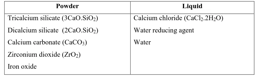
Chemical Composition
Biodentine™, available in a capsule containing the good ratio of powder and
[image:44.595.95.530.619.749.2]liquid.
Table 2: Composition of biodentine
Powder Liquid
Tricalcium silicate (3CaO.SiO2)
Dicalcium silicate (2CaO.SiO2)
Calcium carbonate (CaCO3)
Zirconium dioxide (ZrO2)
Iron oxide
Calcium chloride (CaCl2.2H2O)
Water reducing agent
Materials and Methodology
25 Properties of the different components:
• Tricalcium silicate (3CaO.SiO2): It is the main component of the powder. It
regulates the setting reaction.
• Dicalcium silicate (2CaO.SiO2): It acts as second main core material
• Calcium carbonate (CaCO3): It acts as filler.
• Zirconium dioxide (ZrO2): It is added on top to provide the radio-opacity to
the cement.
• Calcium chloride (CaCl2.2H2O): It is an accelerator.
• Water reducing agent (Superplasticiser): It is based on polycarboxylate but
modified to obtain a high short-term resistance. It reduces the quantity of water
required by the mix (water / cement), decreases viscosity and improves handling of
cement.
Active and collaborative research between Septodont and several universities
for years led to a new calcium-silicate based formulation Biodentine™, which is
suitable as a dentine replacement material whenever original dentine is damaged. The
Active Biosilicate Technology™ is a proprietary technology developed according to
the state-of-the-art pharmaceutical background applied to the high temperate ceramic
mineral chemistry [15].
Dentin Bonding Agents
In 1955, Buonocore was the first one who predicted that the surface of the
enamel can be altered by acid, which renders it more receptive to adhesion. He
discovered that the human enamel could be bonded to that of acrylic resin after 85%
Materials and Methodology
26 Dentin adhesive development was very slow until 1950s. Bowen synthesized
a surface active comonomer that can act us a go-between water resistant chemical
bonds of resins to dentinal calcium. But these commercial stuffs based on this
comonomer resulted in very poor clinical performance. There has been differences
observed over the past 45 years in dental bonding systems is application chemistry
technique, mechanism & effectiveness. This accompanied development of enhanced
composite resins & ceramic esthetic dental materials & in raising demand by patients
for esthetic dentistry. All indirect restorations are candidates for bonding. The
evolution of bonding agents has increased as the demand for bonded esthetic
restorations has continued to increase [16].
Etch-and-Rinse Adhesives
Etch-and-rinse adhesives were developed in mid 1990’s, can readily be
recognized by an initial etching step, the so-called conditioning step, followed by a
compulsory rinsing phase. Another frequently used name for this category of
adhesives is “total-etch” adhesives which are, however, less appropriate because
self-etch adhesives can also etch and demineralize tooth tissue [17].
Etch-and-rinse systems typically consisted of three separate application steps:
(a) conditioning; (b) priming; and (c) adhesive resin application. An adhesive system
which follows this method is called a three-step etch-and-rinse adhesive. In searching
for fewer application steps and simplification, a two-step etch-and-rinse design has
been devised combining the priming and bonding steps into one. Another frequently
used name for a two-step etch-and rinse adhesive is “one-bottle
adhesive”misleadingly suggesting a single application step [13]
Materials and Methodology
27 Table 3: Composition of Adper single bond
BisGMA, HEMA, Dimethacrylates, Ethanol, Water, Photoinitiator and
Methacrylate functional copolymer of
polyacrylic
Polyitaconic acids
Universal bond
The seventh generation bonding systems was introduced in late 1999. The
seventh generation or one-bottle self-etching system represents the latest
simplification of adhesive systems. With these systems, all the required ingredients
for bonding are delivered from a single bottle. This greatly simplifies the bonding
protocol because it states that consistent bond strengths are achieved by completely
eliminating the errors that could normally be introduced by the dental professionals
who had to combine the separate components with other more complicated systems.
However, incorporating and placing all of the chemistry required for a viable adhesive
system into a single bottle, and having it remain stable over a reasonable period of
time, poses a significant challenge [18].
These inherently acidic systems tend to have a considerable amount of water
Materials and Methodology
28 Furthermore, once placed and polymerized, they are generally more hydrophilic than
two-step self-etching systems; this condition makes them more prone to water
sorption, limits the depth of resin infiltration into the tooth and creates some voids.
The advantage of this generation was not any mixing required and the bond strengths
were consistent. However, the seventh generation adhesives have proven to have the
lowest initial and long term bond strengths of any adhesive on the market today that
may be considers as disadvantage. Seventh generation adhesives involve the
application of etch, primer, and adhesive which have already been mixed, followed by
light curing the tooth. The clinical and scientific data on these adhesives proves that
they are hydrophilic and humiliate in a more rapid rate. In addition, the chemistry
mast be acidic, as etch is involved in this liquid, and this has been shown to adversely
[image:48.595.202.432.443.706.2]react with the composite initiator systems [19].
Table 4: Composition of Universal bond
MDP Phosphate Monomer
Materials and Methodology
[image:49.595.212.423.83.317.2]29 FIGURE 3: MATERIALS USED IN THIS STUDY
METHODOLOGY
For evaluating the shear bond strength, the root portion of each tooth was
embedded into an acrylic mold with the occlusal surface of tooth parallel to the base.
Molds were then filled with cold cure acrylic resin (DPI-RR cold cure) leaving the
crown portion of tooth alone exposed. The mid-coronal portion of the occlusal
surfaces of dentin was exposed by a flat cut perpendicular to the long axis of the tooth
with a fine diamond disc in high speed with a copious water spray. Specimens were
then stored in distilled water.
The mid coronal dentin of the occlusal surfaces of the premolar tooth are
ground flat and a cavity of 6mm width and 2mm depth reference are prepared in the
flat surface of dentin (figure 4a,b) and it was then filled with a base material such as
biodentine for group A (n=20) and RMGIC for group B (n=20) (figure 5a,b) as per
manufacturer instructions. Once the material sets, for etch and rinse group (subgroups
A1 & B1) (n=10 for each subgroups), the filled cavity surface was etched with 35%
Materials and Methodology
30 Bonding agent was then applied over the base for 10 seconds and then it is dried with
gentle air stream for 5 seconds and light cured for 10 seconds. For self etch group
(subgroups A2 & B2), bonding agent is applied over the base for 10 seconds and dried
with mild air for 5seconds and light cured for 10seconds. Once the application of
bonding agent is done, a PVC (Poly Vinyl Chloride) mold of 3.5mm diameter by 3
mm height was positioned perpendicular over the base. It was then filled with
composite resin which was then cured for 20 seconds. After that, the mold is removed
with BP (Bard Parker) blade (figure 6).
(a)
[image:50.595.186.447.312.723.2](b)
Materials and Methodology
31 (a)
[image:51.595.114.493.83.473.2](b) (c)
FIGURE 5 a, b, c: SHEARBOND SAMPLES AFTER RESTORATION OF CAVITY WITH BIODENTINE AND RMGIC
[image:51.595.232.399.547.727.2]Materials and Methodology
32 Shear bond strength is evaluated with Universal Testing Machine (INSTRON)
using a steel knife edge (1mm thickness) at a cross head speed of 0.5 mm/ minute
(figure7). Each sample was mounted in universal testing machine with the dentin
surface parallel to the machines trajectory. A compressive load was applied, using a
steel knife-edge placed over the sample's tooth-restoration interface so that the force
of the shear was applied directly to the bond interface. Load was applied such that
crosshead moving at speed of 0.5 mm/minute (figure 8 a, b, c, d). Load was applied
[image:52.595.217.412.350.639.2]until restoration failure occurred and values were recorded.
Materials and Methodology
33
(a) RMGIC SB (b) RMGIC UB
[image:53.595.148.505.80.501.2]
(c) BIO SB (d) BIO UB
FIGURE 8a, b, c, d: SAMPLES UNDER SHEAR STRESS
For evaluating microleakage for 40 samples with 10 samples for each
subgroups (A1, B1, A2, B2). Standardized class II cavities preparation (figure9a,b)
was made involving the proximal and occlusal surfaces using No.245 tungsten carbide
bur in a high-speed airotor handpiece with water spray. All internal line angles were
rounded. The overall dimensions and depths of cavities were standardized (occlusal
floor, width 4 mm, length 5 mm; axial wall, width 4 mm, height 3 mm; gingival floor,
width 4 mm, depth 2.5 mm. The proximal boxes ended in dentin, just below the CEJ
Materials and Methodology
34 material(Biodentine) and resin modified glass ionomer cement (Fuji II LC GIC) in
cervical wall to the thickness of 3 mm using the open sandwich technique (figure
10a,b,c). Once the material sets the filled cavity is applied with adhesives.
(a)
[image:54.595.230.399.212.435.2](b)
Materials and Methodology
35 (a)
(b)
[image:55.595.156.469.77.622.2](c)
FIGURE 10a,b,c: MICROLEAKAGE SAMPLES AFTER CAVITY BASE RESTORATION
For etch and rinse group, the cavity surface was then etched with 35%
phosphoric acid etchant for 15 seconds. It was then blot and dried for 10 seconds.
Bonding agent was then applied over the filled cavity surface for 10 seconds and then
Materials and Methodology
36 For self etch group, bonding agent was applied over the filled cavity surface
for 10 seconds and dried with mild air for 5seconds and light cured for 10seconds.
After that the cavity was restored with composite resin which was then cured for 20
seconds (figure 11).
FIGURE 11: RESTORATION WITH COMPOSITE RESIN
All teeth were finished to contour with composite finishing bur 7901 and
polished using soflex discs in a low-speed contra angle micromotor handpiece. All
specimens were subjected to 1000 thermo cycles (figure 12 a,b,c). Each cycle
consisted of 30 seconds at 6°C ± 2°C and 30 seconds at 60°C ± 2°C. Teeth were then
impermeabilized using sticky wax (figure 13 a,b,c,d). One coat of nail varnish was
applied on the entire tooth except 1 mm from the restoration margin. Teeth were then
placed in 5 % methylene blue dye (figure 14) for 12 hours at room temperature. After
removal of the specimens from the dye solution, the superficial dye was removed with
a pumice slurry and rubber cup (figure 15). The specimens were then sectioned
longitudinally with double-sided diamond discs and two sections were obtained
Materials and Methodology
37 magnification (figure 17) and scoring for microleakage was done according to scoring
criteria (figure 18 a,b,c,d) by Rekha et al [28]
1. No dye penetration.
2. Partial dye penetration.
3. Dye penetration along the gingival wall, but not including axial wall.
4. Dye penetration to and along the axial wall
(a)
[image:57.595.199.429.268.450.2]
(b) (c)
Materials and Methodology
38
(a) (b)
[image:58.595.111.515.82.479.2](c) (d)
FIGURE 13a, b, c, d: MICROLEAKAGE SAMPLES AFTER COATED WITH NAIL VARNISH AND WAX
[image:58.595.209.420.535.724.2]Materials and Methodology
[image:59.595.199.430.110.343.2]39 FIGURE 15: MICROLEAKAGE SAMPLES AFTER IMMERSED IN 5%
METHYLENE BLUE DYE
[image:59.595.183.446.455.659.2]Materials and Methodology
Materials and Methodology
41
(a) SCORE 1 (b) SCORE 2
[image:61.595.360.510.134.346.2]
(c) SCORE 3 (d) SCORE 4
Results
42
RESULTS
Ten samples from each group were tested for shear bond strength and the
values were recorded. Mean shear bond strength was calculated from the recorded
[image:63.595.130.547.235.533.2]values and statistical analysis was done with the paired“t”test.
Table 5: Shear bond strength values for all groups
SAMPLE
NUMBER GROUP A1 GROUP A2 GROUP B1 GROUP B2
1 13.80 5.57 9.42 21.18
2 11.83 12.54 5.73 10.52
3 19.03 3.30 5.35 7.01
4 13.17 3.95 19.46 20.18
5 15.36 2.24 12.33 5.65
6 8.40 10.43 12.62 9.23
7 4.79 2.66 2.62 17.56
8 22.03 12.63 3.09 11.73
9 14.64 8.12 11.49 13.49
10 1.83 11.39 12.96 5.36
Results
[image:64.595.132.501.141.387.2]43 Graph 1: Distribution of scores across various groups and samples
Table 6: Mean shear bond strength of Biodentine and RMGIC groups
Groups N Mean value
Group A1 – BD ASB 10 12.488
Group A2 – BD UB 10 7.283
Group B1 – RM ASB 10 9.507
Group B2 – RM UB 10 12.191
N – sample size
Table 6 shows Mean shear bond strength of Biodentine and RMGIC of
groups. Mean shear bond strength was found to be higher in Group A1 and Group B2
[image:64.595.120.514.435.605.2]Results
[image:65.595.174.458.131.305.2]44 Table 7: Comparison of mean shear bond strength of Biodentine and
RMGIC groups
Groups P values
Group A1 Vs group A2 0.03*
Group B1 Vs group B2 0.14
Group A1 Vs group B1 0.15
Group A2 Vs group B2 0.04*
P – 0.05, * - significant
Table 7 shows that comparison of mean shear bond strength of Biodentine and
RMGIC groups. On comparison within the groups, group A1 - Biodentine with Adper
single bond showed better shear bond strength than with group A2 - Universal bond
which was statistically significant (P = 0.03). Group B2 - RMGIC with universal
bond showed better shear bond strength than with group B1 - Adper single bond
which was not statistically significant (P = 0.14). On comparing between the groups,
group A1 - Biodentine with Adper single bond shows better shearbond strength than
group B1 - RMGIC which was not statistically significant (P = 0.15) and group B2 -
RMGIC with Universal bond shows higher shear bond strength than group A2 -
Biodentine which was statistically significant (P = 0.04).
Microleakage
Ten teeth from each group were evaluated for microleakage using
stereomicroscope. They are then categorized by using scoring criteria and results were
Results
[image:66.595.103.523.91.469.2]45 Table 8: Microleakage values for all groups
SAMPLE NUMBER GROUP A1 GROUP A2 GROUP B1 GROUP B2
1 4 4 3 2
2 4 4 2 2
3 3 2 3 4
4 4 3 4 3
5 4 4 3 4
6 1 4 3 2
7 4 3 4 4
8 3 3 3 4
9 4 2 1 3
10 1 4 1 2
Table 8 shows microleakage scores for all 10 samples in each group.
[image:66.595.124.511.554.739.2]Results
[image:67.595.157.475.97.282.2]46 Table 9: Mean microleakage of Biodentine and RMGIC of the study groups
Groups N Mean value
Group A1 – BD ASB 10 3.2
Group A2 – BD UB 10 3.3
Group B1 – RM ASB 10 2.7
Group B2 – RM UB 10 3
N – sample size
Table 9 shows mean microleakage score of Biodentine and RMGIC groups.
Least mean microleakage score was seen in Group B1 (2.7) and Group A2 showed
[image:67.595.162.472.103.276.2]maximum mean microleakage score (3.3)
Table 10: Comparison of mean microleakage of Biodentine and RMGIC of the study groups
Groups P values
Group A1 Vs group A2
0.42
Group B1 Vs group B2
0.19
Group A1 Vs group B1
0.27
Group A2 Vs group B2
0.13
P – 0.05, * - significant
Table 10 shows on comparison within the groups, group A1 - Biodentine with
[image:67.595.196.437.464.674.2]Results
47 which was not statistically significant (P = 0.42). Group B2 - RMGIC with universal
bond showed more leakage than with group B1 - Adper single bond which was not
statistically significant (P = 0.19). On comparing between the groups, group A1 -
Biodentine with Adper single bond shows more microleakage strength than group B1
- RMGIC which was not statistically significant (P = 0.27) and group B2 - RMGIC
with Universal bond shows less microleakage than group A2 - Biodentine which was
not statistically significant (P = 0.13).
Thus the result showed that Biodentine with Adper single bond had a better
shear bond strength with a statistical significance and RMGIC with Adper single bond
shows less marginal leakage when compared to other groups but there was no
Discussion
48
DISCUSSION
Dentin loss is perhaps one of the most important losses which hamper the
integrity of the tooth structure to a significant extent. To be in the coronal portion or on
the radicular one, the dentin loss must be substituted with an artificial material, which
can restore the physiological integrity of the tooth structure. Over the past years, many
materials have been for this purpose of study. While referring to the dentin loss in the
coronal part, such as in case of deep carious lesions, materials like Glass-Ionomer
Cement have been used extensively, but with its limitation of not stimulating any
reparative dentin formation on its own [7].
Restorative procedures in subgingival cavities in primary and permanent teeth
should possess a good marginal seal, high bond strength and should be less technique
sensitive [34].
Most of these were accomplished with the advent of the resin modified glass
ionomer restoration. Though glass ionomer forms a chemical bond with the tooth
structure in addition to the mechanical bond, its ability to release fluorides and prevent
recurrent caries, its limitations prevent its use in cavities with bleeding or in other
clinical conditions where proper isolation cannot be achieved. Thus RMGIC developed
in an attempt to improve mechanical properties, decrease setting time, and attenuate
moisture sensitivity. RMGIC developed with a hybrid of glass ionomers and composite
resin, and thus contain acidbaseand polyme