•*•
*
*
*
*
*
*•*
*Commission of the European Communities
Biological indicators
for the assessment
of human exposure
to industrial chemicals
Report
1987
. Commission of the European Communities
Biological indicators
for the assessment
of human exposure
to industrial chemicals
Alkyl lead compounds
L. Alessio, A. Deii'Orto, A. Forni
Dimethylformamide
R. Lauwerys
Mercury
V. Faa, G. Bertelli
Edited
by
L. Alessio, A. Berlin,
M. Bani, R. Roi
Commission of the European Communities
Joint Research Centre
lspra Establishment
1-21020 lspra (Varese)
Directorate-General for Employment and Social Affairs
Health and Safety Directorate
Directorate-General Science, Research and Development
Published by the
COMMISSION OF THE EUROPEAN COMMUNITIES
Directorate-General
Telecommunications, Information Industries and Innovation
BAtiment Jean Monnet
LUXEMBOURG
LEGAL NOTICE
Neither the Commission of the European Communities nor any person acting on
behalf of the Commission is responsible for the use which might be made of the
following information
Cataloguing data can be found at the end of this publication
Luxembourg: Office for Official Publications of the European Communities, 1987
ISBN 92-825-7627-2
Catalogue number: CD-NA-1 0704-EN-C
©
ECSC- EEC- EAEC, Brussels • Luxembourg, 1987
Ill
( '
;:.A~~l
r,.5'
'i
7
.b
Preface of the first volume
The evaluation of the exposure of workers to dangerous agents is one of the measures insuring a better health protection. This evaluation is called monitoring.
Two approaches are available for the monitoring :
- ambient monitoring already in use for many years and
- biological monitoring of more recent development.
The need for clear definitions and for establishing the respective roles of these two types of monitoring has become necessary recently. In 1980 in Luxembourg at an international seminar organized jointly by the CEC and the United States authorities (Occupational Safety and Health Administration and the National Institute for Occupational Safety and Health) on the Assessment of Toxic Agents at the Workplace, the following definitions were agreed :
- ambient monitoring is the measurement and assessment of agents at the
workplace and evaluates ambient exposure and health risk compared to an appropriate reference;
- biological monitoring is the measurement and assessment of workplace agents
or their metabolites either in tissues, secreta, excreta, expired air or any combination of these to evaluate exposure and health risk compared to an appropriate reference.
In addition, the term "Health Surveillance" was also defined as the periodic medico-physiological examinations of exposed workers with the objective of protecting health and preventing occupational related disease. The detection of established disease is outside the scope of this definition.
The definitions of biological monitoring and health surveillance separate components of a continuum which can range from the measurements of agents in the body through measurements of metabolites, to signs of early disease. A problem left unresolved concerns the precise place within these definitions of certain biochemical tests such as zinc protoporphyrin (ZPP), delta aminolaevulinic acid dehydrase (ALA-D), delta aminolaevulinic acid (ALA) in the blood and urine, etc., which are, in fact, indicators of metabolic effects which have occurred as a consequence of exposure.
Ambient monitoring is carried out for different reasons, for example :
a. determining ambient concentrations in relation to an established legal standard or consensus guideline;
b. determining the relationship, if any, between the concentrations of agents at the workplace and the health of the workers;
c. ensuring the effectiveness of control measures;
d. evaluating the need for controls in the vicinity of specific emission sources; e. indicating trends in relation to an improvement or determination at the workplace; f. providing an historical record.
Biological monitoring measures or evaluates exposure from all routes. It sometimes allows a better evaluation of health risk than ambient monitoring especially in cases where exposure through different routes has to be considered.
IV
The two types of monitoring are complementary in increasing the protection of workers' health. If both are carried out simultaneously, information should be produced on the relationships existing between external exposure and concentration of the substance in biological samples, and between this concentration and early effects.
Detailed knowledge of the metabolism of the toxic agent in the human organism and of the alterations that occur in the critical organ is essential in selecting the parameter to be used as indicator.
Unfortunately, however, such knowledge is usually insufficient and thus limitations exist in most biological monitoring programmes.
The conditions necessary for successful biological monitoring are: - existence of indicators,
- existence of analytical methods that will guarantee technical reliability in the use of these indicators,
- possibility of measuring the indicators on readily accessible biological specimens, - existence and knowledge of dose-effect and dose-response relationships.
In carrying out a biological monitoring programme, it is indispensable to know exactly what the characteristics and behaviour of the indicators under study are in relationship to length of exposure, time elapsed since beginning and end of exposure, and all physiological and pathological factors other than exposure that could give a false interpretation of the results obtained.
Conditions for biological monitoring application include adoption of analytical methods yielding values comparable throughout the different laboratories.
This long time adopted approach has already permitted the CEC to standardize in 1972 a method for erythrocyte ALAD determination and develop programmes for inter-laboratory comparisons for lead and cadmium determination in biological media.
The Council of Ministers of the European Communities in adopting in 1978 the First Action Programme on Safety and Health at Work proposed by the Commission stressed the need to increase protection against dangerous substances; it emphasized the need to promote new monitoring and measuring methods for the assessment of individual exposure, in particular through the application of sensitive biological indicators.
In August 1982 the Council adopted a directive on the protection of workers exposed to lead. The monitoring of blood lead levels as well as the determination of ALAU, ALAD and ZPP are among the tools to be used for monitoring worker exposure to lead. A comparison of the results with action levels and limit values allows appropriate action to be taken.
Considerable data concerning the biological monitoring of a number of industrial chemicals has been published in the international literature.
Nevertheless, the difference in approaches used in the research, the variety of analytical methods and the frequent discordances in the results, usually make it difficult to formulate a conclusive synthesis permitting the transfer of literature data into practice.
The aim of this series dedicated to human biological monitoring of industrial chemicals in occupational health is based on the considerable experience acquired by the authors in the specific topics.
For the draft of the monographs, the following outline, suggested by R.L. Zielhuis and R. Lauwerys, has been used :
- a review of metabolism and/or mechanism of action;
- potentially useful biological parameters for evaluation of exposure and/or body burden and/or early reversible effects;
- a critical evaluation of each parameter : predictive validity in regard to exposure;
quantitative relationship between levels of external exposure and internal exposure, and between exposure and effects;
limitations of the test;
- a proposal for one or several tests for biological monitoring.
v
follow this outline strictly in every single one of the monographies. It is hoped that future research will fill these gaps.
It must be recognized that the biological monitoring approach for other toxic agents must still be developed and that considerable research is still necessary.
The Council in the above mentioned action programme and in the directives recently adopted in this field stressed the need to provide adequate information at all levels. It is considered that these monographs will be of benefit to the occupational health physicians, the industrial hygienists, the employers and the trade-union representatives, by giving the scientific rationale on which a number of biological monitoring programmes are based.
The Editors
VI
£~r-Preface of the second volume
Last year we published a series of monographs in one volume under the title "Human biological monitoring of industrial chemicals series" in which Benzene, Cadmium Chlorinated Bydrocarbon solvents, Lead, Manganese, Titanium and Toluene were discussed. When preparing these documents each author was asked to pay particular attention to the problem of the quantitative relationships between the levels of external and internal exposure and between exposure and effects.
In a number of cases information on the levels of biological indicators which are indicative of current exposure without short term detectable health effects is available.
However we were already confronted with the impossibility to determine at present if the levels for biological indicators without short term detectable health effects can also be considered as adequate with respect to longer term effects.
In preparing the present series of monographs it became apparent that for a number of biological indicators corresponding to biochemical tests, it is not yet possible to establish if these are indicative of early reversible effects and would thus qualify for terminology of "biological monitoring" as defined in the preface of the first volume. For many substances, extensily used in industry, biological indicators are being developed but still require extensive assessment before possible routine application.
As the object of these monographs is to provide up to date scientific information not only for the chemical substances for which biological indicators could be rised routine, but also for the many more substances for which biological indicators are at the early stage of development it was considered advisable to change the title of the series to "Biological indicators for the assessment of human exposure to industrial chemicals".
We hope that this new title will avoid giving the impression to the reader that for all the substances presented in this volume and the substances ones, biological indicators can be already routinely applied.
The Editors
VII
Preface of the third volume
Following the established yearly frequency, we are happy to present the third volume in the series of monographs on I I Biological Indicators for the Assessment of Human Exposure
to Industrial Chemicals", which are addressed to occupational health physicians, industrial hygienists and, in general, to all who are concerned with prevention in the workplace. The original title of the first volume of the series I I Human Biological Monitoring of Industrial
Chemicals Series" was changed in the second volume and this change is now further justified by the four monographs making up the third volume : alkyl lead compounds, dimethylformamide, mercury and organophosphorus pesticides.
As in the previous volumes, the scope of the publication has not been limited to the most widely known and used toxic industrial agents. It was felt that consideration should also be given to other substances, where recent scientific advances have suggested the need to verify how far assessment of exposure using biological indicators is reliable in real industrial situations. One of the aims of this series is, in fact, to stimulate further research, especially applied research, that would have the task of validating, on large groups of workers, preliminary scientific observations that are usually obtained from studies on relatively small groups of subjects and often in controlled experimental exposure situations.
Eighteen monographs have now appeared in this series published by the Commission of the European Communities. The previous two volumes covered fourteen monographs on acrylonitrile, aluminium, benzene, cadmium, chlorinated hydrocarbon solvents, chromium, copper, lead, manganese, styrene, titanium, toluene, xylene, zinc.
Highly competent scientists from the following European scientific and research institutes have contributed in preparing the monographs: Cattedra di Medicina del Lavoro deii'Universita di Parma (Italy), Clinica del Lavoro "L. Devoto" deii'Universita di Milano (Italy), Coronel Laboratorium, Universiteit van Amsterdam (the Netherlands), lnstitut fOr Arbeits- und Sozialmedizin der Universitat Erlangen-NOrnberg (F.R. Germany), Unite de Toxicologie lndustrielle et Medicale, Universite de Louvain, Bruxelles (Belgium). It is planned in the future to extend cooperation to other scientific institutes and thus involve a wider number of scientists and experts.
The fourth volume, which is already under way, will include monographs on "Aldrin, Dieldrin and Endrin" by N.J. Van Sittert, Shell lnternationale Petroleum (the Netherlands); "Non-Substituted Aliphatic Hydrocarbons" by K.H. Cohz, Danish National Institute of Occupational Health, Hellerup (Denmark); "Arsenic" by V. Foa, Clinica del Lavoro, University of Milan, (Italy); "Vanadium" by K.- H. Schaller, lnstitut fOr Arbeits- und Sozialmedizin, University of Erlangen-NOrnberg (F.R. Germany).
The Editors
VIII
Editorial Board
Prof. Lorenzo Alessio
Institute of Occupational Health Clinica del Lavoro L. Devoto University of Milan
Dr. Alexander Berlin
Commission of the European Communities Directorate General Employment and Social Affairs Health and Safety Directorate
Luxembourg Dr. Renato Roi Dr. Mirto Boni
Commission of the European Communities Joint Research Centre - ECDIN Group lspra (VA) Italy
List of Authors
ALKYL LEAD COMPOUNDS L. Alessio, A. Deii'Orto, A. Forni DIMETHYLFORMAMIDE
R. Lauwerys MERCURY
v.
Foa, G. BertelliContents
Preface ... Ill
Alkyl Lead Compounds ... .... ... ... 3
Dimethylformamide ... .. ...
17
Me~u~ ... . ... 29
Biological indicators
for
the
assessment
of
human
exposure
to
industrial
chemicals
Alkyl
Lead Compounds
5
Summary
The main route of absorption of alkyl lead compounds is via the respiratory tract but the skin, too, offers an excellent penetration route for both tetraethyllead (TEL) and tetramethyl led (TEM). The toxic effects of alkyl lead compounds involve mainly the central nervous system. Only slight alterations in heme synthesis have been demonstrated, which contrasts with the situation in the case of inorganic lead.
Lead in urine (PbU) is at present the only biological indicator that can be used for assessment of occupational exposure to organic lead compounds.
Abbreviations TEL
TEM PbB PbU PbU-EDTA ALAD EP ALAU CPU
Tetraethyl lead T etramethyl lead Lead in blood Urinary lead
Amount of chelatable lead excreted with 24-h urine after administration of CaNa2EDT A
Delta-aminolevulinic acid dehydratase activity of erythrocytes Erythrocyte protoporphyrin
7
Alkyl Lead Compounds
Introduction
Alkyl lead compounds must be discussed separately from inorganic lead because of the quite different mode of absorption, metabolic transformation and pathological pattern which involve the central nervous system in particular. Even though a large number of alkyl lead compounds exists only tetraethyl lead (TEL) and tetramethyl lead (TEM) are of practical importance in industry where, due to their exceptional antiknock properties, they are used as additive in petrol for internal combustion engines to increase the "octane number". The concentration of alkyl lead compounds in petrol varies according to the regulations in force in different countries: an EEC Directive requires that Member States impose a maximum lead limit of 0.15 to 0.40 g/1 (1978).
Alkyl lead compounds used as additive in petrol constitute one of the largest sources of inorganic lead pollution of the air, especially in urban areas. The quantity of organic lead compounds emitted with combustion fumes does, however, account for only a small part of the metal contained in the petrol (between 0. 70/o and 1 .1 Ofo) (Grand jean and Nielsen, 1979). Therefore the problem of environmental pollution due to the use of alkyl lead compounds as antiknock agents seems to be mostly the result of emission of inorganic lead with motor vehicle exhaust. However, a study by Nielsen et al. (1978) revealed significantly higher encephalic concentrations of trialkyl lead compounds in subjects not occupationally exposed to alkyl lead compounds living in urban areas, compared with the encephalic concentrations detected in subjects living in rural areas.
Occupational exposure may occur during the production, transport and mixing of TEL and TEM with gasoline, but only a relatively small number of cases of poisoning has been reported (Grandjean and Nielsen, 1979). Hazardous situations may occur during the cleaning of leaded gasoline storage tanks (Kitzmiller et al., 1954; Akatsuka, 1973). Other subjects particularly exposed are filling stations attendants, staff in garages and underground parking lots and motor mechanics who use gasoline to clean their hands (Leavitt, 1934; Gutniak et al., 1964).
Gasoline sniffing has been the cause of several cases of alkyl lead compounds poisoning diagnosed among children (Grandjean and Nielsen, 1979).
Physico-Chemical Properties
Tetraethyl lead
Formula
Constants Molecular weight: 325 MP: -136.8°C
BP: 198 to 202°C with decomposition
0: 1.659 at 18°C
vapour pressure : 1 mm Hg at 38.4°C
8
T etramethyl lead Formula
Constants Molecular weight: 267.33 MP: -27.5°C
BP: 110°C
0: 1.99
Vapour pressure: 22 mm Hg at 25°C
Colourless liquid, slightly soluble in benzene, ethyl alcohol and ethyl ether.
Effects on Humans
The toxic effects of alkyl lead compounds involve the central nervous system.
Absorption of high quantities of TEL over a short period or of lower quantities over prolonged periods gives rise to a pattern of intoxication affecting the nervous system. In the less severe forms, the characteristic clinical symptoms are loss of appetite, nausea, metallic taste in the mouth, troubled memory and sleep (Hamilton et al., 1925; Sanders, 1964; Akatsuka, 1973). As the intoxication progresses, other clinical symptoms appear (Machle, 1933) : the patient undergoes profound character changes, with recurrent episodes of complete disorientation, hallucinations, facial muscle contraction, muscular hyperactivity and increased resistance to physical fatigue. These episodes may suddenly change to maniacal symptoms similar to "delirium tremens" or to violent convulsions that may lead to coma or death. The period between the cessation of exposure and appearance of the initial symptoms may vary from a few hours in the most severe, rapidly evolving cases, to 8-10 days in mild cases (Kehoe, 1983).
The neurological changes that occur during TEL intoxication appear to be in the encephalus. Some authors suggest that triethyllead (the toxic metabolite of TEL) acts by inhibiting cell respiration, probably through uncoupling of oxidative phosphorylation (Cremer, 1959; Cremer et al., 1961; Nielsen et al., 1978).
According to Galzigna et al. (1975) "the action of alkyl lead derivatives of lead can be explained by their multimodal effects on the CNS and particularly by the interference with cholinergic, adrenergic and serotonine-ergic processes. The toxic action of triethyl lead might be related mainly to a transformation of norepinephrine into an aminochrome which is stabilized by acetylcholine leaking from cholinergic neurones after inhibition of pseudo-cholinesterase. The combined effects result in the formation of a psychotogenic complex particularly active at the central nervous system level''.
According to a more recent hypothesis trialkyllead compounds elicit increased neurone excitability probably because of a changed distribution of the
cr
ions via the cell membranes (Cremer, 1984). The increased neurone activity gives rise to an increased glucose utilization and consequently an accumulation of piruvic and lactic acids. In addition, TEL and TEM may have a direct effect on the mitochondria, inhibiting the entry of substrates (Skilleter, 1975) and A TP synthesis.The neurotoxic action of diethyl lead seems to be far less marked than that of trialkyl compounds.
TEM intoxication appears to resemble TEL intoxication, even though it is usually less severe (Sprigman et al., 1973; Gething, 1975).
Up to 1979 about 1 00 cases of fatal TEL intoxication had been reported but no fatal cases of TEM intoxication appear to have occurred (Grandjean et al., 1979).
As regards other clinical effects, contrary to the situation with inorganic lead, during intoxication by alkyl lead compounds no evidence of significant alterations in blood chemistry, abdominal colic or peripheral neuropathy has been recorded.
Finally, it has been recently suggested, on an experimental basis, that alkyl lead compounds may exert a genotoxic effect. Experiments performed on Drosophila Me/anogaster have in fact shown an increased incidence of non-disjunction and chromatid breaks (Ahlberg et al., 1972; Ramel, 1973).
A study made on human lymphocytes treated in vitro with trialkyl lead compounds (the toxic metabolites of tetra alkyl lead compounds) demonstrated cytogenetic effects, such as an increase in sister chromatid exchanges (SCE) and reduced chromosome length, at concentrations of the toxic in the culture similar to those found in blood and tissues of exposed subjects (1 J.tmol/1) (Grandjean and Andersen, 1982).
9
(Robinson, 1974 b) does not permit any definite conclusions to be drawn, since the population in this study was of a relatively young age (mean age 31.5 years, range 20-58 years).
Furthermore, in Robinson's studies (197 4 b, 1976) the mortality and morbidity figures for workers exposed to TEL are the same as in unexposed workers; the author examined two groups of subjects employed in the production of TEL for more than 20 years whose exposure was "within a range termed 'safe' by current industrial medicine standards".
Metabolism
Absorption
In working environments TEL and TEM are easily absorbed via the respiratory tract but, due to their high liposolubility they are also easily absorbed via the skin. TEM is absorbed less rapidly than TEL via this route (Kehoe, 1983). The digestive system is not a major route of absorption in the working environment.
Distribution
In blood, lead was found predominantly in the lipid fraction obtained by extraction with chloroform-methanol :the mean proportion of lead extracted in blood lipids in three patients with TEL poisoning was 73.1 %, which was three times as high as the proportion in blood lipids in normal subjects (24%) and in subjects with inorganic lead poisoning (24%) (Beattie et al., 1972).
The alkyl compounds are transported from the blood to the liver where they undergo metabolic degradation by endoplasmic reticulum enzymes (Cremer, 1959). A small proportion is also metabolized in the kidney and encephalus, while probably no significant metabolization takes place in the blood, spleen and muscles (Bolanowska et al., 1971). Cremer (1959) demonstrated experimentally that TEL is degraded to triethyl lead, diethyl lead and metallic lead according to the following reaction:
(CH3CH2)4Pb -+ (CH3CH2bPb + -+ (CH3CH2bPb + + -+ Pb + +
The conversion of TEL to trialkyl compounds is relatively rapid, but conversion to diethyl lead and Pb + + is slow. The formation of diethyl lead was not confirmed by Bolanowska (1968) in experimental animals, but appears to have been demonstrated in man (Chiesura, 1970; Yamamura et al., 1982).
High quantities of triethyllead concentrate in the brain, reaching much higher levels than TEL. High amounts are also found in blood and liver. Contrary to inorganic lead triethyl lead does not have a particular affinity for the bone tissue.
The metabolic transformations by TEM are probably similar to those described for TEL, since TEM is transformed to a trialkyl compound, i.e., trimethyl lead.
The lower toxicity of TEM compared to TEL appears to be due to slower absorption and slower metabolic transformation, with a consequently lower concentration of neurotoxic metabolites in the central nervous system.
Excretion
The degradation products of TEL and TEM are mainly eliminated with the urine. It has also been demonstrated that tetraalkyl compounds can be partially eliminated with expired air (Heard et al., 1979). The portion excreted with the feces is mostly due to the high biliary excretion of organic lead compounds (Kehoe, 1931 ). Excretion via the hair, perspiration and maternal milk appears to be minimal (Jensen, 1984).
Biological Indicators
10
Blood lead levels are generally not significantly high and rarely exceed 50 /Lg/1 00 ml (Muller, 1953; Kehoe, 1964). In workers exposed daily to low concentrations of TEL or TEM, the mean lead concentration in blood was 41-45 /Lg/1 00 ml (De Treville et al., 1962; Robinson, 1976).
In the subjects studied by Kitzmiller et al. (1954) the PbB levels were below 60-70 J.Lg/1 00 ml even in the presence of severe intoxication. Lead levels around 400 J.Lg/1 00 ml were found in cases of fatal human poisoning (Bolanowska et al., 1967; Mizoi et al., 1973). In subjects exposed to alkyl lead compounds blood lead binds mainly with the lipid components: the mean perc~ntage of lead in the lipid fraction of whole blood was about three times higher in the subjects exposed to alkyl lead compounds compared to "normal subjects" or subjects with metallic lead poisoning (Beattie et al., 1972). The correlation between severity of symptoms and lipid fraction lead levels seems to be closer than the correlation between symptoms and total blood lead levels.
In a study on 5 workers currently exposed to TEL, no statistically significant correlation was found between PbB and atmospheric lead concentrations measured with personal samplers (Cope et al., 1979).
In workers moderately exposed to TEL and TEM the mean urinary levels of lead were 90 to 150 J.Lg/1 with a maximum of 310 J.Lg/1 (De Treville et al., 1962; Robinson, 1974a; Robinson, 1976).
Lead excreted with the urine consists essentially of organic lead, mainly diethyllead. This finding results from a study made on 6 subjects with acute intoxication who were examined at varying intervals (from 3 to 20 days) after accidental exposure to TEL (Chiesura, 1970). Determination of lead in urine was made using the method proposed by Bolanowska (1968) to differentiate triethyl lead (benzene-extractable), Pb + + ion (precipitable as oxalate) and alkyl lead compounds in general (not precipitable as oxalate). Similar results were reported in a study by Yamamura et al. (1982) on a worker with acute TEL intoxication. The patient was a 48 years old man and urinary lead determinations were done serially between 20 and 196 days after the exposure and blood lead determination was performed once a week. Twenty days after the accident the blood lead level was 52.3 /Lg/1 00 ml and the urinary lead concentration was 586 /Lg/1.
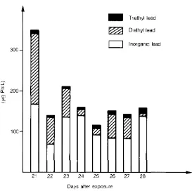
In this case determination of the various chemical forms of lead (by atomic absorption after specific treatment and extraction) consisted of 50% diethyllead, 480/o inorganic lead and about 20/o triethyllead. On the 28th day, total lead consisted of 85% inorganic lead, 10% diethyl lead and 5% triethyl lead (Fig. 1 ) .
300
:::::J :0
a... 200
Ol
:::i..
100
..
T nethyl lead~
D1ethyl leadD
lnorgan1c lead21 22 23 24 25 26 27 28
Days after exposure
[image:19.586.154.427.511.783.2]11
It should be observed, however, that the patient had probably undergone chelation therapy on the 4th and 1Oth day as appears from the graph illustrating this paper.
A relationship seems to exist between urinary lead levels and appearance of symptoms of TEL intoxication. In 49 workers with no clinical signs of intoxication, lead in urine was, in the majority of cases, below 200 ~-tg/1 and only exceptionally exceeded 300 J.tg/1. In 45 subjects with evident symptoms of intoxication PbU was never lower than 200 J.tg/1 and in many cases reached much higher levels, up to 850 J.tg/1 (Faa et al., 1970). Chiesura and Danieli (1969) observed that, when urinary lead exceeds 200 J.tg/1, there is a marked increase in the incidence of gastro-intestinal symptoms (loss of appetite, nausea, vomiting, gastric complaints, bad taste in the mounth) and neuropsychic symptoms
Table 1. TEL distillation workers: % frequency of signs and symotoms compared to PbU
PbU ug/1
N. cases
G.l. (1) Neuropsychic CNS (3) Arterial (4)
symptoms symptoms (2) alterations hypotension
Weigth loss (5)
%
Pallor %
Diarrhoea
and/or No signs
. or symptoms
% % % % const1pat1on % 011 0
< 110 110-150 160-200 210-250 260-300 310-350 360-400 410-450 >450 34 152 211 251 90 57 25 17 15 7.2 10.9 21.1 24.4 42.1 36 35 40 10 14 24.3 31 38.6 48 47 33.3 10 9 12.3 28 30 32 35 10 6 4.5 3.8 6.7 16.6 8.7 16
(1) Lack of appetite, nausea and vomiting, gastric trouble, bad taste 1n the mouth (2) Asthenia, headache, insomnia, troubled dreams, dazed state. dizziness. (3) Tremour, Romberg test fluctuations, restlessness, excitability, moodiness.
8.5 10 17 34.4 36.8 28 41 27 3 3.2 4.2 7.5 10 7 8 6 20 0.7 2.3 4.7 10 12.2 12 24 13 94.1 71.7 70 53 39 28 40 35 3 40
(4) Systolic pressure reduced by 15-20 mm Hg compared to 7-15 days previously.
(5) 2-3 Kg weight loss over a relatively short period (about 1 month). from Chiesura and Danieli (1969)
(weakness, headache, insomnia, troubled dreams, dazed state, dizziness) (Table 1). The objective signs of intoxication (CNS alterations, weight loss, arterial hypotension) become particularly frequent when PbU exceeds 600 J.tg/1, according to Gherardi and Vidoni (1965). On the question of "limit values" for PbU the ACGIH (1963) and Fleming (1964) suggest that a PbU level above 110 J.tg/1 should be considered as an indicator of exposure above "normal"; when a value of 150 J.tg/1 is registered, the worker must be removed from the job. Kehoe (1983) on the other hand, considers that a PbU level of 150 J.tg/1 is indicative of "harmful! degree of lead absorption'~; if the PbU values reach 180 J.tg/1, even if the subject is asymptomatic, he must be moved away from the job involving exposure.
More recently Grandjean (1984) raised doubts as to whether a value of 150 J.tg/1 can adequately protect against adverse effects.
As far as the relationship between PbU and atmospheric concentrations of alkyl lead compounds is concerned, Lynch (1975) found that "results from fixed station samplers throughout the manufacturing areas had not correlated well with routine medical examinations and urinary excretion levels for lead ... No correlation could be established between personal and fixed station monitor results; however, the mobile units gave significantly higher values. An approximately linear relationship between breathing zone lead levels and urinary excretion was found when the sum of the weekly average organic and inorganic lead TLV coefficients were related to the corresponding average urine excretion".
However, these findings are not of practical importance since the author used, to evaluate the environmental exposure, a coefficient obtained with an unspecified method. Cope at al. (1979) did not find any correlation between atmospehric lead levels and PbU. PbU measured after i.v. CaNa2EDTA treatment was altered even some time after cessation of exposure to alkyl lead compounds. However, it is not possible with this test to establish whether the subject was exposed in the past to alkyl lead compounds or to inorganic lead. Administration of 1 g EDTA i.v. in 45 subjects with TEL intoxication and base line PbU between 40 and 300 J.tg/1 caused a marked increase in urinary lead levels (PbU- EDTA
12
In a more recent study made on a subject with acute TEL intoxication after administration of 1 g EDTA i.v. a 5-fold increase in urinary inorganic lead levels was observed, whereas no significant increase in elimination of diethyl and triethyl lead was seen (Yamamura et al., 1982).
Administration of CaNa2EDTA in treatment of TEL intoxication may therefore serve to accelerate elimination of inorganic lead from the organism, but it apparently does not have any effect on the elimination of triethyl and diethyl lead.
In intoxication by alkyl lead compounds the changes in heme synthesis are less marked than might be expected on the basis of the internal dose tests, especially PbU, probably because these compounds are slowly degraded to inorganic lead.
Miller et al. (1972) found a reduction in erythrocyte ALAD levels in 27 subjects exposed to TEL who had no symptoms of intoxication and who had normal ALAU levels. In the exposed subjects the ALAD levels were about one-third of the levels in the control group. The mean PbB levels were 42.5 ± 10 JLg/1 00 ml (Table II).
Table 2. ALAD and PbB levels in subjects exposed to TEL and TEM and in a control group.
Exposed subjects Control group p*
n=27 n=9
ALAD
nM PBG/hr/1 010 220±80 677 ±230 <0.001
red cells
PbB ug/1 00 ml 42.5±10 15±5 <0.01
* Stat1st1cal comparison was us1ng Student's test for mdependent samples from Millar et al. (1972)
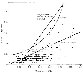
In a group of 123 asymptomatic subjects exposed to TEL and TEM, Robinson (1974a) demonstrated a significant correlation between ALAU and PbU (r = 0.52; p
<
0.001) and a linear type increase in the urinary metabolite. However, for corresponding levels of PbU the ALAU levels in these subjects were definitely lower than in workers exposed to inorganic lead (Fig. 2).2.5 2.0
E
0 0 OJ E<{ 1 5
_j <( >. Cii c ;::: ::J 1 0 0.5 Haeger-Aronsen, deKretser & Waldron (Estimated)
;f
• I
I
I
J
Stopps. I
I
I
. I
/
•
/
.
.
/
/ •
/ *
/
*
/
*~·
.
A'.
/
•••/
• , /
*
Dav1s & Andel man/
• *
•
~
. * / .
•• . / . • • • • • c)(-•• ...-:.. • •
;,"""' I
·?·~-···
. . ..
__ ., .. ··7*
·.!-:- ·-· ••••
.
.
.
~··-··.·-
·:-···
.
• • -·4-
·- :-
--=----··· -··.
·0··
···--· • -··- -·
.
~*··
.
··-0 ··-05 010 0.15 0.20
Unnary Lead. mg/hter
..
0.25 0 30 0.35
[image:21.599.127.475.288.361.2] [image:21.599.125.472.477.782.2]13
In subjects with signs and symptoms of moderately severe TEL intoxication Beattie et al. (1972) found normal ALAU levels, a slightly increase in EP and CPU and a clear reduction in ALAD. Contrary to the usual observations these subjects had high PbB levels which in one case exceeded 90 JLg/100 mi. The baseline PbU levels were between 512 and 762
JLg/1.
A study carried out on 13 subjects exposed to TEL for more than 8 years revealed normal EP levels (x = 21.12 JLg/1 00 ml ± 2 s.d. 9. 7). The PbB levels in the subjects who had clinical symptoms of intoxication such as headache and dizziness were between 62 and 155 JLg/1 00 ml (X = 114.2 JLg/1 00 ml) (Gutniak et al., 1964).
Foa et al. (1970) found abnormal EP levels in subjects who had ceased work involving exposure to alkyl lead compounds two months before. The EP levels and length of employment were significantly correlated and the highest EP levels were found in workers with the longest exposure to TEL.
Conclusions
It is clear from the above that PbU is the only biological test that can be used for the assessment of exposure in subjects working with organic lead compounds.
Various studies have in fact shown that a relationship exists between PbU levels and severity of symptoms. In addition, on the basis of studies reported in the literature, it seems that clinical symptoms appear when PbU exceeds 200 JLg/1 (Chiesura and Danieli, 1969). It does not seem, however, that the test can be used to assess the degree of exposure through inhalation because the levels are not correlated (or only moderately correlated) with atmospheric lead concentrations; this could be due to the fact that the substance is effectively absorbed through the skin.
Need for Further Research
Relationship between PbU levels and state of health
Research should be undertaken on the performance of sensitive neurologic instrument tests and behavioural tests in subjects with occupational exposure of varying degrees, considering even low levels, to establish limit values.
Research of this type necessitates standardization of the methods of urine collection and standardization of the way the results are expressed.
Indicators of internal dose
Further research should be undertaken to ascertain:
- the existence of a possible correlation between lead content of total blood (PbB) and the portion of lead bound to the lipid fraction;
- the existence of a correlation between lead levels in the lipid fraction of the blood and symptoms;
- the existence of a possible correlation of the levels of organic lead in urine with PbB and with lead levels in the lipid fraction;
- the existence of a possible variation in the PbU levels during the course of the day, so as to establish the most appropriate time to collect urine samples;
- the behaviour of PbU after cessation of exposure if possible in conjunction with assessment of clinical symptoms;
14
References
AHLBERG J., RAMEL C., WACHTMEISTER C.A. (1972) Organolead compounds shown to be genetically active. Ambia 1, 19
AKATSURA R. (1974) Tetramethyllead poisoning. Sangyo lgaky, 15, 3
BOLANOWSKA W. (1968) Distribution and excretion of triethyllead in rats. Brit. J. lndustr. Med. 25, 203
BOLANOWSKA W., PIOTROSKI J., GARCZYNSKI H. (1967) Triethylled in the biological material in cases of acute tetraethyllead poisoning. Arch. Toxicol. 22, 278
BOLANOWSKA W., WISNIEWSKA-KNYPL J.M. (1971) Dealkylation of tetraethyllead in the homogenates of rats and rabbits tissues. Biochem. Pharmacal. 20, 2108
BEADlE A.D., MOORE M.R., GOLDBERG A. (1972) Tetraethylled poisoning. Lancet II, 12 CHIESURA P. (1970) Escrezione urinaria di cataboliti del piombo tetraetile nell'uomo. Med. Lavoro 61, 437
CHIESURA P., DANIELl A. (1969) Piomburia e giudizio di intossicazione da piombo tetraetile. lndagine in operai addetti alia produzione. Med. Lavoro 60, 129
COPE R.F., PANCAMO B.P., RINEHART W.E., TER HAAR G.L. (1979) Personnel monitoring of tetraalkyl lead in the workplace. Am. Ind. Hyg. Assoc. J. 40, 372 CREMER J.E. (1959) Biochemical studies on the toxicity of tetraethyllead and other organa-lead compounds. Brit. J. lndustr. Med. 16, 191
CREMER J.E. (1984) Possible mechanism for the selective neurotoxicity in: Grandjean P.: Biological effects of organolead compounds. CRC Press Inc. Boca Raton, Florida (U.S.A.), p. 121
CREMER J.E., CALLAWAY S. (1961) Further studies on the toxicity of some tetra and trialkyl lead compounds. Brit. J. lndustr. Med. 18, 277
DAVIS J.R., ANDELMAN S.L. (1967) Urinary delta-aminolevulinic acid (ALA) levels in lead poisoning. I. A modified method for the rapid determination of urinary delta-aminolevulinic acid using disposable ion exchange chromatography columns. Arch. Environ. Health 15, 53 DE KRETSER A.J., WALDRON H.A. (1963) Urinary delta aminolevulinic acid and porphobilinogen in lead exposed workers. Brit. J. lndustr. Med. 20, 35
DE TREVEILLE R.T.P., WHEELER H.W., STERLING T. (1962) Occupational exposure to organic lead compounds. Arch. Environ. Health 5, 532
EUROPEAN ECONOMIC COMMUNITY DIRECTIVE, 78/611/EEC, July 22, 1978 FLEMING A.J. (1964) Industrial hygiene and medical control procedures. Arch. Environ. health 8, 266
FOA' V., CAVAGNA G., MANFREDI M. (1970) Valutazione della piomburia nella diagnosi di intossicazione da piombo tetraetile. Med. Lavoro 61, 491
GALZIGNA L., FERRARO M.V., MANANI G., VIOLA A. (1973) Biochemical basis for the toxic effects of triethyl lead. Brit. J. lndustr. Med. 30, 129
GETHING J. (1975) Tetramethyllead absorption: a report on human exposure to a high level of tetramethyl lead. Brit. J. lndustr. Med. 32, 329
GHERARDI M., VIDONI G. (1969) Giorn. Med. leg. lnfortunistica e tossicologia 11, 61 Quoted by Chiesura and Danieli (1969)
GRANDJEAN P., ANDERSEN 0. (1982) Toxicity of lead additives. Lancet II, 333 GRANDJEAN P., NIELSEN T. (1979) Organolead compounds: environmental health aspects. Residue Reviews 72, 97
GRANDJEAN P. (1984) Organolead exposure and intoxications in: GRANDJEAN P.: Biological effects or organolead compounds, CRC Press Inc. Boca Raton, Florida (U.S.A.), p. 229
GUTNIAK 0., KOSIOLOWA H., KOWALSKI E. (1964) Free protoporphyrin content of erythrocytes in chronic tetraethyl lead poisoning. Lancet I, 1137
HAEGER-ARONSEN B. (1960) Studies on urinary excretion of delta-amino-levulinic acid and other haem precursors in lead workers and lead intoxicated rabbits. Scand. J. Clin. Lab. Invest. 12 (suppl. 47), 1-128
HAMILTON A., REZNIKOFF P., BURNHAM G.M. (1925) Tetraethyllead. JAMA 84, 1481 HEARD M.J., WELLS A. C., NEWTON D., CHAMBERLAIN A.C. (1979) Human uptake and metabolism of tetraethyl and tetramethyl lead vapour labelled with Pb-203. International Conference Management and Control of Heavy Metals in the Environment, London, 18-21 September 1979), p. 103
JENSEN A.A. (1984) Metabolism and toxicokinetics in: GRANDJEAN P.: Biological effects of organolead compounds. CRC Press 1nc. Boca Raton, Florida (U.S.A.), p. 108 KEHOE R.A., THAMANN P. (1931) The behaviour of lead in the animal organism. II. Tetraethylled. Am. J. Hyg. 13, 478 (1931)
KEHOE R.A. (1964) Free protoporphyrin content of erythrocytes in chronic tetraethyllead poisoning. lancet II, 594
15
KITZMILLER K.V., CHOLAK J., KEHOE R.A. (1954) Treatment of Inorganic Lead (TEL) Intoxication with CaEDTA. Arch. lndustr. Hyg. 10, 312
LEA VITI
F.
H.
(1934) Case oflead poisoning from gasoline impregnated with tetraethyllead. Arch. Neutral. Psychiatr. 32, 1109LINCH A.L. (1975) Biological monitoring for industrial exposure to tetraalkyllead. Am. Ind. Hyg. Ass. J. 34, 214
MACHLE W.F. (1935) Tetraethyllead intoxication and poisoning by related compounds of lead. JAMA 105, 578
MILLAR J.A., THOMPSON G.G, GOLDBERG A., BARRY P.S.I., LOWER E.H. (1972) Delta-aminolevulinic acid dehydrase activity in the blood of men working with lead alkyls. Brit. J. lndustr. med. 29, 317
MIZOI Y., TATSUNO Y., HISHIDA S., MORGAKI T. (1973) Tetraalkyl lead poisoning. Japanese J. Leg. Med. 27, 371 (in Japanese)
MORTENSEN A. (1942) J. Ind. Hyg. 24, 285
Quoted by WALDRON and STOFEN in: Subclinical lead poisoning. Academic Pres, London-New York (1974)
MULLER K. (1953) Zur Kasuistik der Bleitetraethyi-Vergiftung. Zbl. Arbeit 3, 8
NIELSEN T., JENSEN K.A., GRANDJEAN P. (1978) Organic lead in normal human brains. Nature 274, 602
RAMEL C. (1973) The effect of metal compounds on chromosome segregation. Mutat. Res. 21, 45
ROBINSON T.R. (1974.a) Delta-aminolevulinic acid and lead in urine of lead antiknock workers. Arch. Environ. Health 28, 133
ROBINSON T.R. (1974.b) 20-year mortality of tetraethyllead workers. J.O.M. 16, 601 ROBINSON T.R. (1976) The health of long service tetraethyllead workers. J.O.M. 18, 31 SANDERS L.W. (1964) Tetraethyllead intoxication. Arch. Environ. health 8. 270 SKILLETER D.N. (1975) The decrease of mitochrondrial substrate uptake caused by trialkyltin and trialkylled compounds in chloride media and its relevance to inhibition of oxidative phosphorylation. Biochem. J. 146, 465
SPRINGMAN P., BINGHAM E., STEMMER K.l. (1963) The acute effects of lead alkyls. Arch. Environ. Health 6, 469
Biological indicators
for
the
assessment
of
human
exposure
to
industrial
chemicals
Dimethylformamide
19
Summary
DMF vapours are absorbed through the lung and also through the skin. Direct skin contact with DMF solution represents a frequent circumstance of exposure in industry. DMF exerts its main toxic action on the liver and an early manifestation of excessive uptake is in the development of alcohol intolerance.
N-hydroxymethyi-N-methylformamide (DMF-OH) has been identified as a urinary metabolite of DMF. The concentration of N-methylformamide (NMF) in urine of workers exposed to DMF is much less than that of DMF-OH. The latter, however, is also measured as NMF by gas chromatography along with the small proportion of NMF present in urine. Observations on workers have clearly demonstrated that for a substance like DMF, which can enter the organism not only by inhalation but also through skin contact, biological monitoring is much better than ambient monitoring for assessing exposure.
DMF-OH
+
NMF analysis in urine (both detected as a single NMF peak by gas chromatography) currently appears to be the best method to reach this goal. In view of their short biological half-life , it is recommended that the urine sample be collected at the end of the exposure period.21
Dimethylformamide
Introduction
Chemical and Physical Properties
Dimethylformamide (DMF) is a colourless liquid at normal temperature. Several of its chemical and physical properties are given in Table I.
Table 1. Physico-chemical properties of dimethylformamide
Boiling point (760 mm Hg): 153°C Vapour pressure (25°C): 3.7 mm Hg Water solubility: infinite
q
Formula: HCN (CH 3)2
1 ppm: 3 mgfm3
Effects on Humans
The human data on the toxicity of dimethylformamide (DMF) are limited.
Prolonged skin contact may cause local irritation (Chary, 197 4; Martelli, 1960; Potter, 1973; Rein! and Urban, 1965).
The main target organ following acute or long-term exposure to DMF is the liver (Potter, 1973; Rein! and Urban, 1965; To lot et al., 1968); the gastric mucosa and the pancreas may also be affected (Chary, 1974). Nausea, vomiting, abdominal cramps, loss of appetite, hepatomegaly and increased activity of various serum enzymes (GOT, GPT, AP, OCT, 'Y-GT) have been reported in workers exposed to DMF for various periods of time. In man, an early manifestation of excessive exposure is the development of alcohol intolerance (Chivers, 1978; Lyle et al., 1979). In workers exposed to DMF, various symptoms such as palpitations, anxiety, headache, flushing of the face and the trunk, nausea and even vomiting mqy occur when consumption of alcoholic beverages occurs during or within a few hours after the end of exposure.
The available data suggest that, provided skin contact is prevented, long term exposure to an airborne concentration below 10 ppm will not lead to the occurrence of biological signs of hepatic cytolysis (Krivanek et al., 1978; Lauwerys et al., 1980). Nevertheless, at this exposure level some individuals may still present symptoms of alcohol intolerance (Lauwerys et al., 1980; Yonemoto and Suzuki, 1980).
22
Metabolism
Human data indicate that DMF absorption occurs not only through inhalation of vapours but also by direct skin contact with the liquid form (Maxfield et al., 1975; Kimmerle and Eben, 1975b; Lauwerys et al., 1980). We have found that in workers from an acrylic fibre factory skin absorption was more important than inhalation in the overall exposure to the solvent when no personal protective devices were used (Lauwerys et al., 1980). DMF vapour can also be absorbed through contact with skin. Maxfield et al. (1975) have found that when a relatively inactive man exposes a large surface area to vapour concentrations around 10 ppm for 6 hours, cutaneous absorption may account for one quarter to one third of his total metabolite excretion during and for the 24 hours following the exposure.
DMF is rapidly metabolized in vivo. A negligible fraction of the absorbed dose is excreted unchanged in urine and in the gastrointestinal tract. Until recently, it was believed that the biotransformation of DMF in vivo, in rat, dog and human consisted of a progressive demethylation mediated by the microsomal mixed function oxidases to yield N-methyl-formamide (NMF) and N-methyl-formamide (F) (Kimmerle and Eben, 1975a,b; Krivanek et al., 1978; Lauwerys et al., 1980; Maxfield et al., 1975; Scailteur et al., 1981; Yonemoto and Suzuki, 1980).
It has now been demonstrated that the metabolite identified as NMF by gas chromatography (Barnes and Henry, 1974) is mainly N-hydroxymethyi-N-methylformamide (DMF-OH), a stable carbinolamine which breaks down in the injector of the gas chromatograph to give NMF (Scailteur et al., 1974; Scailteur and Lauwerys, 1984a,b). By analogy, N-hydroxy-methylformamide (NMF-OH) is considered to be the metabolite initially described as F. Only a very small percentage of the absorbed DMF, however, is transformed into NMF and F (probably less than 5%).
The metabolic pathway leading from DMF to DMF-OH involves hydroxyl radicals. The slight amount of NMF produced in vivo does not seem to result from further DMF-OH biotrans-formation but comes directly from DMF (Scailteur and Lauwerys, 1984a,b). NMF is more toxic than DMF and the differences between DMF and NMF toxicity were difficult to explain when NMF was thought to represent the principal in vivo metabolite of DMF (Kimmerle and Eben, 1975a,b). The metabolic studies (Scailteur et al., 1984; Scailteur and Lauwerys, 1984a,b) which demonstrate that following DMF administration, the main urinary metabolite is in fact DMF-OH and not NMF, now offer a logical explanation for these apparent discrepancies, since DMF-OH has been shown to be less acutely toxic than NMF (Scailteur and Lauwerys, 1984b).
Kimmerle and Eben (1975b) exposed 4 men to 26±8 ppm DMF for 4 hrs and 3 men and 1 woman to 87 ±25 ppm DMF for 4 hrs.
Concentrations of DMF and its metabolites (mainly DMF-OH and NMF-OH measured as NMF and F respectively) in blood and urine were determined.
DMF was no longer detectable in the blood a few hours after exposure. Only after exposure to 87 ppm was it detectable in the urine.
DMF-OH (measured as NMF) was detectable in the urine 4 hours after beginning the exposure. The majority of the substance was eliminated within 24 hours.
The elimination of NMF-OH (measured as F) was delayed; it was detectable in urine up to 72 hours after beginning the exposure.
The same authors also exposed 4 men to 21 ±4 ppm DMF for 4 hrs a day for 5 consecutive days. DMF concentration in the blood decreased rapidly and generally was no longer detectable 4 hours after the end of exposure. DMF-OH concentration (measured as NMF) in blood during repeated exposure varied from one person to another but accumulation did not occur. Urine analysis also showed that during repeated exposure to approximately 20 ppm DMF no accumulation of DMF-OH (measured as NMF) occurs in the body. During exposure, DMF-OH concentration in a 24 hours urine sample remains constant ( ±30 mg/24 hr) but it was no longer detectable 48 hours after the last exposure.
The authors propose to determine DMF-OH (measured as NMF) concentration in 24 hr urine as a routine monitoring method for employees exposed to DMF. They conclude that excretion above 50 mg/24 hr urine indicates exposures exceeding 20 ppm.
23
absorption through both the lungs and skin occurred when no mask was worn. In the case of direct skin application, the subjects wore a mask to avoid inhaling vapour and the amount of undiluted DMF applied to the skin was 0.3 mi.
The type of exposure appeared to determine how soon DMF-OH (measured as NMF) appeared in measurable amounts in the urine. It appeared most promptly when DMF vapour was absorbed through both lungs and skin and almost as quickly when DMF liquid was applied to the skin, but later when DMF vapour was absorbed only through the skin. In the first exposure condition, the peak excretion rate occurred within 5 hours after the beginning of exposure, whereas in the latter condition, it was not reached before 15 hours. Usually, DMF-OH (measured as NMF) had disappeared from the urine on the morning after the skin exposure to liquid DMF and from samples collected 24 to 26 hours after the start of the exposure to DMF vapour, with and without a mask. For exposure to DMF vapour at the TLV concentration (30 mg/m3 or 10 ppm), absorption through the lungs accounted for most (61 to 86%) of the total metabolite excreted during the 24 hour interval following the exposure. Exposure of large areas of skin to this concentration of DMF vapour can, however, result in the absorption of significant amounts of the compound through the skin.
Only a small amount of DMF inhaled or in contact with the skin may be recovered in the urine as DMF-OH (measured as NMF). In their experiments, the estimated dose recovered ranged from 0.5 to 2%. The authors conclude that the amount of metabolite in isolated urine specimens when collected at a specified time in relation to the work day may serve to monitor overexposure to DMF.
According to their data, the total amount of DMF-OH (measured as NMF) excreted in the urine following 6 hours exposure to 10 ppm DMF is lower than 5 mg. Similar results were obtained by Krivanek et al. (1978). They exposed 8 subjects to DMF vapour at an average concentration of 8.8 ppm for 6 hrs daily for 5 consecutive days. The amount of DMF-OH (measured as NMF) excreted during each 24 hr interval following the beginning of exposure amounted to about 2.5 mg.
In summary, the volunteer studies indicate that DMF-OH (measured as NMF) appears rapidly in the urine of humans exposed to DMF; its biological half-time is short, probably around 12 hrs. Unfortunately, the quantitative data obtained by Kimmerle and Eben (1975b) and Maxfield et al. (Maxfield et al., 1975; Krivanek et al., 1978) are not in good agreement since the values reported by the latter researchers appear to be about 5 times lower than those expected on the basis of the results obtained by Kimmerle and Eben (1975b).
Factors affecting DMF metabolism
Eben and Kimmerle (1976) studied the metabolic interaction between DMF and ethanol in rats, dogs and in men.
A delayed biotransformation of DMF was observed in rats and dogs when ethanol (2.0 g/kg) was administered orally prior to acute exposure to DMF vapours (rats: 209, 104 and 87 ppm for 2 hrs; dogs: 210-240 ppm for 2 hrs). Thus, the DMF concentration in the blood was 2 to 6 fold higher in the animals pretreated with ethanol than in control animals. The administration to rats of a lower oral dose of ethanol (0.2 g/kg) did not influence the metabolism of DMF.
During repeated exposure to DMF (200 ppm 2 h/day on 5 consecutive days) and ethanol (2.0 g/k p.o. once a day for 5 consecutive days) the biotransformation of DMF was also inhibited. Furthermore, they found that in animals, the oxidation of ethanol was also influenced by DMF.
24
Biological Indicators
Several biological parameters can be considered for the evaluation of exposure to dimethyl-formamide.
DMF in blood DMF in expired air
- The sum of N-hydroxymethyl- N-methylformamide (DMF-OH) and N-methylformamide (NFM) in blood. Both metabolites are measured as NMF by gas chromatography. - The sum of DMF-OH and NMF in urine. Both metabolites are measured as NMF. - The sum of N-hydroxymethylformamide (NMF-OH) and formamide (F) in urine. Both
metabolites are measured as F.
DMF in blood
DMF can be measured in blood (Kimmerle and Eben, 1975a). From data published by Kimmerle and Eben (1975b) it appears that DMF concentration in blood increases continuously during exposure and disappears rapidly after the end of exposure. At the end of a 4-hour exposure to average atmospheric concentrations of 21 and 87 ppm, the mean DMF concentration in blood amounted to about 0.3 and 1.4 mg/1 00 ml respectively.
The time of sampling is very critical, which limited the usefulness of the test for the routine control of workers.
DMF in expired air
When skin contact with DMF can be prevented, there is during exposure a significant correlation between environmental exposure and DMF concentration in alveolar air. According to Brugnone et al. (1980), the alveolar concentration amounts on average to 3 mg/m3 when the atmospheric concentration of DMF is 10 mg/m3 .
Sum of DMF-OH and NMF in blood
As indicated above, DMF-OH represents the main oxidative metabolite of DMF, NMF being a minor metabolite. The gas chromatographic method initially developed for NMF determination, measures both NMF and DMF-OH which is demethylated to NMF in the injector of the gas chromatograph.
Unlike DMF, the level of DMF-OH in blood (measured as NMF) seems to remain fairly constant for a few hours after the end of exposure (Kimmerle and Eben, 1975b). The only data available relating DMF-OH (
+
NMF) concentration in blood and exposure to DMF are those of Kimmerle and Eben (1975b) based on 4 volunteers exposed to DMF vapours at concentrations ranging from 21 to 87 ppm. At the end of the exposure, DMF-OH (+
NMF) in blood amounted on the average to 0.2 mg/1 00 ml (21 ppm) and 0.6 mg/1 (87 ppm).More data on humans are required to evaluate the relationship of this parameter with the intensity of exposure.
Sum of DMF-OH and NMF in urine
In workers exposed to DMF, DMF-OH constitutes the main urinary metabolite; only a trace amount of NMF is present in urine. However, the gas chromatographic technique used for urine analysis cannot distinguish between both metabolites since DMF-OH is demethylated to NMF at high temperature (see above).
Data available from both human volunteers and workers exposed have demonstrated that DMF-OH ( + NMF) analysis in urine is useful for monitoring exposure to DMF. However, the results permitting to relate exposure to DMF, and amount of metabolites (DMF-OH
+
NMF) excreted are still very limited.
Furthermore, the results of Maxfield et al. (1975) indicate that individual variation in metabolite excretion is high, probably resulting from differences in the actual amount absorbed as well as factors such as the rate of metabolism and/or renal clearance.
25
The results obtained by Kimmerle and Eben (1975b) on 4 volunteers are summarized in Table II.
Table 2. Urinary excretion of DMF-OH plus NMF 1n 4 volunteers exposed to DMF vapour.
mg DMF-OH + NMF excreted within 24 hours after Exposure beginning of exposure
Subject 1 Subject 2 Subject 3 Subject 4
26 ppm 4 hr 26 21 27 22
87 ppm 4 hr 94 90 111 95
21 ppm 4 hr on the average 25 to 30 mg per each (5 consecutive days) 24 hr period (4 subjects)
Catenacci et al. (1980) Yonemoto and Suzuki (1980) and Wicarowa and Oadah (1980) examined workers exposed to dimethylformamide and found a relationship between the environmental concentration of the solvent and the amount of DMF-OH
+
NMF (both measured as NMF) excreted in 24 hours. Their quantitative results, however, are very different since, for an exposure to 10 ppm DMF, the first group of authors found an excretion of 12- 15 mg DMF-OH+
NMF per 24 hours, whereas Yonemoto and Suzuki (1980) and Wicarova and Dadah (1981) reported an average excretion of 5 and 6 mg of OMF-OH+
NMF per 24 hours, respectively, for the same exposure level.Differences in the percutaneous absorption of DMF and in alcohol consumption, which interferes with DMF metabolism, might explain this discrepancy.
Lauwerys et al. (1980) have carried out a field study in an acrylic fibre factory. DMF vapour concentration was measured at different workplaces with static samplers during a week and the time spent by each worker at different places during each working day was estimated. For each worker and for each day an integrated exposure (concentration x duration) was calculated. It should, however, be recognized that during some operations, skin contact also occurred and that the airborne concentration did not necessarily reflect the total exposure. Urine samples were collected immediately before and after the shift for 5 or 6 consecutive days.
Several observations previously made on volunteers were confirmed:
1. The sum of DMF-OH
+
NMF (both measured as NMF) in urine is a sensitive biological parameter of exposure. The presence of metabolites (mainly DMF-OH) in urine can be easily detected even when the average airborne DMF concentration is below 30 mg/m3 , the current ACGIH TLV (integrated exposure: 180 mg.h.m-3).2. The metabolism of absorbed DMF is rather rapid: DMF-OH
+
NMF concentration in urine is usually greater at the end of the shift than in the morning of the next day. 3. In a group of workers the sum of DMF-OH+
NMF in urine at the end of the shift seemsto reflect the intensity of exposure of the same day.
On an individual basis (N = 116) there was a very low correlation between integrated exposure and DMF-OH
+
NMF concentration (expressed in mg/g creatinine) in urine collected at the end of the shift (r = 0.24 P<
0.01 0) or before (r = 0.16 P>
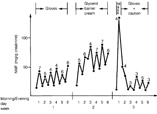
0.05) resuming work the next day (i.e. ± 16 hours after the end of the work).This could be due to individual variation in metabolism (mainly rate of excretion) but also to an error in the estimation of the total exposure since we know that in this type of industry skin contact with DMF solution is an important route of absorption. This was clearly demonstrated by comparing the urinary excretion of DMF-OH + NMF (both measured as NMF by gas chromatography) in workers using different protective devices (Fig. 1 ). The metabolite concentration in urine collected at the end of a day when the workers were equipped with a self-contained breathing apparatus but did not protect their skin (i.e. first day of week 3, in Fig. 1) was about three times higher than during the period when only their skin was protected with gloves (Fig. 1 ).
[image:34.576.110.456.137.224.2]26
Dixon et al. (1983) have noted a seasonal variation in the urinary concentration of DMF-OH
+
NMF (both measured as NMF) in workers exposed to DMF. This change was attributed to seasonal differences in urine volume. Twenty-four hour urine volumes were on the average 13% lower in hot weather than in cold weather.Morni ng/E veni ng day
week
~Gloves~
1 2 3 4 5 6
~
Glycerol~barner cream
1 2 3 4 5 6
2
3
\
• 5. .;J\Aj
1 2 3 4 5 6
3
Fig. 1 Urinary excret1on of DMF- OH + NMF (both measured as NMF by gas chromatography) in workers using different protect1ve dev1ces.
Sum of NMF-OH and F in urine
The experiments on volunteers exposed to DMF vapours (Kimmerle and Eben, 1975b) suggest that the excretion of NMF-OH