This is a repository copy of
Development of an efficient glucosinolate extraction method
.
White Rose Research Online URL for this paper:
http://eprints.whiterose.ac.uk/113468/
Version: Accepted Version
Article:
Doheny-Adams, Timothy, Redeker, Kelly Robert orcid.org/0000-0002-1903-2286, Kittipol,
Varanya et al. (2 more authors) (2017) Development of an efficient glucosinolate extraction
method. Plant Methods. pp. 1-14. ISSN 1746-4811
https://doi.org/10.1186/s13007-017-0164-8
[email protected] https://eprints.whiterose.ac.uk/ Reuse
This article is distributed under the terms of the Creative Commons Attribution (CC BY) licence. This licence allows you to distribute, remix, tweak, and build upon the work, even commercially, as long as you credit the authors for the original work. More information and the full terms of the licence here:
https://creativecommons.org/licenses/
Takedown
If you consider content in White Rose Research Online to be in breach of UK law, please notify us by
Plant Methods
Development of an efficient glucosinolate extraction method
--Manuscript
Draft--Manuscript Number: PLME-D-16-00100R1
Full Title: Development of an efficient glucosinolate extraction method
Article Type: Methodology
Funding Information: Biotechnology and Biological Sciences Research Council
(BB/L002124/1)
Prof Ian Bancroft
Biotechnology and Biological Sciences Research Council
(BB/K020463/1)
Prof Sue E Hartley
Abstract: Abstract
Background
Glucosinolates, anionic sulfur rich secondary metabolites, have been extensively studied because of their occurrence in the agriculturally important brassicacae and their impact on human and animal health. There is also increasing interest in the biofumigant properties of toxic glucosinolate hydrolysis products as a method to control agricultural pests. Evaluating biofumigation potential requires rapid and accurate quantification of glucosinolates, but current commonly used methods of extraction prior to analysis involve a number of time consuming and hazardous steps; this study aimed to develop an improved method for glucosinolate extraction.
Results
Three methods previously used to extract glucosinolates from brassicacae tissues, namely extraction in cold methanol, extraction in boiling methanol, and extraction in boiling water were compared across tissue type (root, stem leaf) and four brassicacae species (B. juncea, S. alba, R. sativus, and E. sativa). Cold methanol extraction was shown to perform as well or better than all other tested methods for extraction of glucosinolates with the exception of glucoraphasatin in R. sativus shoots. It was also demonstrated that lyophilisation methods, routinely used during extraction to allow tissue disruption, can reduce final glucosinolate concentrations and that extracting from frozen wet tissue samples in cold 80% methanol is more effective.
Conclusions
We present a simplified method for extracting glucosinolates from plant tissues which does not require the use of a freeze drier or boiling methanol, and is therefore less hazardous, and more time and cost effective. The presented method has been shown to have comparable or improved glucosinolate extraction efficiency relative to the commonly used ISO method for major glucosinolates in the Brassicaceae species studied: sinigrin and gluconasturtiin in B. juncea; sinalbin, glucotropaeolin, and gluconasturtiin in S. alba; glucoraphenin and glucoraphasatin in R. sativus; and glucosatavin, glucoerucin and glucoraphanin in E. sativa.
Corresponding Author: Timothy William Doheny-Adams, Ph.D University of York
UNITED KINGDOM Corresponding Author Secondary
Information:
First Author Secondary Information:
Order of Authors: Timothy William Doheny-Adams, Ph.D Kelly Redeker
Varanya Kittipol Ian Bancroft Sue E Hartley Order of Authors Secondary Information:
Response to Reviewers: We would like to thank the reviewers for their time and effort and their suggestions for improving the manuscript. We have addressed these suggestions to the best of our ability. Corrections have been highlighted in yellow in the document and the line numbers of those corrections are included below.
REVIEWER #1:
1.The authors should provide the some chromatograms of their real smaples analysis as a supporting materials:
Response: A supporting document with example chromatograms has been uploaded.
2.The size of the column they used should be provided. For example, what is the size of "C18 column" (line 233).
Response: the size of the column has been added (line 237)
REVIEWER #2
LN 103-104: A description of the desulfation process/mechanism and or reference is needed. Why is this a necessary step?
Response: Now reads Samples are subsequently desulfated by ion exchange chromatography on a DEAE sephadex column to remove impurities. Whether it is a necessary step or not and the steps involved are explored later in the manuscript with a direct comparison between desulfated and non-desulfated extractions.
LN112: self-dimerisation via formation of a disulfide linkage during the extraction procedure.
Response: Changed to: In addition, prior to 2002 the major glucosinolate in leaves of E. sativa, 4-mercaptobutyl glucosinolate, was missed due to self-dimerisation via formation of disulfide linkages during extraction [22]
LN113-115: How many glucosinolates possess free thiol groups and the ability to self-react? The main difficulties lie in inhibiting myrosinase activity and degradation via heat or oxidation.
Response:We appreciate the reviewers comment and agree that the vast majority of glucosinolates are not capable of self-dimerization. However, the previous sentence clearly demonstrates that at least one, important, glucosinolate has this potential. Therefore, we feel that this sentence remains valid as stated.
LN156: Table m2 could be converted to text to reduce space.
Response: We are willing to perform this action if the journal is concerned about space, otherwise we are content to let the table remain.
LN160: What were average conditions for grinding prior to milling?
Response: corrected (line 191 and 375) LN190: micron symbol µ
Response: corrected (line 193)
LN192-197: replace incubation with heated (one presumes this was not conducted in an incubation chamber.
Response: incubation has been changed to heated (lines 197, 198)
LN195: magnetic stirrer hotplate corrected (line 198)
LN200: krmp?
corrected to 8000 rpm (line 203)
LN215: small letter p to represent para-nitrocatechol corrected (line 218)
LN215: check consistency re: sulfate vs sulphate
Response: changed all instances of sulphate to sulfate (line 218)
LN218: Is DAE meant to be DEAE? Response: Yes, corrected (line 221)
LN223: For reduction of disulfide linkages, from dimerized please spell out TCEP. Response: changed and added. Now reads: . For the reduction of disulphide linkages, from dimerized desulfoglucosatavin in E. sativa extracts 3g TCEP (Tris(2-carboxyethyl)phosphine hydrochloride powder Sigma, C4706) was (line 226 and 227)
LN228: High performance liquid chromatography Response: amended (line 228)
LN 492: 'krpm' vs rpm?
Response: Changed 8 krpm to 8000 rpm for lines 494 and 496.
LN246: Provide a bold title for Mass Spectrometry Response: added (line 250)
LN257: First mention of TBA and ACN please spell out in first instance Response: amended (line 262 and 263)
LN273: Capital T used for time, check consistency earlier t is used Response: all T= have been changed to t= (lines 278, 636)
1
Development of an efficient glucosinolate extraction method
2
Doheny-Adams T., Redeker K., Kittipol V., Bancroft I., Hartley S. E.
3
4
5
Author information
6
All Authors are employed at the University of York, in the United Kingdom 7
Postal address: 8
Department of Biology 9
University of York 10
Wentworth Way, York 11
YO10 5DD 12
United Kingdom 13
14
Timothy Doheny-Adams: [email protected] 15
Kelly Redeker: [email protected] 16
Varanya Kittipol: [email protected] 17
Ian Bancroft: [email protected] 18
Sue Hartley: [email protected] 19
20
Manuscript Click here to download Manuscript Development of an efficient
glucosinolate extraction method.docx
Click here to view linked References
21
Abstract
22
Background 23
Glucosinolates, anionic sulfur rich secondary metabolites, have been extensively studied because of 24
their occurrence in the agriculturally important brassicacae and their impact on human and animal 25
health. There is also increasing interest in the biofumigant properties of toxic glucosinolate 26
hydrolysis products as a method to control agricultural pests. Evaluating biofumigation potential 27
requires rapid and accurate quantification of glucosinolates, but current commonly used methods of 28
extraction prior to analysis involve a number of time consuming and hazardous steps; this study 29
aimed to develop an improved method for glucosinolate extraction. 30
Results 31
Three methods previously used to extract glucosinolates from brassicacae tissues, namely extraction 32
in cold methanol, extraction in boiling methanol, and extraction in boiling water were compared 33
across tissue type (root, stem leaf) and four brassicacae species (B. juncea, S. alba, R. sativus, and E. 34
sativa). Cold methanol extraction was shown to perform as well or better than all other tested 35
methods for extraction of glucosinolates with the exception of glucoraphasatin in R. sativus shoots. 36
It was also demonstrated that lyophilisation methods, routinely used during extraction to allow 37
tissue disruption, can reduce final glucosinolate concentrations and that extracting from frozen wet 38
tissue samples in cold 80% methanol is more effective. 39
Conclusions 40
We present a simplified method for extracting glucosinolates from plant tissues which does not 41
require the use of a freeze drier or boiling methanol, and is therefore less hazardous, and more time 42
and cost effective. The presented method has been shown to have comparable or improved 43
glucosinolate extraction efficiency relative to the commonly used ISO method for major 44
glucosinolates in the Brassicaceae species studied: sinigrin and gluconasturtiin in B. juncea; sinalbin, 45
glucotropaeolin, and gluconasturtiin in S. alba; glucoraphenin and glucoraphasatin in R. sativus; and 46
glucosatavin, glucoerucin and glucoraphanin in E. sativa. 47
48
Background
49
50
Glucosinolates, B-thioglucoside N-hydroxysulfate derivatives, are secondary metabolites found in 51
brassicaceae and related families [1]. Over 120 glucosinolates, which differ in variable aglycone side 52
chains derived from an alpha-amino acid, have been identified and classified into aliphatic, aromatic 53
and indole glucosinolates [2, 3]. Due to their prevalence in cultivated vegetables, spices, oils and 54
animal feed, glucosinolates and their hydrolysis products have been much studied in the context of 55
their effects on human and animal nutrition [4, 5]. Glucosinolates and their breakdown products 56
have also been a focus of studies in dietary prevention of disorders linked to oxidative stress such as 57
cancer and gastric ulcers [2, 6, 7] and more recently, potential undesirable dietary effects such as 58
genotoxicity of glucosinolate breakdown products in broccoli [8] and Pak Choi [9]. The breakdown of 59
glucosinolates has also been studied because of their potential use as agricultural pesticides in a 60
technique known as biofumigation. In biofumigation a glucosinolate-rich crop is mulched into the 61
field, releasing toxic secondary glucosinolate by-products, in order to reduce the incidence of pests, 62
weeds and diseases in the following arable and horticultural crops [10, 11, 12, 13]. 63
Evaluating biofumigation potential requires rapid and accurate quantification of glucosinolates, but 64
current commonly used methods of extraction prior to analysis involve a number of time consuming 65
and potentially hazardous steps. These steps are (i) lyophilisation, or freeze drying, and tissue 66
disruption, (ii) extraction in water or methanol, (iii) purification of extract, typically by desulfation on 67
DEAE sephadex, and (iv) separation and analysis of (desulfo)glucosinolates. These steps are outlined 68
in figure 1 and discussed in more depth below. This study aimed to improve glucosinolate extraction 69
methods by finding alternatives to commonly used steps which are unnecessary or likely to 70
introduce variability. 71
72
INSERT FIGURE 1 HERE 73
74
Myrosinase, an enzyme found in brassicacae and compartmentalised in cells in close proximity to 75
glucosinolates, is responsible for hydrolysing glucosinolates upon plant tissue disruption. Accurate 76
analysis of glucosinolates therefore requires inactivation of myrosinase prior to tissue disruption. 77
This is achieved by first freezing then freeze drying the tissue which allows disruption by milling or 78
grinding to occur in the absence of water (fig 1). Lyophilisation, or freeze drying, is used to remove 79
water from glucosinolate-containing tissues while preventing myrosinase mediated glucosinolate 80
hydrolysis through thermal inhibition. Publications on freeze drying plant tissue have focussed 81
primarily on the production of heat or its implications in generating oxygen sensitive foodstuffs (e.g.- 82
space, military or extreme-sport foodstuffs and instant coffee) [14]. To our knowledge, no study has 83
yet examined the efficiency of freeze drying in maintaining glucosinolate concentrations. Freeze 84
drying functions on the principle of sublimation: pressure is reduced below the triple point of water 85
(6.12 mbar, 0.01°C) at which point sublimation of ice from the sample occurs. The cooling effect of 86
sublimation should be high enough to ensure the sample remains below 0°C for the initial stage of 87
freeze drying, thus minimizing enzyme-driven glucosinolate hydrolysis. Rapid sample loading and 88
rapid initial pressure drop are also required to avoid sample defrosting before pressure is reduced 89
below 6.12 mbar. Leaves have a high surface area to volume ratio and may defrost quickly, 90
activating myrosinase and reducing final glucosinolate concentration. Despite the importance of the 91
freeze drying process in glucosinolate extraction, many authors do not report details which are likely 92
to affect final concentrations of glucosinolates (e.g.- how samples are transported, temperature of 93
The most commonly used methods for extraction of glucosinolates from plant material are based on 95
the ISO 9167-1 method [15; highlighted in grey in figure 1], which was designed for extraction of 96
glucosinolates from B. napus seed and has been adapted to suit the needs of researchers examining 97
glucosinolate profiles of other plant species and tissue types. Although freeze drying is not explicitly 98
detailed in the ISO 9167-1 method, it is an implicit requirement in order to avoid myrosinase 99
mediated glucosinolate hydrolysis during disruption of leaf, stem or root tissues. Once the plant 100
tissue is prepared, the ISO 9167-1 extraction is carried out at 75°C in 70% methanol for 10 minutes. 101
Heating the sample is thought to be an essential step to denature myrosinase, thus preventing 102
enzymatic hydrolysis of glucosinolates [16]. Samples are subsequently desulfated by ion exchange 103
chromatography on a DEAE sephadex column to remove impurities. Desulfoglucosinolates are then 104
separated and identified using HPLC with a reverse phase C18 column and a UV or MS detector. 105
Hazards associated with boiling methanol [17] and the time required for extractions using this 106
method have led researchers to seek alternatives. Replacing heated methanol with boiling water is 107
reported to have comparable [18, 19], and in some cases better [20], extraction efficiencies. 108
Although most glucosinolates are thermostable for the typical 10-30 minute heating period, indole 109
glucosinolates such as 4-hydroxy-glucobrassicin and 4-methoxyglucobrassicin have been reported to 110
degrade quickly at temperatures below 100°C [21]. In addition, prior to 2002 the major glucosinolate 111
in leaves of E. sativa, 4-mercaptobutyl glucosinolate, was missed because it self-dimerises via 112
formation of disulphide linkages during extraction [22]. A major challenge therefore to ensuring 113
consistent and repeatable GSL analysis is to create extraction conditions in which myrosinase is 114
inactive, and glucosinolates do not self-react or degrade. A single study, conducted exclusively on 115
radish roots, has demonstrated that cold extraction in 80% methanol does not cause appreciable 116
reduction in glucosinolate concentrations compared to more conventional heated extraction 117
methods [23]. However, myrosinase activity can vary dramatically [24] and whether this method is 118
suitable for extraction of glucosinolates from other glucosinolate containing plants has not 119
previously been assessed. 120
A desulfation step is often carried out post extraction to purify desulfoglucosinolates and improve 121
accuracy and identification from HPLC. However, the desulfation reaction of glucosinolates can be 122
affected by feedback inhibition of the enzyme which causes incomplete desulfation of glucosinolates 123
[25]. In addition, rhamnopyranosyloxy-benzyl glucosinolates extracted from M. oleifera have been 124
shown to be completely converted and degraded by the desulfation purification step [26]. Due to 125
these drawbacks, and the additional time and potential error extra steps can introduce, some 126
authors have skipped the purification and desulfation steps entirely [19, 26, 27] (fig 1). 127
We have tested each stage of glucosinolate analysis from the roots, stems and leaves of B. juncea, S. 128
alba, R. sativus, E. sativa and B. napus and suggest a number of adjustments/improvements which 129
can be made to reduce the costs, time and variability associated with glucosinolate analysis. 130
Specifically, this study aims to address the following questions: 131
1) How do lyophilisation conditions affect glucosinolate concentrations? 132
2) Is lyophilisation a necessary step for glucosinolate extraction from green tissues? 133
3) Do extractions in hot methanol, cold methanol and boiling water yield comparable 134
glucosinolate concentrations across a range of brassicacae species and tissue types? 135
4) How do desulfation time and enzyme concentration affect final glucosinolate 136
concentrations? 137
5) Is desulfation a necessary step for glucosinolate extraction from green tissue? 138
139
140
141
Materials and methods
142
Experiment Fig Species Tissue Freeze
drying/tis
sue
disruption
Extraction Desulfation HPLC
1-Effect of freeze drier
on GSL concentration
2 B. napus Leaves FD-A or
FD-B
/mill
Cold
methanol
0.3 U/ml
for 24 H
ISO 9167-1
method
2-Comparison of GSL
extraction from freeze
dried tissue with
extraction from wet
tissue
3 B. napus Leaves FD-A or
-20°C
methanol
Cold
methanol
0.3 U/ml
for 24 H
ISO 9167-1
method
3-Comparison of
extraction methods
6, 7 R. sativus
B. juncea S. alba E. sativa Leaves, Stems, Roots
FD-A Hot
methanol, Cold methanol, Boiling water 0.3 U/ml
for 24 H
ISO 9167-1
method
4-Comparison of
desulfation/purification
methods
8,9 R. sativus
B. juncea S. alba E. sativa Leaves, Stems, Roots
FD-A Cold
methanol
0.3 U/ml
for 12, 24,
48 H, and
5U/ml for
16 H or
filtration ISO 9167-1 Method for desulfoGSL, Herzallah and Holly method for intact GSLs.
Plant material 145
B. napus used in the freeze drying tests were grown in 1 L pots filled with Terra-green in a controlled 146
temperature glasshouse (regulated from 17.6°C to 27.7°C). At 3-4 weeks post germination, leaves 147
were removed and halved down the limits of the midrib, excluding the midrib from the final sample. 148
Leaf halves were immediately frozen in liquid nitrogen and stored at -80°C for a maximum of 1 week. 149
B. juncea (cv. ISCI99), R. sativus (cv. Bento), S. alba (cv. Ida Gold) and E. sativa (cv. Nemat) plants 150
were grown by Barworth agriculture ltd. in a sandy loam soil dominated fields (coordinates: 151
53.000371, -0.290404) from 31/07/2014 to 25/09/2014. Total stem and total leaves were cut from 152
flowering plants and immediately frozen in liquid nitrogen; root samples were gently washed and 153
dried before freezing in liquid nitrogen. Samples were stored at -80°C for a maximum of 2 months. 154
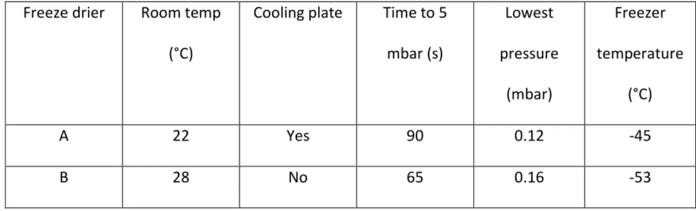
Freeze drying 155
Samples wrapped loosely in aluminium foil were transported on dry ice and loaded into one of two 156
freeze driers (table m2). Maximum loading time was 30 seconds. 157
Freeze drier Room temp
(°C)
Cooling
plate
Time to
5 mbar
(s)
Lowest
pressure
(mbar)
Freezer
temperature
(°C)
Model
A 22 Yes 90 0.12 -45 Lyotrap, LTE
scientific ltd.
1 chamber.
B 28 No 65 0.16 -53 Thermo, Heto
Powerdry LL3000.
4-6 chambers.
Table m2: Freeze drier characteristics 158
Tissue disruption 160
(i) Freeze dried plant tissue was homogenised to a roughly ground powder (approximately
161
0.1cm particle size) using a grinder (Lloytron, E5601BK) Homogenised ground samples were
162
milled at a frequency of 20 Hz for 10 minutes (Retch, MM400) with 2 steel ball bearings to a
163
fine powder (particle diameter <0.1mm). Samples were then sealed and stored at 20°C for
164
up to 9 months.
165
(ii) Frozen fresh B. napus leaf halves (experiment 2, table m1) were placed in 2ml eppendorf vials 166
and stored at -20°C. 1.755ml of 80% methanol precooled at -20°C, 25µl of 5mM sinigrin and 2 small 167
ball bearings were added. Samples were milled for 10 minutes at frequency 20 Hz (TissueLyser II, 168
Qiagen). Final concentrations of methanol were estimated by incorporating average leaf moisture 169
content of fresh B. napus leaves according to equation (1). Final concentration of methanol ranged 170
from 79.3% to 79.9% and leaf moisture content accounted for less than 1% of final liquid volume. 171
C 172
Where cMeOHf is final methanol concentration (%)
173
cMeOHi is initial methanol concentration (90%)
174
VMeOHi is initial methanol volume (1.755 ml)
175
mav is the average moisture content per dry weight (in this case 0.22 ml/g)
176
mdl dry mass of leaf sample (g)
177
Glucosinolate extraction 178
Extractions were carried out in one of three ways (fig 1m). In each case 50µl of a 5mM 179
gluctropaeolin (for B. juncea samples) or 20 mM sinigrin (for all other samples) internal standard was 180
added: 181
Hot methanol extraction (based on the ISO 9167-1 method): 182
0.1g of plant material was preheated at 75°C for 3 minutes in a 20ml falcon tube. 4.95ml of 70:30 183
methanol:water, preheated to 75°C and the internal standard was added. The sample was 184
incubated at 75°C for 10 minutes, and manually shaken every 2 minutes. The sample was then 185
centrifuged at 4000 rpm (Jouan, model:B 3.11) for 10 minutes. Supernatent was stored at -20°C or 186
desulfated directly. 187
Cold methanol extraction (Ishida et al. 2011, [23]): 188
5 ml of 80:20 methanol:water at 20°C was added to 0.1g plant tissue and the internal standard was 189
added. The sample was shaken and left to stand for 30 minutes at room temperature. The sample 190
was then mixed at 70 rpm with a platform rocker for a further 30 minutes (Bibby, STR6) before 191
centrifugation at 4000 rpm (Jouan, model:B 3.11) for 10 minutes. Supernatent was then filtered 192
through a 0.22 µm syringe filter (Millex GP) for direct injection on HPLC, or unfiltered if applied to 193
sephadex column in a purification step. 194
Boiling water extraction (adapted from Herzallah and Holley, 2012, [19]): 195
25 ml of boiling water was added to 0.1g of freeze dried and milled plant tissue in a 150ml 196
erlenmeyer flask and the internal standard was added. Sample was heated at 100°C and stirred with 197
a magnetic stirrer hot plate for 10 minutes. Sample was heated for a further 4 H at 70°C before 198
centrifugation at 4000 rpm (Jouan, model:B 3.11) for 10 minutes. Sample was topped up to 20ml 199
with deionised water. 200
Purification and determination of activity of sulfatase: 201
Sulfatase from Helix pomatia type H-1 (Sigma, S9626) was purified according to Wathalet et al. 202
(1999) [25]. 25 mg of sulfatase was added to 1ml 40% ethanol and centrifuged at 8000 rmp for 1 203
minute (eppendorf centrifuge, 54151). The supernatant was transferred to a fresh 2ml eppendorf 204
tube, 1ml of pure ethanol was added to precipitate the sulfatase before being centrifuged at 8krmp 205
for 1 minute. The supernatant was discarded and the sulfatase pellet air dried and redissolved in 2ml 206
of water. 207
Activity of sulfatase was determined based on the ISO 9167-1 method. 1 ml of buffered 0.15mM 208
sinigrin solution (3ml of 5mM sinigrin, adjusted to 100ml with a solution containing 40ml 0.2% 209
ethylene diamine, 73ml 0.2% acetic acid; adjusted to pH 5.8 ) in a quartz cuvette was placed in a UV 210
spectrometer set to 229nm. At t=0, 25µl of diluted and undiluted purified sulfatase was added to the 211
cuvette and measurements taken over the course of 4 hours. The tangent to t=0 was plotted and its 212
A A (2):
213
Activity U ml 214
W A Ae is the difference between absorbance at equilibrium and
215
absorbance at t=0. 216
The activity for Sulfatase from Helix pomatia type H-1 (Sigma, S9626) given by the supplier is 217
determined by desulfation of p-nitrocatechol sulfate and is an order of magnitude higher than the 218
activity measured for desulfation of sinigrin using this method. 219
Desulfation of glucosinolates 220
As per the ISO 9067-1 method, columns were prepared with 0.5ml sephadex slurry (2g DEAE 221
sephadex beads in 30 ml 2M acetic acid.) and activated with 2ml imizadole formate (6M). Columns 222
were washed twice with 1ml water. The column was washed twice with 1ml 20mM sodium acetate 223
(pH 4.0) and 75µl of purified sulfatase was added (5U/ml or 0.3U/ml). Columns were incubated at 224
room temperature for either 12, 24 or 48 hours before elution of desulfoglucosinolates with two 1ml 225
volumes of water. For the reduction of disulphide linkages, from dimerized desulfoglucosatavin in E. 226
sativa extracts 3g TCEP (Tris(2-carboxyethyl)phosphine hydrochloride powder Sigma, C4706) was 227
added to 1 ml of desulfated extract. Desulfoglucosinolates were stored at -20°C before high 228
performance liquid chromatography analysis. 229
For the high sulfatase treatment, between 0.5 and 1 ml of sample was added due to insufficient 230
sample volume remaining. 231
HPLC 232
A Waters 600E system controller attached to a Waters 717 autosampler, Waters 996 photodiode 233
array detector and SphereClone 5µ ODS(2) column (Phenomonex) were used for separation and 234
detection of desulfo and intact glucosinolates. 235
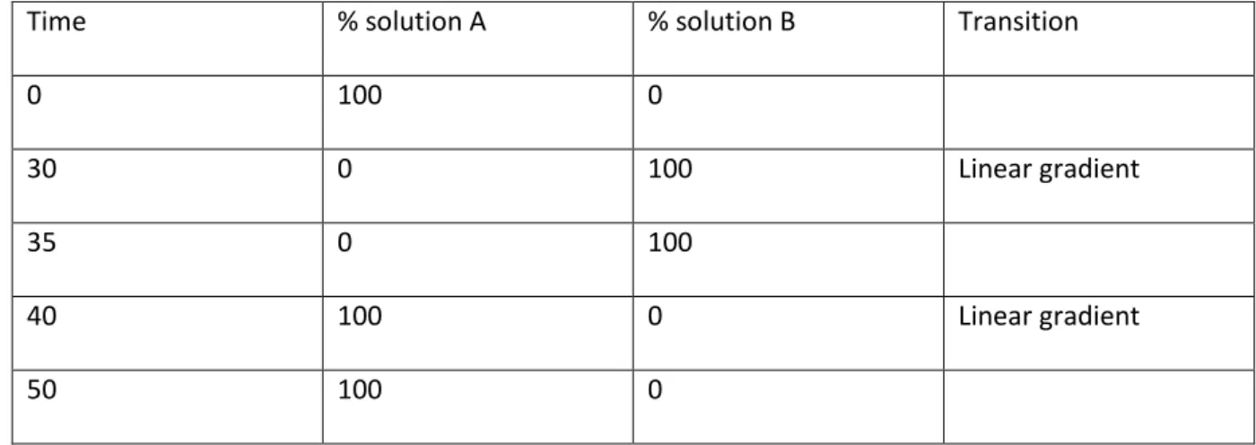
HPLC analysis of desulfoglucosinolates adapted from ISO 9167-1 236
A reverse phase C18 column (Phenomonex, SphereClone 5µ ODS(2), 150mm x 4.6mm) was 237
equilibrated for 30 min with a mobile phase which consisted of 100% diH2O. Flow rate was set to
238
1ml/min and samples separated according to programme for desulfoglucosinolates detailed in table 239
m3. Mobile phase solutions were degassed for 30 minutes in a sonicator (Decon, Sussex England). 240
Solution A: 100% diH2O
241
Solution B: 70:30, diH2O:acetonitrile
242
Time % solution A % solution B Transition
0 100 0
30 0 100 Linear gradient
35 0 100
40 100 0 Linear gradient
50 100 0
Table m3: mobile phase conditions for separation of desulfoglucosinolates.
243
Desulfoglucosinolates were quantified using 229nm wavelength within the UV spectrum. The HPLC 244
PDA detector allowed a full spectrum analysis from 180 to 800nm, allowing comparative UV-Visible 245
spectra analysis, which aided in identifying unknown glucosinolates. Through standard injections and 246
HPLC-MS D
purified standards: sinigrin (sigma aldrich), glucotropaeolin, glucoraphenin, glucoraphanin, 248
glucerucin, glucobrassicin, gluconasturtiin, sinalbin, progoitrin and glucoiberin (phytoplan). 249
Mass spectrometry 250
Major glucosinolates for which no commercial standard is available were identified using an MS 251
detector (Bruker maXis UHR-TOF) with the following settings: 252
Source: Standard electrospray (flow split 1/10 from LC) 253
Nebulizer: 2.0 bar 254
Dry gas: 6.0 L/min 255
Dry gas heater: 250C 256
Capillary voltage: 3500 V 257
Ion polarity: positive 258
Spectra rate: 1Hz 259
HPLC analysis of intact glucosinolates adapted from Herzallah and Holly, 2012 [19] 260
A C18 column (Phenomonex, SphereClone 5u ODS(2)) was equilibrated for 3 h with a mobile phase 261
which consisted of 80 mL (0.02 M) TBA (tetrabutylammonium bromide) and 20 mL ACN 262
(acetonitrile) with detection at 229 nm. The flow rate was set at 1.0 ml/min and separated according 263
to programme for desulfoglucosinolates detailed in table m3. 264
Solution A: 100% TBA (0.02M) 265
Solution B: 70:30, TBA (0.02M):acetonitrile 266
Glucosinolates were quantified using the chromatogram from 229nm and standard curves were 267
constructed using pure sinigrin (sigma aldrich), glucotropaeolin, glucoraphenin, glucoraphanin, 268
glucerucin, glucobrassicin, gluconasturtiin, sinalbin, progoitrin and glucoiberin (phytoplan). 269
In the case of glucoraphasatin in R. sativus leaves and glucotropaeolin in B. juncea minor alterations 270
were made to avoid peaks co-eluting. The mobile phase programme for R. sativus leaves was 100% A 271
for 5 minutes, followed by a 35 minute linear gradient to 66% B followed by a 5 minute linear 272
gradient to 100% B followed by a 5 minute linear gradient to 100% A . For B. juncea leaves, an 273
isocratic 85:15, TBA (0.02M):acetonitrile mobile phase for 70 minutes was used. 274
Determination of myrosinase activity 275
Activity of pure myrosinase was tested in water and 80% methanol solutions containing 0.25mM 276
sinigrin and 0.1 mM ascorbic acid, a myrosinase cofactor (Burmeister et al. 2000). Myrosinase was 277
added at t=0 and absorbance of sinigrin at 229nm was measured over the course of an hour. Activity 278
was measured at room temperature (25°C). 279
Determination of glucosinolate thermostability 280
A 50µl of 10mM sinigrin, 10mM glucotropaeolin, 10mM glucobrassicin solution was added to 0.95ml 281
water or 70% methanol preheated to 100°C or 75°C respectively and sealed in 1.5ml eppendorf 282
tubes. Samples were maintained at either 100°C or 75°C for 5, 10, 30 and 60 minutes and intact 283
glucosinolate concentrations analysed with HPLC following the adapted Herzallah and Holly (2012) 284
method [19]. 285
Calculation of glucosinolate content 286
Glucosinolate content, expressed in µmol/g were calculated according to the ISO 9067-1 method 287
(equation (3)): 288
(3) 289
Where Ag is the peak area corresponding to desulfoglucosinolate;
290
As is the peak area corresponding to internal standard;
291
n is the quantity, in micromoles, of the internal standard; 292
m is the mass of the test portion; 293
Kg is the response factor of the desulfoglucosinolate relative to the internal standard;
294
w is the moisture and volatile matter content, expressed as a percentage by mass of the test sample. 295
Statistical analysis 296
Paired two tailed t-test analysis were carried out on total B. napus glucosinolate content per leaf half 297
in experiments 1 and 2 with Microsoft excel (table m1). For determination of significance of effect of 298
method on final glucosinolate content estimates in experiments 3 and 4 (table m1), repeat measure 299
ANOVA analyses were carried out for each glucosinolate with R statistical software package (version 300
3.3.1). 301
302
Results and discussion
303
1 Lyophilisation 304
Modifications to the ISO9167-1 method (specifically created for the extraction and analysis of 305
glucosinolates from oil rape seed samples) are required for analysis of plant green tissues (leaves, 306
stems and roots). A number of prior-to-analysis steps, such as sampling in the field, cleaning (if 307
required), freezing, crushing, storage or/and shipping and reduction of sample amount have been 308
discussed by Wathelet et al. (2004) and are not revisited here [28]. These preliminary steps are 309
followed by lyophilisation, or freeze drying, to remove water from glucosinolate containing tissues 310
while preventing myrosinase mediated glucosinolate hydrolysis through thermal inhibition. This 311
process allows subsequent tissue disruption without risking glucosinolate degradation. 312
We tested reproducibility of glucosinolate concentrations extracted after lyophilisation in separate 313
freeze driers (table 1). Fresh B. napus leaves were halved, loosely wrapped in foil, flash frozen in 314
liquid nitrogen and transported in dry ice to be dried in separate freeze driers (table 1). Total 315
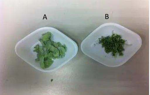
glucosinolate concentrations were significantly higher in samples dried in freeze drier A than freeze 316
drier B (fig 2a). In addition, samples dried in freeze drier B developed a darker hue and deformed 317
more than samples in dried in freeze drier A (fig 2b). Plant tissue samples have been shown to 318
deform during the freeze drying process when temperatures exceed the glass transition state and 319
melting point of water [29]. It is likely that samples placed into freeze drier B may have defrosted 320
before the pressure had reduced below the 6.12 mbar required for sublimation due to higher 321
temperatures and the lack of cooling plate. As a result, enzyme mediated hydrolysis of 322
glucosinolates may have occurred at the initial stage. Additionally, as sublimation slows over time 323
due to the remaining water vapour passing through a dry layer of increasing thickness and because 324
water is increasingly more tissue bound, the sample temperature may have increased to above 0°C 325
in freeze drier B, causing defrosting. 326
327
Freeze drier Room temp
(°C)
Cooling plate Time to 5
mbar (s)
Lowest
pressure
(mbar)
Freezer
temperature
(°C)
A 22 Yes 90 0.12 -45
B 28 No 65 0.16 -53
Table 1: Freeze drier characteristics 328
329
INSERT FIGURE 2 HERE 330
331
These results underline the need for a more substantive study to assess optimal conditions for 332
freeze drying plant tissues for glucosinolate analysis. It is clear that differences in freeze drying can 333
introduce significant variability in retained glucosinolate concentrations (fig 2a). 334
A cold methanol extraction method may be sufficient to 1) inactivate myrosinase and 2) efficiently 335
extract glucosinolates, precluding the need for the lyophilisation step altogether. We tested this by 336
comparing glucosinolates extracted from one half of a B. napus leaf in 80% methanol without freeze 337
drying against glucosinolates extracted from the other half, first dried in freeze drier A and then 338
extracted using the cold methanol extraction method. 339
No significant difference in final glucosinolate concentration was found between the two methods 340
(fig. 3). Freeze drying is an energy intensive and costly process requiring long drying times under 341
continuous vacuum and the significant effect of freeze drier parameters on final glucosinolate 342
concentrations (fig. 2a) highlights a potential source of variation between studies. If long term 343
storage of plant tissue samples is not required, skipping the freeze drying step and extracting 344
glucosinolates directly into cold methanol (-20°C) is cheaper, quicker and less hazardous. 345
346
INSERT FIGURE 3 HERE 347
348
2 Extraction 349
Some authors have highlighted that glucosinolates, specifically indole glucosinolates, are heat 350
sensitive and are significantly degraded in 75°C in less than 10 minutes [21]. This has 351
serious implications for accuracy and reliability of the ISO 9167-1 extraction method, which 352
recommends extractions occur in boiling 70% methanol (75° C) for 10 minutes, as well as the less 353
commonly used boiling water extraction (100° C). In order to first test whether thermal degradation 354
of glucosinolates was likely to occur with these methods we measured the glucosinolate 355
concentrations of pure sinigrin (aliphatic), glucotropaeolin (aromatic) and glucobrassicin (indole) in 356
boiling water (fig 4) and boiling 70% methanol (data not shown). Sinigrin and glucotropaeolin did not 357
significantly decrease over 60 minutes suggesting that extraction in boiling water or methanol is 358
unlikely to affect the concentrations of these glucosinolates. However, glucobrassicin was thermally 359
degraded at 100°C and data from extractions carried out at these temperatures or above (such as 360
with microwave based methods) may underestimate the concentration of glucobrassicin and other 361
indole glucosinolates. Boiling an extract in water for 10 minutes degrades glucobrassicin by an 362
estimated 7%. 363
INSERT FIGURE 4 HERE 364
365
366
Activity of pure myrosinase was tested at 25° C in water and 80% methanol solutions containing 367
0.25mM sinigrin and 0.1 mM ascorbic acid, a myrosinase cofactor [30]. Absorbance of sinigrin at 368
229nm, at room temperature (25°C), was measured over the course of an hour after myrosinase 369
addition. Myrosinase was inactive in 80% methanol (fig 5) suggesting that heating methanol at 75°C 370
for 10 minutes in order to inactivate myrosinase may be an unnecessary step for extracting 371
glucosinolates from plant tissue. 372
INSERT FIGURE 5 HERE 373
Glucosinolates from B. juncea, S. alba, R. sativus and E. sativa leaves, stems and roots were 374
extracted (i) in boiling water for 10 minutes followed by a 4h incubation at 70°C, (ii) in 70% methanol 375
at 75°C, or (iii) in 80% methanol at room temperature (~20°C) for 30 minutes standing followed by 376
30 minutes shaking at 70 rpm. All extracts were centrifuged and desulfated with sulfatase according 377
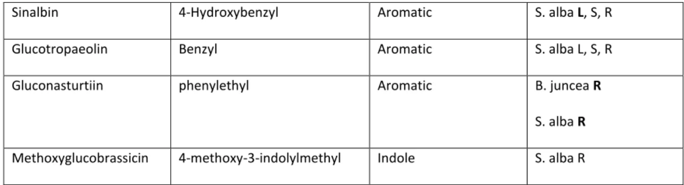
to the ISO 9167-1 method. Major glucosinolates from these species can be found in table 2. 378
Common name Chemical name Structure Species, tissue type
Sinigrin 2-propenyl Aliphatic B. juncea L, S, R
Glucoraphenin 4-methylsulfinyl-3-butenyl Aliphatic R. sativus L, S, R
Glucoraphanin 4-methylsulfinylbutyl Aliphatic E. sativa L, S, R
Glucosatavin mercaptobutyl Aliphatic E. sativa L, S, R
Glucoraphasatin
or hydroxyglucoerucin
4-Methylthio-3-butenyl Aliphatic R. sativus L, S, R
Glucoerucin Methylthiobutyl Aliphatic E. sativa S, R
S. alba, R
Sinalbin 4-Hydroxybenzyl Aromatic S. alba L, S, R
Glucotropaeolin Benzyl Aromatic S. alba L, S, R
Gluconasturtiin phenylethyl Aromatic B. juncea R
S. alba R
Methoxyglucobrassicin 4-methoxy-3-indolylmethyl Indole S. alba R
Table 2: Glucosinolates examined in this study. L, S and R correspond to leaf, stem and root respectively.
379
Letters in bold represent major glucosinolates of those tissues (>10 µmol/g dry weight).
380
381
Figure 6 compares glucosinolate concentrations obtained using the cold methanol method and 382
boiling water method normalised against the ISO 9167-1 boiling methanol method. For most 383
glucosinolates, across most tissue types and species, the three extraction methods yield similar 384
results. We found that extraction with cold methanol produced a significantly higher estimated 385
concentration of sinalbin in S. alba and sinigrin in B. juncea than the hot methanol extraction (fig 6). 386
Surprisingly, given the sensitivity of glucobrassicin to thermal degradation (fig 4), extraction in 387
boiling water did not significantly reduce the concentration of the indole glucosinolate: 388
methoxyglucobrassicin relative to the other two methods. However, glucosatavin was extracted with 389
lower efficiency from leaves of E. sativa using the boiling water method (fig 6). It seems unlikely that 390
this glucosinolate is less thermostable than other glucosinolates and was therefore degraded by the 391
extraction method since reduced extraction efficiencies are not observed for stem and root samples. 392
There are no published explanations or hypotheses that might help to explain the observed lower 393
extraction efficiencies for glucosatavin using the boiling water method. Glucoraphasatin extraction 394
using cold methanol appears to be significantly less effective than the standard ISO method (fig 6), 395
however this was driven by poor extraction efficiencies from R. sativus stems (fig 7). Ishida et al. 396
reported a significant 5% increase in glucoraphasatin concentrations extracted from R. sativus roots 397
using the cold methanol method [23]. In this study, extraction efficiencies of glucoraphenin in R. 398
sativus roots with a cold methanol method were comparable to extraction efficiencies using the 399
boiling methanol method (fig 7). 400
INSERT FIGURE 6 HERE 401
INSERT FIGURE 7 HERE 402
No glucosinolates were detected in a subset of samples extracted in cold water indicating the 403
presence of active myrosinase leading to their degradation (data not shown). However, the cold 404
methanol extraction did not significantly affect the concentration of the internal standard relative to 405
the boiling methanol method (data not shown), providing additional evidence that myrosinase is 406
inactivated in 80% methanol without heating (fig 5). 407
These data demonstrate that 80% cold methanol can be used instead of boiling methanol to extract 408
glucosinolates across a broad spectrum of brassicacae species and tissue types. With the exception 409
of glucoraphasatin in R. sativus shoots, replacing hot 70% methanol with cold 80% methanol did not 410
significantly reduce glucosinolate concentrations, yet marginally increased recovery of sinalbin in S. 411
alba and sinigrin in B. juncea. It is advised, due to reduction in steps and hazard as well as improved 412
or comparable glucosinolate recovery, that a cold methanol extraction is used instead of a boiling 413
methanol extraction for most glucosinolate containing green tissues. 414
3 Purification 415
Purification of extract according to the ISO 9167-1 method is carried out by introducing 1ml of 416
extract to a column containing 0.5ml of sephadex solution. The column is rinsed with a 20mM 417
acetate buffer at pH 4.0 to avoid possible reduction of indole glucosinolates recovery [28]. 75µl of 418
sulfatase solution with an activity above 0.05 U/ml is applied and left to act overnight. We tested the 419
extraction efficiency of the ISO 9167-1 purification step at the described pH 4.0, at 20°C for 12, 24 420
and 48 hours. Complete desulfation of glucosinolates in rapeseed extract required a minimum of 11 421
hours in operating conditions of 30°C and pH 5.8 [25] so it was expected that an overnight 12 hour 422
desulfation period may be insufficient for complete desulfation of samples at room temperature. 423
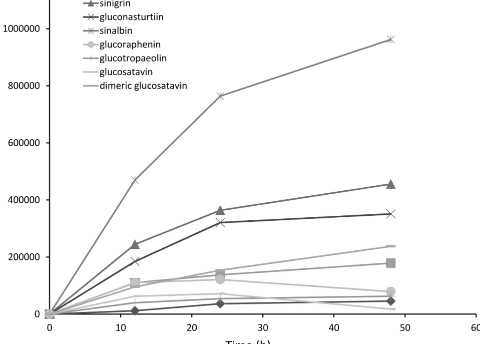
Figure 8 shows absorbance values for representative desulfoglucosinolate solutions from B. juncea, 424
S. alba, R. sativus and E. sativa extracts treated with sulfatase solution for 12, 24 or 48 hours. In 425
most cases, 12 and 24 hour incubation periods were insufficient for complete desulfation of 426
glucosinolates. Glucoraphenin decreased in all R. sativus leaf samples tested, from 24 to 48 hours, 427
while recovery of the internal standard increased, suggesting that specifically this 428
desulfoglucosinolate is degraded during the purification process (fig 8). 429
INSERT FIGURE 8 HERE 430
Not all glucosinolates are desulfated on the column at the same rate [31], meaning that incomplete 431
desulfation of extractions is likely to yield imprecise results: overestimating or underestimating the 432
final concentration of glucosinolates which are desulfated quicker or slower respectively than the 433
internal standard. In addition, relative and total concentrations of glucosinolates and degradation or 434
rearrangement of glucosinolates during this process can also affect final concentrations [26, 31]. Use 435
of higher sulfatase concentrations than outlined in the ISO method has been suggested for 436
glucosinolate analysis in B. napus and B. oleracea [25, 31]. Figure 9 compares relative glucosinolate 437
concentrations from B. juncea, S. alba, R. sativus and E. sativa purified with a low activity sulfatase 438
solution (0.3U/ml) for 12H, 24H and 48H, a high activity sulfatase solution (5U/ml) and intact 439
glucosinolates. All concentrations have been normalised to the intact glucosinolate values. 440
Desulfated glucosinolates concentrations obtained with high concentration sulfatase compared well 441
with intact glucosinolates (fig 9). However, both high sulfatase as well as low sulfatase treatments 442
yielded lower glucoraphenin content estimates. Coupled with the reduction of the recovery of 443
desulfoglucoraphenin from 24H to 48H (fig 8), these data suggest that glucoraphenin is degraded or 444
transformed during the desulfation process. 445
INSERT FIGURE 9 HERE 446
Shorter desulfation times and lower sulfatase concentrations resulted in underestimation of the 447
concentrations of glucoraphenin from R. sativus, glucoraphanin and glucosatavin from E. sativa, 448
sinigrin from B. juncea, and sinalbin from S. alba and an overestimation of the concentrations of 449
glucoraphasatin in R. sativus roots (fig 9). The overnight (12H-24H) incubation with 0.3 U/ml 450
sulfatase solution yields inaccurate results for most major glucosinolates examined in this study. The 451
ISO9167-1 method suggests that a diluted purified sulfatase solution with an activity exceeding 452
0.05U/ml should be used, which is shown to be insufficient for glucosinolate analysis from plant 453
samples and conditions examined in this study (fig 9). Instead, if a desulfation step is carried out, use 454
of a higher concentration of purified sulfatase (in this case, 5U/ml) is advised. 455
In all E. sativa leaf samples tested, recovery of monomeric desulfo-glucosatavin decreased and 456
recovery of dimeric desulfo-glucosatavin increased between 24 and 48 hours. Bennet et al. (2002) 457
previously hypothesised that dimeric glucosatavin is unlikely to be found in vivo and is probably an 458
artefact of the extraction process [22]. We can confirm that glucosatavin forms dimers as a result of 459
the desulfation step of the extraction and that without carrying this step out and instead quantifying 460
intact glucosinolates, no dimeric glucosatavin was detected in these samples. 461
Given that glucoraphenin concentration estimates are lower from methods employing a desulfation 462
step, and that this step is also responsible for the dimerization of glucosatavin, analysis of intact 463
glucosinolates is preferable in most instances. It is out of the scope of this study to compare or 464
improve separation and detection methods but it should be noted that major glucosinolates in this 465
study were accurately measured by a HPLC-UV method adapted from Herzallah and Holley (2012) 466
[19]. For examination of low abundance glucosinolates, and to avoid any potential inaccuracies due 467
to contamination it is advised that an alternative HPLC method such as those suggested in Lee et al. 468
(2013) or Forster et al. (2015) be used instead [26, 32]. 469
470
Suggested method for glucosinolate extraction: 471
Tissue disruption 472
Depending on whether freeze drying is required: 473
1a Freeze samples loosely wrapped in foil in liquid nitrogen and store at -80°C. Transport samples 474
to freeze drier in dry ice. Rapidly load samples onto a cool plate in freeze drier and ensure the 475
pressure drops to below 5 mbar in under 2 minutes. Mill samples once dried and store in airtight 476
containers in the dark. 477
478
or 479
1b Freeze 50mg samples in liquid nitrogen in 2ml eppendorf tubes and store at -80°C (for larger 480
samples use larger tubes). Add a volume of 80% methanol precooled to -20°C ensuring that final 481
methanol concentration remains above 78% according to equation (1) in materials and methods. 482
Add an appropriate volume of internal standard sinigrin or glucotropaeolin (e.g. 100µM final 483
concentration). Disrupt tissue by adding 2 small ball bearings and agitating with a tissue lyser (e.g. 484
tissuelyserII, Qiagen) for 10 minutes at 20 rev/s. Alternatively use a plastic pestle to thoroughly grind 485
the sample taking care that to keep the media below 0°C. Continue directly to 2b. 486
Extraction 487
2a For freeze dried tissue (1a). To 0.1g tissue, add 5ml of 80% methanol and 50µL of 20mM sinigrin 488
solution. Then 489
2b shake sample once and leave to stand for 30 minutes. Shake sample for a further 30 minutes 490
(70 rev/s). Centrifuge at 4000 rpm and transfer supernatant to a fresh tube. 491
Desulfation 492
If desulfation is required, a high concentration sulfatase solution should be prepared by dissolving 493
15-25mg sulfatase in 1ml 40% ethanol and centrifuge at 8000 rmp for 1 minute. Transfer 494
supernatant to a fresh 2ml eppendorf tube and add 1ml of pure ethanol to precipitate the sulfatase 495
and centrifuge at 8000 rpm for 1 minute. Discard the supernatant and air dry the pellet before re-496
dissolving in 2ml of water. Proceed with desulfation according to ISO9167-1 method. 497
498
Conclusions
499
In this study we compared different methods for extracting and purifying glucosinolates from B. 500
napus, B. junea, S. alba, E. sativa and R. sativus green tissues to highlight unnecessary or hazardous 501
steps. We have presented a simplified method for extracting glucosinolates from plant tissues which 502
does not require the use of a freeze drier or boiling methanol, and is therefore less hazardous, and 503
more time and cost effective. The presented method has been shown to have comparable or 504
improved glucosinolate extraction efficiency relative to the commonly used ISO method for major 505
glucosinolates in the Brassicaceae species studied: sinigrin and gluconasturtiin in B. juncea; sinalbin, 506
glucotropaeolin, and gluconasturtiin in S. alba; glucoraphenin and glucoraphasatin (roots but not 507
shoots) in R. sativus; and glucosatavin, glucoerucin and glucoraphanin in E. sativa. 508
Declarations
509
A 510
TDA, KR, and SEH organized the project. VK carried out sample preparation and glucosinolate 511
extractions on B. napus leaves. TDA performed all other experiments, analyzed the data, and wrote 512
the paper; SEH, IB and KR reviewed and edited the manuscript. All authors read and approved the 513
final manuscript. 514
Acknowledgements 515
We are grateful to Catherine Lilley, (University of Leeds), Peter Urwin (University of Leeds), Andy 516
Barker (Barworth Agriculture ltd.) and Helen Barker (Barworth Agriculture ltd.) for collection of leaf, 517
stem and root samples used in this study. We would also like to thank Thomas Hartley (University of 518
York) for statistical advice. We would also like to express our gratitude to the BBSRC for funding this 519
study. Finally, we would like to thank the reviewers for their time and effort in reviewing this 520
manuscript. 521
Competing interests 522
The authors declare that they have no competing interests. 523
Funding 524
This work was supported by UK Biotechnology and Biology Sciences Research Council 525
(BB/L002124/1) and (BB/K020463/1). 526
Ethics approval (NA) 527
Consent for publication (NA) 528
Availability of data and Material 529
Material used in this study is stored at the University of York and is available on request. Datasets 530
analysed in this study are available from the corresponding author on request. 531
532
References
533
[1] Clarke DB. Glucosinolates, structures and analysis in food. Analytical Methods. 2010;2(4):310-25. 534
[2] Fahey JW, Zalcmann AT, Talalay P. The chemical diversity and distribution of glucosinolates and 535
isothiocyanates among plants. Phytochemistry. 2001 Jan 31;56(1):5-1. 536
[3] Mithen RF, Dekker M, Verkerk R, Rabot S, Johnson IT. The nutritional significance, biosynthesis 537
and bioavailability of glucosinolates in human foods. Journal of the Science of Food and Agriculture. 538
2000 May 15;80(7):967-84. 539
[4] VanEtten CH, Daxenbichler ME, Wolff IA. Natural glucosinolates (thioglucosides) in foods and 540
feeds. Journal of Agricultural and Food Chemistry. 1969 May;17(3):483-91. 541
[5] Cartea ME, Velasco P. Glucosinolates in Brassica foods: bioavailability in food and significance for 542
human health. Phytochemistry reviews. 2008 Jul 1;7(2):213-29. 543
[6] Talalay P, Fahey JW. Phytochemicals from cruciferous plants protect against cancer by 544
modulating carcinogen metabolism. The Journal of Nutrition. 2001 Nov 1;131(11):3027S-33S. 545
[7] Shapiro TA, Fahey JW, Wade KL, Stephenson KK, Talalay P. Chemoprotective glucosinolates and 546
isothiocyanates of broccoli sprouts metabolism and excretion in humans. Cancer Epidemiology 547
Biomarkers & Prevention. 2001 May 1;10(5):501-8. 548
[8] Latté KP, Appel KE, Lampen A. Health benefits and possible risks of broccoli an overview. Food 549
and Chemical Toxicology. 2011 Dec 31;49(12):3287-309. 550
[9] Wiesner M, Schreiner M, Glatt H. High mutagenic activity of juice from pak choi (Brassica rapa 551
ssp. chinensis) sprouts due to its content of 1-methoxy-3-indolylmethyl glucosinolate, and its 552
enhancement by elicitation with methyl jasmonate. Food and Chemical Toxicology. 2014 May 553
31;67:10-6. 554
[10] Ngala BM, Haydock PP, Woods S, Back MA. Biofumigation with Brassica juncea, Raphanus 555
sativus and Eruca sativa for the management of field populations of the potato cyst nematode 556
Globodera pallida. Pest management science. 2015 May 1;71(5):759-69. 557
[11] Lord JS, Lazzeri L, Atkinson HJ, Urwin PE. Biofumigation for control of pale potato cyst 558
nematodes: activity of Brassica leaf extracts and green manures on Globodera pallida in vitro and in 559
soil. Journal of agricultural and food chemistry. 2011 Jun 30;59(14):7882-90. 560
[12] Mattner SW, Porter IJ, Gounder RK, Shanks AL, Wren DJ, Allen D. Factors that impact on the 561
ability of biofumigants to suppress fungal pathogens and weeds of strawberry. Crop Protection. 2008 562
Aug 31;27(8):1165-73. 563
[13] Bellostas N, Kachlicki P, Sørensen JC, Sørensen H. Glucosinolate profiling of seeds and sprouts of 564
B. oleracea varieties used for food. Scientia Horticulturae. 2007 Nov 20;114(4):234-42. 565
[14] Ratti C. Hot air and freeze-drying of high-value foods: a review. Journal of food engineering. 566
2001 Sep 30;49(4):311-9. 567
[15] ISO 9167-1, 1992 (NA 057-05-05 AA Joint committee of DIN and DGF for the analysis of fats, 568
oils and products thereof, related and primary products. (2012): Rapeseed Determination of 569
glucosinolate content Part 1: Method using high-performance liquid chromatography (ISO 9167 570
1:1992/DAM 1:2012), German version EN ISO 9167-1:1995/prA1:2012.) 571
[16] Ares AM, Nozal MJ, Bernal JL, Bernal J. Optimized extraction, separation and quantification of 572
twelve intact glucosinolates in broccoli leaves. Food chemistry. 2014 Jun 1;152:66-74. 573
[17] Church AS, Witting MD. Laboratory testing in ethanol, methanol, ethylene glycol, and 574
isopropanol toxicities. The Journal of emergency medicine. 1997 Oct 31;15(5):687-92. 575
[18] Rangkadilok N, Nicolas ME, Bennett RN, Premier RR, Eagling DR, Taylor PW. Determination of 576
sinigrin and glucoraphanin in Brassica species using a simple extraction method combined with ion-577
pair HPLC analysis. Scientia Horticulturae. 2002 Dec 6;96(1):27-41. 578
[19] Herzallah S, Holley R. Determination of sinigrin, sinalbin, allyl-and benzyl isothiocyanates by RP-579
HPLC in mustard powder extracts. LWT-Food Science and Technology. 2012 Jul 31;47(2):293-9. 580
[20] D. F. Stoin and R. D. Dogaru, Bull. USAMV-CN, 2007, 63, 77 82 581
[21] Oerlemans K, Barrett DM, Suades CB, Verkerk R, Dekker M. Thermal degradation of 582
glucosinolates in red cabbage. Food Chemistry. 2006 Mar 31;95(1):19-29. 583
[22] Bennett RN, Mellon FA, Botting NP, Eagles J, Rosa EA, Williamson G. Identification of the major 584
glucosinolate (4-mercaptobutyl glucosinolate) in leaves of Eruca sativa L.(salad rocket). 585
Phytochemistry. 2002 Sep 30;61(1):25-30. 586
[23] Ishida M, Kakizaki T, Ohara T, Morimitsu Y. Development of a simple and rapid extraction 587
method of glucosinolates from radish roots. Breeding science. 2011;61(2):208-11. 588
[24] P A K B M M D K N J B A M
589
activity in different plant samples; optimisation of measurement conditions for spectrophotometric 590
and pH-stat methods. Industrial Crops and Products. 2013 Oct 31;50:58-67. 591
[25] Wathelet JP, Mabon N, Marlier M. Determination of glucosinolates in rapeseed improvement of 592
the official HPLC ISO method (precision and speed). In Proceedings of the 10th International 593
Rapeseed Congress 1999 (p. 185). Gosford, NSW, Australia: The Regional Institute Ltd.. 594
[26] Förster N, Ulrichs C, Schreiner M, Müller CT, Mewis I. Development of a reliable extraction and 595
quantification method for glucosinolates in Moringa oleifera. Food chemistry. 2015 Jan 1;166:456-596
64. 597
[27] Troyer JK, Stephenson KK, Fahey JW. Analysis of glucosinolates from broccoli and other 598
cruciferous vegetables by hydrophilic interaction liquid chromatography. Journal of Chromatography 599
A. 2001 Jun 15;919(2):299-304. 600
[28] Wathelet JP, Iori R, Leoni O, Rollin P, Quinsac A, Palmieri S. Guidelines for glucosinolate analysis 601
in green tissues used for biofumigation. Agroindustria. 2004;3(3):257-66. 602
[29] Karathanos VT, Anglea SA, Karel M. Structural collapse of plant materials during freeze-drying. 603
Journal of Thermal Analysis and Calorimetry. 1996 Nov 1;47(5):1451-61. 604
[30] Burmeister WP, Cottaz S, Rollin P, Vasella A, Henrissat B. High resolution X-ray crystallography 605
shows that ascorbate is a cofactor for myrosinase and substitutes for the function of the catalytic 606
base. Journal of Biological Chemistry. 2000 Dec 15;275(50):39385-93. 607
[31] Hennig K, Verkerk R, Bonnema G, Dekker M. Pitfalls in the desulphation of glucosinolates in a 608
high-throughput assay. Food chemistry. 2012 Oct 15;134(4):2355-61. 609
[32] Lee JG, Bonnema G, Zhang N, Kwak JH, de Vos RC, Beekwilder J. Evaluation of glucosinolate 610
variation in a collection of turnip (Brassica rapa) germplasm by the analysis of intact and desulfo 611
glucosinolates. Journal of agricultural and food chemistry. 2013 Apr 10;61(16):3984-93. 612
613
614
615
616
618
619
Figure legends
620
621
Fig 1: A broad outline of common extraction methods used for glucosinolate analysis. Highlighted in grey is the
622
ISO 9167-1 method which was originally intended for glucosinolate extraction from B. napus seed but is
623
commonly used for glucosinolate extraction and analysis in all glucosinolate containing plant tissues.
624
625
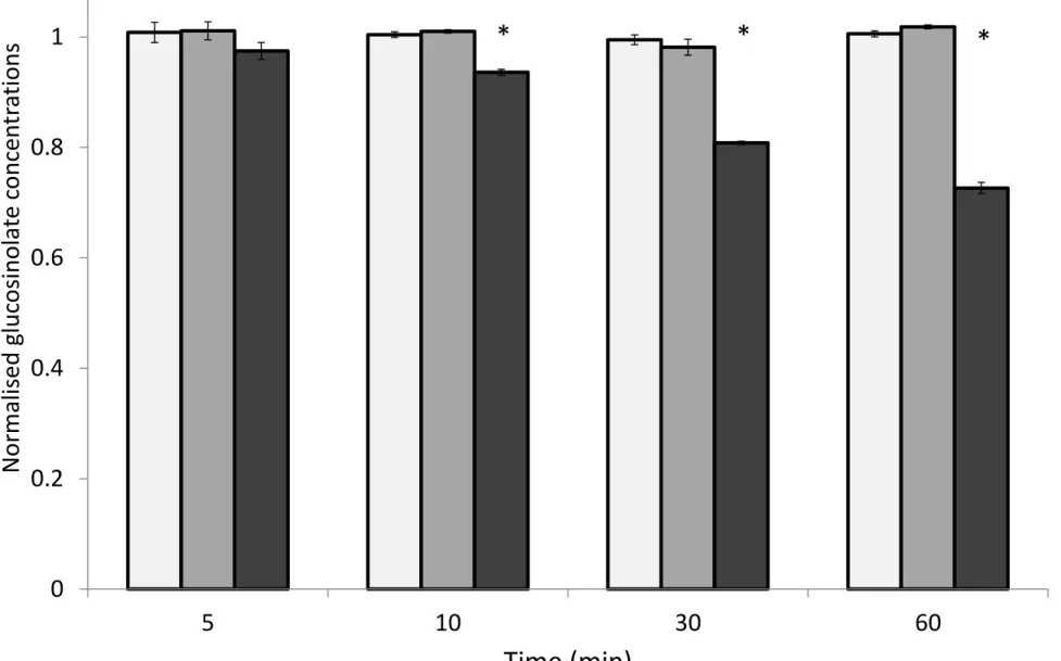
Fig 2: (a) total glucosinolate concentration of B. napus leaf halves dried in freeze drier B are significantly lower
626
(paired t-test, p=0.009) than leaf halves dried in freeze drier A; (b) B. napus leaf tissue dried with freeze drier B
627
is deformed and darker Error bars represent standard error.
628
629
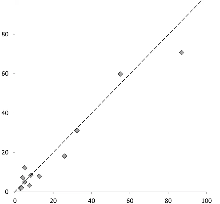
Fig 3: There is no difference in final glucosinolate concentrations between freeze drying or direct extraction in
-630
20°C methanol. B. napus leaves were cut in half and frozen. One half was freeze dried prior to glucosinolate
631
extraction, the other half was extracted directly into -20°C methanol (n=12; paired t-test, p=0.15; R squared =
632
0.96). The dashed line represents equivalence of x and y.
633
634
Fig 4: Concentrations of representative aliphatic (sinigrin) and aromatic (glucotropaeolin) glucosinolates were
635
not reduced over the course of an hour at 100°C. The representative indole glucosinolate (glucobrassicin) is
636
degraded at 100°C. Asterisks represent significant difference from concentration at t=0 (paired t-test, p<0.05).
637
638
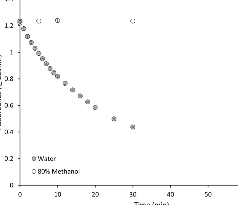
Fig 5: Spectrophotometric analysis of sinigrin hydrolysis kinetics in water and 80% methanol (n=3) by purified
639
myrosinase (0.05mg/ml) at room temperature (25°C).
641
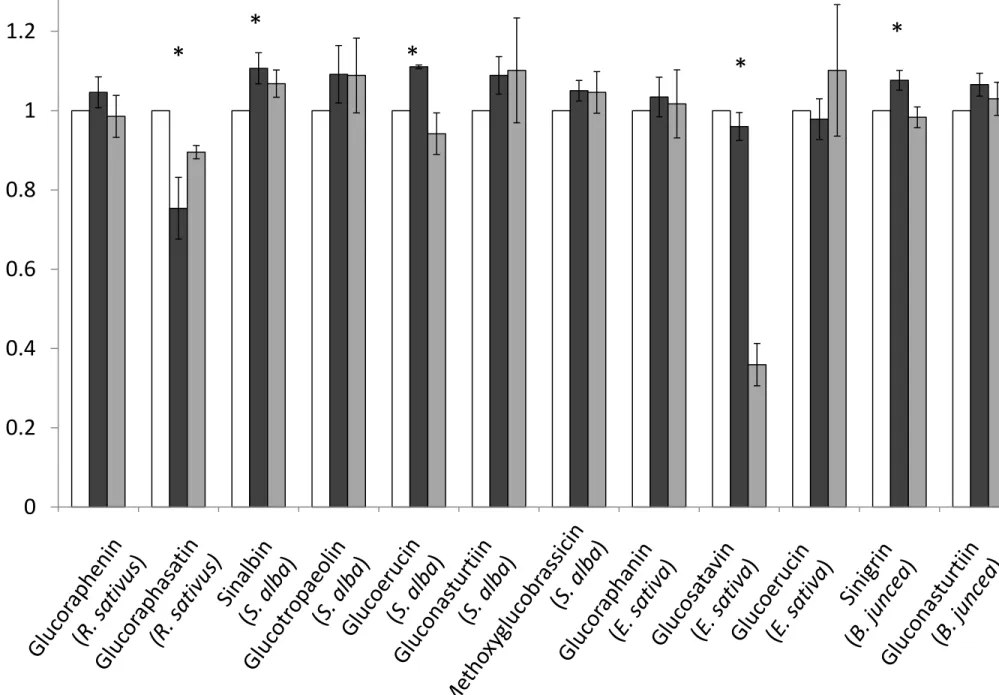
Fig 6: Extraction of glucosinolates µmol/g) in plant tissues across the three extraction methods.
642
Glucosinolate concentrations from the cold methanol and boiling water extraction methods are normalised to
643
the glucosinolate concentrations obtained from the ISO9167-1 (75°C methanol) method (n=4-12). Error bars
644
represent standard error. Asterisks represent a significant effect of extraction method on glucosinolate
645
concentration (repeat measure ANOVA, p<0.05).
646
647
Fig 7: The cold extraction method yields less glucoraphasatin in R. sativus stems relative to the ISO 9167-1
648
(boiling methanol) extraction method (n=4). Values normalised to the ISO method results. Error bars represent
649
standard error. Asterisks represent significant difference from the ISO 9167-1 (boiling methanol) method
650
(paired t test, p<0.05).
651
652
Fig 8: Absorbance values for representative desulfoglucosinolate extracts from B.juncea, S. alba, R. sativus and
653
E. sativa extracts treated with sulfatase solution for 12, 24 or 48 hours. These values are reflective of
654
desulfoglucosinolate recovery and not the initial glucosinolate concentration.
655
656
Fig 9: Desulfoglucosinolate content extracted from B. juncea, E. sativa and R. sativus tissue incubated with 75µl
657
low concentration sulfatase (0.3 U/ml) over 12H, 24H or 48H, and with a high concentration sulfatase (5U/ml)
658
over 24H, normalised to glucosinolate content of the same samples prior to sulfatase treatment. E. sativa leaf
659
samples were treated with TCEP post desulfation to undimerise didesulfoglucosatavin. Asterisks indicate a
660
significant effect of purification method on glucosinolate concen ANOVA
661
indicates the purification method yields a significantly different glucosinolate concentration relative to the
662
intact glucosinolates (paired t-test, p<0.05).
663
Plant tissue freeze dried and milled
Frozen
plant
tissue
disrupted
in -20°C
80%
methanol
Glucosinolates
extracted in 70%
methanol at 75°C
Glucosinolates
extracted in
water at 100°C
Glucosinolates
extracted 80%
methanol at RT
Extract treated with
low (0.05-0.3U/ml)
sulfatase solution
Extract treated with
high ( 0.5-1 U/ml)
sulfatase solution
Filtered without
sulfatase treatment
Tissue
disruption
Extraction
Desulfation
4 6 8 10 12 14
t
o
ta
l
g
lu
co
si
n
o
la
te
c
o
n
te
n
t
(µ
m
o
l/
g
)
A
B
(a)
(b)
20
40
60
80
100
To
tal
g
lu
co
si
n
o
la
te
c
o
n
te
n
t
f
ro
m
f
re
e
ze
d
ri
e
d
e
xt
rac
ti
o
n
(
µ
m
o
l/
g
)
0
0.2
0.4
0.6
0.8
1
1.2
5
10
30
60
N
o
rm
al
is
e
d
g
lu
co
si
n
o
lat
e
c
o
n
ce
n
tr
a
ti
o
n
s
Sinigrin Glucotropaeolin Glucobrassicin