This is a repository copy of Understanding Clostridium difficile Colonization. White Rose Research Online URL for this paper:
http://eprints.whiterose.ac.uk/134869/ Version: Accepted Version
Article:
Crobach, MJT, Vernon, JJ, Loo, VG et al. (4 more authors) (2018) Understanding
Clostridium difficile Colonization. Clinical Microbiology Reviews, 31 (2). e00021-17. ISSN 0893-8512
https://doi.org/10.1128/CMR.00021-17
© 2018 American Society for Microbiology. This is an author produced version of a paper published in Clinical Microbiology Reviews. Uploaded in accordance with the publisher's self-archiving policy.
[email protected] https://eprints.whiterose.ac.uk/ Reuse
Items deposited in White Rose Research Online are protected by copyright, with all rights reserved unless indicated otherwise. They may be downloaded and/or printed for private study, or other acts as permitted by national copyright laws. The publisher or other rights holders may allow further reproduction and re-use of the full text version. This is indicated by the licence information on the White Rose Research Online record for the item.
Takedown
If you consider content in White Rose Research Online to be in breach of UK law, please notify us by
1 Understanding Clostridium difficile colonization
Monique J.T. Crobach,a # Jonathan J. Vernon,b Vivian G. Loo,c Ling Yuan Kong,c Séverine
Péchinéd,Mark H. Wilcox,b Ed J. Kuijpera
Department of Medical Microbiology, Centre for Infectious Diseases, Leiden University
Medical Centre, Leiden, The Netherlandsa,Department of Microbiology, Leeds Teaching
Hospitals NHS Trust & University of Leeds, UKb, Division of Infectious Diseases and
Department of Medical Microbiology, McGill University Health Centre, McGill University,
Montréal, Québec, Canadac , Unités Bactéries Pathogènes et Santé (UBaPS), Université
Paris-Sud, Université Paris-Saclay, France
Running title: Clostridium difficile colonization
2 SUMMARY
INTRODUCTION
DEFINITIONS
Definition of C. difficile colonization
Assessing asymptomatic colonization
MECHANISMS OF C. DIFFICILE COLONIZATION
Disruptions in microbiota
The role of the microbiota: bile acid metabolism
The role of the microbiota: other mechanisms
The role of the immune system: innate immunity
The role of the immune system: adaptive immunity
SOURCES OF C. DIFFICILE HUMAN
ANIMAL AND ENVIRONMENTAL SOURCES OF C. DIFFICILE
Animals
Food
Environment
EPIDEMIOLOGY OF ASYMPTOMATIC COLONIZATION
Infants (0-24 months)
Children (2-16 years)
Healthy adults
Patients at admission to a hospital
Hospitalized patients
3 Healthcare workers
Duration of carriage
Ribotype specific differences
RISK FACTORS FOR C.DIFFICILE COLONIZATION
Risk factors for colonization in a community-setting
Risk factors for colonization at admission
Risk factors for acquiring C. difficile during hospital admission
Risk factors for colonization by toxigenic versus non-toxigenic strains
C. DIFFICILE COLONIZATION AND SUBSEQUENT CDI
INFECTION CONTROL AND ANTIMICROBIAL STEWARDSHIP IMPLICATIONS FOR
ASYMPTOMATIC CARRIERS
CONCLUDING REMARKS AND FUTURE DIRECTIONS
CONFLICTS OF INTEREST
REFERENCES
BIOGRAPHICAL SKETCHES
FIGURE LEGENDS
4 SUMMARY
1
Clostridium difficile is the main causative agent of antibiotic-associated and health care 2
associated infective diarrhea. Recently, there has been growing interest in alternative 3
sources of C. difficile, other than patients with Clostridium difficile infection (CDI) and the 4
hospital environment. Notably, the role of C. difficile colonized patients as a possible source 5
of transmission has received attention. In this review, we present a comprehensive 6
overview of the current understanding of C. difficile colonization. Findings from gut 7
microbiota studies yield more insights in determinants that are important for acquiring or 8
resisting colonization and progression to CDI. When discussing the prevalence of C. difficile
9
colonization among populations and its associated risk factors, colonized patients at 10
admission to the hospital merit more attention as findings from the literature have pointed 11
to their role both in health care associated transmission of C. difficile and a higher risk of 12
progression to CDI once admitted. C. difficile colonization among patients at admission may 13
have clinical implications, although further research is needed to identify if interventions are 14
beneficial to prevent transmission or overcome progression to CDI. 15
16
17
18
19
20
21
22
5 INTRODUCTION
24
Clostridium difficile is a spore-forming, gram-positive rod causing Clostridium difficile
25
infection (CDI), which may range from mild diarrhea to life-threatening pseudomembranous 26
colitis. Clostridium difficile infection has been considered as a healthcare associated 27
infection transmitted primarily from other symptomatic CDI patients. Recent studies, 28
notably based on highly discriminatory techniques like whole genome sequencing, have 29
emphasized that assumptions about the sources and transmission of C. difficile may not be 30
correct (1-3). The realization that a large proportion of CDI cases are not due to transmission 31
from other CDI cases has underlined the need to re-examine the many diverse potential 32
sources of C. difficile, and to determine their contribution to the epidemiology of this 33
disease. Paramount to our understanding is the issue of colonization of C. difficile, which is 34
the subject of this review. 35
36
DEFINITIONS 37
Definition of C. difficile colonization 38
The authors of this review define C. difficile colonization 39
in the absence of CDI symptoms and C. difficile C. difficile
40
toxin (ideally), or a toxigenic strain type, and clinical manifestations of CDI (Figure 1). Clinical 41
presentations compatible with CDI include diarrhea (defined as Bristol stool chart type 5-7, 42
plus a stool frequency of three stools in 24 or fewer consecutive hours, or more frequently 43
than is normal for the individual), ileus (defined as signs of severely disturbed bowel 44
function such as vomiting and absence of stool with radiological signs of bowel distention) 45
and toxic megacolon (defined as radiological signs of distention of the colon, usually >10 cm 46
6 However, as a previous review highlighted, definitions for CDI used in the Infectious Disease 48
Societies of America (IDSA) and European Society of Clinical Microbiology and Infectious 49
Diseases (ESCMID) guidelines differ (5-7). IDSA guidelines accept a CDI diagnosis if C. difficile
50
symptoms are identified in combination with either the presence of a toxigenic strain, free 51
toxin in the stool or histopathological evidence of pseudomembranous colitis, whereas 52
recent ESCMID guidelines require the additional exclusion of alternative etiologies for 53
diarrhea. Differences in definitions for CDI may affect the proportion of patients regarded as 54
asymptomatically or symptomatically colonized instead of having symptomatic CDI. 55
Moreover, the criteria used to define asymptomatic carriage/colonization vary considerably 56
among studies. Strict definitions of colonization have been described (8, 9), including 57
classifying asymptomatic carriers as those testing positive for C. difficile toxins but no signs 58
of CDI for 12 weeks pre- or post-specimen collection, based on a retrospective record 59
review (2). Highly restrictive definitions are difficult to apply in practice, and therefore use 60
of a simplified definition of multiple positive stools from multiple time points to determine 61
colonization has been recommended (10). In contrast, other studies utilized the less strict 62
definition of colonization as a single C. difficile positive stool and the absence of diarrhea 63
(11-13). Clearly, this has implications for who is classified as C. difficile colonized and how 64
asymptomatic cohorts are perceived as potential transmission sources. Donskey and 65
colleagues demonstrated that a single C. difficile positive fecal sample could imply either 66
colonization, transient carriage or even - (10). We thus indicate the 67
importance of further delineation of asymptomatic carriage into transient and persistent 68
colonization, as outlined in a transmission study by Curry et al. (2). Differentiating between 69
repeat, persistent detection (carriage) and point detection (colonization) would enable a 70
7 to prevent CDI spread. However, at the moment longitudinal studies on this topic are
72
lacking. 73
74
Assessing asymptomatic colonization 75
The rates of asymptomatic colonization vary considerably due to the different definitions of 76
diarrhea and laboratory methodological differences. 77
78
Standardization of the definition of diarrhea is essential, since McFarland et al. defined 79
diarrhea (14), whilst others
80
accepted the same number of loose stools, but over a single 24 hour period (12, 15). 81
Therefore, the absence of diarrhea is not synonymous with lack of loose stools, potentially 82
resulting in inconsistent designations of asymptomatic patients. 83
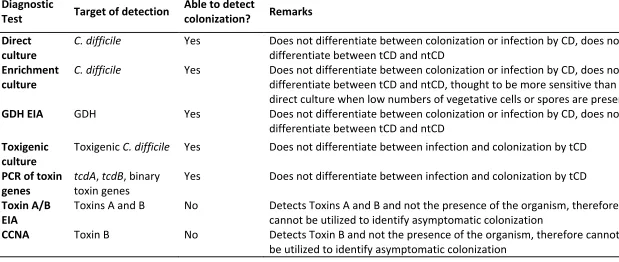
Besides the disparate definitions for diarrhea, assays or methodologies to test for CDI or C.
84
difficile colonization also vary and impact incidence rates of both conditions (13). (See Table 85
1) Methods used for CDI diagnosis can sometimes also be used for diagnosing C. difficile
86
colonization, but on the other hand, some methods used for routinely diagnosing CDI may 87
falsely classify colonized patients with diarrhea (due to a non-C. difficile cause) as CDI 88
patients. 89
Despite its labor intensive and time consuming characteristics and susceptibility to toxin 90
degradation in stool samples with incorrect storage, cell cytotoxicity neutralization assay 91
(CCNA) is frequently considered as the gold standard for CDI due to its high specificity and 92
direct detection of the main virulence factor (toxin) (16, 17). However, as CCNA detects C.
93
difficile toxins and not the presence of the organism itself, its utility is limited in detecting C.
94
8 been used to consider these infants as C. difficile colonized (18), indicating the aberrant 96
association between toxin presence and clinical symptoms in this age group. 97
An alternative gold standard for CDI is toxigenic culture, which includes culture of the 98
organism followed by detection of its in vitro toxin producing capacity by toxin enzyme 99
immunoassay (Tox A/B EIA), CCNA or detection of the toxin genes by nucleic acid 100
amplification test (NAAT). A major study by Planche et al. of greater than 12,000 fecal 101
specimens highlighted no increase in mortality in patients harboring a toxigenic C. difficile
102
strain without the presence of detectable toxin (19), suggesting that free toxin positivity 103
reflects CDI, while toxigenic culture positivity encompasses some patients with colonization. 104
Therefore, the use of toxigenic culture to diagnose CDI could lead to an over-diagnosis of 105
CDI and hence an underestimation of C. difficile colonization. However, if the goal is 106
detection of toxigenic C. difficile colonization in asymptomatic patients, toxigenic culture is a 107
suitable option. 108
As both gold standard methods for diagnosing CDI are time-consuming and laborious, rapid 109
assays are more appealing for CDI testing in daily practice. When rapid assays are used to 110
test for CDI, it is recommended to use them in an algorithm in order to optimize positive 111
and negative predictive values. Concerning the relationship between free toxins and true 112
disease as described above, the algorithm should include a Tox A/B EIA to test for free 113
toxins in stool. However, in clinical practice, rapid assays and especially NAATs, are often 114
used as stand-alone test instead of as part of an algorithm, and this may again lead to C.
115
difficile colonization being erroneously classified as CDI. A study by Polage et al. 116
demonstrated that 39.9% of NAAT positive specimens tested negative for toxin by cell 117
9 diagnosis of CDI and consequently an underestimation of asymptomatic colonization, similar 119
to the situation described above for TC. 120
121
There are some specific limitations that have to be taken into account when assessing C.
122
difficile colonizationIn C. difficile colonization, bacterial loads can be lower than in CDI. 123
Direct culture of the organism is quite sensitive, although detection rates will differ as the 124
sensitivity of the culture media varies. Nonetheless, culture-independent detection 125
techniques, such as enzyme immunoassays, have lower sensitivity and specificity than 126
culture methods. As stools with lower counts of C. difficile could be deemed falsely negative, 127
these assays may lead to underestimation of the asymptomatic colonization rates, making 128
them less suitable for detection of colonization. For example, glutamate dehydrogenase 129
(GDH) screening is regarded as highly specific for detection of C. difficile in clinical 130
specimens (7, 21); however, potential issues have been highlighted with the use of this 131
methodology for reporting asymptomatic colonization (22). In a study by Miyajima et al., 132
only one out of five positives determined by an enrichment culture method was positive by 133
GDH assay (22), probably due to low levels of GDH antigen in non-diarrheal stools, below 134
the lower limits of detection for this assay. 135
136
As the above illustrates, the diagnosis of CDI should not be based on laboratory results 137
alone, but should always be supported by clinical signs and symptoms suggestive of CDI (7, 138
23). This is especially important when methodologies which cannot discern CDI from 139
colonization (stand-alone NAAT, TC) are applied in routine CDI testing. 140
Likewise, we suggest that an optimal diagnostic method for the determination of 141
10 symptoms (i.e. absence of diarrhea, ileus and toxic megacolon per the criteria described 143
above), or the presence of an alternative explanation for these clinical symptoms. The 144
laboratory methods should include (enrichment) stool culture and either toxigenic culture 145
or PCR confirmation. This combination of sensitive techniques, although expensive, will yield 146
more reliable data and support inter-study comparisons. 147
148
MECHANISMS OF C. DIFFICILE COLONIZATION 149
After having defined C. difficile colonization, a closer look at mechanisms that underlie C.
150
difficile colonization is needed. Key factors in acquiring or resisting colonization (and 151
subsequent infection) are the gut microbiota and the host immune response against C.
152
difficile. 153
154
Disruptions in microbiota 155
The gut microbiota has a prominent role in the whole life cycle of C. difficile from 156
germination and colonization to establishing symptomatic disease. Results from studies on 157
the differences in microbial composition in patients with CDI, asymptomatic carriers and 158
non-infected patients can elucidate which alterations determine either the susceptibility to 159
colonization and/or disease development or colonization resistance (defined as the 160
resistance to colonization by ingested bacteria or inhibition of overgrowth of resident 161
bacteria normally present at low levels within the intestinal tract) (24, 25). The optimal 162
method to study the impact of the microbiota in spore germination, colonization and toxin 163
production by C. difficile would be to take luminal samples and biopsies to study both 164
microbiota attached to the intestinal wall and present in the lumen, as C. difficile was 165
11 CDI gut model (26, 27). Also, ideally samples should be examined from different locations 167
along the intestine, because it was demonstrated that in mice, C. difficile spores did 168
germinate and grow in ileal contents, while this was not possible in cecal contents unless 169
the mice had been treated with specific antibiotics (28). Obtaining these samples in human 170
subjects is not feasible, though ingestible remotely controlled capsules that are capable of 171
taking samples from the small intestinal tract are in development. However, most human 172
studies use easy-to-obtain fecal samples for analyzing the intestinal microbiota, although 173
these may actually not optimally reflect the microbial composition in the more proximal 174
intestine where bile acid induced germination of the ingested spores occurs (see below). 175
176
Alterations in gut microbial composition that have been described for CDI patients include a 177
lower species richness and lower microbial diversity compared with healthy controls (29-178
31). Between samples from CDI patients, a greater heterogeneity was observed than 179
between individual samples from healthy controls (31). At the phylum level, Bacteroidetes
180
were less prevalent in CDI patients than in healthy controls, while there was an increase in 181
Proteobacteria. Within the Firmicutes phylum, a decrease in the Clostridia, especially from 182
the Ruminococcaceae and Lachnospiraceae families and butyrate-producing anaerobic 183
bacteria from Clostridium clusters IV and XIVa was noted in CDI patients (31). In addition to 184
these depletions, increases in the orders of the Enterobacteriales and Pseudomonales
185
(Proteobacteria) and Lactobacillales (Firmicutes) were observed (30, 31). Also, in human 186
fecal samples collected prior to onset of a first CDI episode, a decreased diversity, a 187
decrease in the phylum Bacteroidetes and changes within the phylum Firmicutes (a decrease 188
in Clostridiales Incertae Sedis XI and an increase in Enterococacceae from the order 189
12 not develop CDI (32). A reduction in the family Clostridiales Incertae Sedis XI in these
191
samples was demonstrated to be independently associated with CDI development . 192
Moreover, changes in microbial composition comparable to those found in CDI patients 193
have been described for patients with nosocomial diarrhea who tested negative for 194
C.difficile or its toxins. These changes included a comparable decrease in species richness 195
and microbial diversity and again a decrease in butyrate producing bacteria from the 196
Ruminococcaceaea and Lachnospiraceae families in comparison to healthy controls (30, 31, 197
33). This may indicate that patients with nosocomial diarrhea not due to CDI are also 198
susceptible to development of CDI once they are exposed to C. difficile spores. It also 199
suggests that the CDI itself did not much alter the gut microbial composition (31). Among 200
mice that were given clindamycin to render them susceptible to CDI development, luminal 201
samples and biopsies generally confirm the findings in humans and demonstrate a 202
decreased species richness (34). Mice without antibiotic pre-exposure, and therefore 203
undisturbed microbiota, do not develop CDI symptoms after administration of C. difficile
204
spores (34). Also, in mice with CDI a microbiota dominated by Proteobacteria was 205
demonstrated, instead of a Firmicutes and Bacteroidetes dominated microbiota as found in 206
healthy mice (34, 35). 207
208
Alterations in gut microbial composition in C. difficile carriers are less well described, but 209
may give more insight in the mechanisms that allow for colonization whilst protecting 210
against the development of overt disease. One of the few available studies reports a 211
decreased species richness and decreased microbial diversity not only in samples from 8 CDI 212
patients but also in samples from 8 asymptomatic carriers, compared to 9 healthy subjects 213
13 CDI patients and carriers and therefore it is suggested that the absence or presence of 215
certain bacterial taxa is more important in determining the development of CDI or C. difficile
216
colonization than the diversity of species richness alone. In carriers, fewer Proteobacteria
217
and a higher proportion of Firmicutes and Bacteroidetes were found than in CDI patients 218
and so this distribution resembled that of healthy individuals more (29). Another study 219
among 98 hospitalized patients (including 4 CDI patients and 4 C. difficile colonized patients) 220
showed that, compared with CDI patients, a higher level of Clostridiales Family XI Incertae
221
Sedis, Clostridium or Eubacterium was found just before C. difficile colonization was 222
detected, also supporting the notion that the presence of certain bacterial taxa is important 223
to prevent overgrowth or progression from colonization to overt infection (36). Evidence 224
from murine studies also indicates that colonization with certain bacterial taxa may prevent 225
the progression from colonization to CDI; mice precolonized with a murine Lachnospiracea
226
isolate showed significantly reduced C. difficile colonization (37). Similarly, administration of 227
Clostridium scindens in antibiotic-treated mice is associated with resistance to CDI (38). 228
Moreover, in antibiotic-exposed mice who were challenged with C. difficile spores, different 229
patterns in microbiota composition were seen in those that developed severe CDI 230
symptoms versus animals who became only C. difficile colonized (35). In the first group, a 231
shift towards Proteoabacteria was noted, while the latter group had a microbiota that was 232
dominated by Firmicutes (including Lachnospiraceae) resembling that of mice who had not 233
been exposed to antibiotics. The presence of a Firmicutes dominated microbiota seemed to 234
be protective against the development of clinical symptoms in this experiment (35). 235
Interestingly, a recent longitudinal study in a C. difficile colonized infant showed important 236
changes in microbiota composition during weaning. An increase in the relative abundance of 237
14 noted suggesting that these bacterial genera likely account for the expulsion of C. difficile
239
(39). 240
241
In conclusion, there are only a few studies on the intestinal microbiota in patients with 242
asymptomatic C. difficile colonization, which are also very limited in sample sizes. However, 243
these studies and findings from mice studies support the idea that a decreased species 244
richness and decreased microbial diversity appear to allow for colonization, although the 245
presence of certain bacterial taxa seems to protect from progression to CDI. Mechanisms by 246
which the microbiome and in particular the presence of certain bacterial taxa may offer 247
colonization resistance and protection against infection will be described below. 248
249
The role of the microbiota: bile acid metabolism 250
The first step in establishing C. difficile colonization is the germination of spores. Primary 251
bile acids are known to stimulate this germination process (40). The physiological function 252
of primary bile acids is to assist in digesting fat. To be able to do so, after being produced in 253
the liver, primary bile acids are released into and reabsorbed from the small intestine. 254
However, a small amount of the primary bile acids is not reabsorbed and is passed into the 255
colon. In the colon, these primary bile acids are metabolized into secondary bile acids by 256
certain members of the normal gut microbiota. Secondary bile acids inhibit C. difficile
257
growth (40). The capacity to metabolize primary bile acids into secondary bile acids by the 258
-dehydroxylating enzymes is shown in members of the 259
Lachnospiraceae, Ruminococcaceae and Blautia families, all belonging to the phylum 260
Firmicutes (28, 41). A disruption in the intestinal microbiota and depletion of Firmicutes may 261
15 was shown in antibiotic-treated mice, where loss of members of the Lachnospiraceae and 263
Ruminococcaceae families was found to be correlated to a significant loss of secondary bile 264
acids (28). More specifically, this was also shown for one of the members of the 265
Lachnospiraceae family, C. scindens; the administration of this bacterium was shown to 266
restore physiological levels of secondary bile acid synthesis (38). Loss of secondary bile acids 267
and an increase in primary bile acids creates a favorable environment for C. difficile. Support 268
for the role of bile acid metabolism in this susceptibility to C. difficile colonization is 269
obtained from both in vitro and in vivo studies. In vitro, spores are able to germinate in the 270
presence of bile acids concentrations found in feces of CDI patients; however, spore 271
germination and vegetative growth was inhibited in the presence of bile acids at 272
concentrations found in patients after fecal microbiota transplant (FMT) or in mice resistant 273
to C. difficile (28, 42). In vivo significantly higher levels of primary bile acids and lower levels 274
of secondary bile acids were found in feces from CDI patients compared with controls, 275
especially in patients with a recurrent CDI episode (43). Notably, the amount of germination 276
in response to bile acids seems to vary between strains, which may be related to mutations 277
in the CspC germinant receptor (called CspC) that recognizes the primary bile acids (42). A C.
278
difficile mutant completely deficient for the CspC receptor gene was demonstrated to cause 279
less severe clinical symptoms in a hamster model (40). 280
281
The role of the microbiota: other mechanisms 282
Apart from the altered bile acid composition, other mechanisms also induced by disruptions 283
of the microbiota are suggested to play a role in conferring susceptibility to C. difficile. 284
First, disruptions in the microbiota that lead to a diminished production of short chain fatty 285
16 carbohydrates mainly by Lachnospiraceae and Ruminococcaceae, the families that were less 287
abundant in CDI patients and carriers. They may have effect on colonization resistance 288
through reducing the luminal pH (and thereby creating an unfavorable environment for C.
289
difficile) (44) and stimulating the defense barrier as one of the SCFAs (butyrate) is the main 290
energy source of the gut epithelium (45, 46). Amino acids may also play a role in the 291
susceptibility to C. difficile colonization, as they can enhance germination in the presence of 292
secondary bile acids and may influence the immune system. Moreover, the digestion of 293
carbohydrates in the gut results may impact susceptibility for CDI development. 294
Bacteroidetes are mainly responsible for this carbohydrate digestion which results in 295
production of substrates essential for homeostasis of colonocytes (47). A reduction in 296
Bacteroidetes may therefore negatively impact colonic health. 297
Besides the indirect mechanisms described above, the microbiota may also have direct 298
resistant mechanisms against C. difficile. These include competition for niches and nutrients 299
and the production of antimicrobials (48, 49). 300
301
The role of the immune system: innate immunity 302
The precise protective factors of the innate immunity that prevent colonization and 303
progression to CDI are unknown, but are probably less important than the role of the 304
microbiota and bile acid metabolism. Virulence factors of C. difficile induce a rapid innate 305
immune response resulting in an inflammatory response which is necessary to induce 306
adaptive immunity. 307
CDI is characterized by a severe intestinal inflammatory response in which neutrophils 308
17 response (50). After epithelial barrier disruption, TcdA and TcdB trigger inflammatory 310
signaling cascades through activation of NF-kB, AP-1 and inflammasome, and stimulate 311
production of pro-inflammatory cytokines and chemokines in epithelial cells. This promotes 312
the recruitment of immune cells including neutrophils and induces the production of 313
defensins. Surface proteins also trigger an innate immune response. Challenge of 314
macrophages with C. difficile surface proteins (surface layer proteins, SLPs) leads to pro-315
inflammatory cytokine production such as TNF- IL- IL-8 (51). 316
Additionally, C. difficile SLPs interact in vitro with TLR4 leading to dendritic cell (DC) 317
maturation, robust Th1 and Th17 responses with production of IFN and IL-17, and a weak 318
Th2 response leading to antibody production (52). Ryan et al. showed that TLR4 and myeloid 319
differentiation primary-response protein 88 (MyD88) deficient mice were more prone to C.
320
difficile infection (53). C. difficile flagellin FliC also activates an innate immune response via 321
its interaction with TLR5 inducing predominantly activation of p38 MAPK and, to a lesser 322
extent NF-B, resulting in up-regulation of the expression of pro-inflammatory cytokine 323
genes and the production of pro-inflammatory factors (54, 55). In vivo, Batah et al. showed 324
a synergic effect of C. difficile flagellin and toxins in inducing mucosal inflammation (56). 325
In summary, the innate immune response induces an inflammatory response which 326
promotes an adaptive immune response with memory and long-lasting immunity (see 327
below), but its effects on C. difficile colonization are unknown. 328
18 The role of the immune system: adaptive immunity
330
The adaptive immunity against colonization or CDI has mainly been studied for its antibody-331
mediated response whereas the role of the cell-mediated immune response remains 332
unknown. 333
Serum antibodies against somatic antigens and surface components have been found in 334
asymptomatic carriers and patients recovered from CDI (57, 58), which suggests that surface 335
proteins induce an immune response and modulate disease outcome. Vaccination assays 336
with these proteins have been performed in animal models. Parenteral or mucosal 337
vaccination with the S-layer proteins led to specific antibody production but only partial 338
protection in the hamster model (59, 60). Immunization studies that were performed in 339
animals with Cwp84 and the flagellar proteins FLIC and FliD by mucosal route resulted in a 340
significant decrease in intestinal C. difficile colonization in the mouse model and partial 341
protection in the hamster model (61, 62). Likewise, Ghose et al. immunized mice and 342
hamsters intra-peritoneally with FliC adjuvanted with alum, inducing a high circulating anti-343
FliC IgG response in animal sera, full protection in mice against a clinical 072/NAP1 strain, 344
but only partial protection in hamsters against 63 (63). All these results suggest 345
that antibodies against C. difficile surface proteins have a protective role against 346
colonization. At the moment, studies with surface protein-based vaccines to prevent 347
colonization in humans are lacking. 348
Antibodies to TcdA and TcdB do not protect from colonization, but influence disease 349
susceptibility and subsequently the progression from colonization into CDI. Kyne et al. 350
studied anti-TcdA IgG antibody levels in patients who became colonized after C. difficile
351
19 increases in anti-TcdA IgG antibodies than patients who progressed from colonization to CDI 353
(64). 354
Monoclonal antibody (Mab)-based passive immunotherapy directed to toxins was able to 355
protect hamsters from CDI. In humans, two Mabs, one targeting TcdA (actoxumab) and 356
another targeting TcdB (bezlotuxumab) were tested in human clinical trials aimed at the 357
prevention of recurrent disease (65). Bezlotoxumab prevented approximately 40% of the 358
recurrences. A recently published hypothesis suggested that this reduction in recurrences is 359
presumably due to limiting epithelial damage and facilitating rapid microbiome recovery 360
(66), suggesting that reduced (re)colonization may be an important factor, although this 361
should be explored further. Currently, two pharmaceutical firms (Pfizer and Valneva) have 362
vaccine clinical trial development programmes with the two toxins (toxoids or toxin 363
fragments) but no colonization factors as antigens (67); Sanofi Pasteur has recently 364
announced the cessation of its vaccine development programme, which was also based on 365
toxin antigens alone. Therefore these vaccines protect against the toxic effects of C. difficile
366
on the intestinal mucosa, and can thereby hinder the progression from colonization to CDI. 367
368
In conclusion, a rapid innate immune response induces adaptive immunity to CDI, of which 369
the antibody-mediated response is best understood. Antibodies against C. difficile surface 370
proteins are thought to protect against colonization, while antibodies against C. difficile
371
toxins protect against disease, directly by its toxin neutralizing effect and possibly also 372
indirectly by limiting epithelial damage and restoring colonization resistance. 373
20 SOURCES OF C. DIFFICILE - HUMAN
375
Patients with CDI can shed C. difficile not only during the diarrheal episode, but also after 376
completion of therapy. In a study of 52 patients receiving CDI treatment, samples from 377
stool, skin and environmental sites were cultured for C. difficile before treatment, every 2-3 378
days during treatment and weekly after therapy was completed (68). Prior to treatment, 379
100% of stool samples and approximately 90% of skin and environmental samples were 380
culture positive for C. difficile. Stool cultures became C. difficile negative in most patients by 381
the time diarrhea resolved at a mean 4.2 days. However, at the same time, skin and 382
environmental contamination with C. difficile remained high at 60% and 37% respectively. In 383
addition, stool detection of C. difficile was 56% at 1-4 weeks post treatment among 384
asymptomatic patients recovering from CDI. Moreover, 58% had skin contamination with C.
385
difficile 1-4 weeks after completion of treatment and 50% had sustained environmental 386
shedding. Persistent skin and environmental contamination was associated with receipt of 387
additional antibiotic therapy. Prior to treatment, the mean density of C. difficile in stool 388
samples was significantly higher than at the time that the diarrhea resolved, at end of 389
treatment and at 1-6 weeks post treatment. This study highlights that patients with CDI can 390
be a source of C. difficile spores and that they can potentially transmit C. difficile to other 391
patients even after diarrhea has resolved. In addition, similar to animal models, continued 392
antibiotic treatment can trigger patients, in which there is C.
393
difficile overgrowth and excretion of high concentrations of spores (69). 394
395
CDI was historically regarded as a healthcare associated infection transmitted primarily 396
(directly or indirectly) by symptomatic patients, but a growing body of evidence 397
21 One study, using MLST (Multi Locus Sequence Typing) could link only 25% of patients with 399
symptomatic CDI to a previously identified CDI patient (1). A follow-up study of the same 400
large patient cohort (>1200 cases) used whole genome sequencing and was able to link at 401
most only 55% (and more likely only 35%) of new cases to previous patients with CDI (3). A 402
much smaller study (~50 cases) using MLVA (Multiple-Locus Variable number tandem repeat 403
Analysis) found that only 30% of new cases could be linked to previously identified cases (2). 404
One could argue that the inability to link new cases to previous ones might be caused by 405
patients with CDI who are clinically undetected. However, strict criteria were used to 406
determine which samples should be tested for CDI in the large UK study (1, 3); although a 407
toxin EIA was used, which is not as sensitive as a reference test, repeat sampling was carried 408
out according to clinical suspicion of CDI. Depending on the reference test used, the 409
sensitivity of toxin EIA is approximately 60-85%, which means that 15-40% of patients with 410
CDI may go undetected. Nonetheless, this does not account completely for the 45 to 75% of 411
cases that were not closely linked to symptomatic patients (1, 3). This raises the question of 412
what is/are the source(s) accounting for approximately half of new CDI cases? Curry et al. 413
examined patients for C. difficile carriage who were selected to undergo screening for 414
vancomycin-resistant enterococci. They found that 29% of CDIs could be linked to 415
asymptomatic C. difficile carriers (2). 416
417
As asymptomatic carriers and the associated shedding of spores usually goes undetected 418
because of lack of routine screening, they can play a role in spread of C. difficile to the 419
environment and other patients. Although transmission events from one individual 420
asymptomatic carrier may be rare, as was shown in a relatively small study (15), 421
22 they likely outnumber symptomatic CDI patients. A recent study showed that 2.6% of
423
patients who were not exposed to C. difficile colonized patients developed CDI, while this 424
percentage increased to 4.6% in patients who were exposed (70). Unfortunately, however, 425
the case definition of CDI in this study was based on detection of toxin gene rather than 426
toxin, and so over-diagnosis of true cases likely occurred. Asymptomatic carriers who are 427
colonized at admission appear to contribute to sustaining transmission in the ward. Already 428
in 1992, it was recognized that C. difficile strains introduced to the ward by asymptomatic 429
carriers were important sources of onwards health care associated transmission (71), 430
although definitive proof of linkage was hampered by use a non-specific typing technique. 431
More recently, using an epidemiological model of C. difficile transmission in healthcare 432
settings, Lanzas et al. confirmed that patients colonized on admission likely play a significant 433
role in sustaining ward based transmission (72). 434
435
ANIMAL AND ENVIRONMENTAL SOURCES OF C. DIFFICILE 436
Animals 437
Similar to humans, CDI or asymptomatic carriage can occur among domestic, farm and wild 438
animals (73-80). Carriage rates in these studies range from 0-100%. These varied observed 439
rates may be related to different culture methodologies and different study settings. Much 440
of this subject has been reviewed in this journal but new information has emerged on 441
possible transmission from domestic and farm animals (81, 82). 442
443
C. difficile can cause diarrhea in domestic companion animals such as dogs and cats, but 444
asymptomatic transient carriage of C. difficile by household pets is common (11-40%) (73, 445
23 within the same household. A recent study examined the potential for transmission to pets 447
from 8 patients with recurrent CDI (85), but in this study C. difficile was not found in any of 448
the pets. In contrast, Loo et al. studied 51 families with 15 domestic pets that included 9 449
cats, 5 dogs and 1 bird (86). During follow-up visits, toxigenic C. difficile was found in 450
cultures of 2 cats and 2 dogs. Probable transmission occurred in 3 of the 15 domestic pet 451
contacts. None of the domestic pets had diarrhea. Typing by pulsed-field gel electrophoresis 452
showed that the profiles of all 4 domestic pet isolates were indistinguishable or closely 453
related to those of their respective index patients. It is conceivable that household pets can 454
serve as a potential source of C. difficile for humans. 455
456
Transmission from farm animals to humans has been examined using whole genome 457
sequencing using 40 Australian ribotype 014/NAP4 isolates of human and porcine origin 458
(87). A clonal relationship with one or more porcine strains was demonstrated among 42% 459
of human strains underscoring potential interspecies transmission. Similar findings were 460
obtained in a study on 65 C. difficile 078/NAP7 isolates collected between 2002 and 2011 461
that included 12 pairs of human and pig isolates from 12 different pig farms (88). Five 462
(41.7%) of the 12 farmer-pig pairs were colonized with identical and nearly identical C.
463
difficile clones (88); the remaining 7 (58.3%) farmer-pig pairs were not clonal suggesting 464
exposure to different sources such as the environment. 465
466
Food 467
With reports that C. difficile can be detected among farm animals, studies of C. difficile
468
detection in retail food products appeared. 469
24 Studies from Canada and the United States report that C. difficile can be recovered from 471
retail meat including ground beef, ready to eat beef, ground pork, ground turkey, pork 472
sausage, summer sausage, pork chorizo and pork braunschweiger, with prevalences ranging 473
from 20-63% (89-92). 474
However, the prevalence of C. difficile in retail meat products was lower in European 475
countries, ranging from 0-6.3% (93-95). The observed differences in prevalence of C. difficile
476
culture positivity in retail meats in North American and Europe is striking. This may be 477
related to seasonal and temporal changes, or may be true observed geographical 478
differences. 479
480
Using both quantitative and enrichment culture, Weese et al. sought to provide a measure 481
of the degree of contamination from 230 samples of retail ground beef and pork (96). C.
482
difficile was isolated from 28 (12%) and notably, approximately 70% of samples were 483
positive by enrichment culture only. Among the samples that were positive on direct 484
culture, the concentration of spores ranged from 20 to 240 spores/gram. Although the 485
infectious dose of C. difficile is not known, these findings suggest that although C. difficile
486
can readily be recovered from retail meat products, the concentration of C. difficile spores is 487
low. 488
489
Stabler et al. investigated the MLST profiles of 385 C. difficile isolates from human, animal 490
and food sources and from geographically diverse regions (97). Animal and food strains 491
were associated with the ST-1 and ST-11 profiles and these strains have been associated 492
with CDI outbreaks in humans. Although the majority of C. difficile isolates recovered from 493
25 isolates, there have not been any human CDI cases that have been confirmed to be
495
foodborne in origin. 496
497
Environment 498
C. difficile spores can survive in the environment for months or years due to their resistance 499
to heat, drying, and certain disinfectants. Within hospitals, the surface environment is 500
frequently contaminated with C. difficile. C. difficile has been cultured from many surfaces 501
including floors, commodes, toilets, bed pans and high-touch surfaces such as call bells and 502
overbed tables (14, 98). The frequency of environmental contamination depends on the C.
503
difficile status of the patient: fewer than 8% of rooms of culture-negative patients, 8-30% of 504
rooms of patients with asymptomatic colonization and 9-50% of rooms of CDI patients were 505
found to be contaminated with C. difficile, respectively (14, 99, 100). 506
507
To examine environmental sources outside of the healthcare milieu, Al Saif and Brazier 508
undertook a large study of 2580 samples in Cardiff, South Wales from various sources 509
including water, domestic and farm animals, soil, raw vegetables, surface samples from 510
healthcare facilities, veterinary clinics and private residents (101). One hundred and eighty-511
four (7.1%) samples were positive. Water samples gave the highest yield of culture positivity 512
at 36%, followed by soil at 21% and healthcare environments at 20%. C. difficile was found 513
in 59% of lawn samples collected in public spaces in Perth, Australia and toxigenic ribotypes 514
014/NAP4 and 020/NAP4 were predominant (102). A Canadian study demonstrated that C.
515
difficile was found in 39% of sediments sampled from rivers connected to the discharge 516
effluent pipe of waste water treatment plants (103). The most common PCR ribotype was 517
26 519
In summary, C. difficile has been isolated from animals, retail food and the environment. 520
Using ribotyping and whole genome sequencing techniques, there appears to be 521
interspecies and environmental transmission but the directionality of the transmission 522
remains to be elucidated. 523
524
EPIDEMIOLOGY OF ASYMPTOMATIC COLONIZATION 525
After having discussed possible sources of C. difficile and underlying mechanisms of 526
colonization, a description of the epidemiology of colonization, including the prevalence of 527
colonization rates among different populations, is essential. 528
529
Infants (0-24 months) 530
Asymptomatic colonization rates in neonates and infants (<2 years) are widely reported as 531
high, but range between 4-71% (18, 104-108). Although the clinical relevance of C. difficile
532
colonization in infants is considered as less significant, due to low rates of disease in this 533
population (109), its potential as a transmission reservoir for adult populations remains. 534
535
An early study researching the prevalence of C. difficile in the neonate population found 536
that approximately 30% of all newborns were asymptomatically colonized within their first 537
month of life (18). However, these data included four specimens deemed positive with no 538
identifiable organism, only toxin. Nonetheless, the transient nature of colonization at this 539
early stage was highlighted with only 4 of 10 babies who were culture positive in the first 540
week of life remaining positive at 14 and 28 days. A more recent review corroborated these 541
27 approximately 35% of infants under one year of age (105). This large-scale analysis suggests 543
that colonization peaks between 6-12 months, before substantially decreasing towards 544
adult rates. Although this major review provides a valuable assemblage of data, the 545
variability across methodologies used by the included studies should be taken into 546
consideration. 547
548
Geographical differences in infant colonization rates have been identified, with one study 549
indicating a variance of 4-35% across Estonian and Swedish infant populations respectively 550
(108). The colonization rate was inversely associated with an elevated presence of inhibitory 551
Lactobacilli in Estonian subjects, which may be determined by variation in diet and 552
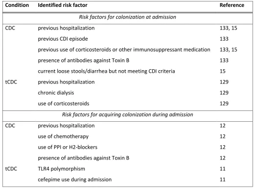
environmental exposure. A US study of hospitalized infants demonstrated a 20% 553
colonization rate (110) whereas Furuichi et al. found no evidence of C. difficile colonization 554
amongst Japanese newborns (111). However, the Japanese data were based on culture only, 555
with no attempt to utilize EIA or NAAT to detect low levels of organism. These studies 556
emphasize the variable epidemiology amongst diverse geographical populations. 557
558
The source of infant colonization is uncertain, with suggestions that the presence of C.
559
difficile in the urogenital tract implicated vaginal delivery as a potential route of 560
transmission to neonates (112). However, later work contradicted this suggestion, failing to 561
detect any C. difficile positive vaginal swabs from post-partum mothers (18, 104). Molecular 562
analysis of both infant and environmental isolates demonstrate likely acquisition from 563
environmental sources and patient to patient transmission (113). 564
28 Infants are rarely diagnosed with CDI. Bolton and colleagues found that almost 50% carried 566
toxin positive strains, but showed no sign of diarrhea, suggesting that although the relevant 567
toxin genes may be present, they may be minimally (or not) expressed and so fail to cause 568
disease; alternatively, absent or immature toxin receptors may explain the infrequency of 569
CDI despite high colonization rates (18). However, understanding toxigenic strain 570
colonization rates may provide a greater insight into the relevance of this population as a 571
reservoir for transmission to adults. Isolates from infants have shown predominance of 572
ribotypes associated with CDI (106). Adlerberth et al. found that 71% of colonized infants 573
had toxigenic strains with more than half identified as ribotypes 001/NAP2 and 014/NAP4 574
that can cause endemic CDI (114). A comparison of C. difficile strains in children (<30 575
months) with those circulating in the ad CDI 576
institution, determined nine shared sequence types among the 20% asymptomatic pediatric 577
subjects (115). This may further implicate infants as a potential reservoir for C. difficile
578
dissemination; nonetheless, no direct transmission events were documented in this limited 579
pilot study. Potential community-based transmission from infant carriers to the adult 580
population was alluded to in a longitudinal study demonstrating colonization in all 10 infants 581
at some point in the first year of life, with 3 infants colonized for 4-9 months (116). 582
583
Children (2-16 years) 584
Meta-analysis of studies examining pediatric C. difficile epidemiologyreported 585
asymptomatic colonization in children older than 1 year at 15%, with prevalence reducing to 586
5% in those greater than 2 years of age (117). One explanation for the reduction in 587
colonization rates after infancy is that by 12 months the distribution of gut flora begins to 588
29 Nonetheless, contemporaneous studies have reported higher rates of up to 30%
590
asymptomatic colonization amongst non-infant pediatric populations (111, 118, 119). 591
Similarly, Merino and colleagues found that around a quarter of US children aged 1-5 years 592
were colonized by C. difficile asymptomatically (120). By using a molecular identification 593
method, classifying groups by the presence of the Toxin A gene (tcdA), the Toxin B gene 594
(tcdB) and binary toxin genes (cdtA/B), they found that although 3/37 asymptomatically 595
colonized children harbored a strain with toxigenic genes tcdA & tcdB, none carried the 596
binary toxin genes cdtA/cdtB. Ferreira et al. (121) found low levels of toxigenic C. difficile in 597
Brazilian children, arguing that the majority of acute diarrhea in this cohort is likely to be 598
associated with entirely different enteropathogens. These epidemiological variations should 599
be considered in the context of widely differing enteric pathogen populations between 600
developing and developed countries. 601
602
Healthy adults 603
Previous studies indicate that the asymptomatic colonization rates amongst healthy 604
individuals range from 4-15% (Figure 2). However, these studies have often been based on 605
point prevalence detection of C. difficile, making a true carriage rate difficult to ascertain. 606
Nevertheless, such a prevalence of even transient colonization by C. difficile suggests 607
significant potential for exposure to the bacterium in the community setting among healthy 608
populations. 609
610
It is important to note the proportions of toxigenic strains because of their importance for 611
transmission and potential for CDI. Work carried out amongst healthy Japanese adults 612
30 (122). However, a more recent US study discovered that all strains contributing to a 6.6% 614
asymptomatic colonization rate were toxigenic (13). This rate is higher than seen in large 615
patient transmission studies (2, 12, 71) suggesting that the healthy adult data may be 616
skewed by relatively small study cohorts (n=149 (122); and n=139 (123)). 617
618
Ozaki et al. identified matching PCR ribotypes amongst a cohort of healthy company 619
employees, as a potential indication of a shared work place as a common source or 620
representing human cross-transmission within this cohort (123). As well, they highlighted 621
the transient nature of colonization, with only 37.5% demonstrating carriage with the same 622
strain within a follow-up period of 1 year. Galdys et al. also found that approximately 33% of 623
participants remained positive with the same strain, in samples submitted one month apart 624
(13). Another study used cluster analysis to highlight that although colonization amongst 625
healthy groups acts as a reservoir for community acquired CDI, it may only occur 626
infrequently between families (124). Although a previous study has implicated the family 627
environment as a source of transmission of C. difficile (125), Kato et al. found only one 628
instance of a shared strain type amongst family members, across 22 families with 1 C.
629
difficile colonized index patient. 630
631
Patients at admission to a hospital 632
Patients at admission to a hospital are a considerable reservoir for C. difficile and, 633
importantly, a potential source of nosocomial transmission. Asymptomatic colonization 634
rates among patients at admission to a hospital range from 3-21% (11, 12, 98, 126-132). 635
(Figure 2) A large study by Clabots and colleagues reported that 9.6% of admissions to the 636
31 but nonetheless accounted for the second most prevalent method of C. difficile
638
introduction, due to their greater numbers (71). A major Canadian study of over 5000 639
admissions demonstrated a lower C. difficile prevalence rate, with 4.05% asymptomatically 640
colonized (133); this rate was very similar in a more recent large-scale study (4.8%) (134). 641
Kong et al. suggested that these low rates may be due to regional distribution, as the 642
majority of C. difficile colonized patients in this multi-institution study were based in 643
hospitals with higher proportions of NAP1-associated CDI (133). 644
645
A recent meta-analysis of studies reporting toxigenic C. difficile colonization rates upon 646
hospital admissions, reported a rate of 8.1% among almost 9000 patients (135). Although 647
this overall rate provides a strong insight into the prevalence of toxigenic C. difficile
648
colonization, the meta-analysis excluded certain large studies due to methodology 649
differences, in order to attain maximum compatibility of data sets. Such exclusions may well 650
have impacted on the reported colonization rates. 651
Two considerably smaller studies have reported higher C. difficile colonization rates, 652
highlighting the potential for sampling bias. Hung et al. found that 20% of 441 patients 653
admitted to a Taiwanese hospital were C. difficile positive, with two thirds carrying toxigenic 654
C. difficile (11), whilst Alasmari and colleagues reported a rate of 21.2% (n=259), with almost 655
75% harboring toxigenic strains (127). Prior healthcare exposure was very common and not 656
statistically different between patients colonized with a toxigenic strain and non-colonized 657
patients (prevalence of prior healthcare exposure 90% and 85%, respectively). However, 658
Leekha and colleagues demonstrated recent health care exposure as a significant risk factor, 659
32 661
Hospitalized patients 662
Determination of hospital C. difficile colonization rates is helpful to understanding the 663
potential for nosocomial transmission. Asymptomatic acquisition during hospital admission 664
has generally been demonstrated to range between 3-21% (11, 12, 14, 71, 98, 131, 136, 665
137). McFarland et al. were able to separate their study cohort into early (<2 weeks) and 666
late (>2 weeks) acquisition relative to hospital admission (14). The majority of patients had 667
early colonization, with a significant increase in disease severity associated with those 668
subjects progressing to CDI after late acquisition. However, this understandably correlates 669
with significant increases in other recognized CDI risk factors, including exposure to 670
antibiotics and multiple comorbidities. 671
672
Nevertheless, a study that involved mainly HIV positive (and younger) participants, 673
demonstrated that all 44 C. difficile negative patients remained non-colonized throughout 674
the period of hospitalization (138). This study population was largely accommodated in 675
single rooms, which could have diminished the impact of positive carriers on transmission. 676
In addition, Guerrero demonstrated that rectal and skin swabs from hospitalized, colonized 677
patients yielded much lower counts than those from subjects with diarrhea, suggesting a 678
reduced transmission potential associated with colonized individuals (8). Furthermore, 679
Longtin and colleagues were able to show a significant decreasing trend in healthcare-680
associated CDI cases after the implementation of contact isolation precautions for colonized 681
patients identified upon admission (134). 682
33 Length of hospital stay not surprisingly is related to the risk of C. difficile colonization; a 684
large study reported a 50% acquisition rate for those patients with a length of stay greater 685
than 4 weeks. For those patients screened negative on admission, the average duration of 686
hospital stay before a positive C. difficile culture, ranges between 12-71 days (11, 14, 137). 687
688
Patients in long-term care facilities 689
Previous reports of C. difficile colonization rates amongst residents of long-term healthcare 690
facilities (LTHF) have ranged widely (4-51%) (139-142). A major caveat in the study reporting 691
the highest colonization rate was that it was conducted during a CDI outbreak (143). 692
Furthermore, two studies that found high rates examined relatively small cohorts (n=68 693
(143) and n=32 (141)). Interestingly, the data from Riggs and colleagues showed 37% of 694
colonized residents harbored the outbreak strain (RT027/NAP1) asymptomatically, whilst 695
‘ O “ -associated strains from the
696
asymptomatic group, including RT027/NAP1, 078/NAP7, 018, 014/NAP4 and 026 (142). 697
These rates must be considered with caution, as the presence of an epidemic strain in a 698
given community is likely to inflate asymptomatic colonization rates. For example, the 699
asymptomatic colonization rate before and post a CDI outbreak was reported to be 6.5% 700
and 30.1%, respectively (p=0.01) (144). 701
702
Arvand et al. identified colonization rates that ranged from 0-10% across 11 nursing homes 703
in Germany and concluded that additional factors influenced the asymptomatic colonization 704
prevalence, including antibiotic exposure rates, comorbidities of residents and the individual 705
(140). Ryan et al. found similar distributions, likely 706
34 colleagues found that nursing home residents were ten times more likely to be colonized 708
with toxigenic strains than non-toxigenic types (140), similar to other reports (122, 139) 709
demonstrating the presence of the toxin genes, tcdA and tcdB, in 70% of strains from the 710
asymptomatic cohorts. Conversely, Rogers et al. found only toxigenic C. difficile in those 711
with asymptomatic colonization (141). In one study where follow up samples from colonized 712
residents (1-3 months after initial screening) were tested, 10/12 displayed persistent 713
carriage by the same C. difficile PFGE type, possibly indicating a less transient nature 714
amongst individuals in LTHFs (143). These data demonstrate the variability across studies, 715
which likely reflect multiple confounders including stringency of infection control 716
procedures, strain type, antibiotic use and comorbidities, and issues such as single room 717
versus shared accommodation. 718
719
Healthcare workers 720
Asymptomatic gut colonization of healthcare workers (HCW) is a potential, but unproven 721
source for C. difficile transmission. HCWs may well have a role in transmission, due to their 722
frequent patient contact, but this could simply be due to transient hand contamination. 723
Kato et al. carried out a large-scale study of Japanese groups including two cohorts of HCWs, 724
and identified 4.2% of hospital employees as colonized by C. difficile (124). Van Nood et al. 725
attempted to clarify whether intestinal colonization was related to the presence of spores 726
HCW O D C. difficile culturepositive
727
on hand print agar plates and fecal samples, respectively (145). Also, in demonstrating that 728
colonization rates were similar across staff working on wards with and without CDI patients, 729
35
HCW U
731
definitive transmission relationships could not be determined. 732
Several studies demonstrated low to non-existent intestinal colonization levels with 0-1% of 733
healthcare workers being C. difficile positive (146-149). Friedman et al. did, however, point 734
out the voluntary nature of study recruitment, and thus HCWs with poorer hand hygiene 735
may have opted out, leading to a nonrepresentative cohort (147). Furthermore, these 736
studies only sampled subjects once. 737
Landelle et al. detected C. difficile spores on the hands of 24% of HCWs who were directly 738
caring for CDI patients (150). Other studies have also shown that after caring for patients 739
with CDI, the proportion of healthcare workers with hand contamination when gloves are 740
not worn ranged from 8 to 59% (14, 151). This highlights the challenge in determining the 741
fecal C. difficile burden, versus HCW hand or environmental 742
contamination as potential sources of transmission. 743
744
Duration of carriage 745
There is a paucity of research reporting duration of asymptomatic C. difficile carriage. Large-746
scale, longitudinal studies are required to investigate length of carriage and the associated 747
determinants. Nonetheless, some research does provide follow up data on asymptomatic 748
hosts. 749
750
Several studies have assessed duration of short term carriage (98, 152, 153). During weekly 751
follow up of 32 asymptomatic subjects, Samore et al. found that 84% remained positive until 752
discharge, although the mean duration of sampling was only 8.5 days (range 7-29 days) (98). 753
36 for up to nine weeks, with no development of CDI during this time (152). Later, when
755
investigating treatment efficacies for asymptomatic carriage, the same investigatorsfound 756
that 60, 80 and 100% lost C. difficile colonization after 40, 70 and >90 days, respectively (in 757
the absence of a targeted intervention) (153). Contemporaneous research demonstrated 758
that only two of six healthy, colonized volunteers retained the same strain one month later 759
(13). Although the data are limited, they indicate the short term, transient nature of 760
symptomless C. difficile colonization, at least in the absence of repeated exposure to C.
761
difficile risk factors such as antibiotics. Nonetheless, variation among patient cohorts and 762
environments must be considered. 763
Longitudinal studies of Japanese healthy populations have followed asymptomatic carriers 764
among students, employees and hospital workers. Kato et al. performed a longitudinal 765
surveillance on 38 asymptomatic carriers for 5-7 months and determined 12 (31.6%) 766
remained C. difficile positive during this time (124). Half of these remained with the same 767
PFGE type, whilst five had acquired a new strain. The remaining participant retained the 768
original strain and acquired a new type. Therefore, only 18.4% of participants retained the 769
same strain after six months, again implying a high rate of transient colonization. 770
Nonetheless, analysis of a single, six-month follow up sample does not permit in-depth 771
analysis of the dynamics of carriage and it remains unclear if carriage was lost after a few 772
days, weeks or months. Testing of 18 asymptomatic subjects in three-month intervals, over 773
one year period found that ten participants (55.6%) only tested positive for C. difficile on a 774
single sampling occasion, indicating loss of carriage within three months; only three (16.7%) 775
were persistently colonized throughout (123). This further supports the suggestion that 776
intestinal colonization in healthy adults is largely a transient phenomenon. Of those testing 777