Effects of an Early Conformational Switch Defect during
X174
Morphogenesis Are Belatedly Manifested Late in the Assembly
Pathway
Emile B. Gordon, Bentley A. Fane
School of Plant Sciences and the BIO5 Institute, University of Arizona, Tucson, Arizona, USA
C-terminal, aromatic amino acids in theX174 internal scaffolding protein B mediate conformational switches in the viral coat protein. These switches direct the coat protein through early assembly. In addition to the aromatic amino acids, two acidic resi-dues, D111 and E113, form salt bridges with basic, coat protein side chains. Although salt bridge formation did not appear to be critical for assembly, the substitution of an aromatic amino acid for D111 produced a lethal phenotype. This side chain is uniquely oriented toward the center of the coat-scaffolding binding pocket, which is heavily dominated by aromatic ring-ring interactions. Thus, the D111Y substitution may restructure pocket contacts. Previously characterizedBⴚmutants blocked as-sembly before procapsid formation. However, the D111Y mutant produced an assembled particle, which contained the struc-tural and external scaffolding proteins but lacked protein B and DNA. A suppressor within the external scaffolding protein, which mediates the later stages of particle morphogenesis, restored viability. The unique formation of a postprocapsid particle and the novel suppressor may be indicative of a novel B protein function. However, genetic data suggest that the particle repre-sents the delayed manifestation of an early assembly error. This seemingly late-acting defect was rescued by previously charac-terized suppressors of early, preprocapsid,Bⴚassembly mutations, which act on the level of coat protein flexibility. Likewise, the newly isolated suppressor in the external scaffolding protein also exhibited a global suppressing phenotype. Thus, the off-path-way product isolated from infected cells may not accurately reflect the temporal nature of the initial defect.
S
caffolding proteins mediate the conformational switches inviral coat proteins that control proper morphogenesis. These switches reduce the thermodynamic barriers in the productive
pathway relative to those that promote off-pathway reactions (1–
3). Since many assembly systems involve the rapid addition of
small elongation units to short-lived nucleation complexes, mor-phogenesis is typically divided into three stages: coat protein
bind-ing, nucleation, and elongation (1–3). While the absence of coat
protein binding can be easily distinguished from other defects
(4–6), the fluid nature of assembly can obscure the differences
between defects in the later stages. An altered nucleation complex could result in the formation of an aberrantly shaped assembled
particle (4,7).
Unlike most assembly systems,X174 morphogenesis is
me-diated by two scaffolding proteins, an internal and an external species. These proteins temporally divide the pathway into two
discernible phases (Fig. 1A). During early morphogenesis, five
copies of the internal scaffolding protein B bind to the underside
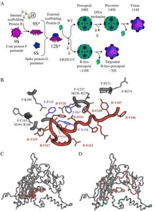
of the 9S coat protein F pentamer forming the 9S* particle (8).
This induces the conformational changes that (i) inhibit 9S parti-cle aggregation, (ii) facilitate DNA pilot protein H incorporation,
and (iii) stimulate coat-spike protein interactions (8–12). During
late morphogenesis, 240 copies of the external scaffolding protein
D organize 12 12S* particles into procapsids (13). As the genome
is packaged, the DNA binding protein J displaces the internal scaf-folding protein. Extrusion is most likely facilitated by the B
pro-tein’s autoproteolytic activity (14). The resulting metastable
pro-virion is infectious (15–17). Spontaneous dissociation of the
external scaffolding protein yields the mature virion.
As seen with the P22, P2,29, and herpes simplex virus (HSV)
internal scaffolding proteins (18–22), the C terminus of the
X174 B protein is most strongly associated with the coat protein
(23–25). In theX174 procapsid crystal structure, six aromatic
amino acid residues mediate approximately 85% (22/26) of these interactions. Most of the participating atoms (17/22) are found in
aromatic rings (Fig. 1B). The results of a previous genetic analysis
demonstrate that nonaromatic substitutions for residue F120, the last amino acid, or the absence of this amino acid, abolish coat
protein binding (5). Substitutions at the other aromatic sites
pro-duced defective conformational switching phenotypes. The mu-tant proteins kinetically trapped early assembly intermediates af-ter coat protein binding, the assembly phase mediated by the internal scaffolding protein, but prior to procapsid formation, which is mediated by the external scaffolding protein. Although the internal scaffolding protein mutations trap different interme-diates within the first half of the assembly pathway, common sup-pressors alleviate the various morphogenetic defects. This suggests a fluid assembly pathway, one in which the scaffolding protein induces a single, coat protein conformational switch, not a series of sequential reactions. In this model, the nature of the improper switch determines the trapped intermediate. Consequently, nei-ther a specific function nor a temporal requirement could be as-signed to any particular amino acid.
In addition to the C-terminal aromatic side chains, two acidic residues, D111 and E113, form salt bridges with coat protein
amino acids R239 and K166 (Fig. 1B). Electrostatic interactions
Received10 October 2012Accepted7 December 2012
Published ahead of print19 December 2012
Address correspondence to Bentley A. Fane, [email protected]. Copyright © 2013, American Society for Microbiology. All Rights Reserved.
doi:10.1128/JVI.02839-12
on November 7, 2019 by guest
http://jvi.asm.org/
govern morphogenesis in other viral systems (3) and have been
thoroughly studied in bacteriophage P22 (4,6,26–28). In the P22
scaffolding protein, two charged residues are central to coat tein binding, whereas other amino acids modulate activity by pro-moting the proper architecture of the coat-scaffolding protein
in-terface (4, 26). Alteration of an amino acid that mediates a
protein-protein interaction, either via a salt bridge or via an aro-matic ring, is typically regarded as a function, or loss-of-contact, mutation. The previously characterized mutations at
ar-omatic sites most likely fall into this category (5). In contrast, the
introduction of aromatic residue into an environment already rich in aromatic content could create a gain-of-contact mutation,
which may compete with wild-type interactions. To investigate
whether charged amino acids are critical toX174 B protein
func-tion, as seen in the P22 scaffolding protein, or play a more subsid-iary role, aromatic and nonaromatic substitutions for D111 and E113 were genetically and biochemically characterized.
MATERIALS AND METHODS
Phage plating, media, buffers, and stock preparation.The reagents, me-dia, buffers, and protocols have been previously described (29).
Bacterial strains, phage strains, and plasmids.The wild-type Esche-richia coli C strain C122 (Su⫺) and the isogenic, amber-suppressing strains BAF7 (supD), BAF5 (supE), BAF8 (supF), and BAF30 (recA) have
FIG 1X174 morphogenesis and the structures of the coat and scaffolding proteins. (A) Wild-type andl(B)D111Yassembly pathways. The 9S* intermediate contains the coat F, internal scaffolding B, and minor vertex H proteins in a respective 5:5:1 ratio. The addition of the major spike protein G pentamer (6S particle) produces the 12S* particle. A total of 240 copies of the external scaffolding D protein organize 12 12S* particles into procapsids. (B) Structure of the coat-internal scaffolding protein binding cleft with theX174 procapsid crystal structure (Protein Data Bank code, 1C3D). Labels depict the protein (F, coat; B, internal scaffolding), amino acid (letter), and position (number) in the primary structure. The three coat protein aromatic amino acid residues—F-F67, F-Y134, and F-F135—that participate in ring-ring contacts with internal scaffolding protein residue B-F120 are highlighted in lavender. (C) Atomic structure of the coat (gray) and internal scaffolding (peach) proteins. (D) Locations of second-site suppressors of internal scaffolding (peach) and external scaffolding (teal) protein defects. The positions of the newly isolated suppressors at amino acids 203 and 314 are highlighted.
on November 7, 2019 by guest
http://jvi.asm.org/
[image:2.585.135.454.65.497.2]been previously described (29,30). The hostslyDmutation confers resis-tance to E protein-mediated lysis (31). The construction of the comple-menting plasmid pXB has been previously reported (5).
Generation ofam(B)and missenseBⴚmutations.Unless otherwise indicated, amber and missense mutants were generated by oligonucle-otide-mediated mutagenesis utilizing the published protocol (32). Mu-tagenized DNA was transfected in BAF30 pXB, which expresses the wild-type B gene. Mutants were identified by complementation-dependent phenotypes. Genotypes were verified by a direct DNA sequence analysis.
Isolation and construction ofam(B)and missenseBⴚmutations, second-site suppressors,su/amstrains used in the cross-suppressor analysis, andsu(B111)-D V75A l(B)D111Ymutant.All protocols were identical to those described in an earlier publication (5), with the excep-tion ofsu(B111)-D V75A l(B)D111Ystrain construction, which was made by recombination rescue. Thel(B)D111Ygene was amplified and cloned as previously described (33). To select for am⫹ recombinants, the
su(B111)-D V75A am(B)D111mutant was plated on cells harboring the clone gene, which was not induced. All genotypes were verified by a direct sequence analysis.
Detection of virion, intermediate particles, and DNA.To generate extracts of infected cells, 100 ml of lysis-resistant cells at 1.0⫻108cells/ml were infected with wild-type or mutantX174 at a multiplicity of infec-tion (MOI) of 5.0 as previously described (34). Subsequent extract prep-aration, rate zonal sedimentation, and protein electrophoresis protocols have already been published (34). For the detection of particles with S values larger than 70, samples were loaded atop 5 to 30% sucrose gradients and spun at 45,000 rpm for 1.0 h in a Beckman SW50.1 rotor. After fractionation into approximately 50 100-l fractions, virally derived pro-tein complexes were detected by UV spectroscopy ( ⫽280 nm). The position of infectious virions (114S) was further determined by plating assays. To detect particles with S values of less than 70, samples were spun at 34,000 rpm for 16 h. After fractionation, material was detected by 15% SDS-PAGE. Modified 15 to 30% sucrose gradients were prepared by the addition of CsCl (3.5%, wt/vol) to the 30% sucrose layer before mixing (13). DNA was extracted directly from particles isolated from sucrose gradient fractions using established protocols (29).
RESULTS
The introduction of a C-terminal aromatic amino acid results in a lethal phenotype.Internal scaffolding protein residues D111 and E113 form respective salt bridges with coat protein residues R239 and K166. To determine whether these interactions were
critical to B protein function, amber (am) mutations were
gener-ated in these codons. Mutants were assayed for plaque formation
on three tRNA informational suppressing hosts (Su⫹), which
in-sert glutamine, serine, or tyrosine at the amber codon during
pro-tein synthesis (29). Thus, a single nonsense mutation can be used
to generate a variety of missense proteins. As determined in plaque assays on the various tRNA informational suppressing hosts, via-bility serves as an indicator of protein function, assessing the con-sequences of altering the wild-type interactions. All three amino acid substitutions for E113 were tolerated, suggesting that the
E113-K166 electrostatic interaction is not essential (Table 1).
Ser-ine and glutamSer-ine, which would either eliminate or substantially alter the electrostatic D111-R239 interaction, were also tolerated, albeit poorly. However, there was a strong lethal phenotype asso-ciated with the introduction of an aromatic tyrosine residue.
The D¡S and D¡Q substitutions at position 111 would alter
the D111-R239 salt bridge and can be regarded as a partial loss of
function. Although the D¡Y substitution would also eliminate
this interaction, the consequences of this substitution appeared to
be much more severe (Table 1). Considering the numerous
aro-matic ring-ring interactions between the coat and internal
scaf-folding protein, the effects of D¡Y substitution may be more
complex. At the center of the coat-scaffolding protein binding cleft, scaffolding protein residue F120 participates in aromatic ring-ring interactions with coat protein residues F67, Y134, and
F135 (Fig. 1B). Altering these contacts strongly inhibits
coat-scaf-folding protein interactions (5). Considering the proximity and
orientation of the D111 side chain toward the cleft’s center, the introduction of an aromatic amino acid at this position may ab-rogate coat-internal scaffolding protein interactions, which would
lead to the intracellular coat protein aggregation (5), or introduce
a novel interaction that redirects assembly. To distinguish be-tween these two hypotheses, the morphogenetic pathway was ex-amined in missense mutant-infected cells.
The D111Y substitution produces an off-pathway procap-sid-like particle.The inefficiency of informational suppression as well as defective protein function can contribute to amber mutant
phenotypes in Su⫹hosts (35,36). Therefore, a D¡Y missense
mutation was generated directly in the viral genome. The resulting
l(B)D111Ymutant exhibited a lethal phenotype but was rescued
by the exogenous expression of a wild-type B gene. Thus, defective protein function is the primary phenotypic determinant. To assay the mutant protein’s effect on particle morphogenesis,
lysis-resis-tant cells were infected with thel(B)D111Ymutant or wild-type
X174 as described in Materials and Methods. Infected-cell
ex-tracts were generated and analyzed by rate zonal sedimentation with 5 to 30% sucrose gradients. In both extracts, particles sedi-menting between 108S and 114S, procapsids and virions, were
detected (Fig. 2A). However, the specific infectivity (PFU/A280) of
thel(B)D111Ymutant peak was approximately 2 orders of
mag-nitude lower than that of the wild-type control (Table 2). In
addi-tion, degraded procapsids (70S particles) were prominent in the
l(B)D111Yextract. The D111Y protein appeared to redirect
as-sembly. All previously isolated internal scaffolding protein muta-tions arrest assembly before procapsid assembly. Thus, the molec-ular phenotype of this mutation is unique.
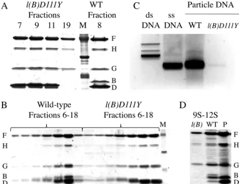
Particle protein composition was examined by SDS-PAGE (Fig. 3A). As the coat protein F remains soluble in thel(B)D111Y
extracts, the D111Y mutation did not block coat protein binding
(5). The wild-type material from 108S to 114S contained both
scaffolding proteins, proteins B and D, indicating a mixture of procapsids and virions. In contrast, the mutant particles from 108S to 114S lacked protein B, the internal scaffolding protein.
This protein composition resembles wild-type provirions (Fig.
1A). However, provirions are infectious and sediment more
quickly at 132S to 140S (16). The mutant 70S material lacked both
scaffolding proteins. In a wild-type infection, 70S degraded pro-capsids result from the loss of the external scaffolding protein D.
However, they retain the B protein (37,38).
To determine whether thel(B)D111Yparticles represent
kinet-ically trapped provirions or a novel off-pathway product, their sedimentation properties were analyzed in 15 to 30% sucrose
gra-dients modified by the addition of CsCl (13), which more
accu-rately separates procapsids (108S) from DNA-filled provirions (132S). The provirion position within the wild-type control gra-dient was determined by identifying the fastest-sedimenting ma-terial with a specific infectivity equal to the prominent, mature
virion peak (Table 2). Again,l(B)D111Yparticles appear to
sedi-ment more slowly than wild-type virions (Fig. 2B) and are less
infectious (Table 2). Wild-type and second-site revertants
consti-tute the background signal in specific-infectivity assays. All viable
on November 7, 2019 by guest
http://jvi.asm.org/
particles detected within the mutant samples were revertants, which provided a valuable internal S value marker. However,
spe-cific infectivity is defined as PFU/A280, regardless of the genotype
of the detected particle. Thus, the reportedl(B)D111Yspecific
infectivity is most likely lower than the assay suggested. All frac-tions were examined by SDS-PAGE. Unlike the wild-type control, the internal scaffolding protein was not detected within any
frac-tion (Fig. 3B).
Thel(B)D111Yparticles are most likely not kinetically trapped provirions, which would be DNA-filled but off-pathway particles. To validate this conclusion, extracts of wild-type- and
mutant-infected cells were prepared, assembled particles (ⱖ70S) were
sep-arated from unpackaged DNA by sucrose gradient sedimentation,
and nucleic acid was extracted (Fig. 3C). Genomic DNA was
abundantly associated with the wild-type particles. However, only trace amounts were detected in the mutant sample, which was 20-fold more concentrated than the wild-type control. The DNA recovered from the mutant sample most likely reflects the
back-ground revertants detected in the specific-infectivity assays. Thus, the mutant D111Y internal scaffolding protein prematurely left the assembly pathway before being displaced by the DNA packag-ing process.
To more accurately gauge when the mutant B protein leaves the assembly pathway, the protein composition of the early, preprocapsid intermediates was determined. Wild-type and mutant 9S and12S particles were isolated as described in Ma-terials and Methods. Since most viral proteins were associated with large assembled particles, fractions had to be pooled and concentrated 25-fold to gather enough material for the
analy-sis. As can be seen inFig. 3D, the internal scaffolding protein was
not detected in the early assembly intermediates isolated from the
[image:4.585.43.548.77.406.2]l(B)D111Yinfection, whereas it was present in the wild-type particles. These data indicate that either the mutant protein prematurely left the assembly pathway before procapsid formation or its association with the viral coat protein F was too weak to withstand the purifica-tion protocol.
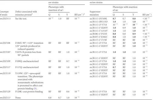
TABLE 1Phenotypes of missense suppressedam(B)andsu/am(B)strainsa
Genotype of strain
Defect associated with missense proteinb
amstrains su/amstrains
Phenotype with insertion of aac:
RF(am⫹)d
Suppressor mutation
Phenotype with insertion of aa:
RF(am⫹)
Q S Y Q S Y
am(B)D111 See the text. 10⫺2 1.0 RF 10⫺4 su(B111)-B F109L 0.7 0.7 0.8 ⬍10⫺5
su(B111)-BN115D 1.0 1.0 1.0 10⫺3 su(B111)-D V75A 10⫺2 10⫺2 10⫺1 ⬍10⫺4 su(B111)-F H203Y 1.0 0.9 1.0 10⫺5 su(B111)-F A314V 1.0 1.0 1.0 10⫺4 su(B106)-F P325S 0.8 0.8 0.9 ⬍10⫺4 su(B107)-F R382C 0.8 1.0 1.0 ⬍10⫺4 su(B117)-F T178I RF RF RF 10⫺4 am(B)F106 F106Y:9S*¡12S* transition RF RF RF 10⫺3 su(B111)-D V75A RF RF 1.0 10⫺2
12S* particle produced in reduced quantity
su(B111)-F H203Y RF RF 1.0 10⫺5
am(B)Y107 Y107S: B protein loss from 9S* particle
RF RF 1.0 10⫺7 su(B111)-D V75A 1.0 1.0 1.0 10⫺5
su(B111)-F H203Y RF RF 1.0 10⫺6 am(B)F109 F109Q: uncharacterized RF RF 0.7 10⫺6 su(B111)-D V75A 1.0 1.0 1.0 10⫺5 su(B111)-F H203Y RF RF 1.0 10⫺5 am(B)F117 F117Q: uncharacterized RF RF 1.0 10⫺5 su(B111)-D V75A 0.7 RF 1.0 10⫺5 su(B111)-F H203Y RF RF 1.0 10⫺4 am(B)Y119 Y119W: 12S*¡procapsid
transition. The phenotype associated with
nonaromatic substitutions is presumed to affect coat protein binding (5).
RF RF 1.0 10⫺5 su(B111)-D V75A RF RF 1.0 10⫺4
su(B111)-F H203Y RF RF 1.0 10⫺6
am(B)F120 F120K: coat protein binding RF RF 0.6 10⫺6 su(B111)-D V75A RF RF 1.0 10⫺6 su(B111)-F H203Y RF RF 1.0 10⫺6
am(B)E113 None 0.8 0.7 1.0 10⫺4
aData are presented as efficiency of plating: assay titer/most permissive titer. The most permissive titer was determined on either BAF 8supF, which inserts tyrosine during protein
synthesis at amber codons, or BAF30 pXB, which expresses a cloned wild-type B gene. Boldface indicates the conditions under which rescue by the suppressor occurred. The names of the amber mutants reflect the wild-type codon and encoded amino acid. Thus,am(B)D111indicates an amber mutation in codon 111 that normally encodes an aspartic acid (D). The names of the second-site suppressors reflect the parental background in which they were isolated, the gene in which the suppressor resides, and the resulting substitution. Thus,su(B111)-D V75Aindicates that the suppressor was originally isolated in theam(B)D111background. “-D” indicates an amino acid substitution in the external scaffolding protein D; “V75A” indicates a valine (V)¡alanine (A) substitution. aa, amino acid.
bThe column summarizes the data published by Gordon et al. (5). Biochemical analysis was conducted with genome-encoded missense mutants. The missense substitution is listed
before the colon, the molecular phenotype is described after the colon. In some instances, the defective phenotypes associated with missense mutants were too weak to biochemically characterized. These are identified with the word “uncharacterized.”
c
Letters indicate the amino acid inserted by the tRNA informational suppressor. Q, glutamine; S, serine, Y, tyrosine.
dRF (am⫹), the reversion frequency of the amber mutation as determined on the Su⫺host.
on November 7, 2019 by guest
http://jvi.asm.org/
Second-site genetic analysis of the D111¡Y substitution yielded novel suppressors.To determine whether viruses could adapt to the mutant internal scaffolding protein, a second-site genetic analysis was conducted. To select for second-site
suppres-sors,am(B)D111was plated on the restrictive Su⫹host, BAF8,
which inserts tyrosine residues at amber sites during protein
syn-thesis. The resulting plaques were stabbed into Su⫹and Su⫺
indi-cator lawns. Putative second-site suppressors were identified by the retention of the amber phenotype. One of the mutations,
su(B111)-F A314V, was very similar to known coat protein
sup-pressors of conformational switch defects (Table 1). However,
su(B111)-F H203Ywould alter the large coat protein␣-helix at the
3-fold axis of symmetry (Fig. 1CandD). Mutations in this helix
are known to suppress defective external scaffolding protein
func-tion (see Discussion). Two suppressors, thesu(B111)-B F109Land
N115Dmutations, resided in gene B. Intragenic suppressors have
not been previously isolated for B protein defects (5,29,39).
Fi-nally, the fourth suppressor, the su(B111)-D V75A mutation,
changes the external scaffolding protein. A full genome sequence
was determined for the su(B111)-D V75A am(B)D111 and
am(B)D111mutations. With the exception of the suppressor
mu-tation, no other changes were found. Thus, the identified suppres-sor was both necessary and sufficient to confer the altered pheno-type.
The location of the suppressor in the external scaffolding pro-tein was unique. The 11 previously isolated suppressors of C-ter-minal, B protein mutations were all located in the viral coat
pro-tein F (5). After being placed into thel(B)D111Ybackground, the
effects of thesu(B111)-D V75A mutation were examined
bio-chemically as described above. As can be seen inFig. 2C, particles
migrated at two speeds, represented by the 108S to 114S and 70S peaks in the sedimentation profile. The material from 108S to 114S was analyzed by SDS-PAGE. Unlike for the wild-type con-trol, which contained a mixture of virions and procapsids, neither scaffolding protein was detected, suggesting that the fractions pri-marily contained virions (data not shown). Moreover, the specific
infectivity (PFU/A280) was comparable to that of the wild-type
control (Table 2). The 70S particles lacked infectivity. Although
the internal scaffolding protein was detected by SDS-PAGE, it appeared to be present at a greatly reduced quantity (data not shown), requiring both silver staining and laser scanning to be visualized. Thus, the suppressor most likely does not act by stabi-lizing the mutant B protein within the procapsid. To further in-vestigate both the nature of the initial defect and possible suppres-sion mechanisms, the genetic analysis was expanded to include the previously characterized coat protein binding mutations, confor-mational switching mutations, and their associated suppressors.
Extragenic suppressors of C-terminal B protein substitu-tions do not exhibit strict allele specificity.With the exception of coat protein binding mutants, previously characterized B proteins with C-terminal mutations exhibited defects in conformational
switching, halting assembly before procapsid formation (5),
whereas thel(B)D111Ymutant generated a postprocapsid,
off-FIG 2Large particles (S⬎50) synthesized in cells infected with the wild type and
l(B)D111Yandsu(B111)-D V75A l(B)D111Ymutants. (A) Sedimentation (5 to 30% sucrose) of extracts of cells infected with the wild type (Œ) andl(B)D111Y
mutant (). The fraction with the highest specific infectivity is labeled 114S, the
sedimentation value of the mature virion. In a wild-type infection, 70S material contains degraded procapsids resulting from the loss of the external scaffolding protein. Lower fractions represent the bottom of the gradient, which would con-tain faster-sedimenting material. (B) Sedimentation (15 to 30% CsCl-modified
sucrose) of extracts of cells infected with the wild type (Œ) andl(B)D111Y
mutant (). The position of the metastable, infectious provirion (132S to 140S) in the wild-type fractions was estimated as described in the text. (C) Sedimentation (5 to 30% sucrose) of extracts of cells infected with the wild type (Œ) andsu(B111)-D V75A l(B)D111Ymutant ().
on November 7, 2019 by guest
http://jvi.asm.org/
[image:5.585.88.238.66.646.2]pathway product. This difference in phenotype could reflect dif-ferent molecular defects. Alternatively, the defects could be fun-damentally related. If the latter, the second-site suppressors of
both the D111Y substitution and the previously characterized
conformational switching mutants should cross-rescue. To test this hypothesis, a cross-suppressor analysis was performed. Three previously isolated suppressors of C-terminally mutated B
pro-teins (5) were placed into theam(B)D111background. In
addi-tion,su(B111)-D V75Aandsu(B111)-F H203Ywere crossed into
previously characterizedam(B)genomes. The coat and internal
scaffolding genes were sequenced in every strain. A full genome
sequence of thesu(B111)-D V75A am(B)F107mutant was
deter-mined. No additional substitutions were identified.
Growth phenotypes were characterized on the Su⫹hosts (
Ta-ble 1). Cross-suppressing activity was defined by plaque forma-tion 2 orders of magnitude above the frequency obtained on the
parental mutant’s restrictive Su⫹host. Two of the previously
iso-lated suppressors of defective B protein function, su(B106)-F
P325Sandsu(B107)-F R382C, restored viability to the missense
suppressedam(B)D111mutant. This observation supports our
fluid assembly model (5). Althoughsu(B117)-F T178Iexhibited
no activity in this background, it is known to be one of the weaker
suppressors (5). Thesu(B111)-F H203Y suppressor was active
only in theam(B)F106background, which may reflect the
later-acting defects associated with substitutions at this site (see Discus-sion). With the exception of nonaromatic substitutions at sites F120 and Y119, which eliminate coat protein binding, the
su(B111)-D V75A mutation rescued all missense suppressed
am(B)mutants.
DISCUSSION
Coat-internal scaffolding salt bridges are less critical than aro-matic-amino-acid-mediated interactions.Six C-terminal
aro-matic amino acids and two acidic residues mediateX174
coat-internal scaffolding protein interactions (24, 25). Four of the
aromatic amino acids—F106, Y107, Y119, and F120 —are
essen-tial for proper internal scaffolding protein function (5).
Substitu-tions at these sites prevent coat protein binding or fail to fully induce the requisite conformational switches for procapsid as-sembly. In contrast, the salt bridges involving D111 and E113
appear to be less critical. Although the E113¡S, Q, and Y and
D111¡S and Q substitutions would eliminate or alter the
respec-tive salt bridges formed with coat protein residues K166 and R239, they do not eliminate viability.
Thus, inX174, charged residues do not strongly govern
coat-scaffolding interactions as observed in bacteriophage P22 (4, 6,
26–28). The coat protein-binding domain of the P22 scaffolding
protein has a helix-turn-helix (HTH) motif (21). The essential
charged residues are located in the C-terminal helix. However,
mutations within the N-terminal helix and the intervening-turn
can alter assembly kinetics and eliminate viability without grossly
disrupting domain structure or protein binding (26). These
ob-servations suggest that some P22 residues perform a subsidiary role, optimizing the architecture of the coat-scaffolding interface.
Salt bridges appear to play this secondary role in the X174
pocket, whereas aromatic ring-ring interactions, mediated pri-marily by F120, govern coat protein binding.
Restructuring the coat-internal scaffolding protein binding pocket.The orientation of the D111 side chain toward the center
of the pocket (Fig. 1B) may explain the uniquel(B)D111Y
pheno-type. An aromatic amino acid at position 111 may compete with F120’s interactions with coat protein residues F67, Y134, and F135. Unlike previously characterized internal scaffolding protein
mutations (5,29,39), C-terminal intragenic suppressors of the
D111Y defect were isolated. This may reflect unique structural rearrangements within the cleft. However, the intragenic suppres-sors were not crossed into other mutant backgrounds to demon-strate allele specificity. The full extent to which the binding pocket can be restructured to support the entire assembly process re-mains to be determined. This will require more targeted and ex-haustive genetic analyses. First, the D111Y mutation, perhaps with
intragenic suppressors, will need to be placed inciswith a
[image:6.585.40.286.79.215.2]non-sense or misnon-sense mutation in codon F120. Additional genetic selections for viability may also be necessary. As the B gene over-laps with other genes and regulatory sequences, the mutant gene may need to be expressed from a plasmid in a complementation-based system.
TABLE 2Specific infectivities of mutant and wild-type particlesa
Gradient and
strain genotype S value
Specific infectivity (PFU/A280)
Specific infectivity normalized to WT
5–30% sucrose gradient
WT 108-114S 5.0⫻1011 1.0 l(B)D111Y 108-114S 2.0⫻1010 0.04 su(B111)-D75V
l(B)D111Y
108S-114S 1.0⫻1012 2.0
15–30%, CsCl-modified sucrose gradient
WT 108S-114S 1.0⫻1012 1.0 132S 1.2⫻1012 1.2 l(B)D111Y 108S-114S 1.6⫻1010 0.016
a
WT, wild type.
FIG 3Protein and DNA compositions of wild-type (WT) andl(B)D111Y
particles. (A and B) Protein compositions of large particles. Fractions analyzed by SDS-PAGE, respectively, correspond to the sedimentation profiles inFig. 2A(5 to 30% sucrose gradients) andFig. 2B(15 to 30% CsCl-modified sucrose gradients). (C) DNA isolated from wild-type andl(B)D111Yparticles.X174 single-stranded DNA (ssDNA) and replicative-form double-stranded DNA (dsDNA) are included as migration references. (D) Protein composition of pooled 9S to 12S fractions, which contain early assembly intermediates, from extracts of cells infected with the wild type and thel(B)D111Ymutant. P, purified procapsids.
on November 7, 2019 by guest
http://jvi.asm.org/
[image:6.585.300.543.442.628.2]Late-acting versus global suppressors ofBⴚconformational switch defects.Thesu(B111)-D V75Amutation may represent a
global suppressor ofB⫺conformational switch defects. It was
ac-tive in all backgrounds except those that prevent coat protein binding. In this two-scaffolding protein system, the external scaf-folding protein may be able to compensate for reduced internal
scaffolding protein function. In a previous study (9), progressive
and targeted genetic selections were employed to lessen and finally eliminate B protein dependency. The primary adaptations in the
sextuple mutantB-free strain involved the overexpression of a
mutant external scaffolding protein D and strengthening of its contacts with the coat protein. This external scaffolding protein override mechanism most likely differs from the mechanism con-ferred by the coat protein suppressors (see below).
In contrast, thesu(B111)-F H203Ymutation, which is located
in the large␣-helix at the 3-fold axis of symmetry, exhibited some
allele specificity. Substitutions in this helix have pleotropic effects. They suppress mutant external scaffolding proteins that (i)
inef-ficiently promote the 12S*¡procapsid transition and (ii) fail to
stabilize the 3-fold-axis-related, procapsid pores through which
genomic DNA is packaged (34,40,41). These are both late-acting
defects. Thel(B)D111Yparticles appeared to be empty, suggesting
that this suppressor may act at the level of pore stabilization.
When crossed into other B⫺ conformational switch-defective
backgrounds, it rescued only the defects associated with the F106Y protein. The F106Y substitution confers a temporally later defect:
12S* particles form, albeit it at low efficiency (5). Considering the
previously defined role of the affected␣-helix, the suppressor may
facilitate procapsid formation at reduced 12S* particle concentra-tions.
Late-acting defects versus belated manifestations and fluid assembly.Thel(B)D111Yassembled particles may indicate that
the D¡Y substitution confers a unique late-acting defect,
indica-tive of a novel B protein function. Alternaindica-tively, the assembled particles may represent the delayed manifestation of an early as-sembly error. In this model, mutant 12S*-like pentamers interact with the external scaffolding protein but redirect morphogenesis
off-pathway (Fig. 1A). Biochemical data favor this interpretation.
The D111Y protein was detected in neither early assembly inter-mediates nor the final off-pathway product. In the complete ab-sence of protein B, wild-type assembly does not continue past 9S and 6S particle formation, which leads to coat protein aggregation
(5). Therefore, the D111Y mutant protein bound the 9S particle,
induced a conformational switch, but prematurely left the assem-bly pathway.
The results of the genetic analyses also indicate that the D111Y defect occurred early in assembly pathway, before procapsid for-mation, but is belatedly manifested after procapsid morphogene-sis. Previously characterized conformational switch mutations ki-netically trapped assembly intermediates before procapsid
formation (5). Although mutations trapped different early
inter-mediates, they were rescued by a set of common suppressors. While the suppressor substitutions are spread throughout the vi-ral coat protein, their nature offered more insights than their spa-tial location. They primarily involved glycine and proline residues, suggesting that an inherently flexible coat protein compensates for the reduced ability of the internal scaffolding protein to induce a conformational switch. These suppressors also rescued the defects associated with D111Y substitution, which is consistent with a previously described fluid assembly model. In that model, the
scaffolding protein induces a single, coat protein conformational switch, not a series of sequential reactions. Incomplete or im-proper switches would kinetically trap intermediates or direct in-termediates of the productive pathway. But as the molecular
characterization of thel(B)D111Ymutant may demonstrate, the
off-pathway product isolated from infected cells may not accu-rately reflect the temporal nature of the initial defect.
ACKNOWLEDGMENTS
This research was supported by National Science Foundation grant MCB 0948399 to B.A.F. and by funds to the University of Arizona Undergrad-uate Biology Research Program from the Howard Hughes Medical Insti-tute (HHMI 52003749) and the BIO5 InstiInsti-tute.
REFERENCES
1.Fane BA, Prevelige PE, Jr.2003. Mechanism of scaffolding-assisted viral assembly. Adv. Protein Chem.64:259 –299.
2.Prevelige PE, Fane BA.2012. Building the machines: scaffolding protein functions during bacteriophage morphogenesis. Adv. Exp. Med. Biol.726: 325–350.
3.Zlotnick A, Fane BA.2010. Mechanisms of icosahedral virus assembly, p 180 –202.InAgbandje-McKenna M, McKenna R (ed), Structural virology. Royal Society of Chemistry, London, United Kingdom.
4.Cortines JR, Weigele PR, Gilcrease EB, Casjens SR, Teschke CM.2011. Decoding bacteriophage P22 assembly: identification of two charged res-idues in scaffolding protein responsible for coat protein interaction. Vi-rology421:1–11.
5.Gordon EB, Knuff CJ, Fane BA.2012. Conformational switch-defective
X174 internal scaffolding proteins kinetically trap assembly intermedi-ates before procapsid formation. J. Virol.86:9911–9918.
6.Parker MH, Casjens S, Prevelige PE, Jr.1998. Functional domains of bacteriophage P22 scaffolding protein. J. Mol. Biol.281:69 –79. 7.Suhanovsky MM, Parent KN, Dunn SE, Baker TS, Teschke CM.2010.
Determinants of bacteriophage P22 polyhead formation: the role of coat protein flexibility in conformational switching. Mol. Microbiol.77:1568 – 1582.
8.Cherwa JE, Jr, Uchiyama A, Fane BA.2008. Scaffolding proteins altered in the ability to perform a conformational switch confer dominant lethal assembly defects. J. Virol.82:5774 –5780.
9.Chen M, Uchiyama A, Fane BA.2007. Eliminating the requirement of an essential gene product in an already very small virus: scaffolding protein B-freeX174, B-free. J. Mol. Biol.373:308 –314.
10. Cherwa JE, Jr, Young LN, Fane BA.2011. Uncoupling the functions of a multifunctional protein: the isolation of a DNA pilot protein mutant that affects particle morphogenesis. Virology411:9 –14.
11. Ruboyianes MV, Chen M, Dubrava MS, Cherwa JE, Jr, Fane BA.2009. The expression of N-terminal deletion DNA pilot proteins inhibits the early stages of phiX174 replication. J. Virol.83:9952–9956.
12. Siden EJ, Hayashi M.1974. Role of the gene beta-product in bacterio-phage phi-X174 development. J. Mol. Biol.89:1–16.
13. Mukai R, Hamatake RK, Hayashi M.1979. Isolation and identification of bacteriophage phi X174 prohead. Proc. Natl. Acad. Sci. U. S. A.76:4877– 4881.
14. Bernal RA, Hafenstein S, Olson NH, Bowman VD, Chipman PR, Baker TS, Fane BA, Rossmann MG.2003. Structural studies of bacteriophage alpha3 assembly. J. Mol. Biol.325:11–24.
15. Fujisawa H, Hayashi M. 1977. Assembly of bacteriophage phi X174: identification of a virion capsid precursor and proposal of a model for the functions of bacteriophage gene products during morphogenesis. J. Virol.
24:303–313.
16. Fujisawa H, Hayashi M.1977. Two infectious forms of bacteriophage phi X 174. J. Virol.23:439 – 442.
17. Fujisawa H, Hayashi M. 1976. Viral DNA-synthesizing intermediate complex isolated during assembly of bacteriophage phi X174. J. Virol.
19:409 – 415.
18. Chang JR, Spilman MS, Rodenburg CM, Dokland T.2009. Functional domains of the bacteriophage P2 scaffolding protein: identification of residues involved in assembly and protease activity. Virology384:144 – 150.
19. Fu CY, Morais MC, Battisti AJ, Rossmann MG, Prevelige PE, Jr.2007.
on November 7, 2019 by guest
http://jvi.asm.org/
Molecular dissection of29 scaffolding protein function in an in vitro assembly system. J. Mol. Biol.366:1161–1173.
20. Oien NL, Thomsen DR, Wathen MW, Newcomb WW, Brown JC, Homa FL.1997. Assembly of herpes simplex virus capsids using the hu-man cytomegalovirus scaffold protein: critical role of the C terminus. J. Virol.71:1281–1291.
21. Tuma R, Parker MH, Weigele P, Sampson L, Sun Y, Krishna NR, Casjens S, Thomas GJ, Jr, Prevelige PE, Jr.1998. A helical coat protein recognition domain of the bacteriophage P22 scaffolding protein. J. Mol. Biol.281:81–94.
22. Weigele PR, Sampson L, Winn-Stapley D, Casjens SR.2005. Molecular genetics of bacteriophage P22 scaffolding protein’s functional domains. J. Mol. Biol.348:831– 844.
23. Burch AD, Fane BA.2000. Efficient complementation by chimeric Mi-croviridae internal scaffolding proteins is a function of the COOH-terminus of the encoded protein. Virology270:286 –290.
24. Dokland T, Bernal RA, Burch A, Pletnev S, Fane BA, Rossmann MG.
1999. The role of scaffolding proteins in the assembly of the small, single-stranded DNA virus phiX174. J. Mol. Biol.288:595– 608.
25. Dokland T, McKenna R, Ilag LL, Bowman BR, Incardona NL, Fane BA, Rossmann MG.1997. Structure of a viral procapsid with molecular scaf-folding. Nature389:308 –313.
26. Padilla-Meier GP, Gilcrease EB, Weigele PR, Cortines JR, Siegel M, Leavitt JC, Teschke CM, Casjens SR.2012. Unraveling the role of the C-terminal helix turn helix of the coat-binding domain of bacteriophage P22 scaffolding protein. J. Biol. Chem.287:33766 –33780.
27. Parent KN, Doyle SM, Anderson E, Teschke CM.2005. Electrostatic interactions govern both nucleation and elongation during phage P22 procapsid assembly. Virology340:33– 45.
28. Zlotnick A, Suhanovsky MM, Teschke CM.2012. The energetic contri-butions of scaffolding and coat proteins to the assembly of bacteriophage procapsids. Virology428:64 – 69.
29. Fane BA, Hayashi M.1991. Second-site suppressors of a cold-sensitive prohead accessory protein of bacteriophage phi X174. Genetics128:663– 671.
30. Fane BA, Head S, Hayashi M.1992. Functional relationship between the
J proteins of bacteriophages phi X174 and G4 during phage morphogen-esis. J. Bacteriol.174:2717–2719.
31. Roof WD, Fang HQ, Young KD, Sun J, Young R.1997. Mutational analysis of slyD, an Escherichia coli gene encoding a protein of the FKBP immunophilin family. Mol. Microbiol.25:1031–1046.
32. Fane BA, Shien S, Hayashi M.1993. Second-site suppressors of a cold-sensitive external scaffolding protein of bacteriophage phi X174. Genetics
134:1003–1011.
33. Novak CR, Fane BA. 2004. The functions of the N terminus of the phiX174 internal scaffolding protein, a protein encoded in an overlapping reading frame in a two scaffolding protein system. J. Mol. Biol.335:383– 390.
34. Uchiyama A, Fane BA.2005. Identification of an interacting coat-external scaffolding protein domain required for both the initiation of phiX174 procapsid morphogenesis and the completion of DNA packag-ing. J. Virol.79:6751– 6756.
35. Floor E.1970. Interaction of morphogenetic genes of bacteriophage T4. J. Mol. Biol.47:293–306.
36. Sternberg N.1976. A genetic analysis of bacteriophage lambda head as-sembly. Virology71:568 –582.
37. McKenna R, Bowman BR, Ilag LL, Rossmann MG, Fane BA. 1996. Atomic structure of the degraded procapsid particle of the bacteriophage G4: induced structural changes in the presence of calcium ions and func-tional implications. J. Mol. Biol.256:736 –750.
38. McKenna R, Xia D, Willingmann P, Ilag LL, Krishnaswamy S, Ross-mann MG, Olson NH, Baker TS, Incardona NL.1992. Atomic structure of single-stranded DNA bacteriophage phi X174 and its functional impli-cations. Nature355:137–143.
39. Burch AD, Ta J, Fane BA.1999. Cross-functional analysis of the Micro-viridae internal scaffolding protein. J. Mol. Biol.286:95–104.
40. Uchiyama A, Chen M, Fane BA.2007. Characterization and function of putative substrate specificity domain in microvirus external scaffolding proteins. J. Virol.81:8587– 8592.
41. Uchiyama A, Heiman P, Fane BA.2009. N-terminal deletions of the phiX174 external scaffolding protein affect the timing and fidelity of as-sembly. Virology386:303–309.