AND CLOSED HEMORRHOIDECTOMY
Dissertation submitted to
THE TAMILNADU DR. M.G.R. MEDICAL UNIVERSITY
In partial fulfillment for the award of the degree of
MASTER OF SURGERY
IN
GENERAL SURGERY
DEPARTMENT OF GENERAL SURGERY
KILPAUK MEDICAL COLLEGE
CHENNAI –600010.
I, Dr.S.KADHIRVEL, solemnly declare that the dissertation titled “ A STUDY OF COMPARISION BETWEEN OPEN AND
CLOSED HAEMORRHOIDECTOMY is a bonafide work doneby me
at Kilpauk Medical College, Chennai during 2016-2017 under the guidance and supervision of PROF. Dr.V.RAMALAKSHMI and
PROF.Dr.R.KANNAN Kilpauk Medical College, Chennai.
This dissertation is submitted to Tamilnadu Dr. M.G.R Medical University towards partial fulfillment of requirement for the award of
M.S. degree (Branch -I) in General surgery.
Place: Chennai
CERTIFICATE BY THE GUIDE
This is to certify that this dissertation entitled
“A STUDY OF COMPARISION BETWEEN OPEN AND CLOSED
HEMORRHOIDECTOMY” is the bonafide original work of
Dr S. KADHIRVEL in partial fulfillment of the requirements for M.S. Branch -I (General surgery) Examination of the Tamilnadu Dr. M.G.R. Medical University to be held in MAY - 2018. The period of study from 2016 to 2017.
Date :
Place : Chennai
DR.V.RAMALAKSHMI M.S
Professor
THE HEAD OF THE INSTITUTION
This is to certify that this dissertation entitled “A STUDY OF COMPARISION BETWEEN OPEN AND CLOSED
HEMORRHOIDECTOMY” is the bonafide original work
DR.S.KADHIRVEL in partial fulfillment of the requirements for M.S.
Branch -I (General surgery) Examination of the Tamilnadu Dr. M.G.R. Medical University to be held in MAY - 2018. The period of study from 2016 to 2017.
DR.R.KANNAN M.S,
Professor and
Head of the Department,
Department of General surgery, Kilpauk Medical College, Chennai - 600010.
Prof. Dr.P.VASANTHAMANI M.D., D.G.O.,MNAMS, DCPSY, MBA
Dean,
I take this opportunity to express my sincere gratitude to our Head of the Department of Surgery Prof. DR.R.KANNAN. M.S., Kilpauk medical college Chennai for his constant guidance and encouragement throughout the period of study without whose help this dissertation would not have been possible.
I am very much thankful to The Dean, DR.VASANTHAMANI.
M.D., D.G.O., MNAMS, DCPSY, MBA Kilpauk medical college,
Chennai for permitting me to do this study and extending all help to conduct this study.
I am very much thankful to my Professor,
DR.V.RAMALAKSHMI. M.S., for her kind help for this study.
I am also very much thankful to my Asst Professors
DR. HARIPRASAD DNB, DR.SETHUKANNAN MS, for their
valuable guidance to this study.
TABLE OF CONTENTS
S.NO. CONTENTS PAGE NO.
1 INTRODUCTION 1
2 AIM OF THE STUDY 3
3 REVIEW OF LITERATURE 4
4 METHODS AND MATERIALS 60
5 RESULTS OF THE STUDY 62
6 DISCUSSION 74
7 SUMMARY AND CONCLUSION 81
8 PROFORMA 82
9 BIBLIOGRAPHY 87
INTRODUCTION
Haemorrhoids is one of the commonest clinical condition that we have come across in our surgical practice. It is one of the commonest troublesome disesase also. From the ancient days-that is from the period of Hippocrates-the piles was treated by many modalities (Smith 1961). Even at present it was claimed that all faculty of medicine-sidha, Ayurvedha and Homeo -Successfully in practices. But most of them are not proven one with scientific data. In Allopathy even though conservative managements are successful, in most of the cases surgery becomes the main stay of treatment.
Epidemiological study based on nation wide health questionnaires and hospital based proctoscopic study shows the prevalence varies from 4.4% to 86% (Emin A Carapeti 1998) and Buie (1960) reported an incidence of 52% in 1000.
The dietary habits have some influence on the haemorrhoids formation and low fiber intake increases the risk of haemorrhoids formation (Graham Stewart 1962). Despite the wide belief, fiber intake and prevalence of haemorrhoids are not associated with a reduction in the haemorrhoids foramtion (Emian carapeti 1998).
The haemorrhoids is usually diagnosed by their external appearance, symptoms from the patients, proctoscopic examination, per rectal examination and sigmoidoscopic examination. To rule out carcinoma rectum all cases should be screened by sigmoidoscopy.
AIM OF THE STUDY
The Aim of this study is to compare these techniques with respect to
Post operative complications such as pain, urinary retention, soiling, wound healing, minor bleeding
Length of hospital stay, Operating time ,
Duration of inability to work,
REVIEW OF LITERATURE
As early as 2250BC- The symptoms of haemorrhoids described by the code of king Hammurabi of Babylon.
In 1700BC. 1552BC. Egyptians were the first people who medically recorded the remedies for haemorrhoids.
In 460 – 375 BC- The treatment of haemorrhoids by cautery and excision was described by Hippocratic. Treatises and he was the first person to use speculum to inspect rectum, which is the base for endoscopy development.
In 25BC- AD - Celsus was described ligation of piles in 50 patient and the use of Aloevera plant for treating haemorrhoids described by Dioscorides in AD 41-68.
In 130-200AD - Marcus Aurelius Galen physician of roman emperor described the treatment of haemorrhoids with ointment, Laxatives and leeches.
In 4th – 5th century Susruta Samhita from India described the use of cautery and clamp. Byzantine physicians used thread to ligate the base of the haemorrhoids and then followed by its excision in 5th - 10th century
haemorrhoids management as well as use of enema in between 1307-1370. The father of. endoscopy philp Bozzini (an Italian – German physician) used an aluminum tube to see the genito-urinary tract in 1806.
Frederick salmon from London constructed the St. Marks hospital mainly for the treatment of fistula and haemorrhoids in 1835. In 1935, E.T.C. Milligan and Morgan did ligation .and excision of haemorrhoids at St. Marks hospital now which is the gold standard treatment for haemorrhoids.
In 1952 modification of the Milligan-Morgan method was introduced by Ferguson. In 1960 rubber-band ligation was introduced. A.G Parks did the closed method of haemorrhoidectomy in 1955.
ANATOMY OF ANAL CANAL
Rectum at its lower end suddenly narrows, begins as Anal canal passing downwards and backwards to end at the anus1,2. It is about 3.8 cm long in adult, its anterior wall being shorter than its posterior wall and in the empty condition its lumen has the form of an antero-posterior or tri radiate longitudinal slit3.
Anal canal-Relations
1. Anterior
Both sexes – perineal body
Males – bulb of penis .and membranous urethra Females – lower part of vagina
2. Posterior
Anococcygeal coccyx tip
3. Lateral
Ischio-rectal fossa
Anal canal inner portions
It is sub divided into three parts:
Upper part (mucous) – 15 mm long.
Upper part
cloaca) and the ectodermal part (derived from the anal pit or proctodeum)6,7
b. Middle part (Transitional zone or pecten)
It is the centre part of the anal canal, which extends about 15 mm below the anal valves. Its epithelium is stratified and is intermediate in thickness between the epithelium lining the upper part and the skin lining the lower part. Transitional zone also overlies part of internal rectal venous plexus and is shiny and bluish in appearance. Its sub mucosa contains fairly dense connective tissue, in contrast with the lax connective tissue, in upper half of anal canal, suggesting a firm support and anchorage of the lining of pecten to the surrounding muscle coats of this part of the anal canal .The transitional zone ends, below at a narrow wavy zone, commonly called the “white line of Hilton”; this line is bluish pink. in colour and is only rarely recognizable macroscopically. It’s only interest lies in the fact that it is situated at the level of interval between the subcutaneous part of external sphincter and the lower border of the internal sphincter ,and on digital examination of anal canal an anal inter sphincteric groove can be felt at this site.
c. Lower part (Cutaneous)
Anal cushions
These are small sub mucous masses comprising of fibro elastic connective tissue, smooth muscle, dilated venous space and arterio venous anastomosis. Usually anal cushions forms at left lateral (3 O’ clock), right posterior (7 O’ clock) and right anterior (11 O’ clock) positions in the upper anal canal. Smaller cushions may also present between them. The opposition of these anal cushions assists the sphincter in maintaining watertight closure of anal canal. Excessive straining at stools may cause enlargement of these cushions and formation of haemorrhoids8.
Anatomical and surgical importance of the dentate (pectinate) line
will thus differ. Internal haemorrhoids develop just above this line. The anal glands open into anal sinuses above the anal valves at this level. and infection in an anal gland may lead to an anal abscess, which may extend into the ischiorectal space or the perianal space .A crack or fissure in the skin of the anal canal extending from the dentate line to the anal verge, and usually lying in the midline, is associated with local inflammation and spasm of sphincter, causing severe pain on defaecation in this sensitive area with its rich somatic nerve supply. A fissure in ano is sometimes caused by rupture of one of the anal valves. In the finer control of continence, stimulation of nerve endings in the region of the dentate line may initiate reflex or voluntary changes in the sphincter tone.
Musculature of anal canal
Muscles of anal canal can be regarded as forming tube within a funnel. The sides of the upper part of the funnel are the levator ani muscles and the stem of the funnel is the external. sphincter, which is continuous with levatorani. The tube inside the stem of the funnel is the internal sphincter.
Internal anal sphincter
diaphragm and ends at the anal orifice. It is 2.5 cm long and 2.5 mm thick. Spasm of this muscle play a major role in fissure and other anal affections.
External anal sphincter
External sphincter sub-divided into deep, superficial and subcutaneous portions, but considered to be one muscle (Goligher) and voluntary in nature and it is made up of striated muscle and surrounds the whole length of anal canal and nerve supply from inferior rectal nerve and perineal branch of 4th sacral nerve.
Anorectal ring
This ring, formed by fusion of deep external sphincter and internal sphincter with puborectalis. In the anal canal examination which is easily felt. Because this puborectalis muscle are absent anteriorly this ring not well marked and damage to this ring cause rectal incontinence
Conjoint longitudinal coat
external sphincter and are attached to the skin around the anus9. The most lateral septum forms the perianal fascia and most medial one, the anal inter muscular septum, is attached to the white line. In addition, some of the strands pierce obliquely the internal sphincter and end in sub mucosa below the anal valves.
Surgical significance of the anal musculature
Integrity of the sphincter mechanism and its nerve supply mainly responsible continence, and on maintenance of the anorectal angle. Incontinence due to damage of pudendal nerve or damage of sphincters10
BLOOD SUPPLY OF THE ANAL CANAL
Arterial supply
Superior haemorrhoidal artery
branch sub divides into two major branches, which run down the right anterior and right posterior aspect of rectum while the left branch continues undivided down the left lateral aspect As the superior haemorrhoidal veins closely accompany the arteries, this arrangement of arterial branches is said by Miles to account for the occurrence in cases of internal haemorrhoids of two main haemorrhoids on the right side but only one on the left side. This arrangement is said to account for the positions of three primary piles but WHF Thompson (1975) has disproved this hypothesis in an injection preparation of cadavers.
Middle haemorrhoidal artery
These artery arise from anterior division of internal iliac or from their inferior vesicle branches and proceed. medially and forward below the pelvic peritoneum in the tissue of the lateral ligaments to reach the rectal branches of superior and inferior haemorrhoidal vessels. Middle haemorrhoidal artery may be absent, double or trible on one or both sides.
Inferior haemorrhoidal artery
communicate with the branches of the inferior haemorrhoidal artery of the opposite side possibly from the middle haemorrhoidal of both sides. Thus, the anal part of mucous membrane is supplied by the inferior haemorrhoidal artery.
Middle sacral artery
Artery arises from the back of the aorta just above its bifurcation and runs down in front of the last two lumbar vertebrae and terminal branches may descend along the anococcygeal raphae of the levator muscle to reach the anal canal and rectum to contribute to their blood supply.
Venous drainage
along their course particularly below the dentate line in the sub-ano dermal tissues.
Lymphatic drainage
Lymphatics above the pectinate line into the internal iliac nodes. Below the pectinate line drain into the medial group of the superficial inguinal nodes.
Nerve Supply
Anal canal above the pectinate line is supplied by the autonomic nerves, both sympathetic (inferior hypogastric plexus, L1, L2) and parasympathetic (pelvic splanchnic S2, S3, S4) nerves. Both nerves carries pain sensation .Area below the pectinate line, it is supplied by somatic (inferior rectal S2, S3, S4) nerves. Contraction and relaxation of internal sphincter by sympathetic nerves and the parasympathetic nerves respectively .Inferior rectal and perineal branch of fourth sacral nerve supplies the external sphincters.
SURGICAL PHYSIOLOGY
discomfort, which is more acute near anal valves. Reflex defecation mainly depends on the highly sensitive area of anal canal. Lower rectum distension causes relaxation of external sphincter in lower animals while in man and other social animals external sphincter remains contracted un till the proper environment available for defecation. Anal canal sensation differentiate between flatus and faeces.
Anal continence
Anal continence mainly depends not only the muscular apparatus but also sensory mechanism which detect the rectal status whether it is empty or loaded with feaces. It is also depends on an acquired capacity to suppress the natural urge to defecate11.
Sensory component
The normal sensation of rectal distension due to faeces generated in the wall of the rectum proper and is mediated via the sacral parasympathetic nerves. This sensory apparatus in the anal canal is important in differentiation of contents of the rectum and to facilitate the appropriate voluntary motor response.
Motor component
Anorectal manometry in haemorrhoids
Normal range of anal sphincter tone is about 60-110 cms of water with episodes of spontaneous fall in resting anal pressures termed sampling reflexes. Higher sphincter pressures with higher than normal distribution of type I muscle fibres in the external sphincter suggest a state of tonic contraction of muscle in some patients. Manometric studies classified the patients with haemorrhoidal disease into those with high anal pressure presenting with bleeding PR most commonly seen in young patient and those with low anal pressure presenting with prolapsed pile mass mostly seen in older individual12
Epidemiology (or) Age Incidence:-
The prevalence of haemorrhoids in US is 4.4% between the age of 45 and 65(Springer).
More common in both men and women and men seems to be affected more than 40% to 60% (Stevan Magilan)13.
practice. The annual rate of office visits for haemorrhoids is 12 for every 1000 patients in US. The prevalence similar between the sexes and increases with age until the seventh decade.
Haemorrhoids are more common between the 20th and the 50th years during active part of life. They are encountered. frequently in old age but are almost unknown in infancy. They are more frequently in men than in women in a ratio of 2:1. But this may mean only that women are more reluctant to submit to examination. Haemorrhoids can occur at any age and affect both of 50years.
FAMILIAL PREDISPOSITION
ETIOLOGY
1. Local
Anorectal deformity, Hypotonic anal sphincter
2. Abdominal
Ascites
3. Pelvic
Gravid uterus, uterine neoplasm, ovarian neoplasm.
4. Neurological
Paraplegia, Multiple Sclerosis.
Etiopathogenesis
Sliding downward of the anal cushion is the correct etiologic theory. Haemorrhoids results from disruption of anchoring and flattening actions of the muscles sub mucosae ani (Treitz’s muscle) and its richly intermingled elastic fibers, downward sliding of the and cushion associated with gravity, straining, irregular bowel habits.
occur as the result of excessive. straining, constipation or low-fiber intake14.
When the man in upright posture lack of valves in portal system and raised intra abdominal pressure were thought to contribute to the development of anal varicosities. Haemorrhoids to be haemangiomatous or to result from changes in the erectile tissue that forms parts of continence mechanism such as hyperplasia of the corpus cavernosum recti. Repeated infection of anal lining secondary to trauma at defecation has been postulated as a cause of weakening and erosion of the walls of the veins of sub mucosa. These hypothesis difficult to accept, as one of the truly incredible properties of anal canal in its resistance to infection.
Low fiber diet have high gut transit time and associated with passage of smaller harder stools that require more straining to expel, which obstruct the venous return as a result engorgement of the anal vein15.
Anal cushion contain high proportion of collagen than muscle fibers and are fragmented and disorganized caudal displacement of anal cushion and mucosal trauma fragmentation of supporting structures leads to loss of elasticity of cushion such that they no longer retract following defecation.
CLINICAL PRESENATATION
There are two cardinal symptoms of internal haemorrhoids, bleeding and prolapse. Pain is not usually considered to be a symptom of uncomplicated internal piles however a history of minimal pain was elicited16.
BLEEDING:-
clothing and ,may suffer several severe haemorrhages in this way (Mackenzie 1981).
Stelzer (1958) emphasized that the blood which escapes from haemorrhoids is bright red in colour and therefore it is arterial rather that venous in character. He explained this fact by his finding of arteriovenous communications in the corpus cavernosum recti. Accroding to the Clark et al (1969) U.K.Jain (1989) bleeding was the first symptom .
PROLAPSE:-
DISCHARGE:-
A mucoid discharge from the rectum can occur in any case with prolapsing piles but is most severe in patients with piles that are in a permanently prolopsed condition, The soiling of the under clothing with mucus and sometimes fecal matter then becomes a trouble some symptom. (Mackenzie 1981). Discharge was the presenting symptom in 34% of patients in clark et al (1969) study, 28% in Emin a carapeti (1998) and only 6% in U.K. Jain (1989) study.
ANAL IRRITATION
Irritation of the perianal skin, due to its becoming moist and soodden from discharge, is almost in variable accompaniment of large third-degree haemorrhoids. It may also occur in idiopathic forms with less severe degree of prolapsed, when its relationship to the piles is far from clear (Mackenize 1981). Anal irritation was present in 24% of patients in clear et al (1969) study, 17% in U,Kk. Jain (1989) study and 26% Emin a carapeti (1998) study.
SYMPTIONS OF SECONDARY ANAEMIA
dizziness on standing, lethargy and pallor due to increasing anemia. (Clark et al 1969)
NATURAL HISTORY AND COMPLICATIONS
Data on natural history of untreated haemorrhoidal disease are scanty. We do not know what proportion of people who suffer at sometime from bleeding, prolapse, pain or pruritis subsequently have no further trouble or have intermittent minor symptoms or what proportion later have severe complications. We do not know why some patients become progressively worse and develop complications.
COMPLICATIONS
Profuse haemorrhage
It is occurs in the early stages of second degree haemorrhoids which is not rare one. The bleeding occurs mainly externally, and also internally when its retracted during that situation rectum is found to contain blood.
Strangulation
leads to strangulation followed by thrombosis ,which produce severe pain.
Thrombosis
After thrombosis haemorrhoid become dark purple or black and feels solid associated with oedema of the anal margin accompanies thrombosis. Once the thrombosis has occurred, the pain of strangulation largely passes off, but tenderness persists.
Ulceration
Strangulation and thrombosis causes Superficial ulceration of the exposed mucous membrane .
Gangrene
Fibrosis
Internal haemorrhoids was converted into fibrous tissue after thrombosis. The fibrosed haemorrhoids is at first sessile, but by repeated traction during prolapse at defaecation, it becomes pedunculated and constitutes a fibrous polyp that is readily distinguished by its white colour from an adenoma, which is bright red. Fibrosis in an external haemorrhoids favours prolapse of an associated internal haemorrhoids.
Suppuration
It is uncommon and occurs as a result of infection of a thrombosed haemorrhoids. Throbbing pain is followed by development of perianal swelling and perianal or sub mucous abscess.
Portal Pyaemia
CATEGORIZATION OF HAEMORRHOIDS
1. First degree piles are cushions that do not descend below the dentate line on straining. By this strict definition everyone, even those without symptoms, fits into this category. The definition therefore has to be qualified to include only those individuals with symptoms, usually bleeding.
2. Second-degree piles are cushions and can be seen at the exterior, only to disappear again immediately straining stops17.
3. Third-degree piles are cushions that descend to the exterior on straining or defecation and remain outside until they are digitally replaced into the anal canal, where they remain until the next bowel movement or possible the next act of straining.
4. Fourth-degree piles is the term sometimes used to describe mucosal-covered internal cushions that are permanently outside the anal verge and return at once outside when they are replaced. It otherwise called procedentia (or)prolapsed haemorrhoids.
External Hemorrhoids
Physical examination
Inspection during straining preferably in left lateral position-Digital rectal examination and anoscopy. Digital rectal examination enables assessment of internal and external haemorrhoidal disease and anal canal tone and exclusion of other lesion especially low rectal or anal canal neoplasms. Anoscopy is the definitive examination, but a flexible procto sigmoidoscopy should always be added to exclude proximal inflammation. or neoplasia18.
Colonoscopy or barium enema should be added if the haemorrhoidal diseases is unimpressive in the history is somewhat uncharacteristic or the patient is older than 40 years or has risk factors for colon cancer such as family history.
MANAGEMENT
Uncomplicated Haemorrhoids
Non Surgical Haemorrhoidectomy
A) Sclerotherapy
B) Rubber band ligation C) Bipolar Diathermy D) Cryotherapy
E) Infra red photo coagulation
Surgical haemorrhoidectomy
A) Milligan Morgan open haemorrhoidectomy B) Ferguson. Closed haemorrhoidectomy C) Stapled haemorrhoidectomy
D) Ligation with Excision
E) Sub mucosal Haemorrhoidectomy
F) Excision with suture of individual piles over a clamp G) Excision of individual piles by suture without clamp H) Excision of the entire. pile bearing area with suture I) Excision with clamp and cautery
MEDICAL MANAGEMENT
The small piles which are discovered during the course of a routine examination for another condition and which have caused no symptoms are usually best left without treatment of any kind. But if the patient has had any complaint referable to his piles then active treatment by injections, rubber band ligation or other measures should be advised18,19. Whatever may be the state of general health there can seldom be any justification for treatment of piles by medical measures alone as is sometimes recommended, like in pregnancy.
Medical measures have usually consisted advice regarding diet to overcome habitual constipation. In recent years the prescription for this purpose unprocessed bran or other preparation like fibrogel to increase the bulk of stools.
Bleeding from first and second degree haemorrhoids often improves with addition of dietary fiber stool softeners, increased fluid intake and avoidance of straining associated pruritis may often improve with improved hygiene.
Regulation of diet and avoidance of prolonged straining at the time of defecation comprise the initial treatment of mild symptoms of bleeding and prolapse.
Increasing the fiber content of the diet to at least 25-35 grams daily with raw vegetables, fruits, whole grain cereals and often hydrophilic bulk forming agents can reduce and often alleviate all symptoms.
UNCOMPLICATED HAEMORRHOIDS
NONSURGICAL HAEMORRHOIDECTOMY
INVASIVE THERAPY
Principles
Three broad methods have developed in parallel with each one relating to hypothesis20,21; they are:
- Prevention of. prolapse by mucosal fixation.
- Prevention of congestion or venous impedance by stretching or by dividing the Internal sphincter.
Mucosal fixation
By creating sub mucosal fibrosis or full thickness ulceration the mucosa. and the sub mucosal vascular cushions can be fixed to the underlying muscle coat . The fibrosis or ,scarring prevents or minimizes prolapse of the cushions through or into the anal canal ,during defecation. Methods of fixation includes: Ligation or suture ,Injection of an irritant sclerosant; creating an ulcer by strangulation, burning or freezing.
A) SCLEROTHERAPY
Injection of sclerosant (most commonly 5% con. sodium tetradechyl sulphate or phenol in almond or arachis oil) has been used for the treatment of Hemorrhoids. 3-5 ml of scterosant injected submucosaly more superficially which leads to fibrosis and reduce the degree of prolapse Deep injection produce. damage to prostate, seminal vesicles and peri rectal sepsis ,recto vaginal fistula.
The first persons to practice injection of haemorrhoids was Morgan in 1869, who treated a case of piles with an injection of persulphate of iron. Colles in 1874 also used this method. Mitchall in 1871 treatment many hundred cases or piles by injection with a solution containing one part of carbolic acid to two parts of olive oil with good result.
of varicose veins by injections, the aim is to damage the intima and produce intravascular thrombosis. No such effect is sought with injection. for veins of a haemorrhoids. Even with a vigorous thrust of the needles into the center of piles, the point usually ends up between the veins and not in their lumen.
Extravasations of RBCs was minimal and there was no evidence of early changes of fibrosis or of thrombosis in any of blood vessels.
Surprisingly, he observed identical reaction after injection of 5% phenol in almond oil and after injections of pure almond oil.
Clinically the fibrous indurations at the site of the injection is the outstanding feature two or three weeks after the injection and it presumably corresponds to the increase in the amount of fibrous tissue as observed histologically by Dukes (1924). The typical fibrous indurations is not present in all patients and this be related to Graham Stewart (1962) observation that as much as half or two- third of a haemorrhoidal injection may subsequently escape though the injected site of mucosa in to the lumen of the bowel.
Two Possible model of action may be postulated.
1. Conceivably, the fibrous tissue that forms surrounds and constricts the veins in the sub mucosa. If the injection has given low in to the pile itself the fibrous tissue may provide a supporting and encasing layer protecting the veins from trauma associated with the passage of faeces. It also contacts on vessels and may even obliterate their lumen or lead to thrombosis as has been shown24,25.
superior haemorrhoids vein and accompanying branching of the superior haemorrhoidal artery in the pile pedicle. This will protect veins of the pile itself from becoming distended by increased backpressure in the portal system during. the exertion of defecation and straining. The consequence of these changes will be to diminish venous congestion in the pile and to reduce the tendency to bleed. In fact this devascularization is the main effect of injection treatment.
3. The fibrosis may also increase the fixation of the pile or its pedicle to the underlying muscular coat and in that way it may reduce the amount of prolapse, but though sometimes very striking this is a much less certain effect. So the injection treatment is chiefly directed at the control of bleeding where bleeding is slight or absent but when the piles are large and fibrosed and prolapsed freely, injection treatment is less likely to be beneficial.
B) RUBBER BAND LIGATION
This type of treatment useful for first second third Haemorrhoids.
from the cylinder and the haemorrhoidal bundle is ligated which is slough out within 10days.
Precaution
Ligature must be placed at 1-2 an above the dentate line to avoid extreme discomfort. Ideally the ligatures should be placed at the top of Haemorrhoidal cushion.
About 25%of patient experience mild dull anorectal discomfort lasting for 2-3 days following the procedure. Mild analgesia and warm. bath is sufficient to relive the discomfort.
Ligation can be performed every 2 – 4 weeks until all symptoms of bleeding or prolapse are alliviated.
Complication
Rectal pain, fever, Inability to void .
Contraindication
This modality of treatment was proposed by Blaisdell(1958) and was developed by Barron (1963) .With this Barron rubber band ligation an assistant is required. Two recent instruments were designed to overcome this problem.
One is Van Hoorn ligating proctosscope and another one is Thomson ligator . No anesthetic is required for rubber band ligation and 2 or 3 haemorrhoids at the same session will be treated by this technique.
A controlled trial conducted by Murie et al (1980), shows the 79% only cured. With an another study conducted by Greca et al 1981 shows 64% only cured.
C) BIPOLR DIATHERMY
Haemorrhoidal tissue ,including mucosa and sub mucosa by using electrical current coagulated by bipolar diathermy . Two second pulse of energy generated by the bipolar diathermy for the treatment. This method is useful for small degree bleeding haemorrhoidal treatment.
Coagulation and ultroid (direct-current)therapy are the another variation on energy to destroy internal haemorrhoids.
at the application site. The depth of the penetration of this injury is usually 3 mm.
Ultroid uses electrical current that is applied 10 minutes to treat one complex.
Local issue destruction and fixation of Haemorrhoidal tissue at appropriate level is the aim of treatment.
No advantage of these type of treatment compared to sclerotherapy.
DIRECT CURRENT THERAPY
Equipment
Mono polar low voltage instrument including a generator unit, attachable handle, single use sterile probes, a grounding pad and a non-conductive anoscope.
Method
CRYO SURGERY
Recent advances in cryogenic techniques have made it easily possible to freeze limited areas of living human tissue in any parts of the body. Such tissue, after being frozen solid, undergoes a gradual necrosis, due partly to thrombosis of the microcirculation23. (Fraser and Gill 1967).
The method of Cryodestruction has in the last few years been applied by many surgeons to the treatment of haemorrhoids (Lewis et al 1969: Lewis 1973; Lloyd Williams et al 1973: O’ Connor 1976). One of the great advantages claimed for this method is that it is painless, rendering it specially suitable for application to outpatient without anesthesia.
INFRARED COAGULATION
The infrared coagulator was developed by Nath et al (1977) for coagulating bleeding points and was adopted to the elective treatment of haemorrhoids by Neiger(1989).
Neiger claims that control of bleeding from haemorrhoids is achieved more rapidly with infrared coagulation than with injection therapy. Laicester et al (1981) also favorable for infrared than injection therapy and he have the success rate of 79% with this method. A controlled trail conducted by Templetion and Ambrose et al 1983 shows that infrared coagulation was equally good with rubber band ligation.
LASER HAEMORRHOIDECTOMY
SURGICAL HAEMORRHOIDECTOMY
Surgical treatment
Indication
Third and fourth degree haemorrhoids
Second degree haemorrhoids that have not been cured by non-operative. treatment
Fibrosed haemorrhoids
Internal-external haemorrhoids when the external haemorrhoids in well defined
Haemorrhoids are complicated by strangulation or associated pathology such as ulceration fissure or fistula.
METHOD OF MILLIGAN MORGAN OPEN
HAEMORRHOIDCTOMY
The ligature and excision operation is virtually identical with that described by Milligan et al (1937).
POSITION OF PATIENT
ANAESTHESIA: SPINAL ANAESTHESIA
Application of skin forceps
The skin covered components of each main piles are now seized in artery forceps and retracted outwards. This has the effect of causing the lower poles of the mucosal-covered parts of the piles to protrude to a varying extent, depending on the degree haemorrhoids formation present.
Application of Mucosa forceps
The purple anal mucosa of each pile is now taken in another forceps and drawn downwards and outwards. This pulls the pile well out of the anus and brings in to view pink rectal mucosa at its upper pole.
Demonstration of triangle of exposure of Milligan
The traction of three piles is maintained till pink rectal mucosa shows not only at the upper part of the piles, but also the mucosal folds running between the piles. This indicates that the piles have been drawn to their maximum extent so that the ligatures will be applied at their upper poles and not in the middle26,27,28.
Isolation of pedicle of left lateral pile
higher level, then latter slips laterally and its place is then taken by a still higher bundle. Finally the mucosa is cut upwards for about 6mm on either side of the pedicle, care being taken to make these cuts parallel or slightly converging as they ascent otherwise a very broad mucosal pedicle may bleed in an embracing manner.
Application of ligature to left lateral pile
A 30 cm strand of No. 15 braided silk is used for this purpose, applied as a simple ligature usually without trans fixation of the pedicle. The strength of the material allows it to be tied with great force, which makes for a very secure ligature despite the fact that it does not transfix the tissues. There is no reason, however, why it should not be inserted. on a as a trans fixation ligature, for catgut is more likely to slip. As at the ligature is tied the ‘mucosa forceps ‘ is removed and applied to the ends of the ligature, so that the left lateral pile is now held by a skin forceps. and a ligature with forceps on its tails. The surgeon gives these forceps to the assistant to hold outwards to the left whilst he turns to the right posterior pile.
ISOLATION AND LIGRATION OF PEDICLE OF RIGHT
POSTERIOR PILE.
the right, whilst the scissors cuts are made with the left hand. Also, in making the skin cut it is important to note carefully the position of the skin wound of the left lateral pile, and to preserve an intact bridge of skin and mucosa running into the anal canal between it and the right posterior pile.
Isolation and ligature of right anterior pile
Lastly, pedicle of the right anterior pile is prepared and tied along the same lines, care being taken to preserve the skin mucosa on either side of it.
Excision of piles and cutting of tails of ligatures
It was noted that as each pile was isolated and ligated it was not excised but, on the contrary, was retracted in position outside the anus till all the piles had been similarly prepared. Now all three are excised leaving. at least 1-2cm of tissue beyond the ligatures, and as the tails are cut short the stump of the piles recede within the anus.
Passage of finger to assess the size of lumen of anal canal
Insertion of dry gauze into anal and canal trimming of anal wounds
To reduce the pile pedicles completely a dry gauze swab is inserted into the anal canal, and the three anal skin wounds are examined in turn. Any loose edges are trimmed with scissors to leave three pear-shaped raw areas, which experience has shown heal more satisfactorily as a rule than do smaller hemorrhoid wounds.
Application of Milton gauze dressing, wool and bandage
The dressing applied is a gauze swab soaked in Milton, bunched up and placed on the anal orifice and external wounds. This is covered with further swabs soaked in Milton, and with dry gauze and wool maintained in position by means of a firm T-bandage.
METHOD OF CLOSED HAEMORRHOIDECTOMY
Position of Patient
This operation is done quite conveniently in the standard lithotomy position.
Anesthesia
of the anal tissues with adrenaline 1 in 2000 dilution in saline is highly desirable for its haemostatic properties during the operation. Insertion of a Hill-Ferguson anal retractor.
Once relaxation of the sphincters has been obtained by local or general anesthesia, a Hill-Ferguson retractor is inserted into the anal canal to open it up and expose fully the haemorrhoids arising from the opposite anal wall29,30.
Incision around the hemorrhoid
The extent of the proposed haemorrhoidectomy on this first pile outline by a light elliptical incision with the scalpel, extending from the level of the anorectal ring above to the perianal region below. This incision is deepened with the scalpel or scissors down to the underlying sphincter musculature, and the haemorrhoids is dissected off these structures from either side and from the perianal end. Haemorrhage is controlled temporarily by suction or gauze swabbing till individual bleeding points can be coagulated with diathermy.
Closing the haemorrhoidectomy wound
Dealing with the remaining hemorrhoids
The remaining two hemorrhoids are dealt with in like fashion, the anal retractor being repositioned to expose each in turn. When all three piles have been removed, the patients is left with three sutured wounds extending out into the perianal region. No dressing is required, but the anal orifice and perianal region is covered with a piece of dry gauze and pad of cotton wool, held in position by a T-bandage31.
Five steps in stapled Haemorrhoidectomy:-
1. Reduce the prolapsed issue.
2. Gently dilate the anal canal to allow it accept the instrument. 3. Place a purse –string suture
4. Place and fire the stapler
5. Control any bleeding from the staple line.
Advantages
Less post –operative pain
Less pain with first bowel movement Early resumption of normal activities.
Disadvantages
Severe pelvic infection Acute rectal obstruction
FORMAL HAEMORRHOIDECTOMY
Six main types of operation are available for the treatment of uncomplicated haemorrhoids.
1) LIGATION AND EXCISION
reduce the size of the pile stump and predispose to slipping of the tie .The entire haemorrhoids was thus left to slough off32.
In 1937 Milligan et al described a low ligation technique similar in essentials to that of miles (1919): they emphasized that the ligated pedicle in their operation was tethered in the lower part of the anal canal by the longitudinal fibers running through the internal sphincter and so was prevented from riding up and leaving a large raw area in the wall of the anal canal this maintains the mucosal cover, they claimed, avoided the risks of stricture formation associated with high ligation33. Their writings have been very influential and as a consequence their techniques is perhaps the most widely used in UK.(Table XI)
1) SUBMUCOSAL HAEMORRHOIDECTOMY
and perianal region. Muco- cutaneous flaps are next raised on either side particular care being taken to divide the mucosal suspensory ligament. Then staring from below the pile is dissected off the underlying sphincter muscles to the upper end of the anal canal, where the slender pedicle is tied off with 3’0 catgut and the rest of the pile excised. The flaps back more of less accurately on the raw area, covering most of it except for the lower portion which extends out into the perianal region where small open wound remains. One or two catgut stitches may be used to bring the flaps together and fix them to the internal sphincter but it is easy to tear the very friable mucosal leaves by so doing.
2) EXCISION WITH SUTURE
A. EXCISION OF INDIVIDUAL PILES WITH SURUTE OVER
A CLAMP
left to granulate and Mitchell claimed that the wounds were invariably soundly healed within 8-10 days, but gave no actual details of the number of cases treated. It will be noted that in this original version the skin was clamped and sutured along with mucosa. Many other surgeons subsequently modified the technique to provide for a preliminary skin cut and the preparation of a pedicle to the pile as in the ligature operation. The pedicle was then treated by Mitchell’s method of clamping and suturing.
This has been a very popular method in USA where it is generally associated with the bane or Earle (1911). Bacon (1949) of philadelphia varied the technique further by passing the suture not over but, as a continuous mattress stitch, under the clamp and stated that this controls haemorrhage satisfactorily. Mitchell’s method has never enjoyed the same popularity in Britain, where the ligature operation has usually held the field34.
B. EXCISION OF INDIVIDUAL PILES AND IMMEDIATE
SUTURE WITHOUT A CLAMP
vaginal speculum is inserted into the anal canal and exposes the haemorrhoids on the opposite wall34. A complete haemorrhoidectomy with high ligation then carried out, and the resulting longitudinal wound on the lining of the canal and in the perianal skin is closed with a continuous suture of 3’0 catgut. The remaining two piles are then dealt with in turn in the same way, leaving the patient with three sutured wounds.
EXCISION OF THE ENTIRE PILE-BEARING AREA WITH
SUTURE(WHITEHEAD HAEMORRHOIDECTOMY)
With large haemorrhoids, the lower skin-covered parts of the piles are said to shrink up subsequently.
EXCISION WITH CLAMP AND CAUTERY
3) UNDERRUNNING OF PEDICLE AND EXCISION
Joshi (1987) modified the excision of pile mass after under running the pedicle. This method is particularly useful in Tropical countries like India. The method minimize the blood loss by ligating the pedicle prior to excision of the pile mass37. In this method there is no need of anesthesia and useful in bleeding piles and anemic patients.
COMPLICATIONS OF HAEMORRHOIDECTOMY
1. Pain – 71%
2 . Retention of urine – 16.4%
3. Reactionary or secondary haemorrhage – 7.6%, 4. Re-operation requirement – 1%
5. Rare complications include: Anal stenosis – 2.9%
Anal fissure – 0.5% Abscess – 0.6% Fistula in ano – 1.2%
Long-term incontinence16%
TREATMENT OF COMPLICATED HAEMORRHIODS
Strangulation, Thrombosis, Gangrene and haemorrhage
In these cases it was formerly believed that surgery would promote portal pyrexia however if adequate antibiotic cover is given from the start this is not found to be so, and immediate surgery can be justified in many patients39.
In same patient early surgery is not advised ,Best adequate pain relief Bed rest with frequent hot baths and warm (or)cold saline impresses with firm pressure usually cause the pile mass to string considerably in 3-4 days.
An anal dilatation technique has is the pasts been used an alternative treatment to surgery for painful strangulated hemorrhoids. However because of the risk of incontinence this is no longer advised40.
MANAGEMENT OF HAEMORRHOIDS IN SPECIAL
CIRCUMSTANCES
Pregnancy
Haemorrhoidal disease can develop for the first time during pregnancy or become exacerbated by the presence of gravid uterus. There is often increased constipation and increased venous compression in the pelvis. Conservative measures should be used if at all possible as symptoms usually rapidly resolve postpartum. Thus, patients should be advised about diet and be prescribed laxatives and topical agents. However, if prolapse and thrombosis occur, there is no clear cut answer. as to whether surgery should be employed. Anal stretch should not be done because of increased risk of incontinence in a patient whose sphincter is already under jeopardy from pregnancy and subsequent birth trauma. A haemorrhoidectomy can be performed using intravenous sedation and local anesthesia provided the pregnancy is otherwise uncomplicated. A closed haemorrhoidectomy in left lateral decubitus during 2nd and 3rd trimesters (Milsom, 1992). If prolapse and thrombosis occur during delivery (Schottler et al) recommend haemorrhoidectomy immediately postpartum.
Inflammatory bowel disease.
haemorrhoids is relatively safe for patients with ulcerative colitis, but hazardous for those with Crohn’s disease. Haemorrhoidectomy in ulcerative colitis is preferred when rectal inflammation is in remission and when it is unlikely that a pouch procedure will be considered in the future.
Immunocompromised states
Drug therapy
Patients who are receiving immunosuppressive therapy such as steroids, chemotherapeutic agents or anti-rejection drugs, and who. develop haemorrhoids, should be treated conservatively as possible. If surgery becomes necessary, appropriate precautions should be taken to prevent sepsis and necrosis of wounds. Thus, a complete bowel preparation should be used and antibiotic prophylaxis should be continued for five days
Leukemia and Lymphoma
METHODOLOGY
Type of Study : Prospective study Sample Size : 54
A prospective randomized study, in which 54 patients with II and III degree Hemorrhoids to be selected.
Group I having 27 patients to be treated with open Hemorrhoidectomy Group II having 27 patients to be treated with closed Hemorrhoidectomy. Open Hemorrhoidectomy to be performed according to Milligan Morgan technique
Closed Hemorrhoidectomy to be performed according to Ferguson technique
SAMPLE SURVEY:
INCLUSION CRITERIA
Age more than 21 years Both Genders
Patients with, G-III ,G-IV hemorrhoids admitted with c/o bleeding per rectum, mass descending per rectum, discharge per rectum.
GII hemorrhoids not cured by non-operative methods
Internal-external haemorrhoids when the external haemorrhoids is well defined
Hemorrhoids satisfying above criteria may be either in primary or secondary positions.
EXCLUSION CRITERIA
Hemorrhoids associated with complicatons (ulceration, recurrent cases, strangulation)
Hemorrhoids associated with other perianal conditions like fissure in ano, fistula in ano, perianal infections
Patients with altered bowel habits, chronic constipation.
Type of analysis : Clinical data analysis
RESULTS OF STUDY
AGE INCIDENCE
SEX INCIDENCE
14
30
38
16
<30 31-40 41-50 >50
NO OF CASES
87% 13%
NO OF CASES
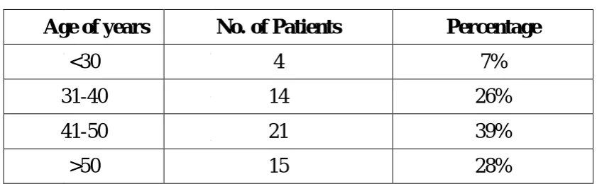
In our study we have taken 54 cases of hemorrhoids in Kilpauk Medical College Hospital during the period from 2016-2017.
The incidence of Haemorrhoids apparently increases with the age. In this study the majority of cases were above 40years of age and constituting about 38%.
Table 1
Age of years No. of Patients Percentage
<30 4 7%
31-40 14 26%
41-50 21 39%
>50 15 28%
SEX INCIDENCE
[image:70.595.98.519.306.436.2]Among the 54 cases, 47 were males and 7 were females and ratio
Table 2
Total no. of Patients Male Female
Figure 11:CLINICAL PRESENTATION
FAMILIAL PREDISPOSITION
Only two patients have the positive family history. Their fathers had previous history of Hemorrhoids.
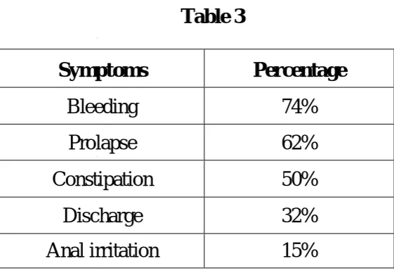
CLINICAL PRESENTATION
Table 3
Symptoms Percentage
Bleeding 74%
Prolapse 62%
Constipation 50%
Discharge 32%
Anal irritation 15%
In our study bleeding and prolapsed were the two cardinal symptoms.
37 35
27
14
10 40
33
27
17
8
BLEEDING PROLAPSE CONSTIPATION DISCHARGE ANAL IRRITATION
SYMPTOMS
[image:71.595.168.449.513.707.2]Bleeding
In this study bleeding was one of the main presenting feature in most of the cases Among the 54 cases the bleeding was the presenting feature in 40 cases.
Prolapse
Prolapse is the main presenting feature in this study. Among 54 cases 33 patients had prolapse.
Constipation
Among 54 patients in this study,27 patients had constipation
Discharge
Among 54 patients in this study , 17 patients have had discharge
Anal irritation
Among 54 patient in this study ,8 patient had anal irritation
Symptoms of secondary anemia
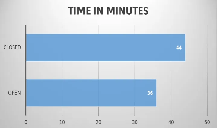
OPERATING TIME IN MINUTES
[image:73.595.129.489.138.351.2]OPERATIVE TIME
Table 4
Open Method Closed method
Operating time 36 ± 7(minutes) 44 ± 8(minutes)
The operating time is shorter in the group I open method . In closed method the operating time was slightly longer
POST OPERATIVE PAIN
The increased pain after haemorrhoidectomy is considered to be the main reason why patients resist operation. There were various modifications in procedures for reduction of post operative pain and
36
44
0 10 20 30 40 50
OPEN CLOSED
studies were conducted to assess the severity of pain. In this study we compared the open technique (Milligan Morgan operation) with closed haemorrhoidectomy (Ferguson method)
IMMEDIATE POST OPERATIVE COMPLICATION
LENGTH OF HOSPITAL STAY
19 6 15 17 12 3 8 11 0 2 4 6 8 10 12 14 16 18 20
PAIN URINARY RETENTION MINOR BLEEDING SEROUS DISCHARGE
COMPLICATIONS
OPEN CLOSED 4.2 4.3 4.4 4.5 4.6 4.7 4.8 4.9 5 5.1 OPEN CLOSEDIMMEDIATE POST OPERATIVE COMPLICATION
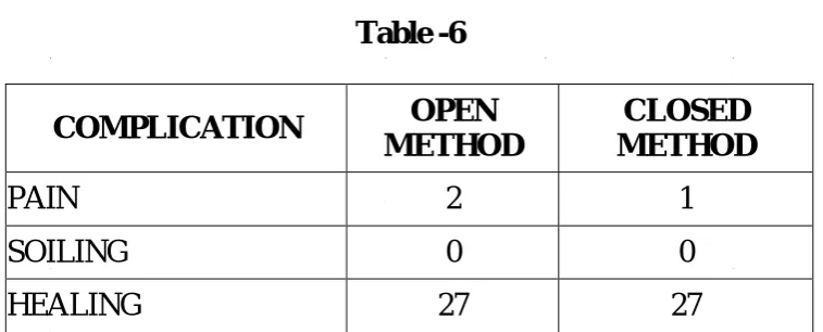
Table -6
COMPLICATION OPEN
METHOD
CLOSED
METHOD 'p' value
PAIN 19(66%) 12(44%) 0.027
URINARY RETENTION 6(22%) 3(10%) 0.102 MINOR BLEEDING 15(56%) 8(32%) 0.016 SEROUS DISCHARE 17(62%) 11(42%) 0.045
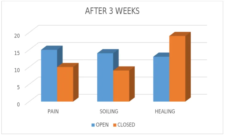
18 15
10
12 10
12
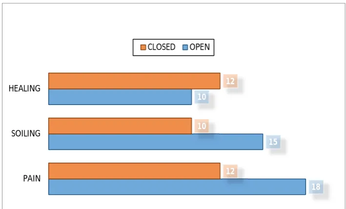
PAIN SOILING HEALING
AT 3 DAYS FOLLOW-UP
Table -6
COMPLICATION OPEN
METHOD
CLOSED
METHOD P VALUE
PAIN 18(66%) 12(44%) 0.027
SOILING 15(55%) 10(37%) 0.045
HEALING 10(37%) 12(44%) 0.024
LENGTH OF HOSPITAL STAY
The length of hospital stay in each group was as follows.
Table 7
Open method Closed method
No of days 5 days 4.5 days
The duration of inability to work 14 days 10 days
Reactionary or secondary haemorrhage
[image:76.595.96.525.452.524.2]Anal Fissure
In both groups no one developed this complication.
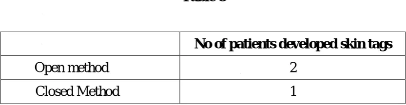
Formation of skin tags
[image:77.595.100.517.310.418.2]The skin tags after open method was found to be about 4% and 2% for closed method.
Table 8
No of patients developed skin tags
Open method 2
Closed Method 1
Anal stenosis
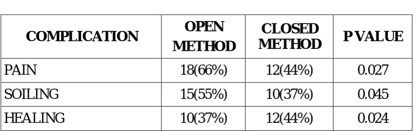
AFTER 3 WEEKS FOLLOWUP
Table 9
COMPLICATION OPEN
METHOD
CLOSED
METHOD P VALUE
SOILING 14 9 0.044
PAIN 15 10 0.028
After three weeks follow up, pain and soiling was found to be more in open method compared to closed method which is statistically significant.
0 5 10 15 20
PAIN SOILING HEALING
AFTER 3 WEEKS
[image:78.595.126.495.136.357.2]AFTER 3WEEKS -HEALING
Table 10
OPEN METHOD CLOSED METHOD P VALUE
13 19 0.025
After 3 weeks follow up healing found to be good in closed method compared to open method which is statistically significant
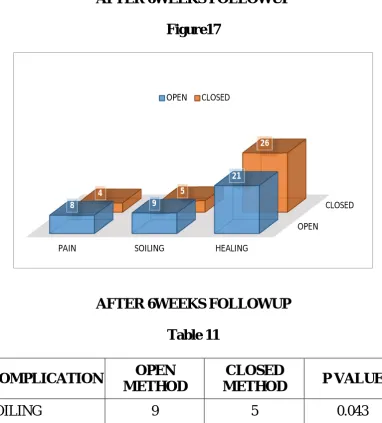
[image:79.595.115.498.319.743.2]AFTER 6WEEKS FOLLOWUP
Figure17
AFTER 6WEEKS FOLLOWUP
Table 11
COMPLICATION OPEN
METHOD
CLOSED
METHOD P VALUE
SOILING 9 5 0.043
PAIN 8 4 0.038
OPEN
CLOSED
PAIN SOILING HEALING
8 9
21
4 5
26
After 6 weeks follow up pain and soiling found to be more in open method compared to closed method which is statistically significant.
AFTER 6 WEEKS FOLLOWUP -HEALING
Table 12
OPEN METHOD CLOSED METHOD P VALUE
21 26 0.007
After 6 weeks follow up healing found to be better in closed method compared to open method which is statistically significant.
[image:80.595.120.499.417.570.2]AFTER 3 MONTHS- FOLLOWUP
Table -6
COMPLICATION OPEN
METHOD
CLOSED METHOD
PAIN 2 1
SOILING 0 0
DISCUSSION
AGE INCIDENCE
Table 13
Authors Over the age of 50 years
Clark et al (1969) 50%
Cuschieri (1996) 50%
Our study (2016- 2017) 28%
According to Clark et al (1969) and Cushieri (1996), half of the patients with haemorrhoids belong to the age group of above 50years. In our study the percentage does not coincide with above authors findings.
SEX INCIDENCE
Table 14
Authors Male Female
Goligher (1969) 66% 33%
Emina Carapeti(1998) 60% 40%
Our study (2011-2012) 87% 13%
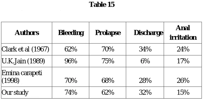
CLINICAL REPRESENTATION
In our study Bleeding and prolapse were the cardinal symptoms in haemorrhoids as listed below, and the bleeding was the presenting feature in 74% of patients and prolapse was the presenting feature in 62% of patients.
[image:82.595.98.521.340.546.2]The study made by the following authors is comparable to that of our study.
Table 15
Authors Bleeding Prolapse Discharge Anal
irritation
Clark et al (1967) 62% 70% 34% 24% U.K.Jain (1989) 96% 75% 6% 17% Emina carapeti
(1998) 70% 68% 28% 26%
Our study 74% 62% 32% 15%
Operative time
Table 16
Open method Closed method
Operative time 36±7 44±8 (minutes)
Post operative pain
Table 17
Study Results
Roetal 1987
Closed : 18 No difference was identified Open : 22
Hoetal 1997
Closed : 33 No difference was identified Open :34
POST PROCEDURE COMPLICATION
Table 18
OPEN METHOD CLOSED METHOD
COMPLICATION OUR STUDY
AINUL HADI ET AL.
OUR STUDY
AINUL HADI ET AL. PAIN 19(66%) 06(24%) 12(44%) 02(08%)
P value in our study is < 0.027 and in Ainul et al. study p value is < 0.01. And both the study shows closed method have less post operative pain compared to open method.
Length of hospital stay - The length of hospital stay shows long in
group I patients but statistically not significant.
Table 19
No.of days
OPEN METHOD (group I) 5 (3-6) CLOSED METHOD (group II) 4.5 (3-5)
Reactionary or secondary haemorrhage
[image:84.595.95.520.552.624.2]FOLLOWUP AT 3 WEEKS
Table 20
COMPLICATION
OPEN METHOD CLOSED METHOD
OUR STUDY
ARBMAN ET AL.
OUR STUDY
ARBMAN ETAL.
PAIN 15(58%) 54% 10(36%) 46% SOILING 14(54%) 78% 9(34%) 27%
P value for pain and soiling at 6 weeks in this study are < 0.028, and <0.025 respectively. Arbman et al. study concluded that at three weeks pain and soiling are higher in open method compared to the closed method. Arbman et al study coincide with our study.
HEALING AFTER 3 WEEKS
Table 21
COMPLICATION
OPEN METHOD CLOSED METHOD
OUR STUDY
ARBMAN ET AL.
S.Y.ET AL
OUR STUDY
ARBMAN ET AL.
S.Y.ET AL
HEALING 48% 18% 18% 70% 86% 75%
[image:85.595.98.522.568.657.2]method when compared to open method as per the tabular column shown above, and the healing is better after three weeks with p value <0.025.
[image:86.595.102.521.256.376.2]FOLLOWUP AT 6 WEEKS
Table 22
COMPLICATION
OPEN METHOD CLOSED METHOD
OUR STUDY
ARBMAN ET AL.
OUR STUDY
ARBMAN ETAL.
PAIN 34% 24% 16% 19%
SOILING 36% 52% 18% 28%
Pain and soiling at 6 weeks follow up is more in open method when compared to closed method as per the results mentioned above. P value in our study for pain and soiling at 6 weeks are <0.038 and <0.043 respectively.
HEALING AFTER 6 WEEKS
Table 23
COMPLICATION
OPEN METHOD CLOSED METHOD
OUR STUDY
ARROYO ET AL.
OUR STUDY
ARROYO ETAL.
[image:86.595.102.522.638.736.2]Healing after 6weeks was better in closed method when compared to open method as shown in the tabular coloumn with p value for our study <0.007.
Formation of skin tags
[image:87.595.122.496.314.433.2]Skin tags formed only in 4 patients in open group and 2 patients in closed group. But the incidence was not significant.
Table 24
Name of procedure No of patients developed
(Skin tags)
Open 4
Closed 2
Anal stenosis and incontinence
SUMMARY AND CONCLUSION
The following are the findings in our study of 54 patients with haemorrhoids treated at Kilpauk Medical College Hospital, Chennai during the period 2016-2017.
1. Post operative pain was significantly lower in closed method when compared to open method.
2. Pain and soiling are lower in closed method compared to open method after 3to 6 weeks follow up.
3. Wound healing was better in closed method compared to open method after 3 to 6 weeks follow up .
4. After 3 months follow up, closed method shows better outcome compared to the open method.
5. The operating time was slightly lower in open haemorrhoidectomy than in closed haemorrhoidectomy.
6. The peak age incidence is in between 40-50 years. There is male preponderance.
7. Most of our patients had bleeding and prolapse as presenting features.
PROFORMA
Name: IP No: Age: Sex:
PRESENTING COMPLAINTS
Bleeding Per Rectum : Yes/No/Duration Nature: Mass Per Rectum : Yes/No/Duration
Straining at defecation : Yes/No/Duration Painful defecation : Yes/No/Duration Constipation : Yes/No/Duration
Discharge Per Rectum : Yes/No/Duration Nature: Anal irritation : Yes/No/Duration
PAST HISTORY
Surgeries
Medical conditions: Diabetes/ Hypertension/ Tuberculosis/ Asthma.
FAMILY HISTORY PERSONAL HISTORY
Bowel/Bladder habits Smoker/Alcoholic
EXAMINATION
GENERAL EXAMINATION: Pallor
Icterus Cyanosis Clubbing
Lymphadenopathy
Vitals: Pulse rate: Blood pressure:
SYSTEMIC EXAMINATION Per abdomen:
Cardiovascular System: Respiratory System: Central Nervous System:
LOCAL EXAMINATION Per rectal: Digital
Proctoscopy:
Colonoscopy for patients more than 40 years of age
DIAGNOSIS:
INVESTIGATIONS
Hb%: TC: DC: ESR: RBS: Blood urea:
Serum creatinine: BT: CT:
Chest x-ray: ECG:
PREOPERATIVE PREPARATION
Overnight fasting; Injection TT;
Shaving of relevant parts;
Proctoclysis enema - on the previous night;
PROCEDURE
Anaesthesia : Position:
Hemorrhiodectomy
Duration at each site: Postop
Antibiotic: Analgesic:
TYPE OF SURGERY
Open hemorrhoidectomy Closed hemorrhiodectomy
OTHER VARIABLES
LENGTH OF HOSPITAL STAY OPERATING TIME
FOLLOW UP- 3 days, 2 weeks ,
4 weeks , 3 months
SCORING SHEET
1) Pain score
No pain 0
Mild pain(relieved by oral medications) 1-3
Moderate pain(relieved by parentral medications) 4-7
Worst pain(relieved only by sedation) 8-10
2) Urinary retention : yes/no 3) Minor bleeding
Surface area of pad soakage <1/4 : yes/no 4) Serous discharge:
BIBLIOGRAPHY
1. Bailey & Love ., Short Practice of surgery 25th Edition ; 2008-page no 1253-1259.
2. Schwartz's principles of surgery -9th edition 1057-1059.
3. Shakelford text book of surgery page 2029-2035Nyhus operative surgery page 1804-1810
4. Cameeroon practice of surgery page 261-268 .
5. Cuschieri.A., Essential Surgical practice Page 635-637-3rd edition.
6. Maingot’s.,Abdominal Operation 10th Edition : page 676-680
7. Sabiston text book of surgery 18th edition. page no 1440-1443.
8. Ambrose ,N.S,Hares MM., Alexandar-williams ,J.et al. Prospective randomized comparison of photocoagulation and rubber band ligation in treatment of haemorrhoids .Br.Med .J.:1983.,286.
10. Arabi ,Y., Alexander-williams ,J. and Keighlely,M.R.B. Anal pressures in hemorrhoids and anal fissure,. AMJ.Surge.; 1977., 134:608.
11. Bates ,T. Rectal Prolapse after anorectal dilatation in the elderly
12. Broader,J.H.,Gunn,I.F. and Alexander-williams ,J.Evalution of a bulk forming evacuant in the management of hemorrhoids Br.J.Surg 1974;61,142
13. Burritt,D. Varicose veins, deep vein thrombosis and hemorrhoids. Epidemiology and suggested etiology. Br.Med.J. 1972 ; 2:556.
14. Chant, A.D.B., May,A. and Wilken, B.J. (1972) Haemorrhoidectomy versus manual dilatation of the anus.Lancet,1972;2:398.
15. Clark, Results of conservative treatment of internal haemorrhoids.Br.Med.J.1967;2:12.
16. Dev P.G. Haemorrhoids : Amelioration without tears, JAMA 1985:2:43