0022-538X/08/$08.00⫹0 doi:10.1128/JVI.01240-07
Copyright © 2008, American Society for Microbiology. All Rights Reserved.
A Highly Divergent Picornavirus in a Marine Mammal
䌤
A. Kapoor,
1,2J. Victoria,
1,2P. Simmonds,
3C. Wang,
4R. W. Shafer,
4R. Nims,
5O. Nielsen,
6and E. Delwart
1,2*
Blood Systems Research Institute, San Francisco, California 941181; University of California, San Francisco, San Francisco,
California2; Centre for Infectious Diseases, University of Edinburgh, Summerhall, Edinburgh EH9 1QH,
United Kingdom3; Division of Infection Diseases, Stanford University, Stanford, California4;
BioReliance Corporation, 14920 Broschart Road, Rockville, Maryland 208505; and
Department of Fisheries and Oceans, Central and Arctic Region, 501 University Crescent, Winnipeg, Manitoba R3T 2N6, Canada6
Received 6 June 2007/Accepted 6 October 2007
Nucleic acids from an unidentified virus from ringed seals (Phoca hispida) were amplified using sequence-independent PCR, subcloned, and then sequenced. The full genome of a novel RNA virus was derived, identifying the first sequence-confirmed picornavirus in a marine mammal. The phylogenetic position of the tentatively named seal picornavirus 1 (SePV-1) as an outlier to the grouping of parechoviruses was found consistently in alignable regions of the genome. A mean protein sequence identity of only 19.3 to 30.0% was found between the 3D polymerase gene sequence of SePV-1 and those of other picornaviruses. The predicted secondary structure of the short 506-base 5ⴕ-untranslated region showed some attributes of a type IVB internal ribosome entry site, and the polyprotein lacked an apparent L peptide, both properties associated with the
Parechovirusgenus. The presence of two SePV-1 2A genes and of the canonical sequence required for cotrans-lational cleavage resembled the genetic organization of Ljungan virus. Minor genetic variants were detected in culture supernatants derived from 8 of 108 (7.4%) seals collected in 2000 to 2002, indicating a high prevalence of SePV-1 in this hunted seal population. The high level of genetic divergence of SePV-1 compared to other picornaviruses and its mix of characteristics relative to its closest relatives support the provisional classifi-cation of SePV-1 as the prototype for a new genus in the familyPicornaviridae.
Previous viral outbreaks in marine mammals, such as mor-billivirus infections in European seals and Eastern U.S. dol-phins, have temporarily reduced their populations (5, 16). Morbillivirus infections have also been detected in marine mammals in the Canadian arctic as evidenced by the detection of neutralizing antibodies in walruses (29). Other infectious diseases known to be circulating in Canadian arctic marine mammals include brucellosis (30), influenza A virus (28), ca-nine adenovirus, and dolphin rhabdovirus (31), but their effects on population morbidity and mortality are largely unknown.
Ringed seals (Phoca hispida), one of the most abundant marine mammals species in the Arctic, are hunted by Canadian Inuit. Fluctuations in the ringed seal population in the Beau-fort Sea of the Northwest Territories have been previously documented (8, 37) and are thought to be associated mainly with climatic conditions during the breeding season. No infor-mation on the number of seals in the Beaufort Sea population is available for the years 2000 to 2002, although ice conditions were normal (Lois Harwood, personal communication).
A recent analysis of lungs, lymph nodes, and nasal swabs from ringed seals hunted in 2000 to 2002 from Ulukhaktok (formerly known as Holman) on the shore of the Beaufort Sea demonstrated the presence of a virus causing strong cytopathic effects (CPE) in Vero cells. The causative agent of CPE passed through a 0.45-m filter was resistant to detergent inactivation
and was therefore thought to be a nonenveloped virus. Here, we analyzed this virus using sequence-independent PCR am-plification and sequence similarity searches. We report the full genome sequence of a novel picornavirus with a deep root on thePicornaviridaefamily phylogenetic tree. Consistent with the nomenclature for other picornaviruses, we suggest the name seal picornavirus 1 (SePV-1) for this new virus and propose that it represent the prototype of a new picornavirus genus.
MATERIALS AND METHODS
Extraction of viral nucleic acids.A nasal swab from a seal hunted in 2002 was used to inoculate Vero cells, which developed CPE. CPE was transferable to a fresh cell culture. The supernatant from this cell culture was used as the input to nonspecifically amplify viral nucleic acids. For enrichment of viral particles from the supernatant of infected cells, 2 ml of culture supernatant was clarified
(5,000⫻gfor 10 min) and filtered through a 0.45-m-pore-size sterile filter
(Millipore HV Durapole) to remove large particles. To concentrate virus
parti-cles, 1.5 ml of filtrate was then centrifuged at 35,000⫻gfor 3 h at 10°C, and the
resulting pellet was resuspended in 100l of water containing 1⫻Turbo DNase
buffer. To remove non-viral-particle-protected DNA from the cultured cells, 20 U of TurboDNase (Ambion) was added, followed by incubation at 37°C for 90 min. Particle-protected nucleic acids were then extracted using a viral RNA extraction kit (Qiagen).
Random amplification, subcloning, and sequencing.Viral RNA was mixed with 50 pmol of primer RA01 (GCCGGAGCTCTGCAGATATCNNNNNNN
NNN), denatured at 75°C for 5 min, and chilled on ice. A reaction mix of 9l
containing 4l of 5⫻first-strand buffer (Invitrogen), 1l of 100 mM
dithio-threitol (DTT), 1l solution containing each deoxynucleoside triphosphate
(dNTP) at 10 mM, 8 units of recombinant RNase inhibitor (Promega), and 200 units of SuperScript II reverse transcriptase (RT) (Invitrogen) was added. The reaction mixture was incubated at 25°C for 10 min and then at 42°C for 50 min. After a denaturation step at 94°C for 3 min and chilling on ice (to reanneal
primer RA01 to cDNA), 2.5 units (0.5l) of 3⬘-5⬘Exo⫺Klenow DNA
polymer-ase (New England Biolabs) was added to extend RA01 and incubated at 37°C for
* Corresponding author. Mailing address: Blood Systems Research Institute, 270 Masonic Ave., San Francisco, CA 94118. Phone: (415) 923-5763. Fax: (415) 276-2311. E-mail: delwarte@medicine.ucsf.edu.
䌤Published ahead of print on 17 October 2007.
311
on November 8, 2019 by guest
http://jvi.asm.org/
1 h, followed by an enzyme inactivation step at 75°C for 10 min. A total of 7.5l of the RT-Klenow-treated product was then used as a template in a subsequent
50-l PCR mixture consisting of 1⫻AmpliTaq Gold PCR buffer II (100 mM
Tris䡠HCl [pH 8.3], 500 mM KCl) (Applied Biosystems), 3 mM MgCl2, each
dNTP at 0.3 mM, 50 pmol of primer RA02 (GCCGGAGCTCTGCAGATATC), and 2.5 units of AmpliTaq Gold DNA polymerase LD (Applied Biosystems). An initial denaturation step for 5 min at 95°C was followed by 40 cycles of PCR (95°C for 1 min, 55°C for 1 min, and 72°C for 2 min). Random PCR products were then separated on a 1.5% agarose gel, and DNA smears ranging in size from approximately 400 to 1,500 bp were excised and extracted using the QIA-quick gel extraction kit (Qiagen). Five microliters of the eluted, purified PCR product was ligated into the pGEMT-Easy vector (Promega Inc.) and introduced
into chemically competentEscherichia coliTOP-10 cells (Topo One Shot;
In-vitrogen). Bacteria were plated onto LB agar plates containing ampicillin, X-gal
(5-bromo-4-chloro-3-indolyl--D-galactopyranoside) and IPTG (isopropyl--D
-thiogalactopyranoside). Ninety-six white colony inserts were sequenced using the T-7 forward primer.
Sequence analysis.Sequence data for all clones were imported into Sequencer 4.1 (Genecode) and trimmed of vector and primer (RA02) sequences. The remaining sequences were then assembled into contigs using an assembly pa-rameter of a minimum 90% base identity with at least a 30-nucleotide overlap. A sequence similarity search was performed using tBLASTx (http://www.ncbi.nlm .nih.gov/BLAST/).
SePV-1 genome sequencing.Sequence contigs of sequences were assembled using Sequencher software. Contigs showing significant tBLASTx hits to
picor-naviruses (E value of⬍0.001) were then linked using PCR. To acquire the 3⬘end
of viral genome, 10l of extracted RNA was mixed with 10 pmol of primer
DT-01 (ATTCTAGAGGCCGAGGCGGCCGACATGTTTTTTTTTTTTTTTT TTTTTTTTTTTTTTVN, where V is A/C/G and N is A/C/G/T), denatured at
75°C for 5 min, and chilled on ice. A reaction mix of 9l containing 4l of 5⫻
first-strand buffer (250 mM Tris䡠HCl [pH 8.3], 375 mM KCl, 15 mM MgCl2)
(Invitrogen), 2l of 100 mM DTT, a 1-l solution containing each dNTP at 10
mM, 8 units (0.2l) of recombinant RNase inhibitor (Promega), and 200 units
of SuperScript II reverse transcriptase (Invitrogen) was then added and incu-bated at 45°C for 30 min, followed by 75°C for 10 min. Two units of RNase H was added, and the reaction mixture was further incubated for 10 min at 37°C. PCR was performed using a virus-specific primer, RdRp-1 (CTGTGCCTGATTTCC CTGAATCT), and DT-02 (ATTCTAGAGGCCGAGGCGGCC). PCR con-sisted of an activation step of 5 min at 95°C followed by 35 cycles of amplification
at 95°C for 1 min, 60°C for 30 s, and 72°C for 2 min. To acquire the 5⬘end of the
SePV-1 genome, 10l of extracted RNA was mixed with 10 pmol of virus
specific-primer VP-R-1 (AGCCATACCCCCTTGGTCTT), denatured at 75°C
for 5 min, and chilled on ice. An RT reaction mix similar to that used for 3⬘rapid
amplification of cDNA ends was added, and the reaction mixture was incubated at 52°C for 30 min, followed by 75°C for 10 min. Two units of RNase H was then added, and the reaction mixture was further incubated for 10 min at 37°C. cDNA was purified using a Qiagen PCR purification kit, and a poly(C) tail was added using terminal deoxynucleotide transferase and dCTP. PCR was performed using the SePV-1-specific primers VP-R-2 (GCGACACGCACACAACTACA) and PPC01 (GGCCACGCGTCGACTAGTACGGGIIGGGIGGGGIGG, where I is deoxyinosine). PCR cycles consisted of an enzyme activation step for 5 min at 95°C followed by 35 cycles of amplification 95°C for 1 min, 60°C for 30 s, and 72°C for 2 min. PCR products were then directly sequenced using the virus-specific primers to acquire viral sequences of both extremities.
Detection of SePV-1 by RT-PCR.One hundred forty microliters of the cell culture supernatants was extracted using a viral RNA extraction kit (Qiagen)
according to the manufacturer’s instructions. The RNA was eluted with 50l of
RNase-free distilled water containing 40 U of RNase inhibitor (RNasin;
Pro-mega). Fifty picomoles of a random octamer oligonucleotide was added to 10l
of RNA, denatured at 75°C for 5 min, and then chilled on ice. A reaction mix of
9l containing 4l of 5⫻first-strand buffer, 2l of 100 mM DTT, a 1-l
solution containing each dNTP at 10 mM, 8 units (0.2l) of recombinant RNase
inhibitor (Promega), and 200 units of SuperScript II reverse transcriptase (In-vitrogen) was then added and incubated at 45°C for 30 min, followed by 75°C for 10 min. PCR was performed using primers VP1F1 (TGATGATGTGTTGGGA AGACTCCA) and VP1R1 (TACCCAGGAAACAAATTTGGCAAT), target-ing positions 1844 to 3341 of the SePV-1 genome. PCR consisted of an AmpliTaq Gold (Applied Biosystems) activation step for 5 min at 95°C followed by 35 cycles of amplification at 95°C for 1 min, 55°C for 30 s, and 72°C for 2 min. The PCR products including the entire VP1, 2A, and 2B sequences were separated in 1% agarose gels and purified with a QIAquick gel extraction kit (Qiagen). Nucleo-tide sequences that were 1,428 nucleoNucleo-tides long were determined by using a 373A
DNA autosequencer (PE-Applied Biosystems) using primers VP1R1 and VP1F1.
Phylogenetic analysis.Translated sequences from the coding region of SePV-1 and other picornaviruses were aligned using ClustalW with default settings (10). Amino acid sequence divergence of SePV-1 from other picornaviruses was de-termined for the 3D (pol) region and by sliding-window analysis of the nonstruc-tural region using the program Sequence Distance within the Simmonic2005 v1.6 sequence editor (34).
Phylogenetic analysis on aligned regions within the NS region was carried out by neighbor joining of translated amino acid distances implemented in the program MEGA2 (24). Trees were rooted by their longest branch in the absence of a naturally occurring outgroup for the picornavirus sequences; the most
similar positive-stranded RNA viruses, such as members ofComoviridaeand
Sequiviridae, were too divergent for defensible alignments in the regions ana-lyzed. Confidence in phylogenetic groupings was obtained by generating consen-sus trees derived from 100 sets of bootstrap-resampled sequence data.
RNA structure determination.Prediction of RNA secondary structure in the
5⬘-untranslated region (5⬘UTR) and 3⬘UTR was carried out using a standard
minimum free-energy method (MFOLD) (46) and PFOLD (21). For estimation of mean folding energies (MFEs) of fragments of the SePV-1 genome and control sequences, the program ZIPFOLD was used with default settings (46). Consecutive fragments of 498 bases overlapping by 249 bases were generated from aligned complete genome sequence data sets of each of the four virus groups (sequences listed below). MFEs were determined by MFOLD (46), and the sequence order-dependent component to folding was determined by com-parison with sequence order-scrambled copies of each native sequence. The program NDR (34) was used for sequence order randomization to retain any biases in dinucleotide frequencies that might exist among native sequences. MFE results were expressed either as MFE differences (MFEDs), i.e., the percentage difference between the MFE of the native sequence from that of the mean value
of the 50 sequence-order-randomized controls, or as aZscore, which is the
position of the MFE of the native sequence within the distribution of MFEs of the randomized sequences expressed as the number of standard deviations from
the mean value of the randomized sequences; thus, values between⫺2 and⫹2
fall within the range of 95% of their MFE value (43).
Picornavirus sequences.For determination of sequence divergences between SePV-1 and those of other picornaviruses, two or more representative serotypes from each species within each of the nine currently classified genera of picorna-viruses were aligned. Sequences used for the comparison comprised the
follow-ing: for the genusAphthovirus, GenBank accession numbers NC_011450, FMD
VALF, FDI320488, NC_004004, NC_002554, FMV7572, NC_004915,
AY687334, NC_003992, FDI251473, AY593843, NC_011451, AY593853, and NC_011452 for foot-and-mouth disease virus (FMDV) and accession numbers
NC_003982 and ERVPOLY for equine rhinitis A virus; for the genus
Enterovi-rus, accession numbers NC_003988 for simian enterovirus, NC_002058,
HPO132960, POL2LAN, POL544513, POL3L37, HPO293918, NC_001428, and CXA24CG for human enterovirus species C, NC_002347, NC_001360, NC_000881, NC_001657, NC_002601, NC_001342, NC_001656, and NC_001472 for species B, NC_001612 and ETU22522 for species A, NC_001430 and AY426531 for species D, BEVVG527 and AY508697 for bovine enterovirus, and
NC_001617 and NC_001490 for human rhinovirus; for the genusHepatovirus,
accession numbers NC_003990, AJ225173, AY517471, and AY275539 for avian encephalomyocarditis virus, SHVAGM27 for simian hepatitis A virus (HAV),
and HAVRNAGBM and NC_001489 for human HAV; for the genus
Cardiovi-rus, accession numbers NC_001479 and XXEVCG for encephalomyocarditis
virus and NC_001366 and AB090161 for Theiler’s virus; for the genusErbovirus,
accession numbers NC_003077 and NC_003983; for the genusParechovirus,
accession numbers AF538689, AF327921, and AF327920 for Ljungan virus (LV) and L02971, NC_001897, AF055846, NC_008286, AB252582, NC_003976, AF538689, AF327922, AF327921, EF051629, DQ315670, AM235749, and
AB084913 for human parechoviruses (HPeV); for the genusKobuvirus, accession
number NC_004421 for bovine kobuvirus and NC_001918 and DQ028632 for
Aichi virus; for the genusTeschovirus, accession numbers PEN011380 and
AF296117; for currently unclassified viruses, accession numbers EF382778, DQ249301, DQ249300, DQ249299, and EF093502 for duck hepatitis virus (DHV) and DQ641257 for Seneca Valley virus (SVV); and for members of the
proposed “Sapelovirus” genus, accession numbers NC_003987 for porcine
en-terovirus A, AY064708 for simian picornavirus 1, and NC_006553 for duck picornavirus.
For RNA structure comparisons, the following complete genome sequences
were used: for the genusAphthovirus, GenBank accession numbers AF154271,
FMDVALF, NC_002554, NC_003982, NC_003992, NC_004004, and NC_002527
for FMDV; for the genusCardiovirus, accession numbers NC_001479 and MNG
on November 8, 2019 by guest
http://jvi.asm.org/
POLY for encephalomyocarditis virus and NC_001366 for Theiler’s virus; for the
genus Enterovirus, accession numbers NC_001428, NC_001430, NC_001472,
NC_001490, NC_001612, NC_001617, NC_001752, NC_001859, NC_002058,
NC_003986, NC_003988, POL3L37, and SVDMPS; for the genusErbovirus,
accession number NC_003983; for the genusHepatovirus, accession numbers
NC_003990 for avian encephalomyocarditis virus, SHVAGM27 for simian HAV,
and NC_001489 for human HAV; for the genusKobuvirus, accession numbers
NC_004421 for bovine kobuvirus and NC_001918 for Aichi virus; and for the
genus Teschovirus, accession numbers AB038528, AF231769, AF296087,
AF296091, AF296093, AF296115, AF296119, and NC_003985.
Nucleotide sequence accession number.The annotated genome sequence re-ported here has been deposited in GenBank under accession number EU142040.
RESULTS
Sequencing of new virus.Virions in CPE-causing cell culture supernatant were first purified from larger particles by filtra-tion, and contaminating cellular DNAs were digested by DNase, while viral nucleic acids remained protected from di-gestion within viral capsids (1, 15). Total nucleic acids were then extracted and purified, and viral RNA was amplified fol-lowing reverse transcription with an oligonucleotide primer containing a randomized 3⬘-end sequence followed by second-strand synthesis using the same primer with Klenow DNA polymerase. The fixed portion of this oligonucleotide was then used to PCR amplify the DNA fragments with complementary ends (see Materials and Methods). The random PCR product was purified and subcloned, and the inserts of 96 plasmids were sequenced. Sequence data were analyzed using tBLASTx (i.e., translated in all six frames and aligned against the entire sim-ilarly translated GenBank database).
The viral RNA-derived sequences revealed the presence of four contigs showing sequence similarities to picornavirus pro-teins with amino acid identities ranging from between 22 and 41%. Except for the extremities of the contigs, each base of the contigs was sequenced at least twice from different overlapping
subclones. The three gaps between the four contigs were am-plified from the randomly primed cDNA using primers lo-cated within contigs followed by direct PCR product se-quencing. The 5⬘and 3⬘extremities of the viral genome were acquired using rapid amplification of cDNA ends (see Ma-terials and Methods).
Relationship to other picornaviruses.The SePV-1 genome was 6,693 nucleotides long, excluding the poly(A) tail, and encoded a 2,027-amino-acid-long polyprotein. The genome showed a relatively low G⫹C content of 44% [excluding the poly(A) tail] compared to other picornaviruses. Its composi-tion was most similar to those of HPeV and LVs in the Pare-chovirus genus (40% and 42%, respectively), DHV (44%), teschoviruses (44% to 45%), and equine rhinitis A virus equine rhinitis A virus (46 to 48%); greater than that of HAV (38%); and generally lower than those of other picornaviruses (e.g., FMDV [52 to 55%], cardioviruses [48 to 49%], enteroviruses [39 to 49%], and kobuviruses [55% to 60%]).
[image:3.594.43.540.77.316.2]To investigate its sequence relationship with other picorna-viruses, alignments of the SePV-1 polyprotein were made in the P1 and 2C-3D regions of the genome with representative members of each picornavirus genus (listed in Materials and Methods). Regions of amino acid sequence similarities were most apparent in the nonstructural region of the SePV-1 ge-nome, such as the region surrounding highly conserved motifs in the active site of the RNA-dependent RNA polymerase. Alignment of the P1 region was more problematic, with several regions with no detectable homology between genera and a frequent necessity to introduce long gaps to align homologous sites. Although it is possible that both the 3D and P1 align-ments could be optimized further, this would have only a small effect on sequence similarity values (Table 1 and Fig. 1A) or phylogenetic relations (Fig. 1B).
TABLE 1. Pairwise amino acid sequence identities between SePV-1 and other picornavirus genera in the P1 and 3D regionsa
Virus
% Sequence identity with:
SePV-1 Parechovirus DHV Aphthovirus Cardiovirus Erbovirus Teschovirus SVV Enterovirus Sapelovirus Hepatovirus Kobuvirus
P1 region
Parechovirus 25.6 61.1
DHV 24.3 28.9 78.3
Aphthovirus 11.6 12.1 12.4 61.8
Cardiovirus 9.3 12.2 12.5 25.9 70.7
Erbovirus 10.3 12.3 12.6 30.2 31.5 73.5
Teschovirus 9.4 11.5 11.8 20.0 23.4 21.6 79.6
SVV 10.6 11.8 10.7 25.2 31.9 29.4 20.2 NA
Enterovirus 11.0 10.9 12.3 18.9 20.1 21.0 17.6 19.8 54.2
Sapelovirus 10.5 11.5 12.4 19.8 21.2 22.3 18.7 21.1 34.8 49.1
Hepatovirus 12.4 12.2 11.0 13.9 14.2 14.1 13.0 13.4 15.5 13.4 69.9
Kobuvirus 12.0 12.7 12.5 15.5 18.0 18.4 14.8 17.6 16.9 16.1 12.8 63.7
3D region
Parechovirus 29.3 64.2
DHV 30.0 35.2 97.9
Aphthovirus 20.5 20.5 20.3 83.0
Cardiovirus 21.6 21.6 20.3 40.5 72.6
Erbovirus 19.7 21.4 18.7 35.2 39.8 96.3
Teschovirus 19.7 21.5 23.5 33.8 36.1 36.5 97.2
SVV 23.4 22.3 21.7 43.6 55.7 37.6 38.1 NA
Enterovirus 21.5 23.8 22.9 27.7 28.7 28.3 30.5 28.7 73.1
Sapelovirus 22.1 24.2 23.8 29.2 28.7 29.0 32.5 29.0 53.5 61.0
Hepatovirus 19.3 21.6 20.1 20.8 22.0 19.5 22.2 22.2 23.8 24.5 62.4
Kobuvirus 20.1 22.2 18.7 31.8 31.2 30.3 28.2 29.8 32.0 33.8 22.7 80.7
a
Boldface type, averages of pairwise amino acid sequence identities between SePV-1 and members of different picornavius genera. Italics, averages of pairwise amino acid sequence identities between members of the same picornavirus genus. NA, not applicable.
on November 8, 2019 by guest
http://jvi.asm.org/
Both the P1 and 3D regions of SePV-1 showed the greatest similarity to DHV (24% and 30%, respectively) and to the parechoviruses LV and HPeV (26% and 29%) (Table 1). Sim-ilarity was less than 13% (P1) and 24% (3D) with all other picornavirus genera (Table 1). The level of sequence identity between SePV-1 and other picornaviruses was within the range seen between other genera (average of 17.6% and range of 10% to 35% for P1 and average of 23% and range of 19 to 56% for 3D) (Table 1). Many intervening regions between conserved motifs in the P1 and 2C-3D region dis-played no detectable sequence similarities between genera. This is demonstrated by the sliding window of amino acid sequence divergence between different genera of picornavi-ruses (Fig. 1A).
In the 2C-3D structural gene region, pronounced dips in amino acid divergence between SePV-1 and other genera cor-responded to conserved motifs within 2C, the end of 3C, and
four regions in 3D (Fig. 1A). SePV-1 showed no close se-quence relatedness to any of the picornavirus genera or the currently unclassified SVV or DHV sequences, with the pos-sible exception of marginally greater similarity to parechovi-ruses and DHV (6, 17, 38, 39). In the P1 region, SePV-1 showed a more apparent greater degree of sequence similarity with parechoviruses and DHV (Fig. 1A, black and gray lines, respectively) than other genera.
For analysis of sequence relationships between SePV-1 and different picornavirus genera in the P1 and nonstructural gene regions, the following regions were identified as being reliably alignable and showing unequivocal amino acid similarities. These comprised sequences between positions 912 and 1229 (P1 region A) (all genome positions are numbered according to the SePV-1 sequence), 1599 and 1995 (P1 region B), 3603 and 4044 (2C region C), 5001 and 5165 (3C region D), 5253 and 5423 (3D region E), 5670 and 5879 (3D region F), 5913
FIG. 1. (A) Sliding-window analysis of pairwise-translated protein distances. Mean divergences between SePV-1 and each member of each classified genus were calculated between translated sequential fragments for successive 12-amino-acid fragments, incrementing by 3 amino acids. The 2A-2B region is not shown due to difficulties in obtaining a reliable alignment between genera. (B) Phylogenetic analysis using neighbor joining of different P1 and nonstructural protein regions A to H (positions indicated by solid bars in A). The same color code is used in both analyses.
on November 8, 2019 by guest
http://jvi.asm.org/
[image:4.594.46.539.65.473.2]and 6167 (3D region G), and 6263 to 6521 (3D region H) (Fig. 1A). Phylogenetic trees constructed from picornavirus se-quences from each region demonstrate consistent branching patterns with generally good differentiation of the nine cur-rently classified genera, although the proposed “Sapelovirus” genus showed a substantially greater similarity to the Entero-virusgenus than to other picornaviruses in both structural and nonstructural regions (Fig. 1B). An exception to the otherwise consistent branching patterns of the trees was SVV, which generally grouped within or as an outlier to the cardioviruses but fell within theAphthovirusgenus in region E. In this region, members of the proposed “Sapelovirus” genus also became interspersed with enteroviruses (Fig. 1B).
The phylogenetic relationship between SePV-1 and other picornavirus genera was similarly highly consistent where all regions except E grouped closest to the parechovirus genus and DHV (Fig. 1B). In regions A, B, F, G, and H, these sequences grouped in bootstrap-supported clades. Given the anomalous position of SVV in region E, the lack of grouping of SePV-1 with parechoviruses in this region may be the result of a lack of phylogenetic resolution in this relatively short frag-ment. Indeed, analysis of regions D and E combined restored the SePV-1/parechovirus/DHV grouping (data not shown). The relationships between SePV-1 with DHV and parechovi-ruses differed in different genomic regions. In the P1 region, the three virus groups formed a much more distinct, well-defined clade than in nonstructural regions of the genome (trees C to H), a difference also apparent from the similarity scan (Fig. 1A) (see above). Within the SePV-1/parechovirus/ DHV grouping, DHV split earlier in the lineage in P1, while in the 2C-3D region, SePV-1 was consistently ancestral to the other viruses.
Despite the frequent bootstrap-supported grouping of SePV-1 with parechoviruses and DHV in the nonstructural 2C-3D region and the existence of identifiable homology else-where in the capsid-encoding region, the newly discovered virus was nevertheless highly divergent from each of these virus
groups and indeed similar to sequence divergences that exist between other picornavirus genera, supporting the possibility that a new picornavirus genus was identified.
Polyprotein.A methionine codon starting at nucleotide po-sition 507 was found in a standard Kozak context (RNNA UGG) and used to deduce the start of the polyprotein (22). Picornavirus structural and nonstructural proteins are typically generated by cleavage with virus-encoded proteinases. The hypothetical cleavage map of the SePV-1 polyprotein was de-rived from an alignment with other picornaviruses whose ex-perimentally determined or hypothetical protease cleavage sites have been reported (Fig. 2). The presence of cleavage sites at interdomain junctions was sought based on the prefer-ence of picornaviruses for Q and E at the P1 position and a small amino acid residue (e.g., G, S, R, M, A, and N) at the P1⬘ position (2, 14). Prediction of the cleavage sites in Fig. 2 were therefore determined by scanning for pairs of amino acids fitting this patterns around the interdomain regions inferred based on the amino acid alignment. The predicted cleavage sites result in a typical picornavirus gene order of VP0–VP3– VP1-2A1-2A2-2B–2C–3A–3B–3C–3D (Fig. 1A and 2).
The absence of a predicted maturational cleavage site be-tween 1A and 1B (VP0) was consistent with HPeV, DHV (6), and LV (14) in which VP0 is not cleaved.
An analysis of the N terminus of the 2A protein of SePV-1 revealed a sequence corresponding to the canonical cotransla-tional cleavage site DxExNPG P (Fig. 2). Cleavage at this site is cotranslationally mediated in cisby 2A and is present in cardioviruses, erbovirus, teschovirus, and aphthoviruses as well as in LV and DHV but not in HPeV, hepatoviruses, and the kobuvirus Aichi virus (7, 12, 18, 44, 45). Proteolytic cleavage at this site would therefore release a small 2A1 protein (Fig. 2). Based on the hypothesized cleavage sites, SePV-1 therefore appears to contain two structurally unrelated 2A proteins in a manner analogous to that of LV (13, 14, 17) but unlike HPeV, which contains a single 2A protein (11). Additionally, DHV
FIG. 2. Predicted location of polyprotein cleavage sites and expected lengths of resulting proteins. The P1 positions are highlighted in boldface type, and the cleavage sites are indicated by vertical bars. The amino acids following vertical bar are the P1⬘position. Genomes used were DHV-1 (GenBank accession number 110611895), DHV-2 (accession number 78128452), HPeV5 (accession number 111607003), HPeV2 (accession number 7674174), HPeV4 (accession number 83702491), HPeV3 (accession number 24898927), HPeV31 (accession number 61097778), and LV (accession number 21309880).
on November 8, 2019 by guest
http://jvi.asm.org/
may encode a third 2A protein absent in both LV and SePV-1 (6, 17, 38).
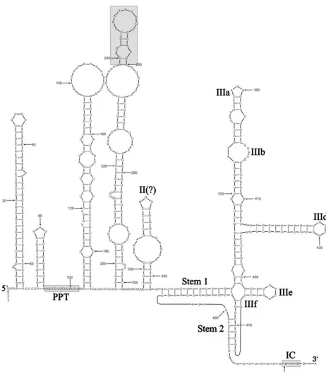
Untranslated terminal regions. The length of the SePV-1 5⬘UTR until the polyprotein initiation AUG was unusually short at 506 nucleotides and included two terminal uracils required to covalently link the RNA to the VPg (3B) protein, a characteristic common to all picornaviruses (3). The
[image:6.594.63.525.64.590.2]second-ary structure of the 5⬘UTR RNA was predicted using a com-bination of a thermodynamic folding energy minimization al-gorithm (MFOLD) and a stochastic context-free grammar method (PFOLD), independent algorithms that produced gen-erally concordant results for the main structural features (Fig. 3). The analysis carried out is necessarily limited by the avail-ability of only a single SePV-1 sequence from the 5⬘UTR; the
FIG. 3. RNA secondary structure of the SePV-1 5⬘UTR predicted by MFOLD and PFOLD. The positions of the a polypyrimidine tract (PPT), a 22-nucleotide region with sequence identity to DHV, and the polyprotein start codon are indicated by shaded boxes. The type IVB IRES has been annotated as previously proposed (9).
on November 8, 2019 by guest
http://jvi.asm.org/
following therefore presents provisional predictions that will have to be confirmed once more sequences from this region become available. For this reason, the presentation of results has concentrated on the more obvious structural features or those supported by similarities to structurally similar internal ribosome entry sites (IRESs) of other viruses.
The first stem-loop was remarkably large, with a duplex region of 28 base pairs, including an uninterrupted pairing of 15 consecutive bases. This stem-loop was much larger than the 5⬘-terminal loops of most other picornaviruses. The down-stream stem-loop was similarly highly stable (duplex length of 9 nucleotides), with a terminal loop of only three unpaired bases. This configuration would likely prevent the formation of the pseudoknot (Cpk) found in a homologous position in HPeV (27) (Fig. 3). Of uncertain functional significance was a polypyrimidine tract (14 bases between positions 90 and 102) in the predicted unstructured region downstream of the second stem-loop (Fig. 3, gray box).
Although neither MFOLD nor PFOLD can predict tertiary RNA structure interactions, sequences at the 3⬘ end of the 5⬘UTR can be convincingly modeled onto the previously pro-posed type IV IRES (9). Specifically, the predicted structure was most closely similar to IRES structures of members of the SapelovirusandTeschovirusgenera and to DHV, together clas-sified as type IVB IRES elements. SePV-1 thus contains the highly conserved IIIe stem-loop (with the unpaired GAYA sequence), a CpG dinucleotide pairing (IIIf), a longer-range interaction to form stem 1, and, finally, a pseudoknot pairing between positions 491 and 495 and upstream positions 467 to 471). As well as structural conservation, there were also several regions of sequence identity between SePV-1 and other viruses with a type IVB IRES (Fig. 4), including both paired and unpaired bases, and a large number of covariant sites between
the six sequences analyzed that lend further bioinformatic sup-port for the proposed structure.
The proposed IRES structure for SePV-1 lacked a tetraloop containing the GNRA sequence, a common feature of most other type I, type II, and type IV IRES sequences. In type IV IRESs, the tetraloop is normally found as the four unpaired bases in stem-loop IIId, which was GGGG in SePV-1. How-ever, a GGGGG pentaloop was found in a homologous posi-tion in the simian picornavirus type IVB, indicating that either it is not absolutely required or the sequence requirement for this element is slightly less stringent than originally proposed (perhaps GNRR).
The 3⬘UTR was also the shortest one reported for a picor-navirus, with a length of 34 nucleotides, the next biggest being from rhinoviruses, at 40 nucleotides. No folded RNA struc-tures were detected.
Myristylation and leader peptide.Myristylation plays a cru-cial role in virion morphogenesis of most picornaviruses and involves the covalent linkage of myristic acid to an N-terminal glycine residue in a canonical GxxxT/S motif. SePV-1 contains a putative myristylation sequence starting at position 16 rela-tive to putarela-tive methionine amino termini. This location for myristylation most resembles that of HPeV at position 13, while DHV has a canonical sequence at position 31 and LV has one at position 3. The non-near-terminal position of the puta-tive myristylation site of SePV-1 indicates that, as proposed for DHV and as shown for HPeV, myristylation may not occur and that alternative modifications at the amino terminus of the polyprotein may direct it to a lipid environment (14, 17, 36).
Leader peptides of variable length are found in the Cardio-virus,Aphthovirus,Teschovirus, Kobuvirus, andErbovirus gen-era, for which various roles have been proposed (18, 32, 33, 44, 45). HPeV and LV are not thought to encode a leader peptide,
FIG. 4. (A) Alignment between the 3⬘end of the 5⬘UTR of SePV-1 and other viruses with type IVB IRESs. These include DHV, members of the proposedSapelovirusgenus, duck picornavirus (DPV), porcine enterovirus A (PEVA), simian virus 2 (SV2) (23, 38), and porcine teschovirus (PTV). Conserved pairings around the region of pseudoknot formation are shown as solid lines; variable pairings are indicated by dotted lines. Regions of duplex formation have been annotated as previously proposed (9). Nucleotide positions showing covariance (compensatory changes) between virus sequences are underlined. Highly conserved, unpaired based are shown in gray boxes.
on November 8, 2019 by guest
http://jvi.asm.org/
[image:7.594.44.545.71.277.2]while DHV may encode a short leader peptide (6). SePV-1, like HPeV and LV, is not expected to encode a leader peptide.
GORS. Previous analyses of RNA structure formation among the different picornavirus genera indicated that mem-bers of the parechoviruses were unstructured, with no signifi-cant differences in MFEs between native and sequence order-scrambled sequences (34). The lack of genome-scale ordered RNA structure (GORS) in theParechovirusgenus contrasted with the high levels of sequence order-dependent RNA struc-ture in the genera Aphthovirus, Kobuvirus, and Teschovirus. Since the possession of GORS varies between genera, we con-ducted a more detailed comparison of RNA structure between human parechoviruses and LV (Parechovirus genus), DHV, and SePV-1 (Fig. 5). Each of the four parechovirus-like virus groups showed low MFEDs averaged over the length of the genome, similar to values previously determined for the “un-structured” enteroviruses and hepatoviruses and distinct from those genera with thePicornaviridaepreviously shown to pos-sess GORS. Analysis of subgenomic regions revealed that the sequence order-dependent RNA structure within SePV-1 and other parechovirus-like viruses was in the 5⬘and 3⬘noncoding regions (data not shown). Recomputation of MFEDs for se-quences confined to the coding region produced mean values of 0.06% for SePV-1 and similarly lower values for HPeV, DHV, and LV (⫺0.57% to 0.95%).
Prevalence and diversity of SePV-1. Cell culture superna-tants derived from 18 out of 108 seals hunted in 2000 to 2002 showed CPE and were tested for SePV-1 RNA using RT-PCR of the complete VP1, 2A, and 2B regions of SePV-1. Eight supernatants were positive (7.4% of animals). Specificity of the PCR was confirmed by sequencing 1,428 bases of the amplifi-cation products, which also revealed the presence of minor
variations between isolates, ranging from 0.2 to 3.7% (average, 1.5%) nucleotide differences.
DISCUSSION
We have genetically characterized the genome of a new picornavirus isolated from arctic ringed seals. Although the hunted animals from which SePV-1 was recovered appeared healthy, with normal body mass indexes, and did not exhibit any specific pathologies after cursory examination, it remains unknown whether this virus may, on occasion, be pathogenic. An extensive regulatory system of wildlife management pre-cludes the inoculation of SePV-1 in this species to test for associated symptoms. Its high prevalence in 2000 to 2002 may represent recent spread or a more stable endemic relationship within this seal population. The absence of a GORS whose presence has been correlated with viral persistence in infected hosts (34) supports the possibility that this virus results in a transient infection, with its high prevalence therefore reflecting a recent epidemic spread. Whether this virus is transmissible to humans by hunting and consuming these seals, sometimes raw, is unknown and will require the testing of exposed populations for antibodies to SePV-1.
Picornaviruses have traditionally been identified and classi-fied based on biophysical/antigenic properties. Viral genome sequences have also been used for taxonomy purposes. The family Picornaviridae is currently divided into nine genera (Aphthovirus,Cardiovirus,Enterovirus,Hepatovirus, Parechovi-rus,Rhinovirus,Erbovirus,Kobuvirus, andTeschovirus) (18, 35). Rhinoviruses have recently been reclassified as a new species within the Enterovirus genus, while sapeloviruses, SVV, and DHV may become new Picornaviridae genera. The nearest genetic neighbors of SePV-1 are the parechoviruses (HPeV and LV) and the currently unclassified DHV, which has been recently proposed as the prototype member of a new picorna-virus genus (6, 17, 38). Relative to the parechopicorna-viruses, SePV-1 has a more basal phylogenetic root than DHV1 in the non-structural 2C and 3D regions, while DHV1 was more basal in the P1 region. SePV-1 is therefore divergent enough from other picornaviruses in both regions to qualify as the prototype of another picornavirus genus.
SePV-1 contains some but not all the characteristics of HPeV, LV, or DHV1. All four viruses show similar G⫹C contents (40 to 44%), but unlike the parechoviruses HPeV and LV, which have type II IRESs, SePV-1 and DHV1 contain 5⬘UTR structures similar to those of sapeloviruses and are classified as type IVB (9). Alternative relationships between these viruses existed in other attributes. For example, like HPeV and DHV but unlike LV, its canonical myristylation site appears to be too far from the C terminus of VP0 to be functional. The two 2A proteins of SePV-1 resemble the ge-netic organization of LV 2A1 and 2A2 proteins and are distinct from HPeV’s single 2A or DHV’s three 2A proteins. Like DHV1 and LV but unlike HPeV, its 2A1/2A2 boundary contains the canonical site required for cotranslational cleavage. Like HPeV and LV but unlike DHV1, the SePV-1 genome appears to be missing a leader protein. SePV-1 therefore also exhibits a unique mixture of picornavirus genetic characteristics.
[image:8.594.55.272.66.322.2]Microarrays consisting of highly conserved viral sequences
FIG. 5. Comparison of GORS for SePV-1 using MFED. The vi-ruses used are similar to those in Fig. 1 and listed in Materials and Methods.
on November 8, 2019 by guest
http://jvi.asm.org/
have been used for the identification of both known and novel viral species (20, 40–42). The level of sequence similarity be-tween SePV-1 and the most closely related oligonucleotides on the latest version of the microarray was lower than that ob-served when the severe acute respiratory syndrome coronavi-rus was identified by cross-hybridization with preexisting se-quences (42) (data not shown). It is therefore unclear if such divergent picornaviruses as SePV-1 would be detected using microarrays based on preexisting viral sequences, but the in-clusion of SePV-1 sequences on future microarrays will allow further searches for new viruses in this region of viral sequence space.
Picornaviruses can replicate in numerous mammals and birds, and picornavirus-like viruses have been found in insects (18). A recent study (4) using degenerate PCR primers target-ing conserved amino acids in the highly conserved RNA-de-pendent RNA polymerase gene (26) also found highly diverse picornavirus-like viral sequences in marine seawater, including a virus causing the lysis of a toxic bloom-forming alga (25) and one found in clams (19). The new picornavirus described here is distinct enough from those already known to infect mammals and birds to represent a novel genus clearly anchored in the Picornaviridae family. To our knowledge, it is also the first sequenced picornavirus shown to infect a marine mammal.
REFERENCES
1.Allander, T., S. U. Emerson, R. E. Engle, R. H. Purcell, and J. Bukh.2001. A virus discovery method incorporating DNase treatment and its application to the identification of two bovine parvovirus species. Proc. Natl. Acad. Sci.
USA98:11609–11614.
2.Blom, N., J. Hansen, D. Blaas, and S. Brunak.1996. Cleavage site analysis in picornaviral polyproteins: discovering cellular targets by neural networks.
Protein Sci.5:2203–2216.
3.Cheney, I. W., S. Naim, J. H. Shim, M. Reinhardt, B. Pai, J. Z. Wu, Z. Hong, and W. Zhong.2003. Viability of poliovirus/rhinovirus VPg chimeric viruses and identification of an amino acid residue in the VPg gene critical for viral
RNA replication. J. Virol.77:7434–7443.
4.Culley, A. I., A. S. Lang, and C. A. Suttle.2006. Metagenomic analysis of
coastal RNA virus communities. Science312:1795–1798.
5.de Swart, R. L., T. C. Harder, P. S. Ross, H. W. Vos, and A. D. Osterhaus.
1995. Morbilliviruses and morbillivirus diseases of marine mammals. Infect.
Agents Dis.4:125–130.
6.Ding, C., and D. Zhang.2007. Molecular analysis of duck hepatitis virus type
1. Virology361:9–17.
7.Donnelly, M. L., G. Luke, A. Mehrotra, X. Li, L. E. Hughes, D. Gani, and M. D. Ryan.2001. Analysis of the aphthovirus 2A/2B polyprotein ‘cleavage’ mechanism indicates not a proteolytic reaction, but a novel translational
effect: a putative ribosomal ‘skip. ’ J. Gen. Virol.82:1013–1025.
8.Harwood, L. A., and I. Stirling.1992. Distribution of ringed seals in the
southeastern Beaufort Sea during late summer. Can. J. Zool.70:891–900.
9.Hellen, C. U. T., and S. de Breyne.2007. A distinct group of hepacivirus/
pestivirus-like internal ribosomal entry sites in members of diverse
Picorna-virusgenera: evidence for modular exchange of functional noncoding RNA
elements by recombination. J. Virol.81:5850–5863.
10.Higgins, D. G., A. J. Bleasby, and R. Fuchs.1992. CLUSTAL V: improved
software for multiple sequence alignment. Comput. Appl. Biosci.8:189–191.
11.Hughes, P. J., and G. Stanway.2000. The 2A proteins of three diverse picornaviruses are related to each other and to the H-rev107 family of
proteins involved in the control of cell proliferation. J. Gen. Virol.81:201–
207.
12.Jia, X. Y., D. F. Summers, and E. Ehrenfeld.1993. Primary cleavage of the HAV capsid protein precursor in the middle of the proposed 2A coding
region. Virology193:515–519.
13.Johansson, E. S., B. Niklasson, R. B. Tesh, D. R. Shafren, A. P. Travassos da Rosa, and A. M. Lindberg.2003. Molecular characterization of M1146, an American isolate of Ljungan virus (LV) reveals the presence of a new LV
genotype. J. Gen. Virol.84:837–844.
14.Johansson, S., B. Niklasson, J. Maizel, A. E. Gorbalenya, and A. M. Lind-berg.2002. Molecular analysis of three Ljungan virus isolates reveals a new,
close-to-root lineage of thePicornaviridaewith a cluster of two unrelated 2A
proteins. J. Virol.76:8920–8930.
15.Jones, M. S., A. Kapoor, V. V. Lukashov, P. Simmonds, F. Hecht, and E.
Delwart. 2005. New DNA viruses identified in patients with acute viral
infection syndrome. J. Virol.79:8230–8236.
16.Kennedy, S.1998. Morbillivirus infections in aquatic mammals. J. Comp.
Pathol.119:201–225.
17.Kim, M. C., Y. K. Kwon, S. J. Joh, A. M. Lindberg, J. H. Kwon, J. H. Kim, and S. J. Kim.2006. Molecular analysis of duck hepatitis virus type 1 reveals a novel lineage close to the genus Parechovirus in the family Picornaviridae.
J. Gen. Virol.87:3307–3316.
18.King, A. M. Q., F. Brown, P. Christian, T. Hovi, T. Hyypia¨, N. J. Knowles, S. M. Lemon, P. D. Minor, A. C. Palmenberg, T. Skern, and G. Stanway.
2000. Picornaviridae. Academic Press, San Diego, CA.
19.Kingsley, D. H., G. K. Meade, and G. P. Richards.2002. Detection of both hepatitis A virus and Norwalk-like virus in imported clams associated with
food-borne illness. Appl. Environ. Microbiol.68:3914–3918.
20.Kistler, A., P. C. Avila, S. Rouskin, D. Wang, T. Ward, S. Yagi, D. Schnurr, D. Ganem, J. L. Derisi, and H. A. Boushey.2007. Pan-viral screening of respiratory tract infections in adults with and without asthma reveals unex-pected human coronavirus and human rhinovirus diversity. J. Infect. Dis.
196:817–825.
21.Knudsen, B., and J. Hein.1999. RNA secondary structure prediction using stochastic context-free grammars and evolutionary history. Bioinformatics
15:446–454.
22.Kozak, M. 1986. Point mutations define a sequence flanking the AUG initiator codon that modulates translation by eukaryotic ribosomes. Cell
44:283–292.
23.Krumbholz, A., M. Dauber, A. Henke, E. Birch-Hirschfeld, N. J. Knowles, A. Stelzner, and R. Zell.2002. Sequencing of porcine enterovirus groups II and
III reveals unique features of both virus groups. J. Virol.76:5813–5821.
24.Kumar, S., K. Tamura, I. B. Jakobsen, and M. Nei.2001. MEGA2:
molec-ular evolutionary genetics analysis software. Bioinformatics17:1244–1245.
25.Lang, A. S., A. I. Culley, and C. A. Suttle.2004. Genome sequence and characterization of a virus (HaRNAV) related to picorna-like viruses that infects the marine toxic bloom-forming alga Heterosigma akashiwo. Virology
320:206–217.
26.Liljas, L., J. Tate, T. Lin, P. Christian, and J. E. Johnson.2002. Evolutionary and taxonomic implications of conserved structural motifs between
picorna-viruses and insect picorna-like picorna-viruses. Arch. Virol.147:59–84.
27.Nateri, A. S., P. J. Hughes, and G. Stanway.2002. Terminal RNA replication
elements in human parechovirus 1. J. Virol.76:13116–13122.
28.Nielsen, O., A. Clavijo, and J. A. Boughen.2001. Serologic evidence of influenza A infection in marine mammals of arctic Canada. J. Wildl. Dis.
37:820–825.
29.Nielsen, O., R. E. Stewart, L. Measures, P. Duignan, and C. House.2000. A morbillivirus antibody survey of Atlantic walrus, narwhal and beluga in
Canada. J. Wildl. Dis.36:508–517.
30.Nielsen, O., R. E. Stewart, K. Nielsen, L. Measures, and P. Duignan.2001. Serologic survey of Brucella spp. antibodies in some marine mammals of
North America. J. Wildl. Dis.37:89–100.
31.Philippa, J. D., F. A. Leighton, P. Y. Daoust, O. Nielsen, M. Pagliarulo, H. Schwantje, T. Shury, R. Van Herwijnen, B. E. Martina, T. Kuiken, M. W. Van de Bildt, and A. D. Osterhaus.2004. Antibodies to selected pathogens in free-ranging terrestrial carnivores and marine mammals in Canada. Vet.
Rec.155:135–140.
32.Ryan, M. D., and M. Flint.1997. Virus-encoded proteinases of the
picorna-virus super-group. J. Gen. Virol.78:699–723.
33.Sasaki, J., S. Nagashima, and K. Taniguchi.2003. Aichi virus leader protein
is involved in viral RNA replication and encapsidation. J. Virol.77:10799–
10807.
34.Simmonds, P., A. Tuplin, and D. J. Evans.2004. Detection of genome-scale ordered RNA structure (GORS) in genomes of positive-stranded RNA viruses:
implications for virus evolution and host persistence. RNA10:1337–1351.
35.Stanway, G., F. Brown, P. Christian, T. Hovi, T. Hyypia¨, A. M. Q. King, N. J. Knowles, S. M. Lemon, P. D. Minor, M. A. Pallansch, A. C. Palmenberg, and T. Skern (ed.).2005. Family Picornaviridae. Elsevier/Academic Press, Lon-don, United Kingdom.
36.Stanway, G., N. Kalkkinen, M. Roivainen, F. Ghazi, M. Khan, M. Smyth, O. Meurman, and T. Hyypia.1994. Molecular and biological characteristics of
echovirus 22, a representative of a new picornavirus group. J. Virol.68:8232–
8238.
37.Stirling, I., W. R. Archibald, and D. Demaster.1977. Distribution and
abun-dance of seals in the eastern Beaufort Sea. J. Fish. Res. Board Can.34:1126–
1129.
38.Tseng, C. H., N. J. Knowles, and H. J. Tsai.2007. Molecular analysis of duck hepatitis virus type 1 indicates that it should be assigned to a new genus.
Virus Res.123:190–203.
39.Tseng, C. H., and H. J. Tsai.2007. Molecular characterization of a new
serotype of duck hepatitis virus. Virus Res.126:19–31.
40.Urisman, A., R. J. Molinaro, N. Fischer, S. J. Plummer, G. Casey, E. A. Klein, K. Malathi, C. Magi-Galluzzi, R. R. Tubbs, D. Ganem, R. H. Silver-man, and J. L. Derisi.2006. Identification of a novel gammaretrovirus in prostate tumors of patients homozygous for R462Q RNASEL variant. PLoS
Pathog.2:e25.
on November 8, 2019 by guest
http://jvi.asm.org/
41.Wang, D., L. Coscoy, M. Zylberberg, P. C. Avila, H. A. Boushey, D. Ganem, and J. L. DeRisi.2002. Microarray-based detection and genotyping of viral
pathogens. Proc. Natl. Acad. Sci. USA99:15687–15692.
42.Wang, D., A. Urisman, Y. T. Liu, M. Springer, T. G. Ksiazek, D. D. Erdman, E. R. Mardis, M. Hickenbotham, V. Magrini, J. Eldred, J. P. Latreille, R. K. Wilson, D. Ganem, and J. L. DeRisi.2003. Viral discovery and sequence
recovery using DNA microarrays. PLoS Biol.1:E2.
43.Workman, C., and A. Krogh.1999. No evidence that mRNAs have lower folding free energies than random sequences with the same dinucleotide
distribution. Nucleic Acids Res.27:4816–4822.
44.Wutz, G., H. Auer, N. Nowotny, B. Grosse, T. Skern, and E. Kuechler.1996. Equine rhinovirus serotypes 1 and 2: relationship to each other and to
aphthoviruses and cardioviruses. J. Gen. Virol.77:1719–1730.
45.Yamashita, T., K. Sakae, H. Tsuzuki, Y. Suzuki, N. Ishikawa, N. Takeda, T. Miyamura, and S. Yamazaki.1998. Complete nucleotide sequence and
genetic organization of Aichi virus, a distinct member of the
Picornaviri-daeassociated with acute gastroenteritis in humans. J. Virol.72:8408–
8412.
46.Zuker, M.2003. Mfold Web server for nucleic acid folding and hybridization
prediction. Nucleic Acids Res.31:3406–3415.