Int. J. Electrochem. Sci., 13 (2018) 4981 – 4990, doi: 10.20964/2018.05.83
International Journal of
ELECTROCHEMICAL
SCIENCE
www.electrochemsci.orgGraphitic Carbon Nitride Sputtered with Silver Nanoparticles
for Efficient Photocatalytic Degradation of Rhodamine B Dye
Lifeng Cui†,*, Tingting Pu†, Xueyou Fang†, Jialing Song, Shasha Li, Junjie Wang, Chaochuang Yin, Huancong Shi, Shifei Kang
Department of Environmental Science and Technology, University of Shanghai for Science and Technology, Shanghai 200093, China
†
These authors contributed equally to this work and should be considered co-first authors.
*
E-mail: lifeng.cui@gmail.com
Received: 31 Janary 2018 / Accepted: 9 March 2018 / Published: 10 April 2018
Graphitic carbon nitride (g-C3N4) powder was synthesized using a thermal polymerization method.
Silver nanoparticles were uniformly deposited on thin sheets of g-C3N4 using a simple sputtering
method. Pure g-C3N4 and Ag/g-C3N4 were tested for photocatalytic degradation performance of
Rhodamine B dye under visible light. The degradation tests show that photocatalytic activity of heterostructured Ag/g-C3N4 was greatly enhanced compared with that of pure g-C3N4 even with as
little as 0.08 wt% added silver. The percentage of silver used is significantly lower than that used in previous studies of Ag/g-C3N4 composite catalysts. The Ag/g-C3N4 catalyst also shows excellent
stability over multiple reaction cycles.
Keywords: Photocatalytic degradation, Graphitic carbon nitride, Silver nanoparticles, Sputtering method, Electrochemical impedance spectroscopy.
1. INTRODUCTION
responsed photocatalysts for efficient utilization of the solar energy has become a research hotspot in recent years [8,9].
G-C3N4, as a n-type semiconductor (Eg = 2.7 eV) which contains no metallic elements, has
attracted enormous research interests because of its extensive applications in lithium ion battery [10], dye-sensitized solar cells [11], hydrogen evolution [12] and pollutant degradation [13]. In order to overcome its intrinsic shortcoming of low efficiency and to enhance its catalytic performances, different types of g-C3N4-based heterostructures with various band edge positions have been
investigated by many researchers. To date, a few reports in the literature have studied Ag/g-C3N4
nanocomposites for photocatalytic degradation. Zhang prepared Ag modified g-C3N4 composite
photocatalysts by photodeposition method. When the amount of Ag was as high as 54%, the composite catalyst Ag/g-C3N4 showed the best activity for diclofenac degradation [14]. Bu successfully deposited
Ag nanoparticles (NPs) onto mesoporous g-C3N4 using photo-assisted reduction method. They found
that 3 wt% Ag in the Ag/g-C3N4 composite demonstrated the best photocatalytic RhB degradation
efficiency attributed this improvement to the enhanced separation of photo-generated electrons and holes [15].
In this study, we deposited silver nanoparticles on g-C3N4 nanosheets using a simple sputtering
method. The TEM studies showed that the silver nanoparticles were dispersed evenly, and the size was uniform. It is worth mentioning that the amount of silver used in this paper is as low as 0.08 wt% and the heterostructured catalyst shows excellent performance in degradation test of RhB under visible light.
2. EXPERIMENTAL SECTION
2.1. Reagents
Ultrapure deionized water (>18 MΩ cm), produced with a Millipore system, was used for the preparation of all experimental solutions. All chemicals were of reagent grade and were used without further purification or treatment.
2.2. Preparation of g-C3N4 and Ag/g-C3N4 sample
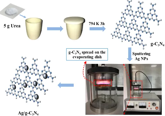
The g-C3N4 powder was prepared using a thermal polymerization method. Briefly, 5g of urea
was added into a crucible with a cover, and heated to 794 K in a muffle furnace at a heating rate of 15 K per minute from room temperature. The temperature was maintained at 794 K for 3 h. After naturally cooling down to room temperature, the residual g-C3N4 product was collected and grounded
into yellow powder.
Ag/g-C3N4 heterostructured photocatalyst was prepared by a simple sputtering method. As
shown in Fig. 1, 0.5 g of as-prepared bulk g-C3N4 was grounded into powder and then added into 50
dish, and then the evaporating dish was placed in a sputtering apparatus. The sputtering current was set to a relatively small value (2 mA) and then the apparatus was turned on to start the sputtering. After a certain time of sputtering, the instrument was turned off and the sample was collected. The Ag/g-C3N4
samples, which were sputtered with 5, 10, 15, 20 and 300 s, were labeled as AgCN5, AgCN10, AgCN15, AgCN20 and AgCN300, respectively.
The percentage of silver in the AgCN10 catalyst was estimated to be 0.08 wt% by comparing the weight of the sample on the evaporating dish before and after sputtering.
5 g Urea 794 K 3h
Ag/g-C3N4
g-C3N4
g-C3N4spread on the
[image:3.596.161.441.208.405.2]evaporating dish Sputtering Ag NPs
Figure 1. Preparation flow chart of Ag/g-C3N4
2.3. Characterization methods
Crystal structures of the products were identified by X-ray powder diffractometer (XRD) with Cu Kα irradiation (λ = 1.54 Å) at 40 KV, 30mA, and a scan rate of 2° min-1
. The morphologies and microstructures of the photocatalyst samples were characterized by transmission electron microscopy (TEM, JEOL, JEM-2010). The X-ray photoelectron spectroscopy (XPS) measurements were done on a Thermo Scientific ESCALAB 250 instrument meter with Al Kα source. The binding energies (Eb) were calibrated internally by the carbon deposit C1 s binding energy at 284.6 eV. The UV-vis absorption spectra of the samples were collected with a UV-vis spectrophotometer (Shimadzu, Japan, UV-2401) in the range of 200-800 nm. BaSO4 was the reflectance sample.
2.4. Evaluation of photocatalytic activity
The photocatalytic activities of the as-prepared g-C3N4 nanosheets and Ag/g-C3N4 were
cooled by water circulation, and the temperature was maintained at 293 K. The aqueous samples were collected at regular intervals and filtrated through 0.45 μm membranes. The photodegradation over RhB was also performed without any photocatalyst under the same condition.
2.5. Photoelectrochemical measurements
Transient photocurrent responses and the electrochemical impedance spectroscopy (EIS) measurements were performed with a CHI 660E potentiostat in a standard three-electrode cell system. An Ag/AgCl electrode was used as the reference electrode and a Pt plate was used as the counter electrode. The working electrode was prepared by taking active material (Ag/g-C3N4 composites),
conductive additive (carbon black), and binder (PVDF) at the weight ratio (8:1:1) and mixed together into a N-methyl pyrrolidone solvent to obtain the slurry. The resulting slurry was coated onto the ITO plate (an active area of 1 cm2) and dry at 333 K in an oven for overnight. A 0.5 M Na2SO4 solution
was used as the electrolyte. A 300 W Xe lamp equipped with a UV cut-off filter (λ ≥ 420 nm) served as the visible light source. The photoresponsive signals of the samples were measured under chopped light at 0.0 V.
3. RESULTS AND DISCUSSION
Figure 2. (a) Power XRD patterns, (b) UV-visible absorbance spectra and plots of (αhν)2 versus photon energy (b inset) of g-C3N4 and AgCN10.
To investigate the crystal structure of pure g-C3N4 nanosheets and AgCN10 powder, XRD was
carried out. From Fig. 2a, the XRD pattern of pure g-C3N4 is indexed as JCPDS No. (87-1526) [16].
Compared with the pure g-C3N4, after being sputtered for 10 seconds, there is no obvious peak of Ag
[image:4.596.93.507.415.606.2]
44.2◦, 64.4◦, 77.4◦ can be indexed to the diffraction of (111), (200), (220), and (311) lattice planes of crystalline silver (JCPDS No. 65-2871) [17]. These results confirmed the existence of Ag NPs on the g-C3N4 nanosheets, and they are consistent well with the HRTEM observations.
The optical properties of the g-C3N4 nanosheets and AgCN10 were carried out by the UV-vis
absorption spectra (Fig. 2b). Compared with the pure g-C3N4, AgCN10 showed increased absorption
of visible light after the sputtering of Ag NPs. As shown in Fig. 2b inset, the optical bandgaps of g-C3N4 and AgCN10 were calculated as 2.98 eV and 2.90 eV. The band gap of AgCN10 reduces after
the sputtering of Ag NPs that promoted the utilization of low-energy photons in degradation of RhB. The morphology and microstructure of the AgCN10 photocatalyst were investigated by TEM. Fig. 3 shows the TEM micrograph of AgCN10. The uniform dispersion of numerous dark grey colored spots with the diameter of ~10 nm indicate a high deposition of Ag NPs on the surface of g-C3N4. The
inset figure in Fig. 3 shows the lattice image of the Ag NPs, the characteristic values of lattice constant are 0.24 and 0.20 nm corresponding to the typical (111) and (200) crystal phase of Ag, respectively [18,19].
Ag (111) 0.24 nm
Ag (200) 0.20 nm
[image:5.596.156.441.334.531.2]50 nm
Figure 3. TEM of AgCN10 (inset: high-magnification TEM image of a Ag NP).
XPS spectra of AgCN10 were obtained for the analysis of the structure and surface of the composite catalyst (Fig. 4). The survey scan spectrum of AgCN10 showed peaks for C, N and Ag (Fig. 4a). The C 1s (Fig. 4b) spectra of AgCN10 could be fitted to four peaks at 284.7, 285.1, 288.3, and 288.6 eV, which could be attributed to the sp2 C-C bonds, C-NH2 species, N=C-N coordination and the
[image:6.596.92.508.68.400.2]
Figure 4. XPS spectra of AgCN10: (a) survey XPS spectrum; (b) C 1s; (c) N 1s; (d) Ag 3d.
In order to investigate the excitation and transfer of photo-induced charge carriers among photocatalysts, the transient photocurrent responses and EIS measurements were performed. This photocurrent increases when exposed to a light source, and it indicates that the photo-generated electrons migrate in the bulk materials to produce photocurrent (Fig. 5a) [24]. The photogenerated current densities of pure g-C3N4 and AgCN10 under visible light were 0.81 and 1.37 μA cm−2,
respectively. The higher photocurrent intensity led to lower recombination speed of photo-induced electrons and holes, so we could find out the AgCN10 photocatalyst owned better separation rate of photo-generated electron-hole pairs compared with pure g-C3N4 photocatalyst. Fig. 5b shows the EIS
date of AgCN10 and g-C3N4 nanosheets. The diameter of the Nyquist semicircle of AgCN10 is smaller
than that of pure g-C3N4 [25], and implies that the AgCN10 has a smaller charge transfer resistance,
which reflects the effective separation of electrons and holes.
RhB was chosen to evaluate the photocatalytic performances of Ag/g-C3N4 photocatalyst at
room temperature. Before visible light irradiation, the RhB solutions containing photocatalyst were stirred in the darkness for 0.5 h to achieve adsorption equilibration. Fig.6a shows the RhB degradation curves with catalyst, in which C0 is the RhB concentration after adsorption equilibration, and C
Figure 5. Photocurrent responses and EIS spectra of pure g-C3N4 and AgCN10 under visible light
irradiation.
It is shown from the photocatalytic results that the AgCN10 photocatalyst was more efficient for the degradation of RhB than Ag/g-C3N4 composite with other sputtering time. Furthermore, the
result in Fig. 6b shows no strong reduction in photocatalytic activity after four consecutive cycling tests. The results indicated that the photocatalyst AgCN10 exhibited good stability for the photodegradation of RhB under visible light.
Figure 6. (a) Photocatalytic degradation curves of g-C3N4 and Ag/g-C3N4 samples and (b)
Recyclability of AgCN10 for RhB dye degradation under visible light irradiation (λ ≥ 420 nm)
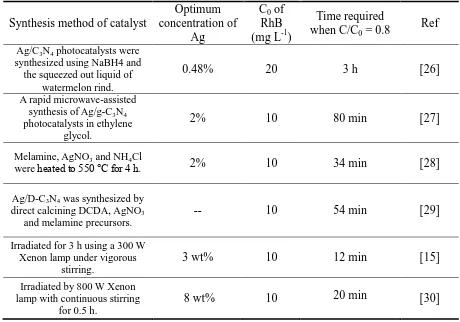
The recent literature on the degradation of RhB with Ag/g-C3N4 composite was
investigated, as shown in Table 1. Comparing with pure g-C3N4, modifying g-C3N4 with Ag has
greatly improved the photocatalytic degradation efficiency, which demonstrates that Ag/g-C3N4
[image:7.596.96.505.72.257.2] [image:7.596.93.510.431.600.2]
Table 1. Comparison of RhB degradation using Ag/g-C3N4 composite
Synthesis method of catalyst
Optimum concentration of
Ag
C0 of
RhB (mg L-1)
Time required when C/C0 = 0.8
Ref
Ag/C3N4 photocatalysts were synthesized using NaBH4 and the squeezed out liquid of
watermelon rind.
0.48% 20 3 h [26]
A rapid microwave-assisted synthesis of Ag/g-C3N4 photocatalysts in ethylene
glycol.
2% 10 80 min [27]
Melamine, AgNO3 and NH4Cl
were heated to 550 °C for 4 h. 2% 10 34 min [28]
Ag/D-C3N4 was synthesized by direct calcining DCDA,AgNO3
and melamine precursors.
-- 10 54 min [29]
Irradiated for 3 h using a 300 W Xenon lamp under vigorous
stirring.
3 wt% 10 12 min [15]
Irradiated by 800 W Xenon lamp with continuous stirring
for 0.5 h.
8 wt% 10 20 min [30]
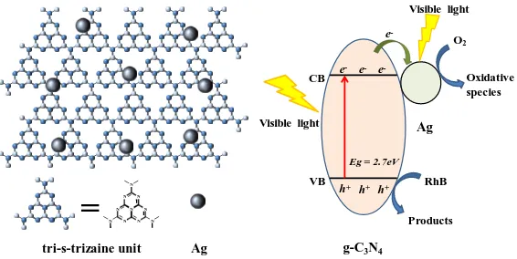
Based on the above results, we propose a tentative reaction mechanism for photocatalytic degradation of RhB over the Ag/g-C3N4 composites (Fig. 7). The g-C3N4 nanosheets can be excited to
generate electrons and holes under visible light irradiation. In the Ag/g-C3N4 composites, a Schotky
barrier junction can be produced by Ag NPs and g-C3N4 nanosheets, which can effectively enhance
both recombination time and separation of photo-induced electrons and holes. Photo-induced charge carriers can react with OH- and O2 to form •OH and •O2-, and the active species are promoted due to
surface plasmon resonance (SPR) effect of Ag. The possible photocatalytic reactions and degradation of RhB are listed in the following:
Ag/g-C3N4 + hv → Ag/g-C3N4 (e- + h+) (1)
OH- + h+ → •OH (2)
e- + O2 → •O2- (3)
tri-s-trizaine unit Ag
=
N N N N N N N N N NEg = 2.7eV
CB Products O2 Oxidative species RhB VB
Visible light
e- e
-e
-h+ h+
h+
e
-g-C3N4
Ag
Visible light
Figure 7. A diagram of the Ag/g-C3N4 composite (left) and Schematic diagram illustrating the
photocatalytic degradation of RhB under visible light irradiation (right).
4. CONCLUSION
A novel visible-light-driven heterostructured Ag/g-C3N4 photocatalyst was synthesized using a
simple sputtering method. Although the amount of silver used in this work is very small (0.08 wt%) compared with previous studies of Ag/g-C3N4 composite catalysts, a great photodegradation catalytic
performance was achieved. The TEM spectra shows that numerous Ag NPs about 10 nm in diameter were uniformly deposited on the surface of g-C3N4 nanosheets. The photodegradation performance of
Ag/g-C3N4 photocatalyst was significantly improved compared with that of pure g-C3N4. The
mechanism of enhanced photocatalytic performance toward RhB degradation was experimentally investigated via UV-vis DRS, photocurrent responses and EIS spectra. The enhanced photocatalytic performance was attributed to combining effects of good interfacial junction between Ag NPs and g-C3N4 nanosheets, which led to increased optical absorption in the visible region, quick separation and
transportation of photo-induced electrons and holes, and long lifetime of charge carriers as well.
ACKNOWLEDGMENTS
This work was supported by National Natural Science Foundation of China (Grant No. 51528202, 51671136 and 51502172) and International Science and Technology Collaboration Project of Shanghai (Grant No. 17520710300).
References
1. I.Ali, Chem. Rev., 112 (2012) 5073.
2. Y.H. Sang, Z.H. Zhao, M.W. Zhao, P. Hao, Y.H. Leng and H. Liu, Adv. Mater., 27 (2015) 363. 3. D.P. Zhang, S.H. Cui and J. Yang, J. Alloy. Compd., 708 (2017) 1141.
4. Y.W. Gao, Y. Wang and H. Zhang, Appl. Catal. B., 178 (2015) 29.
5. H.J. Huang, J. Zhang, L. Jiang and Z.G. Zang, J. Alloy. Compd., 718 (2017) 112.
6. D.J. Yang, C.C. Chen, Z.F. Zheng, H.W. Liu, E. R. Waclawik, Z.M. Yan, Y. Huang, H.J. Zhang, J.C. Zhao and H.Y. Zhu, Energy Environ. Sci., 4 (2011) 2279.
[image:9.596.158.441.70.212.2]
8. C. Yang, S.C. Wu, J.H. Cheng and Y.C. Chen, J. Alloy. Compd., 687 (2016) 804.
9. Z.W. Tong, Y. Dong, L. Zhen, Y.H. Nan, F. Ding, Y.C. Shen and Z.Y. Jiang, Acs Nano., 11 (2016) 1103.
10. M.J. Shi, T.H. Wu, X.F. Song, J. Liu, L.P. Zhao, P. Zhang and L. Gao, J. Mater. Chem. A., 4 (2016) 10666.
11. J. Xu, G.X. Wang, J.J. Fan, B.S. Liu, S.W. Cao and J.G. Yu, J. Power Sources., 274 (2015) 77. 12. X.Y. Fang, J.L. Song, H.C. Shi, S.F. Kang, Y. Li, G.W. Sun and L.F. Cui, Int. J. Hydrogen
Energ., 42 (2017) 5741.
13. J.Z. Wang, S.Y. Dong, C.F. Yu, X. Han, J.Y. Guo and J.H. Sun, Catal. Commun., 92 (2017) 100. 14. W. Zhang, L. Zhou and H.P. Deng, J. Mol. Catal. A: Chem., 423 (2016) 270.
15. Y.Y. Bu, Z.Y. Chen and W.B. Li, Appl. Catal. B., 144 (2014) 622.
16. L. Zhou, W. Zhang, L. Chen, H.P. Deng and J.L. Wan, Catal. Commun., 100 (2017) 191. 17. X. He, R.H. He, A. L. Liu, X.Y. Chen, Z.L. Zhao, S. Feng, N. Chen and M. Zhang, J. Mater.
Chem. C., 2 (2014) 9737.
18. H.M. Dai, N. Cao, L. Yang, J. Su, W. Luo and G.Z. Cheng, J. Mater. Chem. A., 2 (2014) 11060. 19. S. Sareen, V. Mutreja, S. Singh and B. Pal, RSC Adv., 5 (2015) 184.
20. J.H. Li, B. Shen, Z.H. Hong, B.Z. Lin, B.F. Gao and Y.L. Chen, Chem. Commun., 48 (2012) 12017.
21. B.H. Long, J.L. Lin and X.C. Wang, J. Mater. Chem. A., 2 (2014) 2942.
22. M. Yan, Y.Q. hua, F.F. Zhu, L. Sun, W. Gu and W.D. Shi, Appl. Catal. B., 206 (2017) 531. 23. L. Ge, C.C. Han, J. Liu and Y.F. Li, Appl. Catal. A., 409 (2011) 215.
24. L. Li, Y.H. Qi, J.R. Lu, S.L. Lin, W. J.An, Y.H. Liang and W.Q. Cui, Appl. Catal. B., 183 (2015) 133.
25. D.S. Wang, Z.X. Xu, Q.Z. Luo, X.Y. Li, J. An, R. Yin and C. Bao, J. Mater. Sci., 51 (2016) 893. 26. K. Tian, W.J. Liu and H. Jiang, Acs. Sustain. Chem. Eng ., 3 (2015) 269.
27. T. Sun, H.Y. Jiang, C.C. Ma, F. Mao and B. Xue, Catal. Commun., 79 (2016) 45. 28. J. Jin, Q. Liang, C.Y. Ding, Z.Y. Li and S. Xu, J. Alloy. Compd., 691 (2017) 763. 29. C.Y. Min, C. Shen, R. Li, Y. Li, J.L. Qin and X.F. Yang, Ceram. Int., 42 (2016) 5575. 30. Z.J. Li, J.H. Wang, K.X. Zhu, F.L. Ma and A. Meng, Mater. Lett., 145 (2015) 167.