COMPARISON OF SHEAR BOND STRENGTH
OF TWO DIFFERENT CERAMIC REPAIR
SYSTEMS WITH TWO DIFFERENT SURFACE
TREATMENTS ON METAL SURFACE-AN
IN-VITRO STUDY
Dissertation submitted to
The Tamil Nadu Dr. M.G.R. Medical
University
In partial fulfilment of the degree of
MASTER OF DENTAL SURGERY
BRANCH I
PROSTHODONTICS AND CROWN &
BRIDGE
CERTIFICATE
This is to certify that the dissertation entitled “Comparison of shear bond strength of two different ceramic repair systems with two different surface treatments on metal surface – An in-vitro study” is a bonafide record of the work done by Dr Soumya Mohan B Post graduate student during the period 2015-2018 under my guidance and supervision. This dissertation is submitted in partial fulfilment of the requirements for the award of MASTER OF DENTAL SURGERY IN, BRANCH I (PROSTHODONTICS AND CROWN AND BRIDGE) under THE TAMIL NADU Dr.M.G.R MEDICAL UNIVERSITY, CHENNAI. It has not been submitted (partial or full)
for the award of any other degree or diploma.
GUIDE
Dr.T.SREELAL, M.D.S Professor and Head, Department of Prosthodontics,
Sree Mookambika Institute of Dental Sciences, Kulasekharam
CO-GUIDE
Dr.Aparna Mohan, M.D.S Reader,
Department of Prosthodontics,
CERTIFICATE II
This is to certify that this dissertation work titled ³&RPSDULVRQ RI
shear bond strength of two different ceramic repair systems with two
different surface treatments on metal surface - An in-YLWURVWXG\´of the candidate Dr Soumya Mohan B with registration Number 241511302
for the award of MASTER OF DENTAL SURGERY in the branch of Prosthodontics and Crown and Bridge, [Branch- I]. I personally verified the urkund.com website for the purpose of plagiarism Check. I found that the uploaded thesis file contains from introduction to conclusion pages and result shows 3 percentage of plagiarism in the dissertation.
Guide & Supervisor sign with Seal.
Date:
DECLARATION
I hereby declare that this dissertation entitled “Comparison of shear bond strength of two different ceramic repair systems with two
different surface treatments on metal surface – An in-vitro study” is a bonafide record of work undertaken by me during the period 2015-2018 as a part of post graduate study. This dissertation, either in partial or in full, has not been submitted earlier for the award of any degree, diploma, fellowship or similar title of recognition.
Dr. Soumya Mohan B MDS Student,
Department of Prosthodontics, Sree Mookambika Institute of Dental sciences,
SREE MOOKAMBIKA INSTITUTE OF DENTAL SCIENCES, KULASEKHARAM
ENDORSEMENT BY THE PRINCIPAL / HEAD OF THE INSTITUTION
This is to certify that the dissertation entitled “Comparison of shear bond strength of two different ceramic repair systems with two different surface treatments on metal surface – An in-vitro study” is a bonafide research work done by Dr. Soumya Mohan under the guidance
of Dr.T.Sreelal, M.D.S, Professor and Head, Department of
Prosthodontics and Crown and Bridge, Sree Mookambika Institute of
Dental Sciences, Kulasekharam.
Date: Dr.Elizabeth Koshy, MDS, Principal,
Sree Mookambika Institute of
Dental Sciences, V.P.M Hospital Complex,
Padanilam, Kulasekharam, Kanyakumari
ACKNOWLEDGEMENT
“At times our own light goes out and is rekindled by a spark from another
person. Each of us has cause to think with deep gratitude of those who
have lighted the flame within us.”
I bow in gratitude to the Almighty God, the creator, who has helped
me to sustain these best and toughest years of my life. For providing me the
immense strength and willpower to complete this work and to whom I owe my
very existence. I also thank the Almighty for His unparalleled grace, superior
protection and guidance throughout the lows and highs of my post graduate
journey. Thank you God for keeping my ship sailing and not failing me in my
journey.
I would like to express my deepest appreciation and sincere thanks to
my guiding light, my mentor and Guide Dr.T.Sreelal., Professor and HOD,
Department of Prosthodontics, Sree Mookambika Institute of Dental Sciences,
Kulasekharam. I thank him from the bottom of my heart for his time, patience
and unyielding faith in me. His constant and timely advice, support and
guidance inspired me to aspire for perfection and has helped me to complete
this dissertation. I could not have imagined for having a better advisor and
mentor for this study. I attribute the level of my master’s degree to his encouragement and effort and without him this thesis too would not have been
study under his guidance. I also thank him in particular with whom I started
this thesis work and many rounds of discussions on my project with him,
helped me a lot. He not only helped me with my thesis work but also has
influenced me with his level of knowledge, which he has selflessly passed on
to me. With his pearls of wisdom, he made the subject interesting and
understandable for our budding minds. He has constantly forced me to remain
focussed on achieving my goal. He is an example of a true mentor and guide.
With all the gratitude that I feel and warm regards that I can muster, I thank
you sir for imbibing the precious seeds of knowledge, patience, discipline and
duty, that is a treasure for a lifetime.
I am deeply indebted to Dr.Giri Chandramohan, MDS, Reader,
who with his vast knowledge has helped me in my work theoretically as well
as clinically. He is a person with lots of clinical and laboratory knowledge and
at the same time he is very helpful and approachable.
Hand in hand was the constant and perpetual guiding of my
co-guide, Dr. Aparna Mohan, MDS, Reader. She acted as a strong pillar of
support who inspite of her own hectic schedule found time in correcting this
dissertation. She was always available to clear my doubts and guide me to the
light of knowledge with her valuable guidance, suggestions and tireless pursuit
for perfection. She has a strong personality and a heart full of affection which
helped me pass the tough times during my undergraduate and post graduate
matured person under her wings of knowledge. Her words always inspired me
and brought me to a higher level of thinking.
I also extend my gratitude to Dr.Allen Jim Hines MDS, Senior
Lecturer, who is always approachable for any help at any time and he is the
one who taught me how to manage anxious and apprehensive patients giving
me an insight of his psychological approach.
My heartfelt and sincere thanks to Dr.James Rex (Reader), for his
valuable help and guidance both in my undergraduate and postgraduate
journey.
I express my sincere thanks to Dr.Sarath Babu K PhD Associate
Professor,Department Of Pharmacology, Dr. Vinod Krishnan MDS, Head of
the Department, Department of Orthodontics Sri Shankara Dental College,
Akathumuri, Varkala for their valuable guidance and support for the
completion of this dissertation.
I express my sincere thanks to Dr. Elizabeth Koshy MDS, Principal,
Sree Mookambika Dental College, Kulashekaram for allowing me to utilize
the clinical material and facilities for the completion of this dissertation.
This endeavour would have been impossible without the help,
guidance and inspiration of Dr Roy Joseph, scientist G, Division of polymeric
sciences and Dr Manoj Komath and Nishad, Scientists, Division of
Technology, Trivandrum, who inspite of their own hectic departmental work
found time for helping me in completing my study, I am indeed, indebted to
them.
It gives me immense pleasure to thank Dr.Ebinu A, my co-PG, who has
been with me throughout my postgraduate life more like an younger brother,
guiding and encouraging me during the happy and hard moments making this
journey a memorable one.
This acknowledgement seems lacking without the mention of my dear
seniors Dr.Vivek B Chandran,Dr. P RajKumar and Dr. Eshona Pearl and
my fellow Post graduates Dr. Jithin G N, Dr. Amalorpavam V, Dr
Ponjayanthi and Dr Femin David for their support, motivation and
encouragement when I slow down.
I am also thankful to Mr.Bibin Sekhar, technician and ceramist
SMIDS and to Mrs. Sunitha and Mrs. Geetha Assistants, Department of
Prosthodontics, SMIDS, for their sincere work that helped me a lot all through
my three years of Post Graduate life.
I am also thankful to Mr.Srinivasan, Mrs.Jayalekshmi Srinivasan,
Mr.Vijesh and Miss Priya, Office staffs, SMIDS for their sincere work that
helped me a lot all through my three years of Post Graduate life.
A wave of fond emotions sweep over me as I struggle to gather the
feel for my father Late R. Rajamohanan, my mother Mrs Bindhu G, my
husband Mr Jayakrishnan Nair G, my son Master Navneeth J, my in-laws
Mr Gopalakrishnan Nair and Mrs Girija Nair, my sister Mrs Remya
Mohan B ,my brothers Mr Renji Mohan R and Mr Achuth Vimal my
brother in laws Mr Praveen Thachat and Mr Mohanakrishnan Nair and my
sister in law Mrs Tanusree Pillai for their confidence in me and their
sacrifices, strength and encouragement at all times has been an inspiration and
is solely responsible for refurbishing my life and profession.
“In the end, though, maybe we must all give up trying to pay back
the people in this world who sustain our lives. In the end, maybe it's wiser
to surrender before the miraculous scope of human generosity and to just
keep saying thank you, forever and sincerely, for as long as we have
SPECIAL ACKNOWLEDGEMENT
I take this opportunity to thank specially our Chairman
Dr.C.K.VELAYUTHAN NAIR MS, Sree Mookambika Institute of Dental Sciences, our Director Dr.REMA.V.NAIR MD, Sree Mookambika Institute
of Dental Sciences and our Trustees Dr.R.V.MOOKAMBIKA MD, DM,
Dr.VINU GOPINATH MS, MCH and Mr.J.S.PRASAD, Adminstrative
officer for giving me the opportunity to utilize the facilities available in this
CONTENTS IN CONCISE
SL.NO INDEX
1 LIST OF FIGURES
2 LIST OF TABLES
3 LIST OF GRAPHIC DIAGRAMS
4 ABSTRACT
5 INTRODUCTION
6 AIMS AND OBJECTIVES
7 REVIEW OF LITERATURE
8 MATERIALS AND METHODOLOGY
9 RESULTS AND OBSERVATIONS
10 DISCUSSION
11 SUMMARY AND CONCLUSION
CONTENTS
SL.NO
INDEX
PAGE
NO.
1 ABSTRACT 1-4
2 INTRODUCTION 5-10
3 AIMS AND OBJECTIVES 11
4 REVIEW OF LITERATURE 12-25
5
MATERIALS AND
METHODOLOGY 26-31
6
RESULTS AND
OBSERVATIONS 32-40
7 DISCUSSION 41-48
8
SUMMARY AND
List of Figures
FIGURE -1
Wax pattern made for
Cobalt-Chromium metal block
FIGURE -2
Lukadent Casting Machine
FIGURE -3
Casting process
FIGURE-4
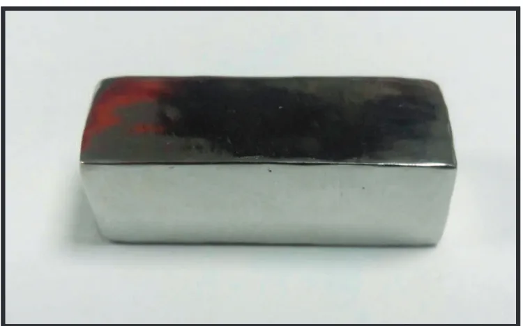
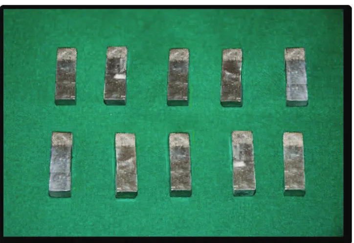
Cobalt-Chromium metal block
FIGURE-5
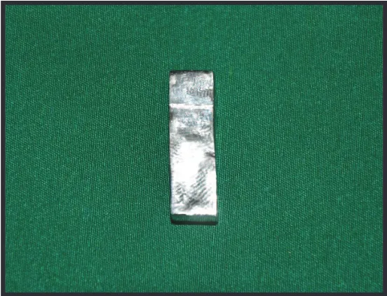
Cobalt-Chromium metal blocks
FIGURE-6
6mm marked on Cobalt-Chromium
metal block
FIGURE-7
Intra Oral Sandblaster
FIGURE-8
Alumina particles (110µ)
FIGURE-9
Sandblasted Cobalt-Chromium blocks
FIGURE-10
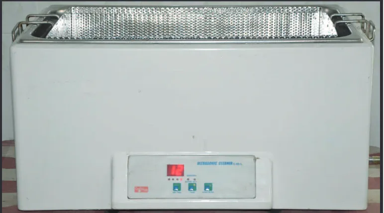
Ultrasonic cleaner
FIGURE-11
Sandblasted Cobalt-Chromium metal
blocks in ultrasonic cleaner
FIGURE-12
Demineralised water
FIGURE-13
Nd:YAG intra oral laser unit
FIGURE-14
Laser etching process
FIGURE-15
List of Figures
FIGURE-16
Laser etched Cobalt-Chromium
blocks
FIGURE-17
Ivoclar ceramic repair kit
FIGURE-18
Ivoclar ceramic repair kit
FIGURE-19
Ivoclar-Heliobond application
FIGURE-20
Ivoclar- Monobond application
FIGURE-21
Shofu Ceramic repair kit
FIGURE-22
Shofu Ceramic repair kit
FIGURE-23
Shofu metal primer application
FIGURE-24
Shofu Cera resin bond 1 & 2
FIGURE-25
Ceramic repair system curing done
using composite curing light cure unit
FIGURE-26
Cobalt-Chromium metal block with
light cured ceramic repair system
FIGURE-27
Scanning electron microscopic image
of sandblasted specimen
FIGURE-28
Scanning electron microscopic image
of Laser etched specimen
FIGURE-29
Scanning electron microscope
FIGURE-30
Ion sputter machine
FIGURE-31
Gold coating on sandblasted
Cobalt-Chromium metal block
FIGURE-32
Gold coating on laser etched
Cobalt-Chromium metal block
FIGURE-33
List of Figures
List of Tables
[image:21.612.141.493.114.666.2]LIST OF TABLES
TABLE 1
Mean Max load and Shear Bond Strength values of different groups
TABLE 2
Comparison of mean Max load and Shear Bond Strength values of Group-I with other groups
TABLE 3
Comparison of mean Max load and Shear Bond Strength values of Group-II with other groups
TABLE 4
Comparison of mean Max load and Shear Bond Strength values of Group-III with other groups
TABLE 5
Comparison of mean Max load and Shear Bond Strength values of Group-IV with other groups
TABLE 6
Multiple comparison of mean Max load and Shear Bond Strength values of different groups
TABLE 7
Comparison of mean Max load and Shear Bond strength values between Group-I and Group-II
TABLE 8
List of Abbreviations
1 Co -Cr Cobalt Chromium
2 Nd :YAG Neodymium yttrium aluminum garnet
3 ANOVA Analysis Of Variance
List of Graphic Diagrams
LIST OF GRAPHIC DIAGRAMS
GRAPH NO:1
Graph depicting Mean Shear Bond Strength values of different groups
GRAPH NO:2
Graph depicting Comparison of mean Max load and Shear Bond Strength values of Group-I with other groups
GRAPH NO:3
Graph depicting Comparison of mean Max load and Shear Bond Strength values of Group-II with other groups
GRAPH NO:4
Graph depicting Comparison of mean Max load and Shear Bond Strength values of Group-III with other groups
GRAPH NO:5
Graph depicting Comparison of mean Max load and Shear Bond Strength values of Group-IV with other groups
GRAPH NO:6
Graph depicting Multiple comparison of mean Max load and Shear Bond Strength values of different groups
GRAPH NO:7
Graph depicting comparison of mean Max load and Shear Bond strength values between Group-I and Group-II
GRAPH NO:8
Abstract
1
Introduction
Metal Ceramic Restorations are widely used in restorative dentistry with a high
degree of success. On occasions, fractures do occur in ceramics due to trauma,
flexure of metal or fatigue of ceramic. Fractured porcelains will affect
aesthetics & function of prostheses which may warrant patients to seek
immediate treatment1.
Ceramic fracture may result from trauma, fatigue,
occlusal prematurity, para- functional habits, poor abutment preparation,
inappropriate coping design and incompatibility of coefficient of thermal
expansion between ceramic and the metal structure. One option is to remake
the restoration. This is both expensive and time consuming. Intra-oral chair
side porcelain repair system is a quick, painless and highly patient acceptable
procedure, without the removal of restoration or fabrication of new restoration1.
Three kinds of fractures have usually been monitored at metal ceramic
restorations: simple fractures (formed only within porcelain and metal does not
get out of surface), mixed fractures (as well as porcelain fractures, metal gets
out of surface), complex fractures (metal completely gets out of
surface).Porcelain fractures are the most common cause of removing the
prosthesis.
Fractured porcelain affects patients negatively in terms of aesthetic and
function and requires to be changed. In this case, two different treatment
options come to mind. The primary and ideal treatment option involves
Abstract
2
alternative method is repair of fractured area with composite resin intraorally.
Intraoral repair method offers some advantages such as, economic cost and
time savings. But, the bond between restoration remained in the repaired area
and repair material should be strong and resistant to the functional loads. In
order to improve the bond between composite and fractured surfaces, many
mechanical and chemical bond methods have been developed. To provide the
mechanical bond; many surface treatments including roughening with diamond
drills, sandblasting with aluminium oxide have been used for both metal and
ceramic surfaces46.
Intraoral repairs often involve bonding composite to fractured
porcelain. Newer adhesive systems, currently referred to as multipurpose
systems, include materials with recommended procedures for repair of
porcelain33.Of the many ceramic repair systems commercially available,
Ivoclar ceramic repair system and Shofu ceramic repair system are used for the
present study.
Aims and Objectives
The aim this study was to evaluate & compare the shear bond strength of two
different Ceramic Repair Systems (Ivoclar & Shofu) with two different surface
treatments (Sandblasting & Laser etching) on metal surface. The objectives
were to estimate the most efficient ceramic repair system based on the obtained
shear bond strength values and to estimate the most efficient surface treatment
Abstract
3
Methodology
Twenty Cobalt Chromium blocks were made and ten such blocks were
sandblasted with intra oral sandblaster and the remaining ten blocks were laser
etched with ND: YAG laser. Five sandblasted blocks were coated with Ivoclar
ceramic repair system and remaining five sandblasted blocks were coated with
Shofu ceramic repair system. Five laser etched blocks were coated with coated
with Ivoclar ceramic repair system and remaining five laser etched blocks were
coated with Shofu ceramic repair system. All the twenty blocks were tested on
universal testing machine for evaluating shear bond strength.
Results
Group I (Sandblasted cobalt chromium metal surface coated with Ivoclar
ceramic repair system) has the highest shear bond strength compared to all
other groups.
Summary and Conclusion
Within the limits of the present study it can be concluded that
:-Ø Group I – sandblasted cobalt – chromium metal blocks coated with
Ivoclar ceramic repair system showed highest shear bond strength than
all other three groups.
Ø Surface treatment with sandblasting or air abrasion with 50µ alumina
Abstract
4
Ø For sandblasted groups, Ivoclar ceramic repair system gave the better
shear bond strength value.
Ø For laser etched groups, Shofu ceramic repair system gave the better
shear bond strength value.
However further clinical research is suggested in order to prove
it as a reliable and successful treatment modality.
Introduction
5
De Chemant in 17 century introduced ceramics to dentistry for
making denture teeth. Dental ceramics have various advantages like colour
stability, radio-opacity, esthetics, and coefficient of thermal expansion similar
to dentin, good compressive & abrasive resistance but also have drawbacks like
low tensile strength, low edge strength & high brittleness. Subsequently,
innovations were attempted to strengthen dental ceramics. The prevalence of
fracture of metal ceramic restorations at the metal ceramic interface is
approximately 2–5% and has been reported as the second greatest cause for the
replacement of restorations after dental caries1.
Ceramic failures can occur as simple, mixed or complex,
fractured porcelains will affect aesthetics & function as they occur most
frequently in regions that are quite visible, which may warrant patients to seek
immediate treatment. Removal & reconstruction of the prosthesis is a costly
affair & hence it is better to attempt repair with composite resins intra-orally,
especially in less severe cases2,3.
Most metal ceramic restorations may fracture in the form of
chipping or deveneering of ceramic due to bond failure between ceramic and
metal surface. Fracture can be from trauma, occlusal prematurity,
para-functional habits, poor abutment preparation, inappropriate coping design and
incompatibility of coefficient of thermal expansion between ceramic and the
metal structure.
Intra-oral chair side repair is a quick, painless and highly
Introduction
6
restoration or fabrication of new restoration. Repair of fractured metal ceramic
crowns aims to re-establish the function & esthetics of restorations by using
various repair materials. For the repair material to withstand functional loads,
the bond between the repair material & remaining restoration must be strong &
durable3,4,5. Intraoral repairs often involve bonding composite to fractured
porcelain. Newer adhesive systems, currently referred to as multipurpose
systems, include materials with recommended procedures for repair of
porcelain33.
Many repair agents such as cyanoacrylates, acrylic and
composite resins were used but were partially successful due to esthetic and
mechanical limitations. The earlier repair systems generally used two
component silane coupling agents (silane and acid). It was designed to
chemically bond composite to the silica (SiO2) component of ceramic but had
low shear bond strength. The recently introduced repair systems have 10
methacryloyloxydecyl dihydrogen phosphate (MDP), which recommends
physical alteration of ceramic and metal substrates in conjunction with
chemical agents such as metal primer, ceramic primer and improved silane
coupling agents to promote adhesion of resin to fractured metal ceramic
restorations1,2.
A strong resin bond relies on micromechanical interlocking
and chemical bonding to the ceramic surface, which requires roughening and
cleaning for adequate surface activation. To promote a satisfactory adhesion
Introduction
7
substrate must be performed. There are many surface treatments available to
improve mechanical retention, out of which Sandblasting & Laser Etching are
the two commonly used. These type of treatments improves the bond strength
between the repair material & the surface of a fractured prosthesis8.
In the present in-vitro study, the sandblasting of ceramic
surfaces that are to be repaired is performed with a high speed stream of
purified aluminium oxide particles (50µm) delivered by air pressure (30 to 40
psi) for approximately 15 seconds.
For doing sandblasting in patient’s oral cavity, intraoral
chairside sandblasters are available, the procedure is same as stated above.
Care should be taken to avoid injuries to the surrounding soft tissues. As well
as control the emission & spread of aluminium oxide particles over the
operative area. This can be accomplished by using rubber dam isolation & high
power suction systems.
In Laser etching, the surfaces of specimens are irradiated
with Neodymium: YAG laser. Then a silane coupling agent is applied onto
the surface & a ceramic repair system is bonded onto it. Recently some new
surface conditioning methods on substrate surface has been developed like
laser irradiation on zirconia or porcelain, nanostructure alumina coating on
zirconia & ceramics and chemical vapour deposition of chloro-silanes &
sulphur hexafluoride onto zirconia or ceramic surface.
Introduction
8
mixture of feldspar and quartz. They are frequently used to veneer metal frameworks and indirect restorations, such as inlays, onlays, and laminates11,13.
Introduction
9
Introduction
10
In addition, companies have intended to strengthen the bond between composite resin and metal ceramic surfaces by various primary and bond systems included in repair sets in themselves through developing adhesive systems. It has been intended to improve existing repair systems to exclude use of surface treatment application procedure and loss of time46.
Aims and Objectives
11
AIMS & OBJECTIVES
Aims
To evaluate & compare the shear bond strength of two different ceramic repair systems (Ivoclar & Shofu) with two different surface pre-treatments (sandblasting & laser etching) on metal surface.
Objectives
ü To compare the shear bond strength of two different commercially available ceramic repair systems.
ü To evaluate the efficiency of surface treatment based on the obtained shear bond strength values.
Review of literature
12
Intraoral chair side repair system is a quick, painless & highly
patient acceptable procedure, without removal of a restoration or fabrication of
new restoration. There are very limited studies conducted to evaluate the shear
bond strength of repair systems after different surface treatments1,2. The
porcelain laminate veneer technique is widely used in prosthetic dentistry,
because anaesthesia is not required for the preparation, removal of tooth tissue
is reduced, and the aesthetic results are excellent. The key to this technique is the
adhesion of composite to porcelain. The adhesion of dental resin to porcelain is
achieved by the etching of the porcelain surface with hydrofluoric acid and by
the use of silane coupling agents.
Newer generations of silane systems are composed of two or three
solutions; one is the silane coupler, and the other is the acid component of the
solution. Heat treatment or acid catalysis increases the bond strengths of the
polymers to ceramics treated with silane coupling agents because of the effective
initiation and progress of the formation of siloxane bonds between the silane
coupling agent and the porcelain surface. Heat treatment is not a clinically
available system, so acid solutions of these newer silane agents are used to
facilitate the reaction of silane coupling agent to the porcelain surface. Surface
treatments that do not require hydrofluoric acid etching of the porcelain, such as
phosphoric acid etching or roughening with diamond burs, have been used.
However, earlier systems relied on the mechanical retention of the composite
Review of literature
13
review had shown that there are many different surface pre-treatments and
ceramic repair systems available.
Kalra A et al. 2015 1 evaluated the shear bond strength of two intra-oral porcelain repair systems to repair metal- ceramic restoration after three
different surface treatments and concluded that the shear bond strength of
ceramic repair system with 40% phosphoric acid etching showed highest value
as compared to other system and surface treatment used in their study.
Chung K et al. 1997 2 analysed the bonding strengths of
porcelain repair systems with various surface treatments and concluded that the
mean bond strength of composite bonded to base alloy surface after sandblasting
in six porcelain repair systems ranged from 8.0 to 17.0 Mpa. The study suggested
that metal substrates treated with sandblasting and porcelain treated with either
hydrofluoric acid or sandblasting can increase repair strength.
Blatz M et al. 2003 3 reviewed resin ceramic bonding and
concluded that the resin bonded to silica-based ceramics is well documented
through numerous in vitro investigations. Preferred surface treatment methods
are acid etching with hydrofluoric acid solutions (2.5% to 10% for 2 to 3
minutes) and subsequent application of a silane coupling agent.
Santos JG et al. 2006 4 compared shear bond strength of metal
ceramic repair systems and concluded that for the metal substrate
(nickel-chromium alloy) the CoJet Sand/Z100 group showed statistical superiority
Review of literature
14
Plus/Z100 (control group), the CoJet Sand/Z100, and Bistite II DC/Palfique
groups showed the highest shear bond strength values.
Yavuz T et al. 2013 5 evaluated the effects of different surface treatments on shear bond strength in two different ceramic repair systems and
concluded that the shear bond strengths of the resin cements tested on ceramics
after surface treatments varied in accordance with the type of ceramic.
Jochen DG et al.1977 6 evaluated the effect of composite resin repair of porcelain denture teeth and concluded that an effective temporary repair
of fractured porcelain denture teeth involved abrasive treatment of the fractured
tooth surface followed by a composite resin build up. This study also
demonstrated that the best potential retention of the composite build up can be
obtained through abrasive treatment by a coarse diamond stone.
Appledroon RE et al.1993 7 investigated bond strength of composite resin to porcelain with newer generation porcelain repair systems and
concluded that the Clearfil Porcelain Bond system developed a strong bond of
composite resin to porcelain and maintained this bond over time and the
porcelain repair systems that produced the greatest bond strengths generally
produced the greatest number of cohesive failures in the porcelain, with the
exception of the Etch-Free system.
Ferrando JMP et al. 1983 8 studied tensile strength and micro leakage of porcelain repair materials and evaluated the tensile strength and micro
leakage of five restorative resins bonded to porcelain and concluded that
Review of literature
15
tensile strength and the least leakage at the resin porcelain interface. The study
also concluded that bond strength of the repair systems could not be related to
the degree of leakage.
Bello AJ et al. 1985 9 evaluated the bond strength & micro leakage
of porcelain repair materials and concluded that the tensile bond strength and
micro leakage of four porcelain repair materials which were bonded to dental
porcelain to simulate the repair of a ceramic restoration had a direct relationship
and it was found that there is an increase in micro leakage and decrease in bond
strength for the Silanit and Enamelite 500 exhibited the highest strength and the
least amount of micro leakage.
Creugers NHJ et al. 1992 10 analysed an experimental porcelain
repair system & evaluated it under controlled clinical conditions for 6 to 12
months and concluded that the problem of wear and surface deterioration is not
related to the repair system but to the use of micro filled composite resin (Prisma
Fil) in their study. The study concluded that the surface deterioration could have
been minimized if a (submicron) hybrid composite resin had been used.
Sulaiman AHA et al. 1993 11 evaluated the effects of surface treatment & bonding agents on bond strength composite resin to porcelain and
concluded that the most effective surface treatment was a combination of
mechanical roughening with a diamond bur and chemical etching with
hydrofluoric acid which provided slightly greater repair strengths than either
Review of literature
16
Tylka DF et al. 1994 12 conducted a scanning electron microscopic study to compare the photomicrographs for porcelain composite repair systems
and concluded that gross differences in the photomicrographs between the etch
created by the acidulated phosphor fluoride gel and hydrofluoric acid resembled
those published in earlier studies. All samples experienced cohesive failure of
the porcelain to composite-resin bond and that shear bond strength of the repair
by use of hydrofluoric acid or acidulated phosphor fluoride gel showed a greater
cohesive strength.
Thurmond WJ et al. 1994 13 analysed the effect of porcelain
surface treatments on bond strengths of composite resin bonded to porcelain and
concluded that mechanical alteration of a porcelain surface is more important
than agents that promote chemical bonding of composite resin to porcelain.
Porcelain treatment with a combination of aluminium oxide air abrasion and
hydrofluoric acid provided higher bond strengths than treatment with either
procedure alone.
Tulunoglu IF et al.2000 14 evaluated resin shear bond strength to
porcelain and a base metal alloy using two polymerization schemes and
concluded that higher bond strength values were obtained with prepolymerized
resin superstructures compared to in situ polymerized superstructures.
Haselton DR et al.200115 evaluated shear bond strengths of two
intraoral porcelain repair systems to porcelain or metal substrates and concluded
that both porcelain repair systems tested exhibited reasonable bond strengths to
Review of literature
17
Ozcan M et al.2003 16 reviewed on causes for fracture in ceramic
fused to metal restorations and concluded that clinical studies showed the
prevalence of ceramic fractures are between 5 to 10% over 10 years of use.
Acharya GS et al.2012 17 assessed the effect of surface treatments
and bonding regimens on micro tensile bond strengths of repaired composite and
concluded that bond strength obtained by surface treatment with coarse diamond
point is significantly higher than that obtained by surface treatment with silicon
carbide and also that total-etch bonding regimen produced higher bond strength
compared to treatment with silane primer and bonding resin application.
Tomar SS et al.2015 18 evaluated the bond strength of metal crowns with different luting agents after various modes of surface treatments and
concluded that, maximum bond strength was obtained by sandblasting with 110
µm alumina & ultrasonic cleaning, among all types of surface treatments used in
their study and that the best luting agent was resin‑modified glass ionomer
cement.
Silva CB et al.2014 19 studied the influence of surface treatments on bond strength of resin cements to nickel alloy and concluded that the surface
treatment of the metal promoted a more effective bonding of the resin cements
Panavia Fluoro Cement and Rely X ARC to the Ni-Cr alloy when compared to
untreated surfaces.
Kim WH et al.200720 analysed the effect of ceramic surface treatments on the shear bond strength of dental composite resin to all ceramic
Review of literature
18
thermo cycling, airborne-particle abrasion and acid etching had little influence
on bond strengths between composite resin and ceramic materials. On the other
hand, lithium disilicate ceramic and feldspathic ceramic showed higher bonding
strengths after treated with airborne-particle abrasion.
Deepak K et al .201321 compared & evaluated the effect of laser
on shear bond strength of ceramic bonded with two base metal alloys &
concluded that the shear bond strength between ceramic bonded with
Chromium-Cobalt alloys using the laser etching was higher than that with Nickel-Chromium
alloys. And that laser surface treatment produced excellent surface roughness &
achieved good shear bond strength values.
Grover N et al.201522 evaluated the effect of sandblasting and laser surface treatment on the shear bond strength of a composite resin to the facial
surface of primary anterior stainless steel crowns and concluded that laser
surface treatment obtained the highest bond strength.
Gourav R et al.201623 studied the effect of four different surface treatments on shear bond strength of three porcelain repair systems and
concluded that surface treatment with sandblasting exhibited the highest shear
bond strength followed by combined sandblasting & acid etching.
Mohselhifard E et al.201624 compared the effect of Nd: YAG laser
and sandblasting on shear bond strength of a commercial Nickel-Chromium
alloy to porcelain and concluded that Nd: YAG laser increases the shear bond
Review of literature
19
Tjan AHL et al.198725 evaluated the bond strength of composite to
metal mediated by metal adhesive promoters and concluded that the bond
strengths attained with these tested metal primers are in general not high. Further
research is needed for the development of a metal primer with an improved
adhesive strength. Until then, dentists should continue to use the current methods
of undercutting and roughening the metal surface in order to achieve additional
retention.
Aida M et al.199526 studied the adhesion of composite to porcelain
with various surface conditions and concluded that formation of siloxane bond
was important for adhesion between composite resin & porcelain. Scanning
electron microscope study showed that hydrofluoric acid etching gave highest
roughness on porcelain surface.
Bello AJ et al.198527 evaluated the tensile bond strength & micro
leakage of porcelain repair materials and concluded that all materials except
Silanit increased in strength over the 4-week interval of this study. Enamelite
500 exhibited the highest strength at 4 weeks and the least amount of micro
leakage.
Atsu SS et al. 200628 studied the effect of zirconium oxide ceramic
surface treatments on the bond strength to adhesive resin and concluded that
tribochemical silica coating (CoJet System) and the application of an MDP–
containing bonding/silane coupling agent mixture increased the shear bond
Review of literature
20
Beck AD et al.199029 analyzed the shear bond strength of
composite resin porcelain repair materials bonded to metal & porcelain and
concluded that bond strengths of composite resin materials to porcelain are
significantly greater than those to either machined or oxidized alloy. Bond
strengths of Ultra-Bond/Cerinate Prime/Gold Link materials were not
significantly different from Profile/ Fusion Resin/Prisms Universal Bond
materials. Bond strengths varied significantly for different types of porcelain,
and different types of alloys used as substitutes.
Borges AG et al.200330 investigated the effect of etching & air
borne particle abrasion on the microstructure of different dental ceramics and
concluded that hydrofluoric acid etching and airborne particle abrasion with
aluminum oxide increased the irregularities on the surface of IPS Empress, IPS
Empress 2, and Cergogold ceramics. Similar treatment of In-Ceram Alumina,
In-Ceram Zirconia, and Procera did not change their morphologic
microstructure.
Hasegawa T et al.199531 studied the shear bond strength &
quantitative microleakage of a multipurpose dental adhesive system resin
bonded to dentin and concluded that excellent shear bond strength values of 13.9
MPa to 19.5 MPa were obtained and microleakage values obtained were
favorable compared to other dentin bonding systems.
Kelly JR et al.199632 reviewed ceramics in dentistry: its
historical roots & current perspectives and concluded that recent progress
Review of literature
21
complete crowns, partial coverage, and laminate veneer restorations; improved
metal-ceramic esthetics with the advent of opalescent porcelains and framework
modifications; introduction of CAD/CAM and machining as a route to
fabrication of restorations; and improved understanding of the clinical response
of all-ceramic prostheses and of the materials factors that influence clinical
longevity.
Kupiec KR et al.199633 evaluated porcelain surface treatments
& agents for composite to porcelain repair & concluded that the combination of
aluminum oxide air abrasion and hydrofluoric acid treatment of a porcelain
surface provided an optimal surface for composite bonding with the ProBond
system. Silane treatment of porcelain was critical for obtaining suitable bond
strengths of composites to porcelain.
Lacy AM et al.198834 studied the effect of porcelain surface
treatment on the bond to composite and concluded that the silane coupling agent
was effective in establishing a bond between composite and dental porcelain.
Nowlin TP et al.198135 evaluated the bonding of three
porcelain repair systems and concluded that fractured porcelain bars repaired
with Fusion/ Concise displayed significantly greater repair strength ($ < .05) than
bars repaired with Den-Mat and Cervident2.
Pratt RC et al.198936 evaluated bond strength of six porcelain
repair systems and concluded that porcelain repair products are significantly
Review of literature
22
after 3 months, suggesting that porcelain repairs may be an interim clinical
procedure.
Robert JD et al.197937 reviewed repair of porcelain fused
to metal restorations and concluded that the composite resin bonded to porcelain
repair technique will not have as favorable a prognosis, as composite resins were
subjected to more wear, were not color stable, and the chemical bond created
with bonding agents was much weaker than the bond created when porcelain is
fused to metal.
Highton RM et al.197938 studied the effectiveness of
porcelain repair systems and concluded that the surfaces of the fractured and
repaired beams revealed that the failure occurred at the porcelain-resin interface,
which indicates that the bonding agent rather than the resin failed in all
specimens.
Kussano MC et al.200339 evaluated shear bond strength of
composite to porcelain on the basis of surface treatment and concluded that the
combination of sandblasting with acids on porcelain salinization provided the
best results and it was also observed that silane priming considerably improved
bond strength to the porcelain of the composite material.
Al Edris A et al.199040 conducted scanning electron
microscopic evaluation of etch patterns by three etchants on three porcelains &
concluded that etch patterns were noticeably different for the three porcelain and
that Hydrofluoric acid in combination with other acid produced similar etch
Review of literature
23
Arnold DAM et al.198941 evaluated the bond strengths of
intraoral porcelain repair materials & concluded that fusion and scotch prime
materials achieved bond strengths to nonglazed porcelain in excess of the
shearing resistance of the porcelain. Cerinate prime material failed to achieve as
strong a bond.
Bailey JH et al.198942 evaluated porcelain to composite bond
strengths using four organosilane materials & concluded that there is no
significant differences between the flexural strength of 3M Porcelain Repair Kit
product with Scotch prime Ceramic Primer product, the Fusion product, or the
Sybron ‘Kerr Ultrafine Porcelain Repair Bonding System product with the Silux
composite.
Arnold DAM et al.198943 evaluated the bond strengths of
four organosilane materials in response to thermal stress and concluded that
thermocycling caused a statistically significant decrease in the mean shear bond
strength of Command Ultrafine Porcelain Repair system, Enamelite 500, and
Fusion systems. The Scotchprime system maintained consistently high shear
bond strength values under the conditions tested.
Ahamadzadeh A et al.201644 studied effect of silane on shear
bond strength of two porcelain repair systems and concluded that ultra-dent
ceramic repair kit yields higher shear bond strength at ceramic composite
interface compared to pulp dent ceramic repair kit. Use of one or two layers of
silane does not make any significant with regard to the shear bond strength of
Review of literature
24
Khoroushi M et al.200745 analyzed shear bond strength of
composite-resin to porcelain - effect of thermocycling and concluded that silane
treatment of porcelain was critical for achieving durable bond strength between
composite – resin and porcelain.
Ozel GS et al.201646 compared shear bond strength of three
different composite materials to metal and ceramic surfaces and concluded that
Kuraray CL groups revealed highest bond strength resulted from MDP content
and 40% thixotropic acid efficiency in both metal and porcelain substrates. The
bond strength of Ultradent RK group is the lowest among metal groups.
Shah K et al.201647reviewed dental ceramics – past,
present and future and concluded that the increased demand for aesthetics led to
the development of all ceramic restorations. Zirconia is one of the most stable
ceramics and has flexural strength and toughness values almost two times higher
than those produced by glass ceramics.
Kesark P et al.201248 investigated surface hardness of resin
cement polymerized under different ceramic materials and concluded that
surface hardness is one of the most effect methods to evaluate polymerization of
resin cement. Surface hardness decreased significantly moving from top towards
bottom of the specimen. Resin cements polymerized under different ceramic
materials and thicknesses showed statistically significant differences in knop
hardness number.
Young Yoo J et al.201549 evaluated porcelain repair-
Review of literature
25
concluded that airborne-particle abrasion and application of repair system I can
be recommended in the case of a fracture localized to the porcelain. If the fracture
extends to metal surface, the repair system II is worthy of consideration.
Raposo LHA et al.200950 studied ceramic restoration
repair clinically and concluded that the repair performed with composite resin is
an esthetic and functional alternative when extensive fixed partial dentures
cannot be replaced. Adequate bond between ceramics and composite resin is
achieved with a silane coupling agent and an adhesive.
Materials and Methodology
26
Although routine use of ceramics in restorative dentistry is a recent
phenomenon, the desire for a durable and esthetic material is ancient. Most
cultures through the centuries have acknowledged teeth as an integral facial
structure for health, youth, beauty, and dignity. Teeth have routinely been
designated with an equally powerful, if occasionally perverse, role in cultures
where dentitions were purposely mutilated as inspired by vanity, fashion,
mystical and religious beliefs. Therefore, it has been almost universal that
unexpected loss of tooth structure particularly, missing anterior teeth create
physical and functional problems and often psychologic and social
disturbances as well. Although dental technology existed as early as 700 BC
and during the Roman first century BC, it remained virtually undeveloped
until the eighteenth century. Materials for artificial teeth fabrication during the
18th century were (1) human teeth (2) animal teeth carved to the size and
shape of human teeth (3) ivory and (4) “mineral” or porcelain teeth. Other
than for costly human teeth that were scarce, the selection of artificial tooth
materials was based on their mechanical versatility and biologic stability.
Animal teeth were unstable toward the “corrosive agents” in saliva, and
elephant ivory and bone contained pores that easily stained. Hippopotamus
ivory appears to have been more desirable than other esthetic dental
substitutes32.
Porcelain fused to metal crowns have been used as predictable
materials since 1960’s owing to their mechanical strength & low cost.
Materials and Methodology
27
sound crown or bridge with esthetic damage. Intraoral repair using a Bis –
GMA composite light cured resin can be an alternative method that offers
great benefits due to its superior aesthetics color stability and ease of
application. Various techniques for the preparation of exposed surfaces have
also been introduced to improve the bonding qualities between metal or
porcelain substrates and resin composites49.
The objective of the present study was to evaluate the shear bond
strength of two different ceramic repair systems (Ivoclar & Shofu) with two
different surface treatments (sandblasting using intraoral sandblaster & laser
etching using intraoral Nd:YAG laser unit). Scanning electron microscopic
study of the surfaces of the metal blocks were also done to assess differences
between laser etched surface & sandblasted surface.
Materials & Equipment
1) Cobalt Chromium blocks (25mm X 8mm X 8mm) – 20 no’s
2) Pattern wax (GEO Crowax Renfert Company)
3) Investment material ( Castorite Dentarum Company)
4) Cobalt Chromium ingots (Colado cc, Ivoclar Company)
5) Centrifugal casting machine (Luka Dent –Germany)
6) Polyvinyl silicone impression material (Aquasil ,DENTSPLY)
7) Ivoclar Ceramic repair system (Ceramic Repair N Ivoclar Vivadent
Materials and Methodology
28
8) Shofu Ceramic repair system (P&R Repair kit ,CRB Resin bond 1&2
USA)
9) Light cure unit (Suz-Dent (India) Pvt ltd, Naranpura, Ahmedabad)
10) Intraoral Sandblaster (Bio-art Microjato microblaster)
11) Distilled water (Excel Demineralised water)
12) Ultrasonic Cleaner (Confident ultrasonic cleaner CO-80-L)
13) Laser unit (ND: YAG laser) (Fotona fidelis plus III, Slovenia)
14) Universal testing Machine (Model :3345, INSTRON)
15) Scanning Electron Microscope (S 2400, Hitachi)
In the present study, shear bond strength of two ceramic repair systems
are evaluated, they are Ceramic Repair N system kit of Ivoclar Vivadent
Clinical and P&R Repair kit of Shofu. Ceramic repair kit of ivoclar has
monobond N, heliobond, opaquer, three shades of composite resin and
applicator tips. P and R repair kit of shofu has metal primer, ceraresin bond 1
and 2 and applicator tips. Shofu beautifill composite II was coated on cobalt
chromium metal surface after application of P and R repair kit.
Groups
Four groups were there in the present study. Group I, II, III & IV.
Group I - Sandblasted cobalt chromium metal surface coated with Ivoclar ceramic repair system.
Materials and Methodology
29
Group III Sandblasted cobalt chromium metal surface coated with Shofu ceramic repair system.
Group IV Laser etched cobalt chromium metal surface coated with Shofu ceramic repair system.
Methodology
In the present study a stainless steel metal die of 25mm length,
8mm width & 8mm thickness was milled. The die was duplicated using
polyvinyl siloxane impression material (Aquasil, Dentsply) for fabrication of
20 cobalt chromium blocks. Wax patterns (Geo, Crowax) were made with the
duplicated mould. Patterns were then removed from the mould before
investing (Castorite Dentarum Company). The cobalt chromium blocks were
casted using automated centrifugal casting machine (Luka-Dent Germany).
These blocks were divided into two groups of 10 numbers each. Each block
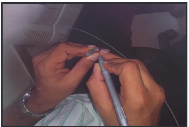
was marked 6mm from the edge using diamond point (shofu 101).
Ten such blocks were sandblasted using of 110µ size of alumina
particles (Aluminox) for 15 seconds using intraoral sandblaster (Bio-art,
Microjato microblaster) and then cleansed using ultrasonic cleaner (Confident
Ultrasonic Cleaner CO-80-L) with distilled water (Excel Demineralised
Water). Ivoclar ceramic repair system (Ivoclar Ceramic Repair N) was bonded
on the sandblasted surface of five blocks (4mm×4mm×4mm) and Shofu
ceramic repair system (P&R CRB Bond 1&2) was bonded on the sandblasted
Materials and Methodology
30
The remaining ten cobalt chromium blocks were laser etched
using ND: YAG laser (Fotona fidelis plus III, Slovenia) in pulsative mode at a
power setting of 6W, 120mj & 50Hz frequency for 60 seconds. Ivoclar
ceramic repair system(Ivoclar Ceramic Repair N) was bonded on the laser
etched surface of five blocks (4mm×4mm×4mm)and Shofu ceramic repair
system(P&R CRB Bond 1&2) was bonded on the laser etched surface of the
remaining five blocks (4mm×4mm×4mm).
A sharp ended chisel tool was made. The study to evaluate shear
bond strength was done in the Division of Polymeric Sciences, Bio Medical
wing of Sree Chithira Tirunal Institute of Medical Sciences and Technology
poojappura, Trivandrum. The cobalt chromium blocks were loaded on to the
large Jig of Universal Testing Machine (Instron model 3345).The tool was
fixed on the Universal Testing Machine. Test method used was compression
mode with a test speed of 1.0mm/min and sample was tested with 5kN load
cell. The shear bond strength was calculated using the formula:-
Shear bond strength = Force/Area
Scanning Electron Microscopic study was done in the Division of
Bio ceramics, Sree Chithira Tirunal Institute of Medical Sciences and
Technology Poojappura, Trivandrum. Scanning electron microscopic (S2400
Hitachi) study of the sandblasted & laser etched surfaces of cobalt chromium
metal blocks were done to assess the difference in roughness of the surface of
Materials and Methodology
31
coated in Ion Sputter machine before scanning electron microscopic
evaluation was done. Then the surface to be studied was marked with a marker
pen. Each surfaces were studied in three magnifications of 500X, 1000X and
2000X. In all the three magnifications the sandblasted surface showed
maximum irregularities and maximum roughness. The laser etched surface
showed maximum smooth uniform polished surface in all the three
magnifications of 500X, 1000X and 2000X. The Scanning electron
microscopic study go hand in hand with the results of present study which
gave maximum value for shear bond strength of sandblasted specimens than
Figures
Fig 1- Wax pattern made for Cobalt-Chromium metal block
Figures
[image:63.612.117.492.415.649.2]Fig 3- Casting process
Figures
Fig 5- Cobalt-Chromium metal blocks
Figures
Fig 7- Intra Oral Sandblaster
Figures
Fig 9- Sandblasted Cobalt-Chromium blocks
Figures
Fig11-Sandblasted Cobalt-Chromium metal blocks in ultrasonic cleaner
Figures
Fig 13- Nd:YAG intra oral laser unit
Figures
Fig 15- Power setting of Laser unit
Figures
Fig 17- Ivoclar ceramic repair kit
Figures
Fig 19- Ivoclar-Heliobond application
Figures
Fig 21- Shofu Ceramic repair kit
Figures
[image:73.612.115.495.405.654.2]Fig 23- Shofu metal primer application
Figures
Fig 25- Ceramic repair system curing done using composite curing light cure unit
[image:74.612.125.494.424.645.2].
Figures
Figures
Figures
[image:77.612.115.500.374.592.2]Fig 29- Scanning electron microscope
Figures
[image:78.612.138.476.371.589.2]Fig 31- Gold coating on sandblasted Cobalt-Chromium metal block
Figures
Fig 33- Universal testing machine
Results and Observations
32
The present study evaluated the shear bond strength of sandblasted
and laser etched cobalt chromium metal surface coated with Ivoclar and Shofu
ceramic repair systems. For all groups the shear bond strength assessment were
done.
Description of Groups
Four groups were there in the present study. Group I, II, III & IV.
Group I - Sandblasted cobalt chromium metal surface coated with Ivoclar
ceramic repair system.
Group II - Laser etched cobalt chromium metal surface coated with Ivoclar
ceramic repair system.
Group III - Sandblasted cobalt chromium metal surface coated with Shofu
ceramic repair system.
Group IV - Laser etched cobalt chromium metal surface coated with Shofu
ceramic repair system.
RESULTS
The data was expressed in mean and standard deviation. Statistical Package
for Social Sciences (SPSS 16.0) version used for analysis. ANOVA (Analysis
of variance) (Post hoc) followed by Dunnet t test were applied to find the
statistical significance between the groups. P value less than 0.05 (p<0.05) is
Results and Observations
33
Table-1: Mean Max load and Shear Bond Strength values of different groups
Groups Treatment Max load
(MEAN±SD)
Shear Bond Strength (MPa)
(MEAN±SD)
Group-I Ivoclar sand blasting 411.81±1.25 25.73±7.84
Group-II Ivoclar laser 126.41±6.03 7.90±3.77
Group-III
SHOFU sandblasting 296.53±3.76 18.53±2.35
Group-IV
SHOFU laser 181.29±6.96 11.23±4.43
According to Table-1:-
a) For Group I (Sandblasted cobalt chromium metal surface coated with
Ivoclar ceramic repair system) the mean maximum load applied is 411N and
the mean shear bond strength value obtained is 25.73 MPa.
b) For Group II (Laser etched cobalt chromium metal surface coated with
Ivoclar ceramic repair system) the mean maximum load applied is 126 N and
the mean shear bond strength value obtained is 7.9 MPa.
c) For Group III (Sandblasted cobalt chromium metal surface coated with
Shofu ceramic repair system) the mean maximum load applied is 296 N and
the mean shear bond strength value obtained is 18.53MPa.
[image:82.612.117.532.133.363.2]Results and Observations
34
Shofu ceramic repair system) the mean maximum load applied is 181N and
the mean shear bond strength value obtained is 11.23MPa.
Group I (Sandblasted cobalt chromium metal surface coated with Ivoclar
ceramic repair system) has the highest shear bond strength value
compared to all other groups.
Table-2: Comparison of mean Max load and Shear Bond Strength values
of Group-I with other groups
Groups Max load (MEAN±SD)
P value Shear Bond Strength (MPa) (MEAN±SD)
P value
Group-I 411.81±1.25 25.73±7.84
Group-II 126.41±6.03* 0.001 7.90±3.77* 0.001
Group-III 296.53±3.76* 0.002 18.53±2.35* 0.04
Group-IV 181.29±6.96* 0.001 11.23±4.43* 0.02
(*p<0.05 significant compared Group-I with other groups)