PREVALENCE AND CLINICAL OUTCOME OF
ANTENATALLY DIAGNOSED RENAL ANOMALIES
Dissertation Submitted by
Dr. THIYAGARAJAN .S
In partial fulfillment of the requirements for the degree of doctor of Medicine (M.D) in Paediatrics
THE TAMILNADU DR.MGR UNIVERSITY
DEPARTMENT OF PAEDIATRICS
PSG INSTITUTE OF MEDICAL SCIENCES & RESEARCH PEELAMEDU, COIMBATORE- 641 004
TAMILNADU, INDIA
DECLARATION BY STUDENT
I hereby declare that this dissertation entitled “PREVALENCE AND
CLINICAL OUTCOME OF ANTENATALLY DIAGNOSED
RENAL ANOMALIES’’ is a bonafide and genuine research carried out by
me under the direct guidance and supervision of Dr. JOHN MATTHAI, M.D., DCH., Professor, Department of PAEDIATRICS, PSG Institute of Medical Sciences and Research, Coimbatore. The dissertation is submitted to the Tamilnadu Dr.MGR Medical University in partial fulfillment of the university regulation for the award of MD degree in PAEDIATRICS. This dissertation has not been submitted in part or full to any other university or for any other Degree or Diploma before this below mentioned date.
Place : Coimbatore Signature of the Candidate
CERTIFICATE BY THE GUIDE
This is to certify that the dissertation entitled “PREVALENCE AND
CLINICAL OUTCOME OF ANTENATALLY DIAGNOSED
RENAL ANOMALIES’’ is a bonafide and genuine research done by
Dr.THIYAGARAJAN .S in the Department of Paediatrics ,PSG Institute of Medical Sciences and Research, Coimbatore in partial fulfillment of the regulations of Dr.MGR Medical University for the award of M.D degree in Paediatrics.
Place : Coimbatore Dr. John Matthai, M.D., DCH.,
Date : Professor ,
Department of Paediatrics,
ENDORSEMENT BY THE HEAD OF THE DEPARTMENT
This is to certify that the thesis entitled “PREVALENCE AND
CLINICAL OUTCOME OF ANTENATALLY DIAGNOSED
RENAL ANOMALIES’’ is a bonafide and genuine research done by
Dr.THIYAGARAJAN .S under the guidance and supervision of Dr. JOHN MATTHAI, M.D., DCH., Professor, Department of Paediatrics, PSG Institute of Medical Sciences and Research, Coimbatore in partial fulfillment of the regulations of Dr.MGR Medical University for the award of MD degree in PAEDIATRICS.
Place : Coimbatore Dr. K. Neelakandan, M.D., (Paediatrics)
Date : Professor & Head,
Department of Paediatrics,
ENDORSEMENT BY THE DEAN
This is to certify that the thesis entitled “PREVALENCE AND
CLINICAL OUTCOME OF ANTENATALLY DIAGNOSED
RENAL ANOMALIES’’ is a bonafide and genuine research done by
Dr. THIYAGARAJAN .S under the guidance and supervision of Dr. JOHN MATTHAI, M.D., DCH., Professor , Department of Paediatrics, PSG Institute of Medical Sciences and Research, Coimbatore in partial fulfillment of the regulations of Dr.MGR Medical University for the award of MD degree in Paediatrics.
Place : Coimbatore Dr.S.Ramalingam, M.D,
Date : Dean
COPY RIGHT
DECLARATION BY THE CANDIDATE
I hereby declare that PSG Institute of Medical Sciences and Research, Coimbatore shall have the rights to preserve, use and disseminate this dissertation in print or electronic format for academic/research purpose.
Place : Coimbatore Signature of the Candidate
ACKNOWLEDGEMENT
I thank God almighty who showered his blessings upon me.
I thank Prof. Dr. John Matthai, M.D., DCH., who was there to guide me all through my journey in thesis and for his expert supervision and constant guidance and unyielding patience in completing this work.
I would like to express my sincere thanks to Prof. Dr Neelakandan, HOD of Dept of Paediatrics, Prof. Dr. AM Vijayalakshmi and Prof. Dr. Jothilakshmi for laying a stable foundation for performing my thesis.
I thank Dr. Pavai Arunachalam, Prof & HOD of Paediatric surgery, who is a very kind hearted and dedicated teacher, who was always willing to lend me a helping hand, for her invaluable guidance, constant encouragement and expert suggestions, in spite of her busy schedule, which was most crucial in overcoming various difficulties.
I also specially acknowledge Dr. Rajesh for his constant support and valuable inputs for this thesis.
I am extremely thankful to Dr. Sara Paul, Dr. Ramesh, Dr. Rajesh who made this work possible with their encouragement and co-operation.
I thank Dr. Ramalingam .S MD, Dean, PSG IMSR, for permitting me to carry out this thesis.
I thank my friends Dr. Liza Susan John, Dr. Mathanki, my colleagues and staffs of the department of Paediatrics , for constantly being a helping hand in my work.
CONTENTS
S.No TITLE PAGE NO
1 Introduction 1
2 Review of Literature 5
3 Aims and Objective 45
4 Methodology 46
5 Results 48
6 Discussion 73
7 Conclusion 82
8 Bibliography
9 Annexure
LIST OF TABLES
Table No
TABLE PAGE NO
1 Sex distribution. 59
2 Prevelance of 2nd trimester anomalies. 63 3 2nd trimester anomalies and their fate in 3rd trimester. 64
4 Anomalies in 3rd trimester. 65
5 3
rd
trimester anomalies and their fate in 1st P.N scan.
67 6
Distribution of 1st postnatal scan anomalies.
68 7 Anomalies persisted during 2
nd
postnatal scan.
70 8
Fate of babies with CAKUT during 3rd post natal
1
PREVALENCE AND CLINICAL OUTCOME OF
ANTENATALLY DIAGNOSED RENAL ANOMALIES
INTRODUCTION:
The congenital anomalies of kidney andurinary tract (CAKUT) cause morbidity in children (1). The uncertainty in the clinical classification of antenatal detected renal anomalies resulted in the aggregation of different entities under the single label acronym congenital anomalies of kidney and the urinary tract. It is one amongthe leading cause of renal failure in childhood, causing a significant burden in young adults. It account for about 40–50% of children having chronic kidney disease (2). CAKUT constitute about 15-20% of all the antenatally diagnosed congenital anomalies (3).
Antenatally detected urinary tract abnormalities are the most common anomaly detected by antenatal ultrasound. CAKUTs comprise a wide range of renal system structural and functional malformations that occur at the kidney, collecting system , bladder and urethra . These anomalies include absence of kidneys, structural abnormalities in the kidney like multicystic dysplastic kidney, hydronephrosis,ureteric dilatation, vesicoureteral reflux , pelvic ureter junction obstructionand posterior urethral valves. (2)
2
weeks of gestation. Before prenatal ultrasound became widely available, children with hydronephrosis often presented with associated symptoms such as infection, pain, hematuria , or palpable mass and surgical intervention was the primary role for alleviation of these symptoms. The use of antenatal ultrasound routinely has led to earlier diagnosis of the asymptomatic patient with an associated shift in treatment to renal preservation rather than symptomatic relief.
The antenatal detected anomalies causes anxiety in parents. They should be counselled for the follow up during postnatal period. Once diagnosed as CAKUT, the parents need to be counselled regarding the outcome of the anomalies. Early antenatal diagnosis is important for counselling the parents for possible intervention and for delivering in the appropriate centre and for further evaluation of the baby. The significance of prenatal diagnosis helps to detect those having anomalies and counsel them. The early evaluation in postnatal period and treatment should started to reduce the adverse outcomes. The risk also should be explained to the parents during counselling.The degree of dilatation of the urinary tract does not always correlate with prognosis. The dilatation of the urinary tract is a dynamic process, which not only can vary over time, but is also influenced by other factors.
3
15 weeks. The renal echogenicity decreases than the echogenicity of liver and spleen by 17 weeks of gestational age . The renal cortex and renal medulla can be clearly distinguished by their hyperechogenic and hypoechogenic respectively at end of 20 weeks of gestation.
The features noted to be documented in ultrasound scan is to locate the both the kidneys, size of the kidneys and the site of location of the kidneys. The USG helps in knowing the structural abnormalities in the kidney and its echogenicity. The bladder is also examined for its size and shape. Amniotic fluid is measured in ultrasound scan. The development of external genitalia also should be looked for. The urine production in the foetus starts around 9 weeks of gestational age. The urine production increases significantly after 16 weeks of gestation. The fetal urine constitutes about 90% of the amniotic fluid around 20 weeks of gestation. The deepest pocket of the amnioticfluid sac is used to measure the amniotic fluid index. It is an objective measurement. (5).
4
detailed historyincluding family history , investigation and treatment. Genetic counselling also should be offered to the parents.
5
REVIEW OF LITERATURE
Development of kidney :
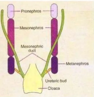
The kidneys are developed from the intermediate mesoderm. Urinary bladder and urethra develop from the urogenital sinus.The development of the urinary tract begins around 3 weeks of gestation. Ureter arises from mesoderm. The collecting part of the kidney is derived from the diverticulum called the ureteric bud. Kidney development has three stages: Pronephros, Mesonephros and Metanephros. Pronephros&mesonephros are transitory while metanephros develops into the permanent kidney (5).
6
Fig 1 : Pronephros , Mesonephros and Metanephros
7
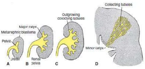
developing kidney. It will lead to conditions like agenesis of kidneys (absence),dysgenesis (abnormal differentiation). The dysgenesis includes aplasia , cystic disease and dysplasia of kidneys.
Fig 2: Development of the renal pelvis, calyces, and collecting tubules of the metanephros. A. 6 weeks. B. At end of the 6th week. C. 7 weeks. D. Newborn
[image:20.595.178.458.171.304.2]8
The common antenatal manifestations of CAKUT in antenatal period include oligohydraminos , variations in morphology of kidneys , ureter and bladder. After birth, CAKUT can be manifested as palpable abdominal pass , decreased urine output , feeding difficulties and with symptoms of urinary tract infections. CAKUT can develop in association with syndromes, but most cases are non syndromic. SyndromicCAKUTs occur with additional involvement of other organs. Few studies have been done on genetic association in development on cakut. The studies on mice model implicates that nonsyndromic human CAKUT may be caused by single-gene defects (2,6). HNF 1B and PAX2 are the most common genes implicated to cause CAKUT. Other cases may be sporadic as a result of many rare genes causing these disease (2).
Abnormalities in development of kidney and urinary tract :
9
1.Fetal kidney anomalies (renal malformations) • Renal agenesis (renal aplasia)
• Multicystic dysplastic kidneys • Renal hypoplasia
2.Fetal ureteric anomalies • Megaureter.
• Ureteropelvic junction obstruction (PUJO). • Ureterovesical junction obstruction (UVJO). • Duplex kidneys/ureters (renal duplications). • Incompetence of UVJ.
3.Fetal vesical anomalies (anomalies of the bladder)
10
Fig:3. Flowchart diagram showing various causes of CAKUT(12).
Due to confusion associated with terminologies for urinary tract
dilatation, the consensus panel recommended avoiding the non specific
terminologies for describing urinary tract dilatation likepyelectasis,
11 Hydronephrosis :
Hydronephrosis is the most common identified renal anomaly (dicke et al, 2006). The obstruction to urine resulting in dilatation of the renal pelvis and calyces is hydronephrosis(9). It literally means “water in the kidney”. Antenatal hydronephrosis is associated with vesicoureteral reflux or urinary tract
obstruction in 14–21% of cases. (10). The majority of the hydronephrosis were
isolated. The dilation of urinary tract in antenatal detected hydronephrosis is
due to large volume of fetal urine.The kidneys of foetus have smallrole in
maintenance homeostasis of salt and water. Urine production increases throughout the gestational age. At 37 weeks the urine volume is reported to be about 50 ml/hour. GFR triples by 3 months of age.The circulatory and the hormonal changes soon after birth leads to decrease in urine output. It results in the resolution of the antenatal detected hyronephrosis and ureter dilation. Some small kinks in ureter causing hydronephrosis also may get resolved over time. Most of the dilation detected in the fetus gets normalized soon after birth and only small percentage of patients will have severe obstruction and end up with surgery (13).
Renal agenesis :
12
agenesis is a lethal condition , a rare entity seen nowadays. The unilateral absent kidney is mostly isolated. It can also be a part of syndromic association. It can be associated with part of VACTERL anomalies.
“VACTERL” anomalies should be considered, if it has at least 3 of the following congenital malformations:
• Vertebral defects – (hemivertebrae, fused vertebrae) • Anal atresia or imperforate anus
• Cardiac defects – VSD is common • TEF - Tracheo-esophageal fistula • Renal anomalies(renal agenesis) and
• Limb abnormalities( radial agenesis, thumb hypoplasia).
True agenesis involves the ipsilateral absence of ureter, and ipsilateral bladder trigone. On USG investigation ther is emptiness in lumbar fossa and the adrenal gland appears enlarged. It classically denotes “lying down adrenal sign”. In individuals with single functioning kidney are attributed to increased risk of chronic kidney disease in adulthood (4, 14,15).
13
includes absence of kidneys in same side or located in abnormal position on same side, mulleriandefectson same side and absence of vagina refers to a syndrome called Mayer RokitanskyKusterHauser (MRKH) syndrome.
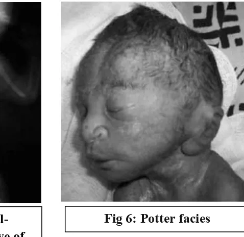
Bilateral renal agenesis is life threatening, with 50% fetuses are stillborn and the rest would die shortly after birth. It occurs in contion potter syndrome. The characteristic features are typical facial appearance, termed potter facies comprises widely separated eyes, low set ears, flat nose, redundant and dehydrated skin , receding chin and limb anomalies. It has prevalence of 1 in 3000 pregnancies.
The ultrasound findings of bilateral renal agenesis are (16):
• Non -visualization of both the kidneys in bilateral renal fossae, as well as in the entire abdominal cavity,
• Lying down adrenal sign,
• Renal fossae occupied by the bowel gas in late 3rd trimester,
• Severe oligohydramnios,
• Nonvisualization of urinary bladder,
• Pulmonary hypoplasia,
14
ARPKD – infantile type , cystic kidney disease, renal hypoplasia and dysplasia of medulla are the other causes of renal failure in neonates with potter syndrome. The babies born with absence of both kidneys will die of pulmonary failure due to hypoplasia of lungs rather than renal failure(4).
[image:27.595.110.391.203.369.2]
Figure 4. USG of fetal abdomen shows that both the renal fossae are not occupied by kidneys, and are occupied by the adrenal glands producing low lying adrenal sign.
[image:27.595.106.515.399.693.2]Fig 5: CXR showing bell-shaped thorax, suggestive of pulmonary hypoplasia
[image:27.595.251.490.424.656.2]15 Ectopic kidney :
The prevalence of ectopic kidneys is about 1 in 1000 pregnancies.
During renal embryogenesis it normally ascends from the pelvis into lumbar
region. Renal ectopia occurs when the process of ascending from sacral to
lumbar region is incomplete. Ectopic kidneyis seen in pelvis , iliac region,
[image:28.595.218.413.395.559.2]thoracic region and contralateral position. The kidneys get fused when the ectopia is bilateral. Fusion is more common in lower pole resulting in horseshoe kidney. Horseshoe kineys is more prevalent in turner syndrome. The incidence of wilms tumour in children with horse shoe kidney is four times more than other children.
Fig 7 : Horse shoe kidney - both fused in the lower pole
Multicystic dysplastic kidney (MCDK):
The developmental malformation of kidney that affect its size, shape or
structure is referred as renal dysgenesis. There are three types of dysgenesis :
16
number of nephrons, it can occur as isolated condition. The term dysplasia
denotes focal , diffuse or segmentally arranged primitive structures resulting in
abnormal metanephric differentiation. Other elements apart from renal tissues
like cartilages can be seen. MCDK can involve the entire kidney or only a certain part. In a kidney when cysts are developed, it is known as cystic dysplasia. When the entire kidney is dysplastic with predominance of cysts, it is known asmulticystic dysplastic kidney (4).
[image:29.595.212.424.544.679.2]The etiology of MCDK is still unclear. Some causes considered are infections, teratogenicdrugs , genetic disturbances and urinary tract obstructions. Mutations in the EYA1, SIX1, and PAX2 genes have been correlated. In a family PAX2 mutation had MCDK(as well as other renal anomalies) affected members that has occurred across three generations. Certain antiepileptic medications, infections such as theenterovirus, cytomegalovirus, and adenovirus have also been considered as contributing factors (18).
17
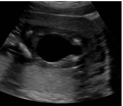
Fig 9 : USG of a MCDK patient
18
In unilateral MCDK , the contralateral kidneys are commonly affected. To evaluvate the kidney function , DMSA and VCUG are usually done. A dimercaptosuccinic acid scan (DMSA) is a nuclear medicine scan that generates tomographic and three-dimensional pictures of the kidneys. It detects the cortical scarring caused by contralateral abnormalities and how the kidneys are functioning. It can be used to distinguish between upper and lower UTI. But DMSA scans are not useful in differentiating contralateral renal abnormalities.
Vesicoureteral reflux causes urine to flow backwards from the bladder into the ureter or even the kidney. It causes pain and also scar the kidney or quite possibly disrupt the function of the kidney . A VCUG is a fluoroscopy exam that determines how the bladder is filled and if reflux occurs (18,19).
The abnormalities that affect the contralateral healthy kidney in MCDK are:
• Vesicoureteral reflux
• UVJ obstruction
• Hydronephrosis
• Ureterocele
• Crossed ectopia
• Echogenic kidney
19 Polycystic kidney disease:
20 Medullary Nephrocalcinosis :
Nephrocalcinosis is defined as renal calcification, is usually associated with hypercalciuric state. 7–64% of preterm neonates with gestational age less than 32 weeks or birth weight less than 1.5 kg , NC was diagnosed . Etiology of nephrocalcinosisin preterm is due to multiple factors. They include prematurity, LBW babies, severe respiratory disease and in imbalance between the stone-promoting factors & stone inhibiting factors.
Etiology :Nephrocalcinosis in preterm:
• Hypercalciuria
• High intake of calcium
• Low phosphorus
• Total Parenteral nutrition
• Diuretics - frusemide,
• Vitamin D
• Glucocorticosteroid
• Hyperoxaluria
• Fat malabsorption,
• Others :
Male, family history of kidney stones
21
The diagnosis is made by ultrasound examination. Spontaneous resolution of nephrocalcinosis occurs in 85 % of children by one year of age. (45). Prematurity itself can be associated with elevated B.P., comparatively smaller kidneys, and tubular dysfunctionin distal tubules. Added to it, nephrocalcinosis in preterm neonates can have long-term problems in renal function.The blood pressure andfunctionof kidneys should be followed up regularly for long term in prematurely born children with neonatalnephrocalcinosis.
Obstructive uropathy:
The urinary tract obstruction at the the junction ureteropelvic junction is the commonest cause of uropathy in babies (20).The incidence is one in 1500 births. Renal pelvis dilation will be seen in the ultrasound investigation done for PUJO. The ureter will not be dilated. The ureter will be involved in conditions like megaureter and UVJO. The ultrasound findings does not correlate clinically. The most of the dilatation gets resolved after birth. The fuctional scan is used for those with persisting anomalies. It is taken prior to surgery to take a decision. DTPA or MAG-3 are used. Ransley at al , concluded in his study that infants having renal pelvis APD greater than 20 mm are at increased risk of functional compromise of the kidney. MRI urogram is available now, it tells about the anatomy and the function of the kidneys The
indications for surgery include <40% differential function of the
22
diameter of the renal pelvis, pain, and infection. Pyeloplasty is the
gold-standard treatment (20). The ultimate goal of the treatment in PUJO is to improve the renal drainage, maintain the kidney function or improving it and relieving from symptoms.
Posterior uretheral valve (21, 22) :
Posterior uretheral valve is also known as congenital obstructing posterior uretheral valve. The m.c.congenital obstructive lesion of urethra is PUV, occurring only in male infants and is associated with morbidities, including urinary tract infection, chronic renal failure, urinary incontinence and even death. Urethral valves arisesfrom the tissues of wolffian duct origin. PUV occurs early in gestation around 5-7 weeks.The incidence is about 1 in 10000-25000 live births (22). The clinical symptoms depends on severity of the obstruction caused by PUV. The fetus will be S.G.A and in USG scan will revealoligohydraminos.
There are 3 types of PUV. Type 1 is the most common type.
Type 1 - Valves representing folds extending inferiorly from the verumontanum to the membranous urethra.
Type 2 - Valves as leaflets radiating from the verumontanum proximally to the bladder neck, and
23
24
In PUV due to the obstruction to urine flow is present , in some patients while voiding high tension is created within the bladder and it may lead to rupture and leads to accumulation of urine in other places.. It includes (1) Calyceal fornix rupture resulting in pararenalurinomas (2) Rupture of bladder intra-peritoneally( accumulated as intraperitoneal fluid, difficult to distinguish in ultrasound from ascites).
VCUGis the imaging of choice for diagnosing the posterior urethral
25
The overall prognosis depends on the degree and duration of
obstruction. Antenatal treatment is by doing vesicoamniotic shunting. It allows
urine to exit the bladder by the shuntcreated and obstruction to urethra is
relieved. This procedure is done by ultrasound guidance and performed in
expert hands. After birth the definitive treatment is surgery transurethral
ablation (fulgration) of the offending valve.
Vesicoureteralreflux :
The most common uropathy in childhood is VUR (vesicoureteral reflux). It means the retrogradeflow of urine tothe ureter and kidney (upper urinary tract) at rest or while voiding. Primary VUR can either be due to position abnormality in ureterovesical junction (UVJ) or in its integrity. The prevalence of the Vesicoureteral reflux in children is not exactly known, as
27
The ureter is attached to the bladder in an oblique direction between the detrusor muscle and bladder mucosa. Reflux is prevented by the flap-valve
mechanism created between them. VUR in children results in significant
pyelonephritis. The inflammatory reaction caused by a pyelonephritic infection
28
nephropathy. VUR can occur as an isolated anomaly or it can occur secondary to other renal pathology. The pathogenic organisms ascend backwards and can result in renal scarring and injury.
29
child to void. After treating the active urinary infection and resolution of clinical symptoms , VCUG should be performed.
Indications for surgery :
• High grade reflux of type 4-5,
• When there is low chance of spontaneous resolution of symptoms,
• Scarring of the kidney,
• Recurrent urinary tract infections,
• Getting fever because of UTI, while on antibiotics continuous prophylaxis.
• Parental decision for surgery.
FDA approved the drug Deflux, a bulking agentfor the treatment of VUR from grades 1-4. 2010 guidelines of American urology association was revised for the patients having breakthrough febrile urinary tract infection to include endoscopy treatment.
30
phase . Their features are consistent with findings of acute pyelonephritis.During follow up , scan 15% of the children had renal scarring.
Cooper et al mentioned in his study that the primary aim of surgical management is to prevent the febrile urinary tract infection and from pyelonephritis. But it is not yet proved that performing surgery will reduce the renal injury. Surgery will be benefitted in patients, having recurrent pyelonephritis and persistent reflux despite of conservative management. (27).
Evaluvation of antenatally detected CAKUT :
31
Antenatal hydronephrosis is present whenthe foetal renal pelvis diameter is more than or equal to 4 mm during the 2nd trimester scan & more than or equal to 7 mm in 3rd trimester scan. It is classified into mild, moderate and severe based on renal pelvic APD size. The upper limit for a normal renal antero-posterior diameter in the third trimester is 7 mm. The AP diameter of more than or equal to 7 mm at 18 weeks ( second trimester)denotes fetus with postnatal reflux or having obstruction. But those having same cut off at late gestation usually do not have significant pathology. There is always chance for inter observer variation and intra observer variation in measuring renal pelvic APD, since it is observer dependent. The renal pelvic AP diameter varies with gestational age, hydration status of mother and in bladder distension. (29).
In a study by Mallik M et al (34), concluded that the there is increasing sensitivity and accuracy of ultrasound screening at the time of 18–22 weeks. When APD of renal pelvis ≥4 mm , then the repeat scan at done after weeks of gestation. If the renal pelvis APD is ≥7 mm in the 2nd trimester then they should be referred to higher centre.
In study by sidhu et al (9), there is spontaneous resolution of antenatal hydronephrosis with renal pelvis APD less than 12 mm and SFU grade 1–2.
In study by lee at al (13), they found that the post natal renal anomaly
32
In a study by Metzger et al (31), they concluded that severe hydronephrosis in postnatal scan has an positive correlation with the risk of urinary tract infection and surgery. There is negative correlation among those who had spontaneous resolutionofhydronephrosis in postnatal scan.
In study by Longpre et al (32), they reported that the risk of morbidity is low in those fetuseswho had minimal pelvic calyceal dilatation of 5-9mm. The morbidity is risk is more in the fetus having severe hydronephrosis with APD of more than 15mm, and these group need regular follow up.
33
after birth during neonatal period. But 1/3rdof the neonates whom found to have moderate hydronephrosisand severe hydronephrosis during the 3rd trimester needed surgical intervention in the post natal period. Nearly eighty percent of the fetuses who were diagnosed in the 2nd trimester had resolution of the findings or improved. These babies had very low morbidity in the postnatal period. (33).
The common c based on SFU gradin diameter. SFU grading the thickness of the co
34
mon classifications for detection of postnatal hy grading and based on size of renal pelvic rading assess the renal pelvic fullness ,calycea the cortex.(Lee et al, 13)
35
36
Infants detected to have unilateral mild hydronephrosis or with bilateral hydronephrosis, whose APD measuring less than 10 mm, or 1-2 grade by SFU classification can be followed only by ultrasound (29). Most of the cases resolve in first two years of life. Hence antibiotic prophylaxis is usually not necessary for mild hydronephrosis (Tombesi et al, 35).
Mallik et al, proposed that infants detected to have renal anteroposterior diametermore than 10 mm or grade 3-4 by SFU 3-4 at birth needs to be monitored closely during follow up period (34).
The other classification previously used for evaluvation of antenatal hydronephrosisare :
Anterior-posterior pelvic diameter (APPD) Grignon (1986), grading
system (9)
Grade Size of pelvis Calyceal dilatation
Grade 1 1 cm Physiological
Grade 2 1–1.5 cm Normal calyces
Grade 3 >1.5 cm Slight dilatation
Grade 4 >1.5 cm Moderate dilatation
37
Blachar (1994), grading system (9)
Grade / Size of pelvis / Features
Grade 0<0.4 cm Normal/ no hydronephrosis
Grade 1 0.4–0.9 cm Detectable hydronephrosis
Grade 2 1–1.5 cm Significanthydronephrosis, rounding of calyces
Grade 3 >1.5 cm Severe hydronephrosis and calyces; cortical atrophy
and distorted renal anatomy.
Sinha et al recommends VCUG is done for patients having unilateral hydronephrosis or bilateral hydronephrosis - pelvis AP diameter more than 10 mm, grade 3-4 by SFU grading or with ureter dilatation. Patients suspected to have lower urinary tract obstruction like PUV, VCUG should be performed early, between 24 to 72 hours of life. The procedure is done at 4 to 6 wks of postnatal life in others (29).
38
Lee et al in a study, reported that 8-38% patients with antenatal hydronephrosis detected to have vesicoureteric reflex, whereas in general population< 1 % . The grade of VUR does not correlate with the severity of hydronephrosis. Some babies will be reported asnormal postnatal ultrasound despite of having VUR (7, 29). The patients with renal pelvis AP diameter more than 10 mm, likely to have severe vesicouretric reflux.
Diuretic Renography:
Diuretic renography is used for the evaluation of renal function and differentiation between the obstructive causes and non-obstructive causes of renal or ureteral dilation. It is a safe procedure and not very expensive. It estimates the relative renal function. The preferred radiopharmaceuticals are 99mTcmercaptoacetyltriglycine (MAG3), ethylenedicysteine (99mTc-EC) or Tc-diethylenetriaminepentaaceticacid. DTPA is relatively low cost and easily available. Radiotracer uptake is reduced inkidneys with reduced function.At 6-8 weeks of age ,renography is done. But in patients with severe HN and thinned cortex, the procedure can be done early. Hydration with iv fluids and catheterizing the bladder are not necessary. Oral hydration is adequate. Urinary catherizationshould be done in patients having poor bladder emptying, bilateral severe reflux or with megaureters.
39
of the drug depends on the hydration status and the composite and differential function of the kidney. Obstruction is ruled out if there is satisfactory elimination of the drug spontaneously, or after giving iv diuretics and after voiding.
When the curve is not declining even after 20 minutes remaining in ascending phase or in plateau phase, radionuclidedrug is not cleared even after giving diuretics and after voiding , then it is said to be obstructive renal curve (36). The differential function of kidneys between 45-55 % were considered normal. The kidney function is impaired, when the differential function is below 35% - 40% in the kidney with drainage obstruction. Obstruction should be considered when it takes long time to eliminate 50 % of the radionuclide drug and in ipsilateral kidney having supranormal function more than or equal to 55 %.
41 Indications for surgery in CAKUT :
42
the renal function is worsening progressively correlating with reduction in reduced by renal differential function by > 5-10 % , then pyeloplasty is recommended. (46).
Antibiotic prophylaxis:
The antibiotic prophylaxis is recommended for the babies who have severe hydronephrosis, hydroureteronephrosis and those who detected to have Vesicouretric reflux. Antibiotics should be given for 1 year in those found to have severe VUR. The recommended antibiotics is cephalexin (dose is 10 mg/kg/day) for first three months of life. For more than 3 months the recommended antibiotics are cotrimoxazole (dose 1-2 mg/ kg/day) and nitrofurantoin( dose is 1 mg/kg/day ) (29).
Antibiotic prophylaxis is recommended by American Urological Association (AUA)for the babies having severe VUR, for grades III-V (48).
In study by Kim et al, they reported that 19 – 36.2 % patients developed urinary tract infection , who were diagnosed to have moderate hydronephrosis or severe hydronephrosis due to obstruction(49). In study by Coelho et al , concluded that risk of infections is very high in babies with renal pelvic AP diameter of >10 mm in the postnatal scan.
43
neither fever nor elevated CRP.(43) The most common organisms causing are E.Coli , Klebsiella species, Proteus etc.
Urine culture is indicated in neonates with urogenital anomalies and in neonatal sepsis to exclude urinary tract infection.5ml of urine sample is obtained in a sterile container by supra pubic puncture,bladder catheterization or clean catch of midstream urine.Urine analysis is done for leucocyte esterase , nitrites and microscopy and findings are correlated with urine culture reports.
UTI may be diagnosed in the presence of one of the following:
(a) >10 WBC/mm3 in a 10 mL centrifuged sample
(b) >104 organisms/mL in urine obtained by catheterization and
(c) any organism in urine obtained by suprapubic aspiration.
Postnatal evaluation of patients with antenatal hydronephrosis in nut shell (29):
Ultrasound scan is postnatal period is donebetween 3 to 7 days of life.
Situations like suspected lower urinary tract obstruction scan is done earlier.
Hydronephrosisin postnatal scan was classified based on SFU grading or renal pelvisAPD classification.
Patients having mild unilateral hydronephrosis / bilateralhydronephrosis
resolution. Clos worsening hydr
Severe grades o
evaluate for und
Diuretic renogr
deciding on sur
The risk of ge parents of infan
Postnatal Mx Of (AN
44
Close monitoring and evaluvation is needed f g hydronephrosis.
rades of hydronephrosis and hydroureteroneph for underlying cause such as obstruction or VUR
enography detects the obstructive conditions on surgery.
of getting urinary tract infections should b f infants with hydronephrosis.
(ANH )Antenatally detected hydronephros
eded for the ones with
ronephrosis should be r VUR.
itions and is useful in
uld be counselled to
45
AIMS & OBJECTIVES
Primary objective :
To study the prevalence and pattern of distribution of antenatally diagnosedCongenital anomalies of kidney and urinary tract ( CAKUT )in live newborns during a one year period.
Secondary objective:
46
METHODOLOGY
:
Study design: Prospective observational study
Study period:1st May 2015 to 30thApril 2016 (1 year)
Study locale: PSG Hospitals , Coimbatore.
All women attending the antenatal clinic of the Obstetrics & Gynaecology department have an antenatal ultrasound during the second trimester (between 20-24 weeks) and third trimester above 30 weeks of gestation. All those who were detected to have CAKUT either during the second or third trimester Ultrasound scan were included in the study, provided they delivered in this hospital. The ultrasound machine used Voluson E8 version 10.0.3 and ultrasound scan was done by the consultant obstetrician trained in sonology, during the antenatal period. All antenatal data were recorded. The antenatal anomalies were classified based on the Society for fetal urology grading and renal pelvis APD classification (29). Written informed consent was obtained from the parents after the baby was born. Both term and pre term babies were included in the study.
47
48
RESULTS
During the study period there were 2614 live births in the hospital, who fulfilled the inclusion criteria. Among them, 111 babies were eligible for the study. 3 parents refused consent. 108 babies (79 males and 29 females) were included in the study. Among them, 54 were found to have renal anomalies in the 2nd trimester scan. 20 of these normalised in the 3rd trimester scan, however an additional 54 with anomalies were detected.
In the second trimester scan anomalies were detected in 54 cases. Unilateral hydronephrosis in 28 , bilateral hydronephrosis in 23 , hydroureteronephrosis in 2 and 1 unilateral multicystic dysplastic kidney . Among the 28 with unilateral hydronephrosis, 14 normalised , 9 persisted , 4 became bilateral hydronephrosis and 1 was reported as multicystic dysplastic kidney during the third trimester scan . In 23 bilateral hydronephrosis , 6 normalised , 5 became unilateral hydronephrosis , 11 remained same and 1 was reported as posterior urethral valve during the third trimester scan.
49
In the 3rd trimester scan, 88 babies were noted to have anomalies. Of them, 51 babies had unilateral hydronephrosis , 29 had bilateral hydronephrosis , 5 had multicystic dysplastic kidneys and 3 had posterior uretheral valve with bilateral hydroureteronephrosis.
All 108 babies had post natal Ultrasound done. In the 1stPost natal scan 65 babies were reported to have normal USG. Among these 65 babies, 34 had unilateral hydronephrosis, and 13 bilateral hydronephrosis in the third trimester scan, while the remaining 18 were those reported abnormal in the second trimester scan, but was normal in the third trimester scam.
50
and 1 was medullary nephrocalcinosis with bilateral hydroureteronephrosis (3rd trimester scan was bilateral multicystic dysplastic kidney).
Among the 43 babies who had abnormal findings in the first scan, 12 cases normalized in the second post natal scan.. 9 unilateral hydronephrosis , 1 bilateral hydronephrosis , 1 unilateral and 1 bilateral hydroureteronephrosis .
Among the 31 with abnormal second scans, 4 were lost to follow up. Of the remaining 27, nine were reported normal in the 3rd post natal scan. 5 with unilateral hydronephrosis , 3 bilateral hydronephrosis and 1 unilateral hydroureteronephrosis.
In the remaining 18 cases, 6 had undergone surgery , 4 persisted as Multicystic dysplastic kidney , 1 had medullary nephrocalcinosis, 1 unilateral bulky kidney, 4 had unilateral hydronephrosis and 2 had bilateral hydronephrosis. They are on follow up.
51
Clinical course of babies who had surgery and their outcome:
Baby 1 presented at 1 month of life with symptoms of UTI. Antenatally diagnosed as Posterior uretheral valve with bilateral HUN was on chemoprophylaxis . Urine culture has grown Escherichia Coli. Sr. creatinine was 0.48 mg/dl. DMSA was done. Cystoscopy and fulgration procedure was done. Discharged on antibiotic prophylaxis. On follow up baby was thriving well .
Usg showing bladder and dilated ureter USG showing Ureterohydronephrosis
MCU showing bilateral VUR
DMSA showed mild reduced cortical function of left kidney and small possible scar in upper pole.
Differential function :
Left kidney : 45% ;
52
Baby 2 with antenatal diagnosis of right PUJO was admitted at 1 ½ months of age following 2nd post natal scan . It was found to have increase in renal pelvis diameter. Right renal pelvis APD was 30 mm with parenchymal thinning. DTPA showed decreased renal function. Dismembered pyeloplasty was done . Post op baby was doing well. At 6 months follow up renal pelvis APD was 8mm .baby doing well , no features of UTI and renal parameters are normal.
Pre op USG showing pelvic calyceal USG – Thinned out parenchyma dilatation
After 3 months After 6 months DTPA : LK – 74% ; RK – 26 %
53
Baby 3 with antenatal diagnosis of bilateral hydronephrosis – Right PUJO and left minimal dilatation was admitted at 3 months of age for surgery. APD was increasing with calyceal dilatation 29 mm .DTPA report was 45 % with obstructive pattern. Dismembered pyeloplasty was done. Baby was doing well at follow up.
Pre- Op: Balloned out pelvis MCUG – No reflux
DTPA : LK - 55% ; RK – 45% function.
54
Baby 4 with antenatally diagnosed b/l HUN with PUV. Fulgration was done on day 2 of life. At 5 months of age Child admitted with for UTI . child was treated with antibiotics . After 2 weeks child presented with recurrent UTI . Child was operated – Cystoscopy + refulgration+ circumcision done. DMSA done .lk -48% ; rk – 52% no evidence of scarring.In further follow up, at 6 months weight was 6.22 kg and renal parameters was normal.
USG showing bilateral UHN with thickened bladder – pre op DMSA: L.K - 48% (no evidence of scarring)
R.K – 52% (no evidence of scarring)
55
Baby 5 with antenatal diagnosis of b/lhydronephrosis – leageft side severe hydronephrosis PUJO and thinning of parenchyma . child was on chemoprophylaxis. At 5 weeks follow up scan showed Left PUJO. At 2 ½ months DTPA showed 29 %. Renal pelvis APD was 25 mm . Dismembered pyeloplasty was done.
Pre-op USG
DTPA : Hydronephrotic , obstructed left kidney with evidence in cortical function Normal functioning , non obstructed right kidney
LK – 29 % ; RK- 71 %
56
Baby 6 with antenatal diagnosis of u/l Hydronephrosis with PCD – 1.74cm. Post natally was on follow up. PN1 scan showed APD of 15 mm .child was given chemoprophylaxis. At 5 months the finding was u/l HUN .on further follow up at 9 months of age child had DTPA – RK 46 % ; LK 54% with decreased perfusion and cortical function – suggestive of partial PUJO. APD was 32mm at time of surgery. Pyeloplasty was done.
Pre-op USG
b/o 6 continued…..
57
MRI images – Suggestive of right PUJO
58
1 baby with nephrocalcinosis in our study was a nicu graduate. Preterm, 33week + 1 day, birth weight of 1.05 kg. Renal parameters were assessed at birth and 5 months of age were normal . Sr. creatinine was 0.38 .Baby was on regular follow up. Thriving well.
59 Table 1 :
Sex distribution
Sex Frequency Percent
Male 79 73.1
Female 29 26.9
Total 108 100
Diagrams
73% 27%
1. Sex Ratio
60
2614; 96% 111; 4% 111; 4%
2. Prevalence of antenatal CAKUT
Total deliveries
61 total
deliveries ; 2614
PN- CAKUT; 2% Other; 43
62
52% 42%
2% 2% 2%
4. Second trimester anomalies
u/l HN
b/l hn
mcdk
u/l HUN
63 Table 2 :
Prevelance of 2nd trimester anomalies
2nd trimester anomalies Frequency
unilateral HN 28
bilateral HN 23
unilateral HUN 1
bilateral HUN 1
unilateral MCDK 1
64 Table 3 :
2nd trimester anomalies and their fate in 3rd trimester
2nd trimester scan anomalies
Total No
Fate of these anomalies in 3rd trimester scan
Normal U/l HN B/l HN U/l HUN B /
l HUN MCDK PUV
U/l HN 28 14 9 4 - - 1 -
B/l HN 23 6 5 11 - - - 1
U/l HUN 1 - - - 1 -
B/l HUN 1 - - - 1
MCDK 1 - - - 1 -
65 Table 4 :
Anomalies in 3rd trimester
3rd trimester scan Frequency Percent
Normal 20 18.5
unilateral HN 51 47.2
bilateral HN 29 26.9
unilateral MCDK 4 3.7
bilateral MCDK 1 0.9
B/L hun+puv 3 2.8
66
18%
47% 27%
3% 5%
5. Distribution of 3rd trimester anomalies
Normal
U/l HN
B/l HN
PUV
67 Table 5 :
3rd trimester anomalies and their fate in 1st P.N scan
3rd trimester scan anomalies
Total No
Fate of these anomalies in the 1st post natal scan
Normal U/l HN B/l HN U/l HUN B /
l HUN MCDK PUV
Normal 20 18 1 - 1 - - -
U/l HN 51 34 11 4 1 - - 1
B/l HN 29 13 7 7 1 1 - -
U/l HUN - - - -
B/l HUN - - - -
u/l MCDK 4 - - - 4 -
b/l MCDK 1 - - - 1
PUV 3 1 - - 2 - -
Total 108 65 20 11 3 3 4 2
68 Table 6 :
Distribution of 1st postnatal scan anomalies
1st post natal scan anomalies Frequency Percent
Normal 65 60.2
u/l HN 20 18.5
b/l HN 11 10.2
u/l HUN 3 2.8
b/l HUN 3 2.8
ul MCDK 4 3.7
Medullary Nephrocalcinosis 1 0.9
unilateral bulky kidney 1 0.9
69
Distribution of 1st postnatal USG – renal anamolies 60%
18% 10%
3%
3% 4%
1% 1%
6. Distribution of 1st P.N scan
Normal
u/l HN
b/l HN
u/l HUN
b/l HUN
u/l MCDK
u/l bulky kidney
70 Table 7 :
Anomalies persisted during 2nd postnatal scan
Follow up cases in PN scan 2 Frequency
Lost to follow up 4
Normal 12
MNC+b/l HN 1
u/l HN 12
b/l HN 6
u/l HUN 1
b/l HUN 2
U/l MCDK 4
71 Table 8 :
Fate of babies with CAKUT during 3rd post natal scan
Fate of Babies With CAKUT At III PN Scan Frequency
Normal in PN scan 3 9
Unilateral HN 4
Bilateral HN 2
Unilateral MCDK 4
Medullary nephrocalcinosis 1
Bulky kidney 1
Surgery 6
Lost to follow up 4
72
Final analysis of 108 samples
Medical renal disease – 4 MCDK, 1 Medullarynephrocalcinosis, 1 Bulky kidney.
80% 5%
5% 4%
2% 4%
7. Final results of 108 cases
Normal
Surgery
Medical Renal disease
u/l HN
b/l HN
73
DISCUSSION
Antenatally detected urinary tract abnormalities are the commonest anomalies detected on prenatal ultrasonography accounting for 20–50 % of all congenital disorders (5).The prevalence of the CAKUT in our study is 42.4 / 1000 live births. The study conducted in a tertiary care centre, in Saudi Arabia by Sallout B et al, reported thatantenatal prevalence of CAKUT is 21.28 per 1000 pregnancies and a birth prevalence of 19.80 per 1000 live births(1). To the best of our knowledge, there is no comparable study published from India.Study by jothi et al incidence of fetal urinary tract anamolies was 0.75%(44).
Antenatally diagnosed hydronephrosisprevalence ranges from 0.6 % to5.4% by Sinha et al(29). In a study done by bondagji, the antenatally detected CAKUT prevalence is 3.26 per1000 births (41).In developed countries, the CAKUT prevalence in live and stillborn infants is 0.3 to 1.6 per 1000(41). In a large study conducted in Europe, multicentre study, the details was taken from 20 registries of 12 countries. The study was done to evaluvate the prevalence of renal anomalies in antenatal ultrasound scan included all live born and stillborn (datas collected from 709030 births- both live and still birth). They found the mean prevalence to be 1.6 per 1000 live births. (41)
74
over there are no studies in high risk population to estimate their prevalence, clinical course and outcome in the developing world (3).
There was a significant male preponderance in our study with 79 males and 29 females, giving a ratio of 2.7:1. In study done by Gokce et al (5), the antenatal renal abnormalitiesappeared to be primarily a problem of boys (187 males, 69 females; with a M:F ratio of 2.71: 1). In a study done in Iran done by SadhegjiBojd et al , they screened 200 patients of post natal hydronephrosis, the male to female ratio is 3.5:1 (40). In brazil study done by Isabel Quirino et al , have male predominance . The study was done on 822 children with CAKUT of which 557 are male and 255 are female (28). In study done by Abishek et al, done in Telegana, India the distribution of their study subjects according to the sex of the baby were 76.1% were male compared to only 23.9% of females.(30)Other studies have also reported a male preponderance.(3, 41, 44).
In the study done by gokce et al (5) the antenatal ultrasound findings is changed in almost one-sixth (16 %) of the cases at postnatal US examination. Another study reported by Najmaldin et al, (37) errors included inability to distinguish between mostly cystic dysplasia and hydronephrosis and between hydronephrosis and normal patients.
75
kidney were termed as unilateral involvement, if both were involved it was termed as bilateral involvement. The abnormalities found in the study are hydronephrosis, multicystic dysplastic kidney (MCDK), medullary nephrocalcinosis, posterior uretheral valve and an abnormal variant with large unilateral kidney. In the antenatal scan the kidney size , parenchyma texture , ureter , bladder , amniotic fluid index was noted. In the post natal scans kidney echotexture , enhanced renal echogenicity , pelvic calyceal dilatation , renal pelvis APD, ureteric dilatation and bladder wall abnormalities were documented. Associated anomalies if any were documented. The most frequently detected anomaly on antenatal ultrasound is hydronephrosis .Most of
the affected patients have no other renal problems. This was termed as isolated
antenatal hydronephrosis. (9).
In our study majority of the detected cases were hydronephrosis. In
second trimester scan 54 out of 108, nearly 50% of the study population had
renal anomalies . The majority was hydronephrosis.
76
In study by jyoti et al hydronephrosis was the most commonly detected anomaly 79% out of 100 cases. On evaluation of babies with moderate to severe hydronephrosis persisting abnormality was seen in 17.4% and 1.1%. 8 babies had surgical intervention of which 20 babies with severe abnormalities. Mild hydronephrosis was resolved in 75% of the cases.
20 cases out of 54 (37 %) had a normal scan at third trimester, but had abnormal record in second trimester were followed up postnatally. Saini et al , reported that all babies detected to havehydronephrosis detected in antenatal scan, for whom it had resolved prenatally should also be evaluated in the postnatal period (3). Lee et al , in his meta-analysis study, (13) reported that in every dilatation of the urinary tract, 36 % of them had high risk for developing uropathy , irrespective of the dilatation . Thus, prenatal diagnosis of any form of hydronephrosis warrants for a critical followuppostnatally for prevention of urinary infections and further renal damage.(29).
77
In our study 1st postnatal scan out of the 108 cases, 65 were normalised (60%). In studyby sidhu et al, their results demonstrate that more than 70% cases of mild hydronephrosis (SFU grades 1–2; APPD less than 12 mm)
resolved, stabilized or improve during follow-up. Ismaili et al concluded in his
study that babies whose hydronephrosis had resolved spontaneously in postnatal period do not require further follow up and they have good outcome (38). In a cohort study of 130 infants with antenatally detected HN and who had normal ultrasound scan in postnatal period were followed for 2 years without any prophylactic treatment with antibiotics . The result was satisfactory as they did not have any worsening of hydronephrosis or urinary tract infectionduring the follow up period (38).Mild variants with APD diameter <10 mm on antenatal scans were transient and resolved spontaneously on subsequent followup scans. In a systemic analysis on 25 studies (9), they demonstrated that isolated antenatal detected hydronephrosisresolved in 98% of the patients with APD less than 12 mm, but only 51 % was resolved for those who had larger APD.
In a study by sidhu et al , they noticed that despite various classification methods was used, babies who had renal pelvic diameter less than 12 mm
mostly normalised and they mostly didn’t have any problems. Since the
measurement of renal pelvis in USG scan is always observer dependent , the
severe hydronephrosis with APD more than 12 mm have a variable outcome
78
In our study 60% of antenatally detected CAKUT resolved spontaneously at birth. In 2 other studies by Elder JS and Koff SA, it was reported that antenatally detected dilatation was transient and resolved spontaneously in more than 50% of cases.27-28 Severe forms of hydronephrosis (APD>12 mm) persisted on postnatal scans. In our study among the persisted hydronephrosis majority were more than 10 mm APD. Majority of the hydronephrosis below 8mm resolved spontaneously.
Cases with hydroureteronephrosis received antibiotic prophylaxis (29). Antibiotic prophylaxis was discontinued at 3 months for patients, if VCUG was normal and no further UTIs occurred (5). In study by Gokce et al , the most commonly detected underlying abnormalities were ureteropelvic junction obstruction (44.8 %), vesicoureteral reflux (30.0 %) and megaureter (9.5 %) in patients with postnatal hydronephrosis.
79
In a study done in Iran by SadeghiBojd et al,200 patients with post natal hydronephrosis were screened. There was male preponderance. Of them who underwent first postnatal control USG , 65% had normal, 18% mild/moderate and 17% severe hydronephrosis. 167 patients had VCUG of whom 20.82% with VUR. DTPA done for 112 patients with following resultsof whom, 50 patients had obstruction and 62 patients showed no obstructive finding. The final results were 54% of 200 patients recovered by conservative therapy, 12.5% by surgery and remaining improved without any surgical intervention.
In study done by Sairam S et al reported that fetalhydronephrosis was identified in 268 out of 11,465 mothers(2.3%) . Out of 268 caes , 216 were found to havemild hydronephrosis (80.6%), whereas the remaining were moderate or severe hydronephrosis( 52 cases out of 268, i.e, 19.4%) . 88 % of the cases had resolution of hydronephrosis in the antenatal itself or during the early neonatal period. No intervention was needed in the mild hydronephrosis group. Surgery was needed in post natal period for one third of the babies who had either moderate hydronephrosisor severe hydronephrosis.Surgery was required in only1 in 1000 births in their study population required surgery.
80
10 mm, grade 3-4 SFU and with ureterohydronephrosis.In the follow up period subsequently 12 was normalised in PN scan 2 and 9 in PN scan 34 were lost follow up (3.7% of the study population) during the study period. 6 babies had surgery . 4 MCDK and 1 Medullary nephrocalcinosis ,one unilateral bulky kidney was a variant in study .other 6 were in follow up . Their follow up status had no issues of renal problems and thriving well. 6 babies had surgery of which 4 had pyeloplasty and 2 had cystoscopy with fulgration.
81
LIMITATIONS OF THE STUDY
• Since the antenatal ultrasound was done by different obstetricians and not the same person, inter observer variation is a major limitation in the study.
82
CONCLUSION
1. The prevalence ofCAKUT in antenatal scans is 4.2%, while in postnatal it is only 1.65%.
2. Only 50% of antenatally diagnosed CAKUT is detected in the 2nd trimester scan. Approximately one third of them, mainly unilateral hydronephrosis normalises in the 3rd trimester scan
3. 50% of unilateral hydronephrosis detected in 2nd trimester scan normalised in 3rd trimester scan, compared to only 6 out of 23 bilateral hydronephrosis(26%).
4. Hydronephrosis is the commonest anomaly picked up in third trimester scan- 58% were unilateral hydronephrosis and 33% were bilateral hydronephrosis.
5. Antenatally diagnosedCAKUT in 65 of the 108 babies (60%) has normalised in the first postnatal scan, while an additional 12 (11%) has normalised in 2nd postnatal scan and 9 (8%) normalised 3rd postnatal scan. Overall 86 of 108 (79%) have normalised by 4 to 6 months.
83
7. Among babies with abnormal first postnatal scan 2 of 20 babies with unilateral hydronephrosis(10%) , 2 of 11 bilateral hydronephrosis(18%) and 2 of 3 with hydroureteronephrosis(66%) required surgery in first 6 months.
14
ARPKD – infantile type , cystic kidney disease, renal hypoplasia and
dysplasia of medulla are the other causes of renal failure in neonates with
potter syndrome. The babies born with absence of both kidneys will die of
pulmonary failure due to hypoplasia of lungs rather than renal failure(4).
[image:97.595.110.391.203.369.2]
Figure 4. USG of fetal abdomen shows that both the renal fossae are not occupied by kidneys, and are occupied by the adrenal glands producing low lying adrenal sign.
[image:97.595.105.518.395.692.2]Fig 5: CXR showing bell-shaped thorax, suggestive of pulmonary hypoplasia
[image:97.595.277.493.423.655.2]15 Ectopic kidney :
The prevalence of ectopic kidneys is about 1 in 1000 pregnancies.
During renal embryogenesis it normally ascends from the pelvis into lumbar
region. Renal ectopia occurs when the process of ascending from sacral to
lumbar region is incomplete. Ectopic kidneyis seen in pelvis , iliac region,
thoracic region and contralateral position. The kidneys get fused when the
ectopia is bilateral. Fusion is more common in lower pole resulting in
horseshoe kidney. Horseshoe kineys is more prevalent in turner syndrome. The
incidence of wilms tumour in children with horse shoe kidney is four times
[image:98.595.218.413.395.559.2]more than other children.
Fig 7 : Horse shoe kidney - both fused in the lower pole
Multicystic dysplastic kidney (MCDK):
The developmental malformation of kidney that affect its size, shape or
structure is referred as renal dysgenesis. There are three types of dysgenesis :
16
number of nephrons, it can occur as isolated condition. The term dysplasia
denotes focal , diffuse or segmentally arranged primitive structures resulting in
abnormal metanephric differentiation. Other elements apart from renal tissues
like cartilages can be seen. MCDK can involve the entire kidney or only a
certain part. In a kidney when cysts are developed, it is known as cystic
dysplasia. When the entire kidney is dysplastic with predominance of cysts, it
is known asmulticystic dysplastic kidney (4).
The etiology of MCDK is still unclear. Some causes considered are
infections, teratogenicdrugs , genetic disturbances and urinary tract
obstructions. Mutations in the EYA1, SIX1, and PAX2 genes have been
correlated. In a family PAX2 mutation had MCDK(as well as other renal
anomalies) affected members that has occurred across three generations.
Certain antiepileptic medications, infections such as theenterovirus,
cytomegalovirus, and adenovirus have also been considered as contributing
[image:99.595.212.424.544.679.2]factors (18).
17
Fig 9 : USG of a MCDK patient
The USG of a Multicystic dysplastic kidney shows multiple cysts ,
giving a appearance of “bunch of grapes”. The DTPA renal scan shows no
renal function.In patients with MCDK conservative management approach is
routinely practiced in many centres. One reason is, this condition is detected
antenatal ultrasound and it can be followed up in the post natal period. Many
researchers have found that multicysticdysplastic kidney regress over the time.
In a study reserachers have noted complete involution rates vary from 19–74%
over 9 months to 10 years. Surgical approach is needed when patients have
complications such as urinary tract infections, bleeding , flank pain ,
hypertension and malignant transformation. In studies done by Nishio et al ,
they found that ultrasound results of MCDK doesn’t necessarily mean the
involution has completely occurred. USG cannot detect small remnants from an