RESEARCH NOTE
Association between the accessory
gene regulator (
agr
) locus and the presence
of superantigen genes in clinical isolates
of methicillin-resistant
Staphylococcus aureus
Hamed Tahmasebi
1, Sanaz Dehbashi
2and Mohammad Reza Arabestani
3,4*Abstract
Objective: Methicillin-resistant Staphylococcus aureus cause to a variety of hard to cure infections. MRSA isolates also, produce an arsenal of virulence factors contribute to severe infections. The aim of this study was to find out the relationship between agr locus and presence of S. aureus superantigens (SAgs).
Results: Clinical isolates in two groups from two different states of Iran were collected. Antibiotic resistance patterns, agr typing, and virulence factor genes prevalence were identified and relationship between them was analyzed using SPSS software version16. Most of the samples were collected from wound 39 isolates in Group 1 and 61 isolates in Group 2. Frequency of MRSA strains was 38.1% in Group 1 and 52.1% in Group 2. Also, the most common resistance among both groups was to penicillin. agr positive isolates were detected in 132 isolates of Group 1 and 104 isolates of Group 2. In Conclusion, a significant relationship between the SAgs frequency and agr locus in both groups has been indicated. The production of superantigens in S. aureus plays an important role in the classification of agr locus, and this locus can affect differently in methicillin-resistant strains.
Keywords: Methicillin-resistant Staphylococcus aureus, Superantigens, Virulence factors, agr locus
© The Author(s) 2019. This article is distributed under the terms of the Creative Commons Attribution 4.0 International License (http://creat iveco mmons .org/licen ses/by/4.0/), which permits unrestricted use, distribution, and reproduction in any medium, provided you give appropriate credit to the original author(s) and the source, provide a link to the Creative Commons license, and indicate if changes were made. The Creative Commons Public Domain Dedication waiver (http://creat iveco mmons .org/ publi cdoma in/zero/1.0/) applies to the data made available in this article, unless otherwise stated.
Introduction
Inappropriate use of antibiotics to treat S. aureus infec-tions have led to the development of antibiotic resistant strains. The first cases of methicillin-resistant S. aureus
(MRSA) were identified in the 1960s, shortly after its introduction into clinical practice [1, 2]. Methicillin resistance is conferred by the mecA gene, which encodes a novel penicillin binding protein (PBP2A) [3, 4]. This protein has a reduced affinity for β-lactam antibiotics. The mecA gene is carried on a mobile genetic element known as the Staphylococcal Cassette Chromosome
mec (SCCmec), which can be horizontally transferred between Staphylococcal strains [5].
Staphylococcus aureus encodes toxin and superanti-gens like hemolysins, enterotoxins, exotoxins, exfoloative toxins, toxic shock syndrome toxin-1 (TSST-1) and leu-kotoxins such as the Panton-Valentine leukocidin (PVL). Different S. aureus strains encode different toxins. Exfo-liative toxins, TSST-1 and PVL are presented only in some clones [6]. Reduced toxicity can hide the bacteria from the immune system, therefore, facilitate more stable and suc-cessful colonization in the host [7]. However, there are a number of undescribed genes in the MRSA strains, which encode virulence factors associated with infections in ani-mals and human. Global regulators such as the accessory gene regulator (agr) system, Staphylococcal accessory regulator (Sar) and S. aureus exoprotein expression (Sae), have been well characterized which could help bacteria to adapt to a hostile environment [8, 9].
The production of S. aureus virulence factors is directly related to methicillin resistance. The mecA gene indirectly activates Autoinducer peptides (AIPs), which
Open Access
*Correspondence: mohammad.arabestani@gmail.com
3 Department of Microbiology, School of Medicine, University
of Hamadan, Hamadan, Iran
play an important role in the production of some regu-latory factors, biofilms and quorum-sensing (QS) [9]. Beceiro et al. state that methicillin resistance induces cell wall alterations that affect the agr quorum-sensing system of the bacteria and consequently reduced viru-lence in a murine model of sepsis [9, 10].
In this research, MRSA and non-MRSA strains were examined with the aim of investigating the relationship between agr regulatory system and virulence factors.
Main text
Methods
Isolation and identification of S. aureus
This cross-sectional study was designed to measure the prevalence of methicillin-resistant Staphylococcus aureus among patients and healthcare workers (three hospitals, four clinical laboratories, and two health-care centers) in Hamadan (Group 1) and Sistan and Baluchistan (Group 2) during July 2015 and August 2016. A multistage sampling method was used to select areas with different climate. Based on the distribution patterns of antibiotic resistance and different charac-teristics of the 28 states, the two states with the most differences in climate were selected. Sampling was done by considering the temperature variation index in different seasons and analyzing this index. Clinical specimens were inoculated on sheep blood agar (Merk, Darmstadt, Germany) and mannitol salt agar (Merk, Darmstadt, Germany), and incubated at 35–37 °C for 18 to 24 h aerobically. Biochemical tests were impli-cated to confirm the suspected isolates [11].
Detection of MRSA and determination of antimicrobial susceptibility profile of each isolate
Antimicrobial susceptibility testing was carried out by the Kirby Bauer disc diffusion method according to the Clinical Laboratory Standards Institute (CLSI) guidelines 2017 on Muller Hinton agar (Merk, Darmstadt, Ger-many). The following drugs were used to determine the antibiotic susceptibility: penicillin (10 U), tetracycline (30 μg), clindamycin (30 μg), gentamicin (30 μg), cipro-floxacin (5 μg), erythromycin (15 μg), chloramphenicol (30 μg), rifampin (5 μg), trimethoprim–sulfamethoxa-zole (10 μg) and linezolid (30 μg). All antibiotic disks were obtained from MAST ® Company, U K. Methicillin
susceptibility was determined using the cefoxitin E-test (Liofilchem, Italy) and oxacillin E-test (AB BIODISK, Sweden). S. aureus ATCC25923 was used as negative control and S. aureus ATCC43300 was used as positive control.
Genomic DNA extraction
Genomic DNA was extracted by Cinnaclon DNA extrac-tion kit (Cinnaclon, Iran) based on manufacturer’s instruction. DNA was yielded and investigated by spec-trophotometry using the Nanodrop (ThermoFishers, USA).
PCR for superantigen genes and screening for strains
The superantigen genes were amplified with specific primers listed by Schlievert and et al. [12] and Jarraud et al. [13] studies.
[image:2.595.58.541.485.705.2]agr typing
Classification of agr system groups was based on the hyper variable domain of agr locus according to Soares et al. [14]. Duplex PCR was performed to type groups based on their product size.
Statistical analysis
Data were organized and analyzed using the Statistical Package for Social Sciences (SPSS) software, version 16. The correlation between phenotypic antibiotic pattern and agr locus, phenotypic antibiotic pattern and superan-tigens genes, superansuperan-tigens genes and agr locus, sources of samples and agr locus in S. aureus isolates was evalu-ated by the Chi-square test and T-test. Statistical signifi-cance was set as a p-value of ≤ 0.05.
Results
Prevalence of clinical isolates
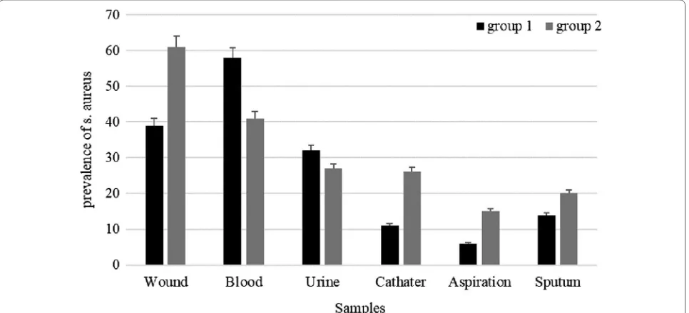
Totally, 1009 clinical samples were collected from patients in Hamedan (Group 1) and Sistan and balu-chistan (Group 2). 160 isolates were collected from Group 1 and 190 isolates were collected from Group 2. In Group 1, the most prevalent isolates were collected from blood 58 (36.25%). Also, in Group 2; most of the samples were isolated from wound 61 (31.10%) (Fig. 1).
Antibiotic resistance profiles and MIC
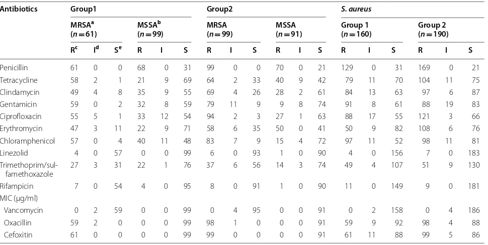
In Group 1, the most prevalent resistance was detected to penicillin (129, 80.62%) and to chloramphenicol (97 isolates, 60.62%). In addition, according to the results of
of E-test strips, 2 (1.2%) isolates intermediate-resistance to vancomycin ≥ 3 µ/ml, 59 (36.87%) isolates resistant to oxacillin ≥ 4 µ/ml and 61 isolates (38.12%) resistant to cefoxitin ≥ 8 µ/ml were identified. Also, 61 (38.12%) MRSA strains were isolated from the clinical and screen-ing samples.
In Group 2, penicillin and ciprofloxacin indicated as the highest resistance, 88.94% (169) and 63.68% (121) isolates, respectively. Moreover, based on the results of E-test strips, 4 (2.1%) isolates showed intermediate-resistance to vancomycin ≥ 3 µ/ml, 98 (51.50%) isolates resistant to oxacillin ≥ 4 µ/ml and 99 isolates (52.10%) isolates resistant to cefoxitin ≥ 8 µ/ml were identified. Also, 99 (52.10%) MRSA strains were isolated from the clinical and screening samples.
In Group 1, most of the MRSA samples were isolated from blood 66.66% (26 isolates) and wound 55.17% [32]. Whereas 0% (0 isolates) and 16.66% [1] of MSSA strains were detected in aspiration and sputum respectively. In Group 2, the most prevalent MRSA isolates were detected in blood 72.13% (44) and wound 46.34% [19], whereas MSSA isolates were identified in aspiration 26.66% [4] and sputum 10% [9] respectively, Table 1.
Superantigens genes profiles
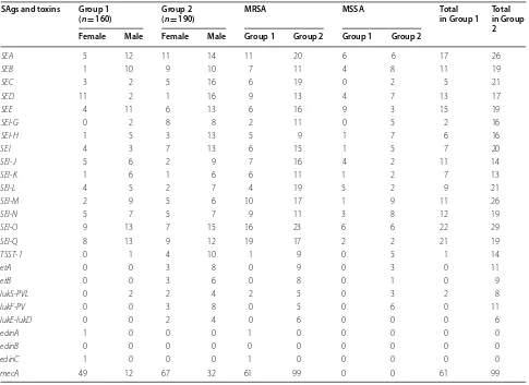
Out of 190 S. aureus isolates of Sistan and baluchistan,
96 (50.5%) of Zahedan, 30 (15.7%) of Khash and 64 (33.6%) isolates of Iranshahr was collected. Also, out of 190 Sistan and baluchistan isolates, seq had the high-est frequency and edinB had the lowest frequency,
Table 1 Antimicrobial resistance profiles of MRSA, MSSA, and S. aureus isolates
Antibiotics Group1 Group2 S. aureus
MRSAa
(n= 61) MSSA
b
(n= 99) MRSA(n= 99) MSSA(n= 91) Group 1(n= 160) Group 2(n= 190) Rc Id Se R I S R I S R I S R I S R I S
Penicillin 61 0 0 68 0 31 99 0 0 70 0 21 129 0 31 169 0 21
Tetracycline 58 2 1 21 9 69 64 2 33 40 9 42 79 11 70 104 11 75
Clindamycin 49 4 8 35 9 55 69 4 26 28 2 61 84 13 63 97 6 87
Gentamicin 59 0 2 32 8 59 79 11 9 9 8 74 91 8 61 88 19 83
Ciprofloxacin 55 5 1 33 12 54 94 2 3 27 1 63 88 17 55 121 3 66
Erythromycin 47 3 11 22 9 71 58 6 35 50 0 41 50 9 82 108 6 76
Chloramphenicol 57 0 4 40 11 48 83 7 9 15 4 72 97 11 52 98 11 81
Linezolid 4 0 57 0 0 99 6 0 93 1 0 90 4 0 156 7 0 183
Trimethoprim/sul-famethoxazole 27 3 31 22 1 76 37 6 56 14 3 74 49 4 107 51 9 130
Rifampicin 7 0 54 4 0 95 8 0 91 1 0 90 11 0 149 9 0 181
MIC (µg/ml)
Vancomycin 0 2 59 0 0 99 0 4 95 0 0 91 0 2 158 0 4 186
Oxacillin 59 2 0 0 0 99 98 1 0 0 0 91 59 9 92 98 4 88
[image:3.595.57.542.481.725.2]which were positive in 29 (15.2%) and 3 (1.5%) isolates, respectively. Moreover, out of 160 S. aureus isolates of
Hamedan, the seq gene found in 22 (13.75%) isolates was more abundant. None of the isolates of S. aureus isolated from Hamadan had etD, etA, etB, lukF-PV and lukE-lukD
genes. The prevalence of SAgs in female patients was higher than male patients. In addition, MDR strains also had the highest frequency of SAgs genes, Table 2.
agr typing
In Group 1 out of 160 isolates of S. aureus, 104 (65%) agr
positive and 56 (35%) agr negative were detected. The fre-quency of agr locus was identified as 27 (25.96%) agrA, 49 (47.11%) isolates agrB, 17 (16.34%) agrC and 11 (10.57%)
agrD. In Group 2, among 190 isolates of S. aureus, 132 (69.47%) were positive for agr and 58 (30.52%) negative for agr. Also, the frequency of agr locus was detected as follows: agrA in 39 (29.54%) agrB in 55 (41.66%), agrC in 29 (21.96%) and agrD in 9 (6.81.96%), Additional file 1: Tables S1, S2.
Statistical analysis
In this study, using t-test and Chi2, there was a significant
relationship between the SAgs and agr locus frequency. And also, a significant relationship was found between phenotypic antibiotic resistance and mecA.
Discussion
Staphylococcus aureus as a threatening agent in hospitals and societies has a diverse range of strategies including antibiotic resistance, virulence factors and precise regu-latory systems which accurately control and synchronize pathogenicity [15]. Therefore, in order to find the rela-tionship among agr types, superantigens production and resistance in MRSA strains, we investigated clinical iso-lates collected from two different regions of Iran, Hama-dan (Group 1) and Sistan and Baluchistan (Group 2).
Among 160 isolates of Group 1 and 190 isolates of Group 2, agrII was the most prevalent type. Strains with agr typeII indicated the highest superantigens production in both groups (p < 0.05). SEl-Q, SEl-O and SEB showed the highest prevalence in Group 1 whereas in Group 2
Table 2 Prevalence of SAgs genes in S. aureus isolates from patients of Group 1 and Group 2
SAgs and toxins Group 1
(n= 160) Group 2(n= 190) MRSA MSSA Total in Group 1 Total in Group 2 Female Male Female Male Group 1 Group 2 Group 1 Group 2
SEA 5 12 11 14 11 20 6 6 17 26
SEB 1 10 9 10 7 11 4 8 11 19
SEC 3 2 5 16 6 19 0 2 5 21
SED 11 2 1 16 9 13 4 7 13 17
SEE 4 11 6 13 6 16 9 3 15 19
SEl-G 0 2 8 8 2 11 0 5 2 16
SEl-H 1 5 3 13 5 9 1 7 6 16
SEI 4 3 7 13 6 15 1 5 7 20
SEl-J 5 6 2 9 7 16 4 2 11 14
SEl-K 1 6 1 6 6 11 1 2 7 13
SEl-L 4 5 2 7 4 19 5 2 9 21
SEl-M 2 9 5 6 10 17 1 9 11 26
SEl-N 5 7 5 7 9 11 3 8 12 19
SEl-O 9 13 7 15 16 23 6 6 22 29
SEl-Q 8 13 9 12 19 17 2 2 21 19
TSST-1 0 1 4 10 1 9 0 5 1 14
etA 0 0 3 8 0 9 0 3 0 11
etB 0 0 3 6 0 8 0 1 0 9
lukS-PVL 0 2 2 4 2 5 0 3 2 8
lukF-PV 0 0 3 8 0 5 0 6 0 11
lukE-lukD 0 0 2 4 0 6 0 0 0 6
edinA 1 0 0 0 1 0 0 0 0 0
edinB 0 0 0 0 0 0 0 0 0 0
edinC 1 0 0 0 1 0 0 0 0 0
[image:4.595.56.541.100.453.2]SEl-O and SEA were observed as the most widespread ones. Also, TSST-1, exfoliative toxins and pantone valen-tine toxins were detected only in Group 2. As a common feature between both groups, superantigen production is more prevalent in MRSA strains than MSSA ones (p < 0.05).
Regarding to the different frequency of antibiotic resistant and pathogenic strains in Groups 1 and 2, it is suggested that different climate conditions may cause extensive changes in resistance and pathogenicity of the bacterium. As MacFadden et al. [16], Singer et al. [17], and Kurenbach et al. [18] studies prove this notion, dif-ferences in the patterns of climate can lead to widespread changes in antibiotic resistance patterns. Consistent with our results, Zhang et al. [19] demonstrated the effects of various environmental conditions on antibiotic resistance and virulence factors in bacteria. Agr typing as a conveni-ent virulence typing method could contribute to a more precise understanding of the pathogenesis and epidemi-ology of staphylococcal infections [20]. Consistent with Collery, Nowrouzian and Chini, superantigen production in S. aureus is directly correlated to agr type of isolates. In so-called studies, the most prevalent superantigens were observed in agr types I and III, while in our study agrII was the predominant one [21–24]. To explain, clonal dif-ferences of strains collected from different regions should be regarded. Based on Guijarro and khelissa studies, environmental clues influences on evolution processes of the organisms and consequently variety in charac-teristics of strains occurs [25–27]. Moreover, in MRSA strains mecA gene leads to some changes in virulence fac-tors of the organism [28]. The activity of this gene affects some structural proteins such as agr, and agr-regulated SAgs such as TSST-1 and SEs, ETs and PVL are influ-enced [13]. As it is demonstrated in this study, there was a significant association between MRSA prevalence and superantigen production and interestingly superantigens which are regulated by agr system were predominated in Group 2. To illustrate, based on many studies, there is a direct relationship between presence of the mecA gene and the bacterial phenotypic resistance [29–31]. Stud-ies by Vitali et al. [32], Duran et al. [33] have shown that the presence of the mecA gene could affect Staphylococ-cus aureus strains in terms of antibiotic resistance pat-terns. The results of antimicrobial resistance studies conducted in Group 2 were shown a high prevalence of resistance to antibiotics. Although multi-drug resistance strains were detected in both groups, MDR strains pre-dominated in Group 2. As well, vancomycin intermediate
S. aureus (VISA) strains in Group 2 were observed more than Group 1. Several factors could be involved in this difference containing age, gender, climatic conditions, food type and regional culture. According to Lundgren
et al. [34], Norris et al. [35] and Wushouer et al. [36] which concluded that, cultural factor is one of the most important causes of antibiotic resistance. Consistent with the above mentioned studies, patients in Group 2 were more interested in taking different drugs, and patients in Group 1 showed less willingness to take medication.
In conclusion: a significant relationship between the SAgs frequency and agr locus in both groups has been indicated. Also, a substantial relevance has been found among phenotypic antibiotic resistance and mecA gene (p < 0.05). The production of superantigens in S. aureus
plays an important role in the classification of agr locus, and this locus can affect differently in methicillin-resist-ant strains.
Limitations
The results of this study suggest that the activity of vari-ous promoters and operons (PII, PIII and egc operon) in
S. aureus is directly related to agr locus. It seems that SAgs play a role as checkpoints of dissemination. In the current study, collaboration of antibiotic resistance with superantigen production has been proved (p < 0.05). However, the accurate mechanism of such a relationship should be unraveled.
Additional file
Additional file 1: Table S1. Characteristics of the agr allelic profiles of Group 1 S. aureus. Table S2. Characteristics of the agr allelic profiles of Group 2 S. aureus.
Abbreviations
agr: accessory gene regulator; MRSA: methicillin resistant S. aureus; SAgs: superantigens; PBP2A: penicillin binding protein; SCCmec: the staphylococcal chromosomal cassette; VISA: vancomycin intermediate-level resistant; VRSA: vancomycin resistant isolates; PVL: Panton-Valentine leukocidin; TSST-1: toxic shock syndrome toxin-1; MGEs: mobile generic elements; CLSI: Clinical Labora-tory Standards Institute.
Authors’ contributions
HT and SD performed microbiological and molecular tests and write the manuscript. MA supervised all of the stages of designing the study, conduct-ing the research and writconduct-ing the manuscript. All authors read and approved the manuscript.
Author details
1 Microbiology Department, School of Medicine, Zahedan University of
Medi-cal Sciences, Zahedan, Iran. 2 Microbiology Department, Faculty of Medicine,
Hamadan University of Medical Sciences, Pajoohesh Junction, Hamadan, Iran.
3 Department of Microbiology, School of Medicine, University of Hamadan,
Hamadan, Iran. 4 Brucellosis Research Center, Hamadan University of Medical
Sciences, Hamadan, Iran.
Acknowledgements
The authors of this article are grateful to Hamadan University of Medical Sci-ences for their financial support in conducting the research.
Competing interests
Availability of data and materials
All the data supporting the findings is contained within the manuscript.
Consent for publication
Not applicable.
Ethics approval and consent to participate
This study was approved by the Ethics Committee of Hamadan University of Medical Sciences (Code No: IR.UMSHA.REC.1395.757).
Funding
This Article was conducted on financial support of vice- chancellor for research of Hamadan University of Medical Sciences. The role of the funding was to supply the acquisition of the necessary materials for the research. There isn’t any funding in the design of the study and collection, analysis, and interpretation of data and in writing the manuscript.
Publisher’s Note
Springer Nature remains neutral with regard to jurisdictional claims in pub-lished maps and institutional affiliations.
Received: 17 December 2018 Accepted: 6 March 2019
References
1. Mino MJ, Ortiz RT, Randad P, Moffatt LT, Jordan MH, Shupp JW. Localiza-tion of superantigen virulence factors in kidney tissue of animals with Staphylococcus aureus-infected burn wounds. J Burn Care Res. 2013;34(1):142–50.
2. Vafaee Mehr M, Alikhani M, Tahmasebi H, Arabestani M. Identification and determination of the relationship between ccr alleles and antibiotic resistance in clinical isolates of methicillin resistant Staphylococcus aureus. J Babol Univ Med Sci. 2017;19(12):28–35.
3. Gardete S, Tomasz A. Mechanisms of vancomycin resistance in Staphylo-coccus aureus. J Clin Invest. 2014;124(7):2836–40.
4. Fleer A, Hemels MA, Paauw A, Krediet TG. Reduced expression of PBP-2A by neonatal mecA-positive coagulase-negative staphylococci (CoNS) blood isolates: beta-lactams are useful first-line agents for the treatment of neonatal CoNS sepsis, restricting the use of vancomycin. J Antimicrob Chemother. 2012;67(7):1616–8.
5. Tahmasebi H, Zeiyni B, Dehbashi S, Motamedi H, Vafaeifar M, Keramat F, et al. The study of blaZ and mecA gene expression in methicillin-resistant Staphylococcus aureus strains and the relationship between the gene expression patterns. J Isfahan Med Sch. 2017;35(443):1062–7. 6. Deodhar D, Varghese G, Balaji V, John J, Rebekah G, Janardhanan J, et al.
Prevalence of toxin genes among the clinical isolates of Staphylococcus aureus and its clinical impact. J Glob Infect Dis. 2015;7(3):97–102. 7. Solanki LS, Srivastava N, Singh S. Superantigens: a brief review with
spe-cial emphasis on dermatologic diseases. Dermatol Online J. 2008;14(2):3. 8. Tuffs SW, James DBA, Bestebroer J, Richards AC, Goncheva MI, O’Shea M, et al. The Staphylococcus aureus superantigen SElX is a bifunctional toxin that inhibits neutrophil function. PLoS Pathog. 2017;13(9):e1006461. 9. Dehbashi S, Tahmasebi H, Zeyni B, Arabestani M. The relationship
between promoter-dependent quorum sensing induced genes and methicillin resistance in clinical strains of Staphylococcus aureus. J Zanjan Univ Med Sci. 2018;26(116):75–87.
10. Beceiro A, Tomás M, Bou G. Antimicrobial resistance and virulence: a suc-cessful or deleterious association in the bacterial world? Clin Microbiol Rev. 2013;26(2):185–230.
11. Bokaeian M, Tahmasebi H. Molecular identification of genes responsible for resistance to aminoglycosides and methicillin in clinical samples of Staphylococcus aureus. J Babol Univ Med Sci. 2017;19(3):38–46. 12. Schlievert PM, Case LC, Strandberg KL, Tripp TJ, Lin Y-C, Peterson ML.
Vaginal Staphylococcus aureus superantigen profile shift from 1980 and 1981 to 2003, 2004, and 2005. J Clin Microbiol. 2007;45(8):2704–7. 13. Jarraud S, Mougel C, Thioulouse J, Lina G, Meugnier H, Forey F, et al.
Relationships between Staphylococcus aureus genetic background,
virulence factors, agr groups (alleles), and human disease. Infect Immun. 2002;70(2):631–41.
14. Soares BS, Melo DA, Motta CC, Marques VF, Barreto NB, Coelho SMO, et al. Characterization of virulence and antibiotic profile and agr typing of Staphylococcus aureus from milk of subclinical mastitis bovine in State of Rio de Janeiro. Arquivo Brasileiro de Medicina Veterinária e Zootecnia. 2017;69:843–50.
15. Thompson TA, Brown PD. Association between the agr locus and the presence of virulence genes and pathogenesis in Staphylococcus aureus using a Caenorhabditis elegans model. Int J Infect Dis. 2017;54:72–6. 16. MacFadden DR, McGough SF, Fisman D, Santillana M, Brownstein JS.
Antibiotic resistance increases with local temperature. Nat Clim Change. 2018;8(6):510–4.
17. Singer AC, Shaw H, Rhodes V, Hart A. Review of antimicrobial resistance in the environment and its relevance to environmental regulators. Front Microbiol. 2016;7:1728.
18. Kurenbach B, Hill AM, Godsoe W, van Hamelsveld S, Heinemann JA. Agrichemicals and antibiotics in combination increase antibiotic resist-ance evolution. PeerJ. 2018;6:e5801.
19. Zhang L, Rogers TJ. Assessment of the functional regions of the superan-tigen staphylococcal enterotoxin B. Toxins. 2013;5(10):1859–71. 20. Mullarky IK, Su C, Frieze N, Park YH, Sordillo LM. Staphylococcus aureus agr
genotypes with enterotoxin production capabilities can resist neutrophil bactericidal activity. Infect Immun. 2001;69(1):45–51.
21. Collery MM, Smyth DS, Tumilty JJ, Twohig JM, Smyth CJ. Associations between enterotoxin gene cluster types egc1, egc2 and egc3, agr types, enterotoxin and enterotoxin-like gene profiles, and molecular typing characteristics of human nasal carriage and animal isolates of Staphylo-coccus aureus. J Med Microbiol. 2009;58(Pt 1):13–25.
22. Collery MM, Smyth DS, Twohig JM, Shore AC, Coleman DC, Smyth CJ. Molecular typing of nasal carriage isolates of Staphylococcus aureus from an Irish university student population based on toxin gene PCR, agr locus types and multiple locus, variable number tandem repeat analysis. J Med Microbiol. 2008;57(Pt 3):348–58.
23. Nowrouzian FL, Dauwalder O, Meugnier H, Bes M, Etienne J, Vandenesch F, et al. Adhesin and superantigen genes and the capacity of Staphylococ-cus aureus to colonize the infantile gut. J Infect Dis. 2011;204(5):714–21. 24. Chini V, Dimitracopoulos G, Spiliopoulou I. Occurrence of the enterotoxin
gene cluster and the toxic shock syndrome toxin 1 gene among clinical isolates of methicillin-resistant Staphylococcus aureus is related to clonal type and agr group. J Clin Microbiol. 2006;44(5):1881–3.
25. Guijarro JA, Cascales D, García-Torrico AI, García-Domínguez M, Méndez J. Temperature-dependent expression of virulence genes in fish-patho-genic bacteria. Front Microbiol. 2015;6:700.
26. Khelissa SO, Jama C, Abdallah M, Boukherroub R, Faille C, Chihib N-E. Effect of incubation duration, growth temperature, and abiotic surface type on cell surface properties, adhesion and pathogenicity of biofilm-detached Staphylococcus aureus cells. AMB Express. 2017;7:191. 27. Corredor Arias LF, Luligo Espinal JS, Moncayo Ortiz JI, Santacruz Ibarra JJ,
Álvarez Aldana A. Relationship between super antigenicity, antimicrobial resistance and origin of Staphylococcus aureus isolated. Colomb Med. 2016;47(1):15–20.
28. Suryadevara M, Clark AE, Wolk DM, Carman A, Rosenbaum PF, Shaw J. Molecular characterization of invasive Staphylococcus aureus infection in central New york children: importance of two clonal groups and incon-sistent presence of selected virulence determinants. J Pediatr Infect Dis Soc. 2013;2(1):30–9.
29. Otarigho B, Falade MO. Analysis of antibiotics resistant genes in different strains of Staphylococcus aureus. Bioinformation. 2018;14(3):113–22. 30. Yilmaz S, Kilic A, Karagoz A, Bedir O, Uskudar Guclu A, Basustaoglu
AC. Investigation of various virulence factors among the hospital and community-acquired Staphylococcus aureus isolates by real-time PCR method. Mikrobiyol Bul. 2012;46(4):532–45.
31. Cameron DR, Howden BP, Peleg AY. The interface between antibiotic resistance and virulence in Staphylococcus aureus and its impact upon clinical outcomes. Clin Infect Dis. 2011;53(6):576–82.
•fast, convenient online submission •
thorough peer review by experienced researchers in your field • rapid publication on acceptance
• support for research data, including large and complex data types •
gold Open Access which fosters wider collaboration and increased citations maximum visibility for your research: over 100M website views per year •
At BMC, research is always in progress.
Learn more biomedcentral.com/submissions
Ready to submit your research? Choose BMC and benefit from:
33. Duran N, Ozer B, Duran GG, Onlen Y, Demir C. Antibiotic resistance genes & susceptibility patterns in staphylococci. Indian J Med Res. 2012;135(3):389–96.
34. Touboul-Lundgren P, Jensen S, Drai J, Lindbæk M. Identification of cultural determinants of antibiotic use cited in primary care in Europe: a mixed research synthesis study of integrated design “Culture is all around us”. BMC Public Health. 2015;15:908.
35. Norris P, Chamberlain K, Dew K, Gabe J, Hodgetts D, Madden H. Public beliefs about antibiotics, infection and resistance: a qualitative study. Antibiotics. 2013;2(4):465–76.