Open Access
Vol 6 No 4
Research article
Analysis of dendritic cells in tumor-free and tumor-containing
sentinel lymph nodes from patients with breast cancer
Nancy J Poindexter
1, Aysegul Sahin
2, Kelly K Hunt
3and Elizabeth A Grimm
11Department of Bioimmunotherapy, The University of Texas MD Anderson Cancer Center, Houston, Texas, USA 2Department of Pathology, The University of Texas MD Anderson Cancer Center, Houston, Texas, USA 3Department of Surgical Oncology, The University of Texas MD Anderson Cancer Center, Houston, Texas, USA
Corresponding author: Nancy J Poindexter, npoindex@mdanderson.org
Received: 22 Jan 2004 Revisions requested: 4 Mar 2004 Revisions received: 29 Mar 2004 Accepted: 5 May 2004 Published: 4 Jun 2004
Breast Cancer Res 2004, 6:R408-R415 (DOI 10.1186/bcr808)http://breast-cancer-research.com/content/6/4/R408
© 2004 Poindexter et al.; licensee BioMed Central Ltd. This is an Open Access article: verbatim copying and redistribution of this article are permitted in all media for any purpose, provided this notice is preserved along with the article's original URL.
Abstract
Introduction Sentinel lymph node (SLN) biopsy allows identification of the first lymph node into which a primary tumor drains. In breast cancer, identification of tumor cells in the SLNs is a predictor of the tumor's metastatic potential. In the present article, we tested the hypotheses that a positive immune response can occur in tumor-free SLNs and that the activation state of dendritic cells (DCs), the major antigen presenting cells within SLNs, predicts the immune status and metastatic potential of the tumor.
Methods Fifty paraffin-embedded SLN sections, 25 tumor-free and 25 tumor-containing, from patients with breast cancer were analyzed by immunohistochemistry to determine the immune maturation state of their DCs. In addition, 12 lymph nodes from noncancer-containing breasts were analyzed. Tissues were stained with antibodies against CD3, MHC class II, CD1a,
CD83, IL-10, and IL-12. Mature DCs were defined by CD83 expression and immature DCs by CD1a expression.
Results We found a trend toward higher numbers of mature CD83-positive DCs in tumor-free SLNs than in tumor-containing SLNs (P = 0.07). In addition, tumor-free SLNs were more likely to contain cells expressing IL-10 (P = 0.02) and, to a lesser extent, IL-12 (P = 0.12). In contrast, when all SLNs, both tumor-free and tumor-containing, were compared with uninvolved lymph nodes, the numbers of mature and immature DCs were similar.
Conclusions Our results suggest tumor-free SLNs are immunologically competent and potentially a site of tumor-specific T-cell activation, as evidenced by the presence of greater numbers of mature DCs and cytokine-producing cells in tumor-free SLNs.
Keywords: CD83, dendritic cells, IL-10, IL-12, sentinel lymph node
Introduction
Tumor-specific T-cell activation begins in the primary tumor when dendritic cells (DCs) encounter antigens in the form of apoptotic or necrotic tumor cells. The DCs engulf dying tumor cells and process their antigens into peptides that are presented in the context of MHC class I and class II molecules [1,2]. The function of a DC is highly influenced by its level of maturation. Immature DCs are capable of anti-gen uptake and processing but cannot, unless given the proper cytokine signals, present antigen to T cells [1,3,4]. After receiving the correct cytokine signals, the mature, peptide-loaded DCs migrate from the tumor to the first draining lymph node, referred to as the sentinel lymph node (SLN). In the SLN, naïve T cells are activated by the pep-tide-loaded mature DCs. These T cells then undergo clonal
expansion, gain effector function, and circulate back to the tumor, where their function is to lyse tumor cells. Evidence to support this process comes almost entirely from in vitro
experiments [1-4].
SLN biopsy allows identification of the first lymph node into which a primary tumor drains. In breast cancer, identifica-tion of tumor cells in the SLNs is a predictor of the tumor's metastatic potential [5,6]. In the present study, we exam-ined SLNs for evidence of immune activation by examining the maturation state of DCs within the SLNs. We defined mature DCs by their expression of the marker CD83 [7,8], while immature DCs were identified by their expression of the marker CD1a. We were interested in determining whether the maturation status of DCs in SLNs was
associated with the tumor status of the SLN, so we com-pared DCs in tumor-free SLNs and in tumor-containing SLNs.
Materials and methods
Study population
SLN tissues from women aged 26–87 years who had a SLN biopsy performed at The University of Texas MD Anderson Cancer Center between 1998 and 2001 were included in the study. Each of the patients had received a diagnosis of breast cancer and had undergone SLN biopsy as part of her surgical treatment. Paraffin-embedded SLN tissues from 50 patients, 25 with tumor-free SLNs and 25 with tumor-containing SLNs, were examined. The tumor status of the SLN was determined by H & E and immuno-histochemical staining. All samples were banked in the Breast Tumor Bank at The University of Texas MD Ander-son Cancer Center.
The study population included six women who received chemotherapy prior to their SLN biopsy: four whose SLNs contained tumors and two whose SLNs were tumor-free. Twelve lymph nodes draining from the unaffected breast of women with breast cancer were similarly processed. All materials were collected under a protocol approved by the MD Anderson Cancer Center Institutional Review Board.
Antibodies
The following antibodies were used for immunohistochem-ical staining: anti-CD3 (clone PS1; BioGenex, San Ramon, CA, USA), anti-HLA class II (clone CR3/43; DAKO, Carpinteria, CA, USA), anti-CD83 (clone HB15A; notech, Marseille, France), anti-CD1a (clone O10; Immu-notech), anti-IL-10 (clone 23738.111; R & D Systems, Minneapolis, MN, USA), and anti-IL-12 (clone 24910.1; R & D Systems). Optimal concentrations were determined empirically, and all antibodies were tested for staining of cells in cytospin preparations of peripheral blood mononu-clear cell (PBMC)-derived immature DCs (PBMCs stimu-lated with granulocyte–macrophage colony-stimustimu-lated factor and IL-4 for 6 days) or mature DCs (PBMCs stimu-lated with granulocyte–macrophage colony-stimustimu-lated fac-tor and IL-4 for 6 days followed by tumor necrosis facfac-tor alpha for 24 hours).
Immunohistochemistry
Tissue sections, 3 µm thick, were cut from tissue blocks of formalin-fixed, paraffin-embedded SLNs. Immunocyto-chemical staining of deparaffinized, fixed slides was per-formed after antigen retrieval by microwave heating in citrate buffer. Slides were incubated with biotinylated goat anti-mouse IgG followed by avidin–biotin–peroxidase com-plex (Vectastain ABC Kit; Vector Laboratories, Berlingame, CA, USA). The peroxidase was developed by 3-amino-9-ethylcarbazole-9 (AEC substrate kit; Vector Laboratories)
and counterstained with Gill's hematoxylin (Vector Labora-tories). Isotype-matched antibodies were used as negative controls. The positive control antibody for each group of slides was anti-CD3.
Because of the localized distribution of DCs within the SLNs, each slide was scored by counting positively stained cells in five high-powered fields (hpfs) under × 400 magni-fication. Results were reported for each SLN as the mean ± standard error of the mean for 5 hpfs. This method has been used by other workers when analyzing the expression of mature DCs in breast tissue [2,9]. All slides were counted without knowledge of the SLN status, and results were confirmed through a second reading by a breast can-cer pathologist (AS).
Statistical analysis
All data sets were tested for normal distribution. Because most data groups were not normally distributed, all data groups were analyzed by the Wilcoxon rank sum test. P < 0.05 was considered significant.
Results
Clinicopathologic characteristics of the patient population
Table 1 presents the clinicopathologic characteristics of the patients whose SLNs were included in this study. Patients ranged from 26 to 87 years of age at the time of diagnosis. No significant difference in median age distin-guished the tumor-free SLN and the tumor-containing SLN groups (56 and 55 years, respectively). Additionally, we examined the estrogen receptor and progesterone recep-tor status, the size, grade, and the stage of disease of the tumors. There were no remarkable differences between the two groups of patients.
Comparison of mature and immature DCs within the SLN
MHC class II, CD1a, and CD83. These slides are representative of the SLNs identified as tumor-free. The CD83-positive cells were found surrounding the paracorti-cal T-cell areas, usually near the sinuses of the SLNs. Although double staining was not performed, examination of sequential sections stained with CD83 and anti-MHC class II antibodies strongly suggested that the cells staining positively for CD83 were also positive for MHC class II.
When SLNs were compared by tumor burden, it was found that tumor-free SLNs contained a higher number of CD83-positive cells than tumor-containing SLNs. The numbers of CD1a-positive cells within these two groups of SLNs were similar. Figure 3 shows the comparison between the num-bers of CD1a and CD83 DCs in tumor-free and tumor-con-taining SLNs. There was a trend (P = 0.07) toward higher numbers of CD83-positive, mature DCs in tumor-free SLNs (median, 10.4 cells/hpf) than in tumor-containing SLNs (median, 4.5 cells/hpf). The numbers of CD1a-positive immature DCs did not differ between the tumor-free and tumor-containing SLNs (P = 0.93).
Comparison of clinicopathologic features of the tumor and DCs in the SLN
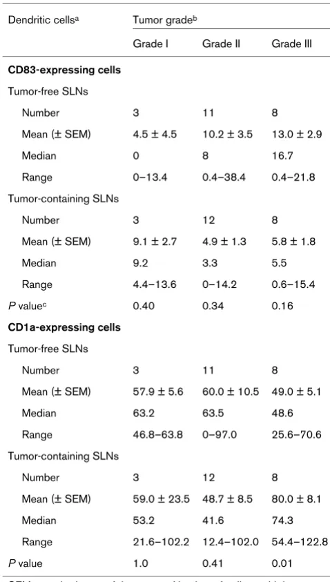
When tumors were analyzed based on hormone receptor status or tumor size, the numbers of CD83-expressing or CD1a-expressing cells in tumor-free and tumor-containing SLNs were not significantly different (data not shown). Notably, however, when tumors were sorted by nuclear grade and SLN status, we found that the number of CD1a-positive, immunologically immature DCs was significantly greater (P = 0.01) in the tumor-containing SLNs draining from grade III tumors than in the tumor-free SLNs draining from grade III tumors (Table 2). These data suggest that the differentiation status of the tumor affected the maturation of the DCs within the tumor.
Cytokine expression in the SLN
[image:3.612.57.558.114.414.2]Pivotal cytokines in the generation of a Th1 response ver-sus a Th2 response are IL-10 and IL-12. DCs will respond to these cytokines and become polarized toward activating Th1 and Th2 [10]. We examined the SLNs for these two cytokines to determine whether there was an indication of this activation. An analysis of cytokine-expressing cells is
Table 1
Clinicopathologic features associated with sentinel lymph nodes (SLNs)
Tumor-free SLNs (n = 25) Tumor-containing SLNs (n = 25)
Patient age (years)
Median 56 55
Range 35–76 26–87
Tumor size (cm)
Median 1.4 1.5
Range 0.1–5 0.6–4.5
Steroid receptor status
Estrogen receptor-positive 17 (68%)a 19 (76%)
Estrogen receptor-negative 4 (16%) 6 (24%)
Unknown 4 (16%) 0
Progesterone receptor-positive 15 (60%) 18 (72%)
Progesterone receptor-negative 5 (20%) 7 (28%)
Unknown 5 (20%) 0
Tumor grade
Grade I 3 (12%) 3 (12%)
Grade II 11 (44%) 12 (48%)
Grade III 8 (32%) 8 (32%)
Unknown 3 (12%) 2 (8%)
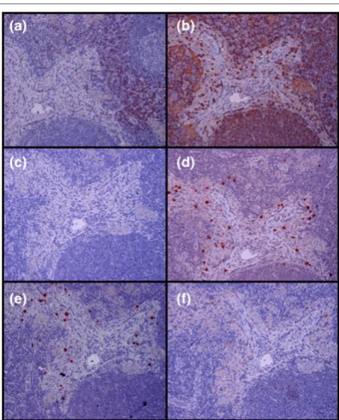
shown in Fig. 4. Tumor-free SLNs contained higher num-bers of IL-10-producing cells (P = 0.02) and IL-12-produc-ing cells (P = 0.12). Examination of sequential slides stained with anti-MHC class II or DC antibodies suggested that the cytokine-positive cells were MHC class II-positive and CD83-positive (Fig. 2). Tumor cells expressing IL-10 were not found in any of the SLNs examined.
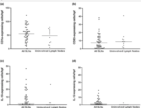
Comparison of SLN and lymph nodes draining from noncancer-containing breasts
To understand whether the numbers of DCs seen in the SLNs were indicative of an activated state due to the pres-ence of tumor, we examined 12 lymph nodes draining from the uninvolved noncancer-containing breast of patients with breast cancer (uninvolved lymph nodes) and com-pared them with the 50 SLNs from the breast cancer patients already analyzed. The numbers of CD1a-positive cells and CD83-positive cells were not significantly differ-ent when these uninvolved lymph nodes were compared with SLNs as a group (Fig. 5a,5b). In addition, when the number of CD83-positive cells in the free and tumor-containing SLNs were compared with the number of CD83-positive cells in the uninvolved lymph nodes, the tumor-free SLNs were very similar to the uninvolved lymph nodes (Figs 3 and 5). When cytokine-expressing cells were examined, SLNs contained significantly higher numbers of both IL-10-expressing cells (P = 0.02) and IL-12-express-ing cells (P = 0.002) than the uninvolved lymph nodes (Fig. 5c,5d). Although the numbers of cytokine-expressing cells in both groups were low, the cytokine expressing cells were more abundant in the tumor-free SLNs.
Discussion
Our experiments were designed to determine whether the SLN was the site of T-cell activation in breast cancer patients. Identification of immunologically mature DCs within the SLN would support this hypothesis. In our
exper-iments, mature DCs were defined by their expression of CD83, a marker that has been shown on DCs capable of antigen presentation and activation of naïve T cells [7,8]. Our results show a definite trend toward higher numbers of CD83-positive DCs in tumor-free SLNs than in tumor-con-taining SLNs. In addition, tumor-free SLNs contained signif-icantly higher numbers of IL-10-expressing cells. Both of these observations support the hypothesis that a tumor-free SLN is immunologically competent and is potentially a site of tumor-specific T-cell activation.
[image:4.612.55.297.91.202.2]What remains unclear from our data is the type of immune response that occurs in the tumor-free SLNs. Identification of both IL-10-expressing and IL-12-expressing cells sug-gests two possibilities. The existence of IL-10 in tumors has been associated with immune suppression of a Th1 response and increased tumorigenicity [11]. But it has been shown convincingly through in vitro studies that IL-10 specifically affects immature DCs by inhibiting upregulation of MHC class II and costimulatory molecules and expres-sion of CD83-positive cells. Mature CD83-expressing DCs are resistant to the effects of IL-10 [12]. Less information is available on the expression of IL-12 in tumor-draining lymph
Figure 1
[image:4.612.316.555.93.389.2]Expression of CD83 in a tumor-free sentinel lymph node (SLN) and of CD1a in a tumor-containing SLN from a breast cancer patient Expression of CD83 in a tumor-free sentinel lymph node (SLN) and of CD1a in a tumor-containing SLN from a breast cancer patient. Paraffin-embedded (a) tumor-free or (b) tumor-containing SLN tissue was ana-lyzed for expression of CD83 or CD1a, respectively, by immunohisto-chemistry. Typical examples of dendritic cell morphology stained with each antibody are shown (magnification, × 400).
Figure 2
Expression of (a) CD3, (b) MHC class II, (c) CD1a, (d) CD83, (e) IL-10, and (f) IL-12 in a tumor-free sentinel lymph node (SLN) from a breast cancer patient analyzed by immunohistochemistry
nodes. Animal, as well as in vitro, studies support the role of IL-12 as a proinflammatory cytokine that drives DCs toward activation of Th1 [13,14]. Clearly, the existence of both of these two cytokines in the SLN and not in unin-volved lymph nodes supports the hypothesis that an active immune response is occurring in the SLNs. Future func-tional studies examining both DCs and T cells are needed to determine whether the SLN is a site of immune suppression, as suggested by the expression of IL-10, or of immune activation, as suggested by expression of IL-12.
Our results differ from those reported by Huang and col-leagues [15], who compared S-100-positive DCs in SLNs and non-SLNs. Their conclusion, based on the morphology
[image:5.612.68.271.98.413.2]and density of the paracortical area and the number of paracortical S-100-positive DCs, was that the SLN was immunomodulated compared with the non-SLN. The SLNs examined in their study had reduced paracortical areas, reduced densities of paracortical DCs, and reduced fre-quencies of S-100-positive DCs with a predominance of immature, poorly dendritic DCs. Because they did not iden-tify the tumor-free SLNs versus the tumor containing SLNs in their study, it is hard to compare our results with theirs. When we group our data and compare all SLNs with lymph
Figure 3
Comparison of the numbers of (a) CD83-expressing cells and (b) CD1a-expressing cells in tumor-free and tumor-containing sentinel lymph nodes (SLNs) of breast cancer patients
[image:5.612.314.553.131.553.2]Comparison of the numbers of (a) CD83-expressing cells and (b) CD1a-expressing cells in tumor-free and tumor-containing sentinel lymph nodes (SLNs) of breast cancer patients. Each data point repre-sents the mean number of cells per high-powered field (in five fields) (magnification, × 400) from a paraffin-embedded section analyzed by immunohistochemistry. The horizontal bars represent the median number for each group. A trend toward a greater number of CD83-expressing cells in the tumor-free SLNs than in the tumor-containing SLNs was seen (P = 0.07); no difference in the number of CD1a-expressing cells distinguished the two types of SLNs (P = 0.93).
Table 2
Comparison of CD83-expressing and CD1a-expressing dendritic cells in tumor-free and tumor-containing sentinel lymph nodes (SLNs) by tumor grades
Dendritic cellsa Tumor gradeb
Grade I Grade II Grade III
CD83-expressing cells
Tumor-free SLNs
Number 3 11 8
Mean (± SEM) 4.5 ± 4.5 10.2 ± 3.5 13.0 ± 2.9
Median 0 8 16.7
Range 0–13.4 0.4–38.4 0.4–21.8
Tumor-containing SLNs
Number 3 12 8
Mean (± SEM) 9.1 ± 2.7 4.9 ± 1.3 5.8 ± 1.8
Median 9.2 3.3 5.5
Range 4.4–13.6 0–14.2 0.6–15.4
P valuec 0.40 0.34 0.16
CD1a-expressing cells
Tumor-free SLNs
Number 3 11 8
Mean (± SEM) 57.9 ± 5.6 60.0 ± 10.5 49.0 ± 5.1
Median 63.2 63.5 48.6
Range 46.8–63.8 0–97.0 25.6–70.6
Tumor-containing SLNs
Number 3 12 8
Mean (± SEM) 59.0 ± 23.5 48.7 ± 8.5 80.0 ± 8.1
Median 53.2 41.6 74.3
Range 21.6–102.2 12.4–102.0 54.4–122.8
P value 1.0 0.41 0.01
SEM, standard error of the mean. a Number of cells per
high-powered field. b The grade was unknown for two tumors in the
tumor-containing SLN group and for three tumors in the tumor-free SLN group. c P values determined by the Wilcoxon rank sum test
nodes draining from the noncancer-containing breast of women with cancer, however, the numbers of DCs in the SLNs and these lymph nodes are not different, suggesting that the SLN is not immunomodulated compared with unin-volved nodes.
Few laboratories have examined the immune status of SLNs of human tumors [15-17]. Most work involving SLNs has been limited to studies of frozen or paraffin-embedded tissue. In one of the few studies in which fresh SLN tissue was available, an attempt was made to determine whether tumor-specific T cells could be categorized as Th1-type or Th2-type cells, based on cytokine profiles [16]. No
classifi-cation was possible because the SLN T cells secreted both interferon gamma and IL-10 when stimulated with autolo-gous tumor cells. However, these T cells were clearly tumor specific because they responded to autologous but not all-ogeneic tumor cells [16]. A more recent study examining the cytokines produced by Staphylococcal enterotoxin-A-stimulated T cells from SLNs that drain primary melanoma showed both Th1 and Th2 cytokines could be measured by an enzyme-liked immunospot (ELISPOT) assay in tumor-free SLNs, whereas tumor-containing SLNs showed no increase in cytokine secretion [17]. The implication from this study was that immune responses were downregulated by micrometastases in the SLN. Both reports support our data and suggest that the SLN is the site of antigen-spe-cific activation and that the tumor status of the node may predict the immune response to the tumor.
The pathologic features of the tumor may affect the immune response in the SLN. It is accepted that the tumor microen-vironment affects the maturation state of the DCs in the tumor. Our data showed that the number of CD1a-express-ing cells in the SLN was associated with tumor grade. When high-grade tumors (grade III) were analyzed, higher numbers of CD1a cells were found in the tumor-containing SLNs. This suggested that the undifferentiated state of the tumor inhibits DC maturation, causing an accumulation of immature DCs unable to activate naïve T cells and resulting in metastasis of the tumor to the draining node. A report by Vitale and colleagues [18] suggested that the lack of an immune response in the high-grade tumor is explained by the downregulation of MHC class I and TAP-1 and TAP-2 proteins in these tumors.
A recent report by Iwamoto and colleagues [9] examining CD83-expressing DCs in 130 human breast tumors demonstrated that the presence of tumor-infiltrating CD83-expressing DCs correlated inversely with lymph node metastasis. Their data strongly suggested that CD83 DCs are involved in the initiation of the primary antitumor response. Our data, which showed a trend toward increased numbers of CD83-positive DCs in tumor-free SLNs compared with tumor-containing SLNs, further sup-port this conclusion, and similarly suggest that the matura-tion state of the DCs predicts the metastatic potential of the tumor. Further experiments to compare the DCs in pri-mary tumors and their SLNs are now underway in our labo-ratory to confirm the relationship between the maturation state of the DCs, the tumor grade, and the tumor metastatic potential.
Conclusions
[image:6.612.60.294.86.458.2]In summary, we found a trend toward higher numbers of CD83-positive DCs in tumor-free SLNs than in tumor-con-taining SLNs. In addition, tumor-free SLNs contain signifi-cantly higher numbers of IL-10-expressing cells. Both of
Figure 4
Comparison of the numbers of (a) IL-10-expressing cells and (b) IL-12-expressing cells in tumor-free and tumor-containing sentinel lymph nodes (SLNs) of breast cancer patients
these observations support the hypothesis that the tumor-free SLN is immunologically competent and is potentially a site of tumor-specific T-cell activation. To establish the prognostic potential of the DCs in the SLN, we are now undertaking functional studies to compare the immune status of DCs isolated from the primary tumors of patients with tumor-free SLNs and those with tumor-containing SLNs.
Competing interests
None declared.
Acknowledgements
The authors thank Sandra Kinney for excellent technical assistance. In addition, they are grateful for the technical assistance provided by the
staff of the Breast Tumor Bank at The University of Texas MD Anderson Cancer Center. The Breast Tumor Bank is supported through a grant from The Nellie B Connally Breast Cancer Research Fund. Statistical analysis was performed with the help of Marcella Johnson, of the Depart-ment of Biostatistics, MD Anderson Cancer Center. This research was supported by Department of the Army Grant DAMD17-00-1-0680 (to NJP) and by funds from the University Cancer Foundation of the Univer-sity of Texas MD Anderson Cancer Center (to NJP).
References
1. Banchereau J, Steinman RM: Dendritic cells and the control of immunity.Nature 1998, 392:245-252.
[image:7.612.60.554.92.472.2]2. Bell D, Chomarat P, Broyles D, Netto G, Harb GM, Lebecque S, Valladeau J, Davoust J, Palucka KA, Banchereau J: In breast car-cinoma tissue, immature dendritic cells reside within the tumor, whereas mature dendritic cells are located in peritu-moral areas.J Exp Med 1999, 190:1417-1425.
Figure 5
Comparison of (a) CD1a-expressing cells, (b) CD83-expressing cells, (c) IL-10-expressing cells and (d) IL-12-expressing cells in sentinel lymph nodes (SLNs) of study patients and lymph nodes draining from noncancer-containing breasts (uninvolved lymph nodes) of women with breast cancer
3. Cella M, Sallusto F, Lanzavecchia A: Origin, maturation and anti-gen presenting function of dendritic cells.Curr Opin Immunol
1997, 9:10-16.
4. Lapointe R, Toso JF, Butts C, Young HA, Hwu P: Human dendritic cells require multiple activation signals for the efficient gener-ation of tumor antigen-specific T lymphocytes.Eur J Immunol
2000, 30:3291-3298.
5. Giuliano AE, Jones RC, Brennan M, Statman R: Sentinel lym-phadenectomy in breast cancer. J Clin Oncol 1997, 15:2345-2350.
6. Leong SPL: Paradigm of metastasis for melanoma and breast cancer based on the sentinel lymph node experience. Ann Surg Oncol 2004, 11:192S-197S.
7. Zhou LJ, Schwarting R, Smith HM, Tedder TF: A novel cell-sur-face molecule expressed by human interdigitating reticulum cells, Langerhans cells, and activated lymphocytes is a new member of the Ig superfamily.J Immunol 1992, 149:735-742. 8. Zhou LJ, Tedder TF: CD14+ blood monocytes can differentiate
into functionally mature CD83+ dendritic cells.Proc Natl Acad
Sci USA 1995, 93:2588-2592.
9. Iwamota M, Shinohara H, Miyamoto A, Okuzawa M, Mabuchi H, Nohara H, Gon G, Toyoda M, Tanigawa N: Prognostic value of tumor-infiltrating dendritic cells expressing CD83 in human breast carcinomas.Int J Cancer 2003, 104:92-97.
10. Kalinski P, Hilkens CMU, Wierenga EA, Kapsenberg ML: T-cell priming by type-1 and type-2 polarized dendritic cells: the con-cept of a third signal.Immunol Today 1999, 20:561-567. 11. Halak BK, Maguire HC, Lattime EC: Tumor-induced
interleukin-10 inhibits type 1 immune responses directed at a tumor anti-gen as well as a non-tumor antianti-gen present at the tumor site.
Cancer Res 1999, 59:911-917.
12. Jonuleit H, Schmitt E, Steinbrink K, Enk AH: Dendritic cells as a tool to induce anergic and regulatory T cells.Trends Immunol
2001, 22:394-400.
13. Heufler C, Koch F, Stanzl U, Topar G, Wysocka M, Trinchieri G, Enk A, Steinman RM, Romani N, Schuler G: Interleukin-12 is pro-duced by dendritic cells and mediates T helper 1 development as well as interferon-gamma production by T helper 1 cells.
Eur J Immunol 1996, 26:659-668.
14. Cella M, Scheidegger D, Palmer-Lehmann K, Lane P, Lanzavec-chia A, Alber G: Ligation of CD40 on dendritic cells triggers pro-duction of high levels of interleukin-12 and enhances T cell stimulatory capacity: T-T help via APC activation.J Exp Med
1996, 184:741-747.
15. Huang RR, Wen D-R, Guo J, Giuliana AE, Nguyen M, Offodile R, Stern S, Turner R, Cochran AJ: Selective modulation of paracor-tical dendritic cells and T-lymphocytes in breast sentinel lymph nodes.Breast 2000, 6:225-232.
16. Chu Y, Hu HM, Winter H, Wood WJ, Doran T, Lashley D, Bashey J, Schuster J, Wood J, Lowe BA, Vetto JT, Weinberg AD, Puri R, Smith JW, Urba WJ, Fox BA: Examining the immune response in sentinel lymph nodes of mice and men. Eur J Nucl Med
1999, 26(Suppl):S50-S53.
17. Leong SPL, Peng M, Zhou Y-M, Vaquerano JE, Chang JWC: Cytokine profiles of sentinel lymph nodes draining the primary melanoma.Ann Surg Oncol 2002, 9:82-87.
18. Vitale M, Rezzani R, Rodella L, Zauli G, Grigolato P, Cadei M, Hick-lin DJ, Ferrone S: HLA class I antigen and transporter associ-ated with antigen processing (TAP1 and TAP2) down-regulation in high-grade primary breast carcinoma lesions.