Open Access
Vol 11 No 1
Research article
Methylation of homeobox genes is a frequent and early epigenetic
event in breast cancer
Stella Tommasi
1, Deborah L Karm
1, Xiwei Wu
2, Yun Yen
3and Gerd P Pfeifer
11Department of Cancer Biology, Beckman Research Institute of the City of Hope, Duarte, California 91010, USA 2Division of Information Sciences, Beckman Research Institute of the City of Hope, Duarte, California 91010, USA
3Department of Clinical and Molecular Pharmacology, Beckman Research Institute of the City of Hope, Duarte, California 91010, USA
Corresponding author: Stella Tommasi, stommasi@coh.org
Received: 24 Oct 2008 Revisions requested: 5 Dec 2008 Revisions received: 18 Feb 2009 Accepted: 27 Feb 2009 Published: 27 Feb 2009
Breast Cancer Research 2009, 11:R14 (doi:10.1186/bcr2233)
This article is online at: http://breast-cancer-research.com/content/11/1/R14 © 2009 Tommasi et al.; licensee BioMed Central Ltd.
This is an open access article distributed under the terms of the Creative Commons Attribution License (http://creativecommons.org/licenses/by/2.0), which permits unrestricted use, distribution, and reproduction in any medium, provided the original work is properly cited.
Abstract
Introduction Aberrant methylation of CpG islands is a hallmark of cancer and occurs at an early stage in breast tumorigenesis. However, its impact on tumor development is not fully determined, and its potential as a diagnostic biomarker remains to be validated. Methylation profiling of invasive breast carcinoma has been largely explored. Conversely, very little and sparse information is available on early-stage breast cancer. To gain insight into the epigenetic switches that may promote and/ or contribute to the initial neoplastic events during breast carcinogenesis, we have analyzed the DNA methylation profile of ductal carcinoma in situ, a premalignant breast lesion with a great potential to progress toward invasive carcinoma.
Methods We have utilized a comprehensive and sensitive array-based DNA mapping technique, the methylated-CpG island recovery assay, to profile the DNA methylation pattern in ductal carcinoma in situ. Differential methylation of CpG islands was compared genome-wide in tumor DNA versus normal DNA utilizing a statistical linear model in the LIMMA software package.
Results Using this approach, we have identified 108 significant CpG islands that undergo aberrant DNA methylation in ductal carcinoma in situ and stage I breast tumors, with methylation frequencies greater than or comparable with those of more advanced invasive carcinoma (50% to 93%). A substantial fraction of these hypermethylated CpG islands (32% of the annotated CpG islands) is associated with several homeobox genes, such as the TLX1, HOXB13, and HNF1B genes. Fifty-three percent of the genes hypermethylated in early-stage breast cancer overlap with known Polycomb targets and include homeobox genes and other developmental transcription factors.
Conclusions We have identified a series of new potential methylation biomarkers that may help elucidate the underlying mechanisms of breast tumorigenesis. More specifically, our results are suggestive of a critical role of homeobox gene methylation in the insurgence and/or progression of breast cancer.
Introduction
Breast cancer is a leading cause of cancer-related mortality in women, claiming over 400,000 lives per year worldwide. At the current breast cancer incidence rates, one in eight women is expected to develop the disease in her lifetime [1]. In spite of the high frequency, however, more than 90% of the breast cancer patients will survive if cancer is detected at an early stage and if treatment is begun promptly. Early detection is therefore extremely crucial for successful treatment and favo-rable prognosis, and emphasizes the need for new screening strategies for prompt intervention.
It is now widely recognized that aberrant epigenetic modifica-tions play a crucial role in altering gene expression and induc-ing tumor formation [2]. Methylation of CpG-rich islands encompassing gene promoter regions is especially relevant for the silencing of important tumor suppressor genes and accounts for a growing number of diseases, including breast cancer [3-5]. Several genes involved in cell cycle regulation and apoptosis (CCND2, CDKN2A/p16, RASSF1A), DNA damage response (BRCA1), cell adhesion (CDH1) and cell signaling (ER, RAR 2) have been reported to undergo pro-moter hypermethylation in breast carcinoma as well as in other
tumor types [6-10]. High levels of some hypermethylated genes can be detected very early, in the ductal lavage and nip-ple aspirates of patients with ductal carcinoma in situ (DCIS) (a stage 0 breast cancer) and stage I tumors, with methylation frequencies comparable with those of more advanced, inva-sive breast cancers [11]. Epigenetic inactivation can also occur, at different levels depending on the gene examined, in benign diseases such as mammary epithelial hyperplasia and intraductal papillomas – but not in disease-free normal breast epithelium, proliferating lactating breast tissue or stromal cells [12]. In some cases, even the normal breast tissue adjacent to the tumor site can display high levels of promoter methylation, indicating that premalignant epigenetic changes have the potential to spread gradually from the tumor epicenter to the surrounding cells or that a field defect exists that promotes tumorigenesis [13,14]. These data altogether support the evi-dence that methylation-driven gene silencing is a frequent as well as a relatively early event in breast tumorigenesis and can be used as a tag to detect breast cancer lesions at their very first appearance.
Based on this assumption, several groups have attempted, in recent years, to establish multigene DNA methylation profiles for the detection and classification of breast cancer. Their studies were mostly restricted to methylation-specific PCR analysis or to the array-based screening of limited target pan-els, and thus failed to interrogate the entire 30,000 CpG island repertoire of the genome [12,15-20]. In addition, these studies as well as genome-wide DNA methylation mapping techniques were usually employed to scrutinize invasive and metastatic breast carcinomas (stage II tumors and higher) [21-25]. These screening methods clearly overlook early epige-netic changes that may precede and/or promote invasive growth formation and offer limited diagnostic applications. Given the scattered and inadequate information available on early-stage breast tumors, it is not surprising that the search for breast cancer-specific methylation biomarkers has barely been translated into new reliable screening tests for the women at risk.
To fill the gaps in this area of research, we have analyzed the global methylation profile of DCIS, a premalignant breast lesion with a high potential to progress toward invasive and metastatic carcinoma, through loss of the myoepithelial cell layer [26]. For DNA methylation analysis, we have utilized a high-throughput methodology, recently developed in our labo-ratory. The methylated-CpG island recovery assay (MIRA) is a very sensitive technique that exploits the strong affinity of the MBD2/MBD3L1 complex to double-stranded CpG-methyl-ated DNA and allows one to detect cell-type-dependent differ-ences in DNA methylation on a microarray platform [27,28]. We have applied this technology to identify a series of novel candidate tumor suppressor genes and potential DNA methyl-ation biomarkers in DCIS. The present study provides an unprecedented snapshot of the global methylation profile in
early-stage breast carcinoma (approximately 28,000 CpG islands were analyzed) and may lead to more accurate diag-nostic tests for the prediction of breast cancer. Moreover, this work draws attention to the potential role of DNA methylation in the misregulation of homeobox genes during breast tumori-genesis. Homeobox-containing transcription factors control vital functional networks during tissue development and differ-entiation, and their aberrant expression has been often associ-ated, in the mammary gland, with both morphological abnormalities and oncogenesis [29-31].
Materials and methods
Specimens
Breast cancer samples of different histological type and grade were obtained from the City of Hope frozen tumor bank (City of Hope, Duarte, CA, USA). Tumors were staged according to the American Joint Committee on Cancer staging system pro-tocol [32]. Tissue sections, derived from various DCIS speci-mens, were stained with H&E and were reviewed by a pathologist to confirm the presence and the extent of the lesions. Breast tissue obtained from non-neoplastic breast quadrants served as the normal control. All patients gave writ-ten informed consent and authorization for use of biological specimens. The present study was approved by the Institu-tional Review Board of the City of Hope Medical Center (IRB# 97134).
Methylated-CpG island recovery assay-assisted microarray analysis
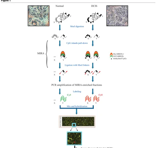
Following the MIRA pulldown, CpG-enriched DNA fragments were ligated to MseI linkers (upper strand sequence, 5'-AGCAACTGTGCTATCCGAGGGAT; lower strand sequence, 5'-TAATCCCTCGGA) and were PCR-amplified for up to 20 cycles by real-time PCR. Two micrograms each of the amplicons from enriched tumor DNA and from MIRA-enriched normal control samples were labeled with Cy5-dCTP and Cy3-dCTP respectively (GE Healthcare Bio-Sciences Corp., Piscataway, NJ, USA), using a BioPrime Array CGH Genomic Labeling kit (Invitrogen, Carlsbad, CA, USA). The purified samples were then mixed, and hybridized to CpG island microarrays, according to the Agilent ChIP-on-chip pro-tocol (version 9.0; Agilent Technologies, Santa Clara, CA, USA). Human CpG island microarrays contain 237,000 oligo-nucleotide probes covering 27,800 CpG islands and were purchased from Agilent Technologies (Santa Clara, CA, USA).
Microarray data analysis
Following hybridization and washing (Agilent ChIP-on-chip protocol, version 9.0), microarray slides were scanned using an Axon GenePix 4000b scanner (Molecular Devices, Sunny-vale, CA, USA) and images were quantified by GenePix Pro 6 software (Molecular Devices, Sunnyvale, CA, USA). Preproc-essing of raw data and statistical analysis were performed as previously described with some modifications [33]. Log2
-transformed ratios on each array were analyzed separately.
Probes were selected as positive if their ratios fell into more than the 98th percentile range on the array. Methylation regions (peaks) were defined as regions that contain a mini-mum of three positive probes. One negative probe was allowed within the peak as long as it was not present at the end of the region. The average of the log2 ratios of the positive
probes within each peak was assigned as the peak score. Peaks were then annotated based on their location relative to known transcripts (UCSC hg18 refseq). Peaks overlapping with the ±1,000 bp region of a known transcription start site to -10 kb of the transcription start site were annotated as upstream of the transcript, and peaks that overlap with the ±1,000 bp region of a known transcription termination site to +10 kb downstream of the transcription termination site were annotated as downstream of the transcript. Peaks falling into known transcripts but not within 1,000 bp of either the tran-scription start site or the trantran-scription termination site were denoted intragenic.
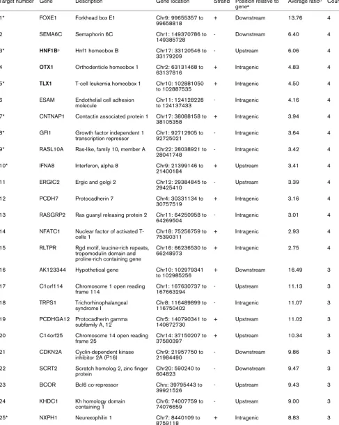
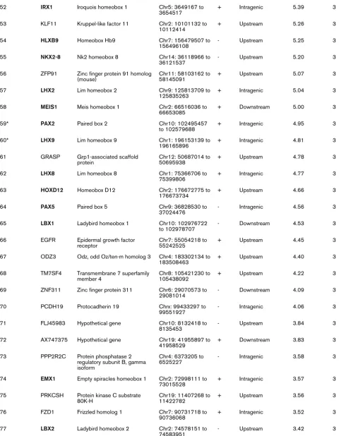
The transcripts that have either upstream, intragenic or down-stream peaks in at least three out of six DCIS samples were selected as interesting targets, and are reported in Table 1. For peaks that fell into uncharacterized regions of the genome, the location of the CpG island was used instead (Table 2). Microarray data were deposited in the Gene Expression Omni-bus repository [GEO:GSE14865].
Combined bisulfite restriction analysis and bisulfite sequencing
Total genomic DNA was isolated from human breast carci-noma and from normal tissues as described above. To analyze the methylation status of cytosines in several hypermethylated microarray targets, DNA (1 g) was treated with sodium bisulfite according to the manufacturer's protocol (EpiTect; Qiagen) and was subjected to combined bisulfite restriction analysis [34]. The PCR primer sequences used to amplify the candidate genes in bisulfite-treated DNA are available upon request. HeLa DNA was methylated in vitro with M. SssI meth-yltransferase and served as a positive control. For sequence analysis, the PCR products obtained after bisulfite conversion were cloned into the TOPO-TA cloning vector (Invitrogen) and up to 12 individual clones were sequenced.
Results
Genome-wide detection of methylated CpG islands in DCIS
The MIRA, used in combination with microarray analysis, is a high-resolution mapping technique and has proven successful in profiling global DNA methylation patterns in lung cancer [33,35]. In the present study we have applied this sensitive method to establish the methylation status of CpG islands in early-stage ductal carcinoma and to investigate its potential role in the initiation and development of breast cancer. Six DCIS were screened for methylation by MIRA-based microar-rays. To ensure consistency with the initial clinical diagnosis, and the presence of epithelial cells in the sample, histological examination of the specimens (both cancer samples and their matching normal) was conducted with the help of a pathologist.
The H&E slides derived from tumor samples displayed a pre-dominant DCIS component (70% to 80%) over normal stro-mal tissue and infiltrating lymphocytes (as shown for DCIS number 4 in Figure 1, upper right panel). Since contamination with low amounts of normal cells is not expected to interfere with the overall interpretation of the methylation data, tumor specimens were not subjected to further microdissection. Nor-mal healthy breast tissues adjacent to the lesions and obtained at the time of the surgical resection were used as a control. When matching normal tissue was not available, a DNA mix-ture from several normal breast tissues was used as a control. To avoid epigenetic variations due to age, reproductive state and/or cancer stage, we pooled together, whenever possible, DNA of normal breast tissues derived from comparable donors (same age frame, tumor stage, estrogen receptor/progester-one receptor status, and so forth).
Table 1
Methylated target genes identified by methylated-CpG island recovery assay-assisted microarray analysis
Target number Gene Description Gene location Strand Position relative to genea
Average ratiob Count
1* FOXE1 Forkhead box E1 Chr9: 99655357 to
99658818
+ Downstream 13.76 4
2 SEMA6C Semaphorin 6C Chr1: 149370786 to
149385728
- Downstream 6.40 4
3* HNF1Bc Hnf1 homeobox B Chr17: 33120546 to
33179209
- Upstream 6.06 4
4 OTX1 Orthodenticle homeobox 1 Chr2: 63131468 to 63137816
+ Intragenic 4.83 4
5* TLX1 T-cell leukemia homeobox 1 Chr10: 102881050 to 102887535
+ Intragenic 4.50 4
6 ESAM Endothelial cell adhesion molecule
Chr11: 124128228 to 124137433
- Intragenic 4.16 4
7* CNTNAP1 Contactin associated protein 1 Chr17: 38088158 to 38105358
+ Intragenic 3.94 4
8* GFI1 Growth factor independent 1 transcription repressor
Chr1: 92712905 to 92725021
- Intragenic 3.64 4
9* RASL10A Ras-like, family 10, member A Chr22: 28038921 to 28041748
- Intragenic 3.42 4
10* IFNA8 Interferon, alpha 8 Chr9: 21399146 to
21400184
+ Upstream 3.41 4
11 ERGIC2 Ergic and golgi 2 Chr12: 29384845 to
29425410
- Upstream 3.39 4
12 PCDH7 Protocadherin 7 Chr4: 30331134 to
30757519
+ Intragenic 3.16 4
13 RASGRP2 Ras guanyl releasing protein 2 Chr11: 64250958 to 64269504
- Intragenic 3.01 4
14 NFATC1 Nuclear factor of activated T-cells 1
Chr18: 75256759 to 75390311
+ Intragenic 2.93 4
15 RLTPR Rgd motif, leucine-rich repeats, tropomodulin domain and proline-rich containing gene
Chr16: 66236530 to 66248973
+ Intragenic 2.75 4
16 AK123344 Hypothetical gene Chr10: 102979341
to 102985256
+ Downstream 16.49 3
17 C1orf114 Chromosome 1 open reading frame 114
Chr1: 167630737 to 167663294
- Upstream 11.13 3
18 TRPS1 Trichorhinophalangeal syndrome I
Chr8: 116489899 to 116750402
- Intragenic 11.07 3
19 PCDHGA12 Protocadherin gamma subfamily A, 12
Chr5: 140790341 to 140872730
+ Upstream 11.02 3
20 C14orf25 Chromosome 14 open reading frame 25
Chr14: 37150207 to 37580397
+ Upstream 10.34 3
21 CDKN2A Cyclin-dependent kinase inhibitor 2A (P16)
Chr9: 21957750 to 21984490
- Downstream 9.86 3
22 SCRT2 Scratch homolog 2, zinc finger protein
Chr20: 590240 to 604823
- Downstream 9.47 3
23 BCOR Bcl6 co-repressor Chrx: 39795443 to
39921526
- Upstream 9.43 3
24 KHDC1 Kh homology domain
containing 1
Chr6: 74007759 to 74076659
- Upstream 9.00 3
25* NXPH1 Neurexophilin 1 Chr7: 8440109 to
8759118
26 CNR1 Cannabinoid receptor 1 (brain) Chr6: 88910156 to 88932281
- Upstream 8.78 3
27 BC039088 Hypothetical gene Chr5: 43050280 to
43054670
- Upstream 8.61 3
28 EVX2 Even-skipped homeobox 2 Chr2: 176653080 to 176656936
- Downstream 8.43 3
29* MT1E Metallothionein 1E Chr16: 55217085 to
55218525
+ Upstream 8.36 3
30* NR2F2 Nuclear receptor subfamily 2, group F, member 2
Chr15: 94674950 to 94683047
+ Downstream 8.24 3
31 HOXC13 Homeobox C13 Chr12: 52618843 to
52626595
+ Upstream 8.08 3
32 HOXD8 Homeobox D8 Chr2: 176702722 to
176704974
+ Upstream 7.97 3
33 SYCP2L Synaptonemal complex protein 2-like
Chr6: 10995049 to 11082527
+ Upstream 7.58 3
34 PCDHGB6 Protocadherin gamma subfamily B, 6
Chr5: 140767953 to 140872730
+ Intragenic 7.37 3
35 ACTA1 Actin, alpha 1, skeletal muscle Chr1: 227633615 to 227636466
- Upstream 7.36 3
36* PRDM14 Pr domain containing 14 Chr8: 71126576 to 71146116
- Intragenic 7.27 3
37* HOXB13 Homeobox B13 Chr17: 44157124 to
44161110
- Intragenic 6.91 3
38 OTX2 Orthodenticle homeobox 2 Chr14: 56337177 to 56346937
- Upstream 6.70 3
39 ZNF711 Zinc finger protein 711 Chrx: 84385652 to 84415025
+ Upstream 6.63 3
40* NR2E1 Nuclear receptor subfamily 2, group E, member 1
Chr6: 108593954 to 108616706
+ Intragenic 6.44 3
41* TAC1 Tachykinin, precursor 1 Chr7: 97199206 to 97207720
+ Upstream 6.37 3
42* CPEB1 Cytoplasmic polyadenylation element binding protein 1
Chr15: 81009005 to 81113783
- Upstream 5.99 3
43 NKAPL Nfkb activating protein-like Chr6: 28335076 to 28336715
+ Upstream 5.86 3
44 NKX6-2 Nk6 homeobox 2 Chr10: 134448309
to 134449527
- Downstream 5.72 3
45* TGIF2 Tgfb-induced factor homeobox 2
Chr20: 34635423 to 34655766
+ Upstream 5.60 3
46 CR596471 Hypothetical gene Chrx: 133511720 to
133522094
+ Upstream 5.56 3
47 EVX2 Even-skipped homeobox 2 Chr2: 176653080 to 176656936
- Intragenic 5.55 3
48 AX747981 Hypothetical gene Chr8: 96148213 to
96154324
- Upstream 5.48 3
49 ST8SIA3 St8 alpha-N-acetyl-neuraminide alpha-2,8-sialyltransferase 3
Chr18: 53170718 to 53187159
+ Intragenic 5.47 3
50 WT1 Wilms tumor 1 Chr11: 32365900 to
32413663
- Intragenic 5.40 3
51 BARHL2 Barh-like homeobox 2 Chr1: 90950167 to
90955382
[image:5.612.60.549.117.729.2]- Upstream 5.39 3
Table 1 (Continued)
52 IRX1 Iroquois homeobox 1 Chr5: 3649167 to 3654517
+ Intragenic 5.39 3
53 KLF11 Kruppel-like factor 11 Chr2: 10101132 to 10112414
+ Upstream 5.26 3
54 HLXB9 Homeobox Hb9 Chr7: 156479507 to
156496108
- Upstream 5.25 3
55 NKX2-8 Nk2 homeobox 8 Chr14: 36118966 to
36121537
- Upstream 5.20 3
56 ZFP91 Zinc finger protein 91 homolog (mouse)
Chr11: 58103162 to 58145091
+ Upstream 5.07 3
57 LHX2 Lim homeobox 2 Chr9: 125813709 to
125835263
+ Intragenic 5.04 3
58 MEIS1 Meis homeobox 1 Chr2: 66516036 to
66653085
+ Downstream 5.00 3
59* PAX2 Paired box 2 Chr10: 102495457
to 102579688
+ Intragenic 4.95 3
60* LHX9 Lim homeobox 9 Chr1: 196153139 to
196165896
+ Intragenic 4.81 3
61 GRASP Grp1-associated scaffold protein
Chr12: 50687014 to 50695938
+ Upstream 4.78 3
62 LHX8 Lim homeobox 8 Chr1: 75366706 to
75399806
+ Intragenic 4.77 3
63 HOXD12 Homeobox D12 Chr2: 176672775 to
176673734
+ Upstream 4.66 3
64 PAX5 Paired box 5 Chr9: 36828530 to
37024476
- Intragenic 4.56 3
65 LBX1 Ladybird homeobox 1 Chr10: 102976722
to 102978707
- Downstream 4.53 3
66 EGFR Epidermal growth factor receptor
Chr7: 55054218 to 55242525
+ Upstream 4.45 3
67 ODZ3 Odz, odd Oz/ten-m homolog 3 Chr4: 183302134 to 183508463
+ Upstream 4.40 3
68 TM7SF4 Transmembrane 7 superfamily member 4
Chr8: 105421230 to 105438092
+ Upstream 4.22 3
69 ZNF311 Zinc finger protein 311 Chr6: 29070573 to 29081014
- Downstream 4.09 3
70 PCDH19 Protocadherin 19 Chrx: 99433297 to
99551927
- Intragenic 4.06 3
71 FLJ45983 Hypothetical gene Chr10: 8132418 to
8135453
- Upstream 3.84 3
72 AX747375 Hypothetical gene Chr19: 41955897 to
41958529
+ Downstream 3.83 3
73 PPP2R2C Protein phosphatase 2 regulatory subunit B, gamma isoform
Chr4: 6373205 to 6525227
- Intragenic 3.58 3
74 EMX1 Empty spiracles homeobox 1 Chr2: 72998111 to 73015528
+ Intragenic 3.57 3
75 PRKCSH Protein kinase C substrate 80K-H
Chr19: 11407268 to 11422782
+ Upstream 3.56 3
76 FZD1 Frizzled homolog 1 Chr7: 90731718 to
90736068
+ Intragenic 3.52 3
77 LBX2 Ladybird homeobox 2 Chr2: 74578151 to
74583951
[image:6.612.60.550.116.734.2]- Upstream 3.42 3
Table 1 (Continued)
the analysis is provided in Materials and methods. Using this approach, we generated a list of over 100 significant CpG islands that display aberrant methylation in early-stage breast cancer and were methylation-positive in at least three out of six DCIS examined (Tables 1 and 2). Consistent with a potential role in transcriptional silencing, anomalous levels of DNA methylation were detected at the 5' end of known genes, in proximity of promoter regions or further upstream. Numerous CpG islands mapping to intragenic or downstream regions of annotated genes, however, were also found to be heavily methylated in DCIS (Table 1).
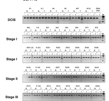
Verification of tumor-specific DNA methylation by combined bisulfite restriction analysis
We next confirmed tumor-specific methylation for several of the targets identified through array analysis using the BstUI combined bisulfite restriction analysis (COBRA) assay. In this assay, bisulfite-converted DNA is PCR-amplified using gene-specific primers and is then digested with the restriction endo-nuclease BstUI, which recognizes the sequence 5'-CGCG. Unmethylated restriction sites are converted to 5'-TGTG by sodium bisulfite and PCR and resist BstUI digestion, whereas methylated sites remain unchanged and are cleaved by the enzyme. The digested fragments displayed on agarose gels are thus indicative of methylated BstUI sites in the region analyzed.
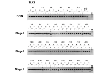
Representative examples of COBRA results are shown in Fig-ure 2. Here, we have inspected the methylation status of the candidate genes ranked numbers 5, 7, 8, 29, 40, 42 and 54 on the list of differentially methylated targets (corresponding to T-cell leukemia homeobox 1 (TLX1), CNTNAP1, GFI1, MT1E,
NR2E1, CPEB1 and homeobox HB9 (HLXB9), respectively). All seven CpG islands appear methylated with high specificity (no or very little methylation detected in normal breast tissue) in the DCIS analyzed, and with methylation frequencies rang-ing from 50% to 83% dependrang-ing on the target gene. No target region scrutinized so far exhibited robust CpG methylation across all six intraductal carcinomas (Table 1 and data not shown). Interestingly, we noticed that one-third of the CpG
islands identified by microarray analysis (26 out of the 81 annotated hits) are associated with members of various home-obox superfamilies (HOX, LHX, NKX, PAX, and so forth) and are preferential targets of de novo methylation in early-stage breast cancer. These master regulators control vital functional networks during tissue development and differentiation, and are misregulated in a variety of malignancies, including breast cancer [29,36].
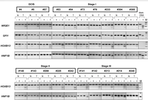
To explore this interesting finding in more detail, we then focused our attention on several homeobox genes. Besides the CpG islands associated with the TLX1 gene and with the
HLXB9 gene (Figure 2), we have examined the methylation status of the HNF1B and HOXB13 candidate gene targets (hit numbers 3 and 37, respectively). An uncharacterized CpG island located on chromosome 7 (CGI 7:48 and hit number 5 in Table 2) was also selected for COBRA analysis. Although some levels of methylation could be detected in normal tis-sues, methylation of HOXB13, HNF1B, and CGI 7:48 CpG islands was clearly more pronounced in intraductal carcino-mas (Figure 3, upper panel and Figure 4). Considering that the same matching controls were negative in other COBRA assays (Figure 2), we can exclude that the methylation signal observed here in normal samples is due to contamination with neighboring cancer cells. Rather, distinct cell populations within the normal breast tissue may display specific DNA methylation profiles, as recently established [37]. Differential promoter hypermethylation in benign breast epithelium derived from cancer patients can also be a function of age in a gene-specific manner [16,38]. Likewise, the occurrence of unmeth-ylated alleles in the cancer samples may reflect the heteroge-neity of CpG methylation within the cell populations of the tumor itself, but also the presence of normal cells in the specimens.
Confirmation of the methylation pattern in invasive breast tumors
To corroborate the DCIS-specific methylation profiles of TLX1, CGI 7:48, HOXB13, and HNF1B, we then extended the COBRA analysis to a series of primary breast tumors of
differ-78 HOXC13 Homeobox C13 Chr12: 52618842 to
52626595
+ Intragenic 3.40 3
79 MT1A Metallothionein 1A Chr16: 55230078 to
55231500
+ Upstream 3.36 3
80 DLX5 Distal-less homeobox 5 Chr7: 96487637 to 96492079
- Intragenic 3.06 3
81 LDOC1L Leucine zipper, downregulated in cancer 1-like
Chr22: 43267113 to 43272669
- Intragenic 2.30 3
aFor definitions of upstream, intragenic, and downstream, see Materials and methods (Microarray data analysis). The transcripts that have either
upstream, intragenic or downstream peaks in at least three out of six DCIS samples were selected as positive targets. *Gene targets verified by combined bisulfite restriction analysis assays or bisulfite sequencing. bAverage of the log
[image:7.612.56.556.109.213.2]2 ratios assigned to positive peaks across the six DCIS. cHomeobox genes are indicated in bold.
Table 1 (Continued)
ent histological type and stage (Figures 3 and 5, lower panels and Figure 4). The TLX1 CpG island was methylated in 13 out of the 16 stage I breast tumors (81%) and in six out of the eight stage II invasive carcinomas (75%). Likewise, the CpG island located on chromosome 7 (CGI 7:48) was methylated in almost every stage I tumor examined (93%) and in 15 out of the 17 more advanced tumors (stages II and III tumors, 88%). Of the 32 invasive breast carcinomas tested (15 cases of stage I tumors, nine cases of stage II tumors and eight cases of stage III tumors) 29 displayed higher levels of methylation relative to their matching controls within the HOXB13 element (91%), whereas the HNF1B CpG island was methylated in 21
tumor samples (66%) (partial COBRA data are shown in Fig-ure 4). Stage I tumors already exhibited a significant degree of methylation (87% for HOXB13 and 73% for HNF1B), con-firming the rapid and early nature of epigenetic reprogramming in breast cancer (Figure 4, representative COBRA analysis and Table 3).
[image:8.612.60.552.114.555.2]Rodriguez and colleagues have reported recently that hyper-methylation of the HOXB13 gene is a late event in breast tum-origenesis [39]. This apparent discrepancy with our results can be ascribed to the different methods used for the analysis (methylation-specific PCR versus our genome-wide DNA Table 2
Methylated CpG islands not associated with known genes
Target number Symbol CpG island locationa Average ratiob Count
1 CGI 1:42 Chr1: 38714506 to 38714991 6.27 4
2 CGI 6:60 Chr6: 106535804 to 106536465 4.69 4
3 CGI 2:112 Chr2: 239419845 to 239420943 4.22 4
4 CGI 4:34 Chr4: 174658599 to 174659018 2.94 4
5* CGI 7:48 Chr7: 35460738 to 35461227 17.26 3
6 CGI 8:121 Chr8: 100054909 to 100056159 16.81 3
7 CGI 10:46b Chr10: 119484483 to 119484981 15.44 3
8 CGI 17:16 Chr17: 44182434 to 44182640 13.96 3
9 CGI 7:351 Chr7: 129205522 to 129209745 11.56 3
10 CGI 6:19 Chr6: 27755772 to 27755984 10.16 3
11 CGI 8:31 Chr8: 81968510 to 81968882 8.45 3
12 CGI 22:48 Chr22: 44655029 to 44655728 7.98 3
13 CGI 2:77 Chr2: 176639821 to 176640909 7.91 3
14 CGI 5:47 Chr5: 176039674 to 176040127 7.78 3
15 CGI 7:29 Chr7: 129210233 to 129210591 7.60 3
16 CGI 3:47 Chr3: 134875805 to 134876344 6.58 3
17* CGI 4:35 Chr4: 24699204 to 24699608 6.22 3
18 CGI 6:18 Chr6: 27571155 to 27571358 6.18 3
19 CGI 7:277 Chr7: 154857318 to 154860615 5.91 3
20 CGI 10:46 Chr10: 102409137 to 102409658 5.84 3
21 CGI 4:21 Chr4: 174657922 to 174658134 5.53 3
22 CGI X:92 Chrx: 136459742 to 136460985 4.81 3
23 CGI 22:18 Chr22: 47762399 to 47762664 4.15 3
24 CGI 1:130 Chr1: 199774402 to 199775940 3.77 3
25 CGI 5:26 Chr5: 3379328 to 3379645 3.42 3
26 CGI 5:87 Chr5: 134853297 to 134854395 3.16 3
27 CGI 8:72 Chr8: 1100465 to 1101480 2.35 3
aFor uncharacterized gene targets, CpG island location was assigned instead of gene location. bAverage of the log
2 ratios assigned to positive
methylation profiling), the different specimens examined in the two studies (invasive-stage breast carcinomas versus DCIS), and the different location of the CpG islands on the HOXB13
gene (promoter versus an intragenic CpG island, target number 37). Methylation of this intragenic CpG island is an early event in breast cancer development and may precede promoter methylation. Whether this intragenic CpG island affects the HOXB13 gene expression remains to be seen.
Two additional targets, the growth factor transcriptional repressor GFI1 gene (target number 8) and the nuclear recep-tor NR2E1 gene (target number 40), were inspected to evalu-ate the level of methylation in early-stage breast carcinomas. In agreement with the MIRA results, we found that the GFI1 CpG island was hypermethylated in 14 out of the 18 tumors exam-ined (six DCIS and 12 stage I tumors, 78%) while the NR2E1
[image:9.612.53.562.87.567.2]target region was methylated in 11 out of the 21 early-stage Figure 1
Outline of the methylated-CpG island recovery assay-assisted CpG island microarray analysis
breast tumors (six DCIS and 15 stage I tumors, 52%) (Figure 4, partial data and Table 3).
To assess the extent of CpG methylation within the HOXB13,
HNF1B, HLXB9 and CGI 7:48 target sequences, primary
breast tumors were also subjected to bisulfite DNA sequenc-ing together with their matchsequenc-ing normal tissues (DCIS case number 57 and stage I case numbers 4b and 233). As expected, there is an evident tendency towards increased methylation in tumor-derived samples; the occurrence of early-stage cancer-specific methylated CpGs is very significant (P
< 0.001, Fisher's exact test) (Figure 6).
Discussion
DCIS is suspected to be a direct, although not obligate, pre-cursor of invasive breast cancer, and aberrant DNA
methyla-tion is believed to play a crucial role in breast tumorigenesis. Considering that epigenetic changes often become apparent in early phases of the disease, we speculated that the identifi-cation of DCIS-specific methylated biomarkers might be cru-cial to elucidate the molecular mechanisms underlying the initiation and development of breast cancer and to conceive effective strategies for early diagnosis.
To acquire valuable information into the epigenetic switches that may promote and/or contribute to the initial neoplastic events, we have analyzed the DNA methylation profile of DCIS, on a MIRA-based CpG island microarray platform. This novel and sensitive genome-wide screening approach has led to the identification of 108 CpG islands that display aberrant levels of DNA methylation in early breast lesions. Of the 81 CpG islands associated with known genes, only 37 map to pro-Figure 2
Verification of tumor-specific methylation of seven candidate target genes
Verification of tumor-specific methylation of seven candidate target genes. These targets were identified by the MIRA-assisted microarray approach (target numbers 5, 7, 8, 29, 40, 42 and 54, corresponding to TLX1, CNTNAP1, GFI1, MT1E, NR2E1, CPEB1 and HLXB9, respectively; Table 1). Genomic DNA from ductal carcinomas in situ (T) and matching normal breast tissues (N) was treated with sodium bisulfite and the target CpG island sequences were amplified using gene-specific primers. Methylation was confirmed by a BstUI combined bisulfite restriction analysis assay, which produces digestion products when BstUI restriction sites are methylated and not converted by bisulfite. HeLa DNA was methylated in vitro
[image:10.612.80.529.93.452.2]moter regions or further upstream (46%). In agreement with recently published data [40], more than one-half of the meth-ylated CpG islands in normal genomes fall within the body of the gene or in downstream regions. The functional role of these intergenic and intragenic CpG-rich elements remains obscure, but it has been suggested that they may constitute short independent transcriptional units [40].
Several targets have been examined by conventional bisulfite methods and found to be differentially methylated in multiple breast tumors, in complete agreement with the MIRA results. Most importantly, these gene candidates display methylation frequencies ranging from 50% to 83% in DCIS and up to 93% in stage I breast cancer, depending on the target gene, and these candidates hold great promise, alone or in combination, for future diagnostic applications. We were also able to iden-tify several genes, such as CDKN2A, PCDHGB6, and WT1
(Table 1) well known to be methylated and transcriptionally silent in breast cancer [10,23]. Many other 'conventional' methylation markers, however, were not represented in our
data set. This apparent discrepancy with previous reports can be ascribed, in part, to the different conditions utilized in micro-array data analysis to define thresholds; consequently, some genes may be classified as false negative simply because they fall below the statistical cutoff points. Digestion of genomic DNA, prior to the MIRA pull down, with the methylation-sensi-tive endonuclease HhaI can also be crucial in excluding those CpG islands that are not methylated at HhaI restriction sites (5'-GCGC), although they are methylated at surrounding CpGs within the target sequence. In addition, genes previ-ously described as methylated in advanced-stage breast can-cer may not be methylated in DCIS.
[image:11.612.127.481.108.434.2]Surprisingly, most of the targets identified in the present study have never been linked to epigenetic errors during breast car-cinogenesis and may shed new light into the molecular mech-anisms underlying the insurgence of breast cancer. The employment of undissected breast tissue that fails to discern the epigenetic contribution of the single cell subtypes and the restricted number of DCIS used in the microarray analysis, Figure 3
Methylation of an uncharacterized CpG island on chromosome 7
however, may represent important limitations to this study and need to be kept in consideration.
Apart from the potential discovery of novel tumor suppressor genes and/or methylation biomarkers, relevant for a better comprehension and management of the disease, the present study has uncovered a broad epigenetic phenomenon that occurs at the onset of breast cancer development. Interest-ingly, we found that 32% of the total hypermethylated CpG islands (26 out of the annotated 81 hits) are associated with members of multiple homeobox gene subfamilies – a surpris-ingly high percentage considering that, so far, only ~300 homeobox genes have been identified in the human genome (roughly 1% of the presumed battery of protein-coding genes) [41]. CpG methylation of homeobox genes has been sporadi-cally observed during breast tumorigenesis; that is, methyla-tion of the HOXB13 gene [39] and members of the HOXA cluster [42,43]. The extent and the recurrence of this epige-netic event in mammary carcinoma, however, have never been emphasized until now. Robust and frequent methylation of
homeobox genes is not restricted to breast cancer, and occurs at significant frequencies (~10% to 20% of all methylated genes) in early-stage lung carcinoma [28,35] – suggesting a common epigenetic pathway involving the homeobox gene network. Yet, the diverse and nonidentical methylation spectra exhibited by DCIS and stage I lung cancer at homeobox gene-associated CpG islands cautions against the existence of a common epigenetic phenotype among different tumor types. This is not surprising since the pattern and function of the homeobox gene networks are exclusive for a particular tissue [44] and no specific expression and/or CpG-island methyla-tion signatures across tumors have so far been reported.
We cannot deduce why homeobox genes become preferential targets of aberrant CpG methylation during breast tumorigen-esis and whether this extensive methylation can shift their finely tuned homeostasis, thus triggering tumorigenesis, or is merely associated with the neoplastic event. The widespread and recurrent nature of this phenomenon, however, seems to suggest that a common mechanistic pathway may exist in can-Figure 4
Combined bisulfite restriction analysis in ductal carcinoma in situ and more advanced primary breast tumors
[image:12.612.58.552.93.426.2]cer cells, which promotes de novo methylation of these targets at the onset of tumor development.
Recent data have unraveled the role of Polycomb repressor complexes in targeting and modulating homeobox genes. At least six independent genome-wide studies have identified several common Polycomb targets in vertebrates and flies,
most of which are represented by homeobox genes and other developmental transcription factors [45]. Interestingly, 43 out of the 81 annotated genes identified in the present study (~53%) and found to be hypermethylated in early-stage breast cancer overlap with known Polycomb targets, strongly sup-porting the PcG link [46]. Moreover, most of these DCIS-spe-Figure 5
Methylation of the TLX1 gene in ductal carcinoma in situ and invasive primary breast tumors
[image:13.612.83.491.97.399.2]Methylation of the TLX1 gene in ductal carcinoma in situ and invasive primary breast tumors. Six cases of ductal carcinoma in situ (DCIS), 16 cases of stage I tumors, and eight cases of stage II tumors were analyzed. After sodium bisulfite treatment, the CpG island within the TLX1 gene was ampli-fied with suitable primers and subjected to the BstUI combined bisulfite restriction analysis assay. Digested fragments on the gel are indicative of methylated BstUI restriction sites (5'-CGCG) within the CpG island. Vertical arrows indicate tumor-specific methylation in the target sequence. N/T, normal/tumor pairs; M, DNA marker; Meth. DNA, HeLa DNA methylated in vitro with SssI methyltransferase, serving as a positive control.
Table 3
Methylation frequencies of selected genes in ductal carcinoma in situ and invasive breast tumors
Ductal carcinoma in situ Stage I tumors Stage II tumors Stage III tumors
TLX1 4/6 (67%) 13/16 (81%) 6/8 (75%)
CGI 7:48 3/6 (50%) 14/15 (93%) 7/9 (78%) 8/8 (100%)
HOXB13 3/6 (50%) 13/15 (87%) 8/9 (89%) 8/8 (100%)
HNF1B 3/6 (50%) 11/15 (73%) 5/9 (56%) 5/8 (63%)
GFI1 5/6 (83%) 9/12 (75%) 7/8 (88%)
[image:13.612.58.554.591.718.2]Figure 6
Bisulfite sequencing data
cific methylated CpG islands are embedded in regions other than promoters, consistent with the finding that the Polycomb repressive complex 2 subunit SUZ12 is distributed across large domains of developmental genes spanning from the pro-moter up to 2 to 35 kb into the gene [46]. SUZ12 is required for the histone methyltransferase activity and silencing func-tion of the EED–EZH2 complex and is upregulated in different tumors, including breast tumors [47]. EZH2, another key Poly-comb repressor complex 2 component, undergoes gene amplification in several tumor types [48] and is overexpressed in prostate cancer and breast cancer [49,50]. EZH2 physically interacts with all three DNA methyltransferases in mammalian cells, and has been suggested to play a crucial role in regulat-ing de novo DNA methylation and its maintenance at target sequences [51].
Further support of this mechanistic connection between Poly-comb silencing and tumor-associated DNA methylation comes from recent studies linking Polycomb occupancy of genes in noncancerous cells and tissues (including embryonic stem cells) with cancer-associated hypermethylation events [28,35,52-55]. Paradoxically, however, several homeobox genes are upregulated rather than downregulated in breast cancer and other tumor types, suggesting that several tiers of regulation, other than DNA methylation, may concur in deter-mining homeobox misregulation. Several genome-wide PcG profiling studies have reported that 10% to 20% of the identi-fied PcG targets are transcriptionally active [46,56,57]. Bracken and colleagues have suggested that, in undifferenti-ated cells, PcG complexes have the potential to target genes poised for silencing as well as target genes predisposed to activation [57]. The transition between alternative modes of PcG regulation may require additional signals upon differenti-ation (and likewise during tumorigenesis), which may include recruitment of additional transcriptional activators and/or com-petition with PcG antagonists, the tritorax group (trxG) pro-teins. These signals may all have a counteracting effect to the PcG-mediated gene repression [57].
In a similar scenario, it is conceivable that many of the home-obox gene-associated CpG islands that become methylated in DCIS might have already switched off their active transcrip-tional state in the normal breast epithelium or its progenitor cells. If that were the case, a hypothesis linking DNA methyla-tion of homeobox gene CpG islands mechanistically to tumor-igenesis would not be sustainable. Unfortunately, no RNA samples from DCIS were available to test the relationship between DNA methylation and gene expression.
Conclusions
Our results strongly suggest that homeobox genes and other developmental transcription factors become preferential tar-gets of de novo methylation in DCIS. Aberrant methylation of these master regulators may play a crucial role in the insur-gence and/or progression of breast cancer.
Competing interests
The authors declare that they have no competing interests.
Authors' contributions
ST designed the study, performed the experiments, inter-preted the data, and wrote the manuscript. DLK performed the COBRA analysis. XW carried out the statistical analysis of the microarray data. YY contributed with the DCIS specimens and supervised the histological analysis. GPP designed the study, interpreted the data, and wrote the manuscript.
Acknowledgements
The authors would like to thank Dr Tibor Rauch and Maricela Covarru-bias for help with MIRA and microarray processing, and thank Dr Therese Ibrahim for help with tumor retrieval. They are also grateful to Dr Ahmad Besaratinia for critical reading of the manuscript and to Dr Ben Paz for helpful discussion. This work was supported by NIH grants to GPP (CA084469 and CA128495).
References
1. Althuis MD, Dozier JM, Anderson WF, Devesa SS, Brinton LA: Global trends in breast cancer incidence and mortality 1973– 1997. Int J Epidemiol 2005, 34:405-412.
2. Ting AH, McGarvey KM, Baylin SB: The cancer epigenome – components and functional correlates. Genes Dev 2006, 20:3215-3231.
3. Baylin SB, Esteller M, Rountree MR, Bachman KE, Schuebel K, Herman JG: Aberrant patterns of DNA methylation, chromatin formation and gene expression in cancer. Hum Mol Genet
2001, 10:687-692.
4. Herman JG, Baylin SB: Gene silencing in cancer in association with promoter hypermethylation. N Engl J Med 2003, 349:2042-2054.
5. Esteller M: Epigenetic gene silencing in cancer: the DNA hypermethylome. Hum Mol Genet 2007, 16(Spec No 1):R50-R59.
6. Yang X, Yan L, Davidson NE: DNA methylation in breast cancer.
Endocr Relat Cancer 2001, 8:115-127.
7. Widschwendter M, Jones PA: DNA methylation and breast carcinogenesis. Oncogene 2002, 21:5462-5482.
8. Szyf M, Pakneshan P, Rabbani SA: DNA methylation and breast cancer. Biochem Pharmacol 2004, 68:1187-1197.
9. Li S, Rong M, Iacopetta B: DNA hypermethylation in breast can-cer and its association with clinicopathological features. Can-cer Lett 2006, 237:272-280.
10. Agrawal A, Murphy RF, Agrawal DK: DNA methylation in breast and colorectal cancers. Mod Pathol 2007, 20:711-721. 11. Krassenstein R, Sauter E, Dulaimi E, Battagli C, Ehya H,
Klein-Szanto A, Cairns P: Detection of breast cancer in nipple aspi-rate fluid by CpG island hypermethylation. Clin Cancer Res
2004, 10(1 Pt 1):28-32.
12. Lehmann U, Langer F, Feist H, Glockner S, Hasemeier B, Kreipe H: Quantitative assessment of promoter hypermethylation during breast cancer development. Am J Pathol 2002, 160:605-612.
13. Yan PS, Venkataramu C, Ibrahim A, Liu JC, Shen RZ, Diaz NM, Centeno B, Weber F, Leu YW, Shapiro CL, Eng C, Yeatman TJ, Huang TH: Mapping geographic zones of cancer risk with epi-genetic biomarkers in normal breast tissue. Clin Cancer Res
2006, 12:6626-6636.
14. Cheng AS, Culhane AC, Chan MW, Venkataramu CR, Ehrich M, Nasir A, Rodriguez BA, Liu J, Yan PS, Quackenbush J, Nephew KP, Yeatman TJ, Huang TH: Epithelial progeny of estrogen-exposed breast progenitor cells display a cancer-like methylome. Can-cer Res 2008, 68:1786-1796.
16. Lewis CM, Cler LR, Bu DW, Zochbauer-Muller S, Milchgrub S, Naftalis EZ, Leitch AM, Minna JD, Euhus DM: Promoter hyper-methylation in benign breast epithelium in relation to pre-dicted breast cancer risk. Clin Cancer Res 2005, 11:166-172. 17. Shinozaki M, Hoon DS, Giuliano AE, Hansen NM, Wang HJ, Turner
R, Taback B: Distinct hypermethylation profile of primary breast cancer is associated with sentinel lymph node metastasis. Clin Cancer Res 2005, 11:2156-2162.
18. Fackler MJ, Malone K, Zhang Z, Schilling E, Garrett-Mayer E, Swift-Scanlan T, Lange J, Nayar R, Davidson NE, Khan SA, Sukumar S: Quantitative multiplex methylation-specific PCR analysis dou-bles detection of tumor cells in breast ductal fluid. Clin Cancer Res 2006, 12(11 Pt 1):3306-3310.
19. Lee JS, Fackler MJ, Teo WW, Lee JH, Choi C, Park MH, Yoon JH, Zhang Z, Argani P, Sukumar S: Quantitative promoter hyper-methylation profiles of ductal carcinoma in situ in north amer-ican and korean women: potential applications for diagnosis.
Cancer Biol Ther 2008, 7:1398-1406.
20. Melnikov AA, Scholtens DM, Wiley EL, Khan SA, Levenson VV: Array-based multiplex analysis of DNA methylation in breast cancer tissues. J Mol Diagn 2008, 10:93-101.
21. Yan PS, Chen CM, Shi H, Rahmatpanah F, Wei SH, Caldwell CW, Huang TH: Dissecting complex epigenetic alterations in breast cancer using CpG island microarrays. Cancer Res 2001, 61:8375-8380.
22. Chen CM, Chen HL, Hsiau TH, Hsiau AH, Shi H, Brock GJ, Wei SH, Caldwell CW, Yan PS, Huang TH: Methylation target array for rapid analysis of CpG island hypermethylation in multiple tissue genomes. Am J Pathol 2003, 163:37-45.
23. Miyamoto K, Fukutomi T, Akashi-Tanaka S, Hasegawa T, Asahara T, Sugimura T, Ushijima T: Identification of 20 genes aberrantly methylated in human breast cancers. Int J Cancer 2005, 116:407-414.
24. Piotrowski A, Benetkiewicz M, Menzel U, de Ståhl TD, Mantripra-gada K, Grigelionis G, Buckley PG, Jankowski M, Hoffman J, Baca D, Srutek E, Laskowski R, Zegarski W, Dumanski JP: Microarray-based survey of CpG islands identifies concurrent hyper- and hypomethylation patterns in tissues derived from patients with breast cancer. Genes Chromosomes Cancer 2006, 45:656-667.
25. Ordway JM, Budiman MA, Korshunova Y, Maloney RK, Bedell JA, Citek RW, Bacher B, Peterson S, Rohlfing T, Hall J, Brown R, Lakey N, Doerge RW, Martienssen RA, Leon J, McPherson JD, Jeddeloh JA: Identification of novel high-frequency DNA meth-ylation changes in breast cancer. PLoS ONE 2007, 2:e1314. 26. Hu M, Yao J, Carroll DK, Weremowicz S, Chen H, Carrasco D,
Richardson A, Violette S, Nikolskaya T, Nikolsky Y, Bauerlein EL, Hahn WC, Gelman RS, Allred C, Bissell MJ, Schnitt S, Polyak K: Regulation of in situ to invasive breast carcinoma transition.
Cancer Cell 2008, 13:394-406.
27. Rauch T, Pfeifer GP: Methylated-CpG island recovery assay: a new technique for the rapid detection of methylated-CpG islands in cancer. Lab Invest 2005, 85:1172-1180.
28. Rauch T, Li H, Wu X, Pfeifer GP: MIRA-assisted microarray anal-ysis, a new technology for the determination of DNA methyla-tion patterns, identifies frequent methylamethyla-tion of homeodomain-containing genes in lung cancer cells. Cancer Res 2006, 66:7939-7947.
29. Coletta RD, Jedlicka P, Gutierrez-Hartmann A, Ford HL: Tran-scriptional control of the cell cycle in mammary gland develop-ment and tumorigenesis. J Mammary Gland Biol Neoplasia
2004, 9:39-53.
30. Lewis MT: Homeobox genes in mammary gland development and neoplasia. Breast Cancer Res 2000, 2:158-169.
31. Chen H, Sukumar S: Role of homeobox genes in normal mam-mary gland development and breast tumorigenesis. J Mam-mary Gland Biol Neoplasia 2003, 8:159-175.
32. Greene FL, Page DL, Balch CM, Fleming ID, Morrow M: AJCC Cancer Staging Manual 6th edition. Springer-Verlag New York, LLC; 2002.
33. Rauch TA, Zhong X, Wu X, Wang M, Kernstine KH, Wang Z, Riggs AD, Pfeifer GP: High-resolution mapping of DNA hypermethyl-ation and hypomethylhypermethyl-ation in lung cancer. Proc Natl Acad Sci USA 2008, 105:252-257.
34. Xiong Z, Laird PW: COBRA: a sensitive and quantitative DNA methylation assay. Nucleic Acids Res 1997, 25:2532-2534.
35. Rauch T, Wang Z, Zhang X, Zhong X, Wu X, Lau SK, Kernstine KH, Riggs AD, Pfeifer GP: Homeobox gene methylation in lung can-cer studied by genome-wide analysis with a microarray-based methylated CpG island recovery assay. Proc Natl Acad Sci USA 2007, 104:5527-5532.
36. Abate-Shen C: Deregulated homeobox gene expression in cancer: cause or consequence? Nat Rev Cancer 2002, 2:777-785.
37. Bloushtain-Qimron N, Yao J, Snyder EL, Shipitsin M, Campbell LL, Mani SA, Hu M, Chen H, Ustyansky V, Antosiewicz JE, Argani P, Halushka MK, Thomson JA, Pharoah P, Porgador A, Sukumar S, Parsons R, Richardson AL, Stampfer MR, Gelman RS, Nikolskaya T, Nikolsky Y, Polyak K: Cell type-specific DNA methylation pat-terns in the human breast. Proc Natl Acad Sci USA 2008, 105:14076-14081.
38. Euhus DM, Bu D, Milchgrub S, Xie XJ, Bian A, Leitch AM, Lewis CM: DNA methylation in benign breast epithelium in relation to age and breast cancer risk. Cancer Epidemiol Biomarkers Prev
2008, 17:1051-1059.
39. Rodriguez BA, Cheng AS, Yan PS, Potter D, Agosto-Perez FJ, Shapiro CL, Huang TH: Epigenetic repression of the estrogen-regulated Homeobox B13 gene in breast cancer. Carcinogen-esis 2008, 29:1459-1465.
40. Illingworth R, Kerr A, Desousa D, Jørgensen H, Ellis P, Stalker J, Jackson D, Clee C, Plumb R, Rogers J, Humphray S, Cox T, Lang-ford C, Bird A: A novel CpG island set identifies tissue-specific methylation at developmental gene loci. PLoS Biol 2008, 6:e22.
41. Holland PW, Booth HA, Bruford EA: Classification and nomen-clature of all human homeobox genes. BMC Biol 2007, 5:47. 42. Raman V, Martensen SA, Reisman D, Evron E, Odenwald WF,
Jaf-fee E, Marks J, Sukumar S: Compromised HOXA5 function can limit p53 expression in human breast tumours. Nature 2000, 405:974-978.
43. Novak P, Jensen T, Oshiro MM, Wozniak RJ, Nouzova M, Watts GS, Klimecki WT, Kim C, Futscher BW: Epigenetic inactivation of the HOXA gene cluster in breast cancer. Cancer Res 2006, 66:10664-10670.
44. Chen H, Sukumar S: HOX genes: emerging stars in cancer.
Cancer Biol Ther 2003, 2:524-525.
45. Ringrose L: Polycomb comes of age: genome-wide profiling of target sites. Curr Opin Cell Biol 2007, 19:290-297.
46. Lee TI, Jenner RG, Boyer LA, Guenther MG, Levine SS, Kumar RM, Chevalier B, Johnstone SE, Cole MF, Isono K, Koseki H, Fuchikami T, Abe K, Murray HL, Zucker JP, Yuan B, Bell GW, Herbolsheimer E, Hannett NM, Sun K, Odom DT, Otte AP, Volkert TL, Bartel DP, Melton DA, Gifford DK, Jaenisch R, Young RA: Control of devel-opmental regulators by Polycomb in human embryonic stem cells. Cell 2006, 125:301-313.
47. Kirmizis A, Bartley SM, Farnham PJ: Identification of the poly-comb group protein SU(Z)12 as a potential molecular target for human cancer therapy. Mol Cancer Ther 2003, 2:113-121. 48. Bracken AP, Pasini D, Capra M, Prosperini E, Colli E, Helin K:
EZH2 is downstream of the pRB-E2F pathway, essential for proliferation and amplified in cancer. EMBO J 2003, 22:5323-5335.
49. Raaphorst FM: Deregulated expression of Polycomb-group oncogenes in human malignant lymphomas and epithelial tumors. Hum Mol Genet 2005, 14(Spec No 1):R93-R100. 50. Ding L, Kleer CG: Enhancer of Zeste 2 as a marker of
preneo-plastic progression in the breast. Cancer Res 2006, 66:9352-9355.
51. Viré E, Brenner C, Deplus R, Blanchon L, Fraga M, Didelot C, Morey L, Van Eynde A, Bernard D, Vanderwinden JM, Bollen M, Esteller M, Di Croce L, de Launoit Y, Fuks F: The Polycomb group protein EZH2 directly controls DNA methylation. Nature 2006, 439:871-874.
52. Ohm JE, McGarvey KM, Yu X, Cheng L, Schuebel KE, Cope L, Mohammad HP, Chen W, Daniel VC, Yu W, Berman DM, Jenuwein T, Pruitt K, Sharkis SJ, Watkins DN, Herman JG, Baylin SB: A stem cell-like chromatin pattern may predispose tumor suppressor genes to DNA hypermethylation and heritable silencing. Nat Genet 2007, 39:237-242.
Lys27 of histone H3 pre-marks genes for de novo methylation in cancer. Nat Genet 2007, 39:232-236.
54. Widschwendter M, Fiegl H, Egle D, Mueller-Holzner E, Spizzo G, Marth C, Weisenberger DJ, Campan M, Young J, Jacobs I, Laird PW: Epigenetic stem cell signature in cancer. Nat Genet 2007, 39:157-158.
55. Hahn MA, Hahn T, Lee DH, Esworthy RS, Kim BW, Riggs AD, Chu FF, Pfeifer GP: Methylation of polycomb target genes in intes-tinal cancer is mediated by inflammation. Cancer Res 2008, 68:10280-10289.
56. Boyer LA, Plath K, Zeitlinger J, Brambrink T, Medeiros LA, Lee TI, Levine SS, Wernig M, Tajonar A, Ray MK, Bell GW, Otte AP, Vidal M, Gifford DK, Young RA, Jaenisch R: Polycomb complexes repress developmental regulators in murine embryonic stem cells. Nature 2006, 441:349-353.