• No results found

Optical measurement of blood flow changes in spinal cord injury

N/A
N/A
Protected

Academic year: 2019

Share "Optical measurement of blood flow changes in spinal cord injury"

Copied!
6
0
0

Loading.... (view fulltext now)

Full text

Figure

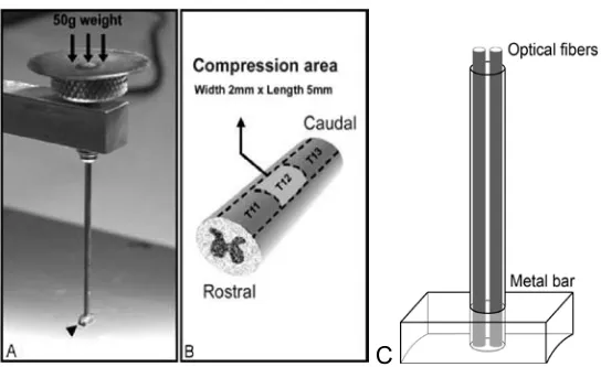
Figure 1. Apparatus for rat spinal cord compression.  (A) compression bar and weight.  (B) section of spinal cord showing compression area (light gray)
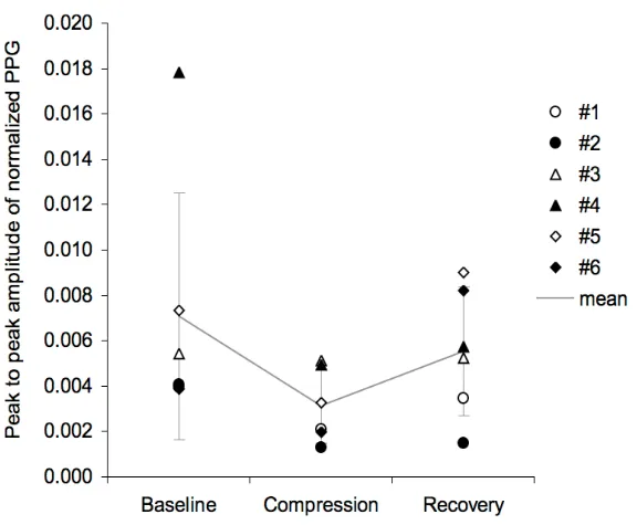
Figure 3. Graph of the PPG amplitude averaged over ‘Baseline’, ‘Compression’ and ‘Recovery’ epochs for all six subjects.

References

Related documents