
0022-538X/80/02-0877/10$02.00/0
Structural Polypeptides
of
the
Granulosis Virus of Plodia
interpunctellat
KATHLEEN A. TWEETEN,' LEE A. BULLA, JR.,2 ANDRICHARD A. CONSIGLI'*
Division of Biology, Section of Virology and Oncology, Kansas State University, Manhattan, Kansas66506,1 andU.S. Grain Marketing Research Laboratory, Science and Education Administration, Manhattan,
Kansas665022
Techniquesweredevelopedfor theisolationandpurificationofthreestructural components ofPlodia interpunctella granulosis virus: granulin, enveloped nu-cleocapsids, andnucleocapsids. Thepolypeptide compositionanddistribution of protein in each viral component were determined by sodium dodecyl sulfate
discontinuous
and gradient polyacrylamide slab gel electrophoresis. Enveloped nucleocapsids consisted of15structuralproteins ranging inmolecularweight from 12,600to97,300. Five of theseproteins,havingapproximatemolecular weights of 17,800, 39,700, 42,400, 48,200, and 97,300, were identified as envelope proteins by surfaceradioiodination of the enveloped nucleocapsids. Present in purified nu-cleocapsidswereeight polypeptides.Thepredominantproteins in thisstructuralcomponenthadmolecularweights of 12,500 and 31,000. Whereas no evidence of
polypeptideglycosylationwasobtained,sixof the viral proteins were observed to bephosphorylated.
Granulosis and nuclear polyhedrosis viruses arestructurally complex viruses belongingtothe family Baculoviridae. They infect a number of lepidopteran insects, causing disease and larval death in infected populations. Because of their effectiveness, the baculovirusesare being consid-ered foruse asbiological insecticides forcontrol
of their insect hosts. As a result, these viruses
have acquired scientific interest, and investiga-tors have beguntoobtain
information
ontheir molecularproperties. Suchknowledgeisneeded fordeveloping
methods of virus identification anddetection and fordetermining
theireffects atthecellular levelonnontargetspecies.The basic structural unit of the granulosis virus (GV) and nuclear polyhedrosis virus (NPV) is a
high-molecular-weight,
covalently closedsupercoiledDNApackaged withina rod-shaped capsid. Thenucleocapsid
issurrounded bya lipidenvelope, anddepositedontheouter surface of the envelope is a thick matrix of protein(1, 10, 21,24).InNPV, several enveloped nucleocapsids are embedded within a protein crystal. This featuredistinguishes
it fromGV,
inwhich the enveloped nucleocapsids are singly
occluded.
Identification of the baculoviruses based on
characteristics of their matrix
proteins
hasbeenshowntobe
inadequate.
Theseproteinsall havemolecular weightsof26,000to30,000(6, 11, 16,
22) and demonstrate
serological
cross-reactivity
t Contribution no. 79-383-j, Kansas Agricultural Experi-mentStation,Kansas StateUniversity, Manhattan,KS66506.
(8, 11, 14). On theotherhand, bothqualitative
andquantitative differences have beenobserved
intheproteincomposition of theenveloped
nu-cleocapsids and nucleocapsids from various
NPVisolates (7, 11, 22).
Comparisons of
GV,
based on polypeptide composition, cannotbe as readily made. Aside from extensive characterization ofgranulin, the majorcomponentoftheGVprotein matrix,the only structural proteins that have been exam-ined are those of theenveloped
nucleocapsids
fromthe GVsofPieris brassicae(5), Trichoplu-sia ni, and Spodoptera frugiperda (22). Like those of
NPV,
they
consisted of 12to 18poly-peptides
ranging
in molecularweight
from12,000 to 90,000. Information on the isolation
and characterization of GV
nucleocapsids
isavailable
only
for the virus which infects P.brassicae(5).
Ourlaboratory isinvestigatingthe molecular biology of the GV of the Indian meal moth Plodia
interpunctella.
Toanalyze
the structural polypeptidecomposition
of thisvirus,
we devel-opedbiophysical
methods for itsseparation
intothree structural components:
granulin,
enve-loped
nucleocapsids,
andnucleocapsids.
These methods,along
with thepolypeptide
composi-tion and distribution of
protein
in each viralcomponent, are
reported
in this paper. OtherstudiesonNPV andGV havenot
provided
directevidence that
specific
viralpolypeptides
arecon-stituents of the viral
envelope.
In thepresent
work, radioiodination which
specifically
labels877
on November 10, 2019 by guest
http://jvi.asm.org/
virus surface proteins was used to identifyand
localize the GV envelope polypeptides.
Modifi-cations of viral polypeptides by glycosylation
andphosphorylation werealsoinvestigated.
MATERIALS AND METHODS
Production and purification of GV. GV was
produced in alaboratory colonyofP.interpunctella rearedaspreviouslydescribed(23).Earlythird instar larvae were infected peroswithGV,andtheviruswas purifiedby differentialcentrifugation,treatmentwith 1% deoxycholate, and velocity sedimentation in su-crosegradients (23,25).
Isolation of theproteinmatrix. Theprotein ma-trixwassolubilizedbyincubating purifiedGVfor10
minat37'C in0.05 Msodium carbonate-0.05 MNaCl, pH10.6.To isolate the matrixproteinsinnondegraded form, the GVwasheated before carbonatetreatment
for30min at700Ctoinactivateendogenousprotease activity (25). The dissociated viruswaslayeredon30 to70%glycerol(vol/vol,in0.01MTris-hydrochloride, pH 7.5) gradients which were centrifuged at 25,000 rpm (SW 41 rotor) for 30 min at 10°C. The matrix proteinswererecovered from the top of thegradient and concentrated inamodel12Amiconultrafiltration cell withaUM-10 membrane.
Isolation ofenvelopednucleocapsids. Purified GV wasincubated in 0.05 Msodium carbonate-0.05 MNaCl, pH 10.6, for5min at roomtemperature. The dissociated virus waslayered on 30 to 70% glycerol (vol/vol, in 0.01 MTris-hydrochloride, pH 7.5) gra-dients which werecentrifugedat 25,000 rpm(SW41
rotor)for1h at 100C.Theband ofenveloped nucleo-capsidswasrecovered from thegradients, diluted with
0.01MTris-hydrochloride,pH 7.5, and centrifugedat
25,000 rpm (SW 41 rotor) for 1 h to remove the glycerol. Theenvelopednucleocapsidswerestored in
0.01MTris-hydrochloride,pH 7.5,at4°C.
Isolation of nucleocapsids. Purified enveloped nucleocapsids wereincubatedin 1% (vol/vol, in0.01
M Tris-hydrochloride, pH 8.5) Nonidet P-40 (NP-40;
Shell Chemical Co.) for30minwith stirring at room temperature. Thenucleocapsids were separated from
thesolubilizedenvelope proteins by sedimentation on
30 to70%glycerol (vol/vol, in0.01 M Tris-hydrochlo-ride, pH 8.5) gradients centrifuged at 30,000 rpm (SW
41 rotor) for 1 h. The band of nucleocapsids was dilutedwith 0.01 MTris-hydrochloride, pH 8.5, and centrifuged at 25,000 rpm (SW 41 rotor) for 1 h to
removeglycerol.Thenucleocapsids were stored in 0.01
MTris-hydrochloride, pH 7.8, at4°C.
Radiolabeling of GV invivo. Radioactively
la-beled GVwasproduced in vivo by injection of1 d(0.1
to 0.5,uCi) ofisotope into larvae at 96 and 120 h after infection. The isotopes used included [3H]thymidine (71Ci/mmol,Schwarz/Mann),'4C-aminoacidmixture
(Schwarz/Mann), and [32P]orthophosphoric acid
(New England Nuclear). GV was purified from in-jected larvae8days afterinfection.
lodination ofenveloped nucleocapsids.
Prepa-rations ofpurifiedenveloped nucleocapsids containing
250
fig
ofproteinin100til
of0.01 MTris-hydrochloride, pH 7.5, were mixed with 501l
of 0.2 M phosphate buffer, pH 7.2,0.5 mCiof['25I]iodine (ICNChemicaland Radioisotope Division), 25tu (0.5 mg) of
Enzy-mobead reagent, and 25 ll of 1% /8-glucose. After incubation atroomtemperature for30min, the En-zymobeadswereremoved bycentrifugation at 250x
g for5min. Unreactediodinewasremoved from the enveloped nucleocapsids by centrifuging the prepara-tionon a30 to70%glycerol gradient for1hat25,000 rpm (SW 41 rotor). The visible band of enveloped nucleocapsids was recovered and dialyzed for 24 h against0.01MTris-hydrochloride, pH 7.5.
Glycosylation of viral components. To label galactosyl residues,amixtureconsisting of250ug of virus (granulin, enveloped nucleocapsids, or nucleo-capsids), 12 U of neuraminidase(Calbiochem), and 25 U of galactose oxidase (Sigma ChemicalCo.) in 200 ,ul of 0.02 Mphosphatebuffer, pH 7.2, and0.1M NaCl
was incubated for 1 h at 37°C. Then, 500 ,Ci of NaB[3H]4 (New England Nuclear) in20 1L of0.01 N NaOHwasadded, and the mixturewasincubatedat roomtemperature for30min. The viralpreparations were then repurified by sedimentation on glycerol gradients. Sendai viruswaslabeled inasimilar man-ner.Controls consisted of viral preparations that
re-ceivednoenzyme treatment.The viral proteinswere
then resolved bypolyacrylamide slab gel electropho-resis andtritium-containing bandsweredetected by fluorography.
Electron microscopy of viral components. Samples ofenvelopednucleocapsids and nucleocap-sidswereplaced onFormvar-coated gridsand nega-tively stained with 1%phosphotungstic acid,pH 7.0. Grids were examined withaPhilips EM 201at60 kV. Polyacrylamide gel electrophoresis of viral polypeptides. Viral proteinsweresubjectedto elec-trophoresis on sodium dodecyl sulfate (SDS)-poly-acrylamide slab gelsby the discontinuous buffer
sys-temof Laemmli(15).Samplepreparation, electropho-resis, andstainingwere as described (25). Molecular weightsweredeterminedby the method of Weber and Osborne(26) withcytochromec(11,700), chymotryp-sinogen (27,500), ovalbumin (43,000), and bovine se-rum albumin(68,500) (Schwarz/Mann) asstandards. Destained gelswerephotographed,andthenegatives were scanned with a Photovolt photometer, model
520-A. Scanswererecorded andpeakareaswere de-termined with a Hewlett-Packard 3380A integrator. Gels were dried with an SE-540 Hoeffer Scientific Instrumentsslabgel dryer.Forautoradiography, dried
gels were placed in contact with Kodak X-Omat R
film (XR-1). Fluorography ofgelswasconducted by
the method ofBonner and Laskey (4). Films were
developed in Kodak D-19, and scans were made as described above.
RESULTS
Purification and characterization of the
protein matrix. It is well-documented that
treatment ofoccludedbaculoviruses with
alka-linecarbonateresults inthesolubilization of the
protein matrix (2, 13, 16). This technique was
used toisolate the matrixproteins from P.
in-terpunctella GV. The solubilized matrix was
separated from other viral components by
cen-trifugation on glycerol gradients. It formed a
J. VIROL.
on November 10, 2019 by guest
http://jvi.asm.org/
GRANULOSIS VIRUS STRUCTURAL POLYPEPTIDES 879
diffuse band which remained on top of the gra-dients (Fig. 1).
Polyacrylamidegelelectrophoresis ofthe iso-lated matrix showed that it consisted of a
mix-tureofpolypeptides ranging inmolecularweight
from 10,000 to 28,000 (Fig. 2B). Most of these
protein bands resulted from the activity of an alkaline protease associated withtheGV(25).If
the protease was inactivated before
solubiliza-tion ofthematrix,theproteinsrecovered on top
of the gradient consisted primarily of a
poly-peptidehavingamolecular weightof 28,000 (Fig.
2A). Also observed in the matrix preparations
were two to three minor polypeptides having
approximate molecular weights of 66,000 to
74,000 (Fig.2AandB).
Purification of enveloped nucleocapsids. Carbonate treatment was also used to release the envelopednucleocapsids from purified GV. After incubation in carbonate, the dissociated viral preparation was centrifuged on glycerol gradients. When
[3H]thymidine-labeled
virus was disrupted, a single peak of DNA-labeled virus was obtained in the gradients (Fig. 1). When"4C-amino
acid-labeled GV wassimilarly treated, a portion of the protein label cosedi-mented with the DNA label while the majority oftheradioactivity correspondingtothe matrixproteins remained on top ofthegradient.
Elec-4~~~~~~~~~~~~~~~~~~4
7- ~~~EN
40 1~~~
6-t
|211y
25-4
0~
U
4--2 4
FRACTONNUMBER
FIG. 1. Isolation ofGVenveloped nucleocapsids (EN) bysedimentation inglycerol gradients. [3H]-thymidine-labeledor14C-aminoacid-labeled GVwas
incubated in 0.05Msodiumcarbonate-0.05 MNaCl, pH 10.6, for15minatroomtemperature. The
disso-ciated GVwaslayeredonto 30to 70%oglycerol
gra-dients (in 0.01 M Tris-hydrochloride, pH 7.5) and centrifugedat25,X000rpm(SW41rotor) for1h.
A B
[image:3.514.312.409.80.263.2]VP28- o
FIG. 2. SDS-polyacrylamide gel electrophoresis of GV matrix proteins. The protein matrix was solubi-lized andrecoveredon topof glycerol gradients as shown inFig. 1.Samples were prepared for electro-phoresisbyboiling in 2% SDS,5% 2-mercaptoethanol, and10%oglycerol. (A) Matrix proteins isolated from GV heatedbefore carbonate treatment for 30 min at 70°Ctoinactivateendogenous protease activity. (B) Matrixproteins isolatedfromprotease-active GV.
tron microscopy ofthe band of virus obtained onthesegradients showed thatit consistedofa homogeneouspreparation ofenveloped nucleo-capsids (Fig. 3A).Fewdegraded viral envelopes were observed, indicating that the carbonate
treatment and sedimentation in glycerol
gra-dients were not detrimental to the structural
integrity ofthis viralcomponent.
Purification of nucleocapsids. GV nucleo-capsidswereisolatedbytreatingenveloped nu-cleocapsids with the nonionicdetergent Nonidet
P-40. Enveloped nucleocapsids, labeled in vivo
with
['H]thymidine
or"4C-amino
acids, weredissociated with the detergent and then
sedi-mented on 30 to 70%
glycerol gradients.
Aftercentrifugation,
essentially
all of the DNA labelwaspresent ina
single
band which sedimentedmoreslowlyin thegradient than the
enveloped
nucleocapsids.
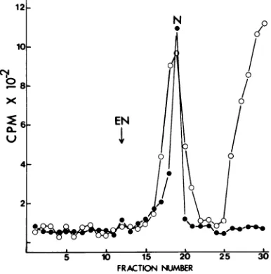
Approximately
43% of the amino acid label cosedimented with the viral DNA label,whereasthe remainderwaslocalizednearthe top of the gradient (Fig. 4). Material from
bothregionsof the
gradient
wasrecoveredandexamined for its
polypeptide composition
(re-sults described
below).
The band of virus wasrecovered from the gradient and examined
by
electronmicroscopytoconfirm that itcontained
nucleocapsids
(Fig.
3B).
Themicrograph
dem-onstratesthat the
nucleocapsids
werestructur-allyintact and free of
contaminating
envelope
fragments.
on November 10, 2019 by guest
http://jvi.asm.org/
[image:3.514.62.256.381.592.2]880 TWEETEN, BULLA, AND CONSIGLI
A
V;p,
B
L... ..
.; .*. .a_ g.
w_' ,sf
..w' .t
-:f y....}
w
.,. . <
J f ju<
., .v>So:
-"
wiywi __.
.. S * W.
[image:4.514.98.433.69.274.2].'
FIG. 3. Electron micrographs ofpurified GVenveloped nucleocapsids and nucleocapsids. Enveloped
nucleocapsids (A)andnucleocapsids(B)wereisolatedasdescribed in thelegendstoFig.2 and4, respectively.
Samples weremounted on Formvar-coatedgrids and stained with 1%phosphotungstic acid,pH7.0. Bar represents400 nm.
12
N
10-~~~~~~~
X,,""l' .
0~~~
4-
2-5 1,5 2'0 30
FRACTIONNUMBER
FIG. 4. IsolationofGVnucleocapsids (N) by sedi-mentationofNonidet P-40-dissociatedenveloped
nu-cleocapsidsinglycerol gradients.
[3H]thymidine-la-beled (0) or '4C-amino acid-labeled (0) enveloped nucleocapsidswereincubatedin 1%Nonidet P-40in 0.01 MTris-hydrochloride, pH 8.5, for30minatroom temperature. Thepreparation was layeredon30to 70oglycerol gradients (in0.01 MTris-hydrochloride, pH 8.5)andcentrifugedat25,000rpm(SW41rotor)
for1h.
Identification of the structural proteins
ofenvelopednucleocapsidsand
nucelocap-sids.Samples ofpurifled enveloped
nucleocap-sids and nucleocapsids were dissociated with
SDS and mercaptoethanol. The proteins were
subjected to electrophoresis in discontinuous
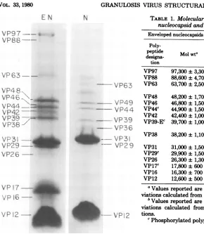
SDS-polyacrylamide gels.Anelectropherogram
ofthe structural polypeptides oftheenveloped
nucleocapsids is shown in Fig. 5 (EN). Fifteen
proteinsranginginmolecularweightfrom12,600
to97,300wereconsistentlyidentified. Avariety
of acrylamide concentrations (10, 12.5, 15 and
20%) wasused toadequatelyseparate proteins
having similarelectrophoretic mobilities.By
us-inggradient gelsranging inacrylamide
concen-tration from 10 to 26% or 7 to 15%, threetofive
additional minor viral protein species were
re-solved. The molecular weights of these minor
structural proteins were 20,000, 36,000, 57,600,
59,800, and 86,000. The average molecular
weight of each of the enveloped nucleocapsid
proteins, determinedbycomparisonwith
molec-ularweightstandards, is presented in Table 1.
Eight polypeptides, ranging in molecular
weight from 12,500 to 64,200, were observed when nucleocapsids were dissociated and elec-trophoresedin 12.5and 15%SDSslab gels (Fig.
5, N). The average molecular weight of each
nucleocapsid polypeptide is shown in Table 1.
Two of the nucleocapsid proteins, VP49 and
VP39, had molecular weights very similar to polypeptides identified as constituents of the
viral envelope (results described below). They
were, however, demonstrated tobe unique
nu-cleocapsid polypeptides by electrophoresison 7 to 15% SDS-polyacrylamidegradient gels. This
gelsystemprovidedforseparation of enveloped
nucleocapsid proteinsVP48 and VP39 eachinto
on November 10, 2019 by guest
http://jvi.asm.org/
[image:4.514.68.261.331.526.2]E N N
VP97--VP8
--
VP63--VP4 8-.
-VP63
VP46
\_
VP44
\=---VP49
VP44-='
-VP42
~
~~~
-VP4
VP39
-7 -VP36
VP31 --
4
_ - VP31VP29
-- VP29VP26
-VP17
-VP 16 --VP12
FIG. 5. SDS-polyacrylamide gelelectr GVenvelopednucleocapsids (EN)andni (N). Numerical designations referto th
weight (x 10-3) ofeachpolypeptide dei
comparison withmolecular weight stand
twodistinctprotein bands (resultsn
Only one of each of these polype
presentinnucleocapsids electrophor
same gel. In addition tothe eight p(
describedabove, minor proteincomp(
ing approximate molecular weights
57,000, and 68,500wereobservedinni
preparations.
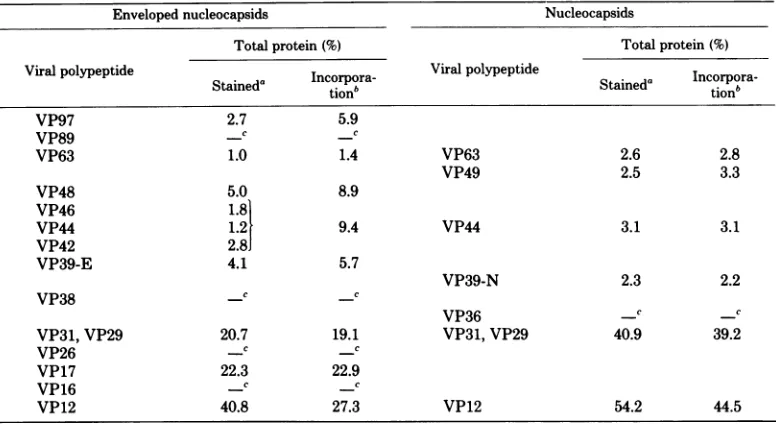
Proteincomposition ofGVenv(
cleocapsids andnucleocapsids. T amount ofeach viral polypeptide in
nucleocapsids andnucleocapsidswa;
from densitometricscansofpolyacry
in which unlabeled or "C-amino a
viruswasresolved. Theresultingdist
proteinamongthe various viral
poly
shown in Table 2. VP12, VP17, V]
VP39, andVP48werethepredomina
in Coomassie-stained enveloped nue
since they represented 40,22, 20, 4,
thetotalproteinin this viralcomponm
[image:5.514.65.355.61.394.2]tively.Theremaining polypeptidesw
TABLE 1. Molecular weights of GV enveloped nucleocapsid and nucleocapsid proteins
Envelopednucleocapsids Nucleocapsids
Poly-
Poly-peptide Mol
wta
peptide MolWtbdesigna-
designa-tion tion
VP97 97,300±3,300
VP88 88,600±4,700
VP63 63,700±2,500 VP63 64,200± 3,000
VP49c 49,300±500
VP48 48,200±1,700 VP46 46,800±1,500
VP44c 44,900±1,500 VP44" 45,600±1,000
VP42 42,400±1,000 VP39-EC 39,700± 1,000
VP39-N 39,600±700 VP38 38,200± 1,100
VP36 36,700±600 VP31 31,000± 1,500 VP31 31,000±700
VP29C 29,900± 1,500 VP29C 30,100±1,300
VP26 26,300±1,300 VP17C 17,800±600 VP16 16,300±700
VP12 12,600±500 VP12 12,500±300 aValues reported are the means and standard de-viationscalculated from 10 separatedeterminations.
bValues reported are the means and standard de-viations calculated from eight separate determina-VP12 tions.
e
Phosphorylated
polypeptides.
inamountsranging from less than 1% (VP88 and
ophoresisof VP38) to 2.8% (VP42). Scans of
Coomassie-e
molecular
stainednucleocapsid proteins
indicated thattermined
bythey
consistedprimarily
of VP12(54%)
andfards. VP29 and VP31 (41%). The other proteins
as-sociated withnucleocapsidswereminor
compo-iotshown). nents which represented from 2.3% (VP39) to ptides was 3.1%(VP44)of the totalnucleocapsid protein.A
esedonthe similar distribution of protein resulted from
olypeptides scans of fluorograms of gels containing
"4C-onentshav- amino acid-labeled viralpreparationsexcept for
of 27,000, VP12. Substantially more of this viral
poly-acleocapsid peptide appeared to be present in enveloped nucleocapsids and nucleocapsids, based on in-elopednu- tensityof Coomassie
staining,
thanon incorpo-'he relative ration of'4C-amino acids (Table2). The lower enveloped percent values obtained by incorporation of sestimated amino acidsmostlikely
canbeaccounted forbylamidegels the
extremely
high arginine content of VP12Lcid-labeled
(manuscript submitted). Polypeptides
of high tributionof argininecontentstainmoreintenselywithCoo-Tpeptides
is massie brilliant blue than do those with lower P29, VP31, argininecontent (D.J.Stubs,M.S.thesis, Kan-ntproteins sasStateUniversity, Manhattan, Kansas,1978). -leocapsids, Identification of viralenvelope proteins.and5%, of Radioiodination is a usefultechniquefor
exam-ent,respec- ining the distribution and organization of
pro-erepresent teinsin animal viruses(19, 27).Agentlein vitro
on November 10, 2019 by guest
http://jvi.asm.org/
[image:5.514.250.457.82.342.2]882 TWEETEN, BULLA, AND CONSIGLI
TABLE 2. Proteincomposition ofGVenveloped nucleocapsidsandnucleocapsids
Envelopednucleocapsids Nucleocapsids
Totalprotein (%) Totalprotein(%)
Viralpolypeptide I Viralpolypeptide
Staineda
tionb
Stained"
Incorpora-VP97 2.7 5.9
VP89 -C -C
VP63 1.0 1.4 VP63 2.6 2.8
VP49 2.5 3.3
VP48 5.0 8.9
VP46 1.8
VP44 1.2 9.4 VP44 3.1 3.1
VP42 2.8J
VP39-E 4.1 5.7
VP39-N 2.3 2.2
VP38 -C -C
VP36 -c _^
VP31,VP29 20.7 19.1 VP31,VP29 40.9 39.2
VP26 -C -C
VP17 22.3 22.9
VP16 _C _C
VP12 40.8 27.3 VP12 54.2 44.5
aPercentage of totalproteindeterminedby calculatingtheareaunder eachCoomassiebrilliant blue-stained peak; values represent the average of six separate determinations.
bPercentage
of totalproteindetermined fromareaundereachpeakresultiilgfromautoradiographyofSDS gels of virus labeled in vivo with'4C-amino acids;valuesreportedarethemeansof three separatedeterminations.cPeak areas less than 1% of the totalprotein.
labeling procedure employing solid-phasebound
lactoperoxidase (Enzymobeadreagent,Bio-Rad)
and['251]iodinewasusedtoidentifyGVenvelope
proteins. The large size ofthe bound enzyme
inhibits itspenetrationintothe viral membrane
so thatonlythoseproteinsexternal tothe viral
envelopearelabeled (18, 27).
To verify the specificity ofthe labeling
pro-cedureused,iodinated enveloped nucleocapsids
were incubated with the proteolytic enzyme
trypsin (2mg/ml)for 2 hat37°Csothat proteins
locatedontheoutersurface of theviral envelope
would be digested (18, 20). After trypsin
treat-ment, the enveloped nucleocapsids were reiso-latedby centrifugation in glycerol gradients. It
wasfoundthat the majority of the iodine label
wasremoved fromthevirus by theproteaseand remained on top of the gradient. Electron
mi-croscopyof the band of virusobtained in these
gradients revealed that itconsisted of particles
with intactenvelopes (results notshown)
indi-catingthat thetrypsin didnotalterthe integrity
ofthe envelopes. It was concluded from these
datathatthe lactoperoxidase labelingwas
spe-cific forproteinsexternaltothe viralenvelope.
Toidentify which viralproteinswerelabeled
bythelactoperoxidaseprocedure, and thuswere
envelope constituents, portionsof the iodinated
enveloped nucleocapsids were dissociated by
SDSandmercaptoethanoland the proteinswere
resolved by electrophoresis in 12.5% discontin-uousSDS
gels.
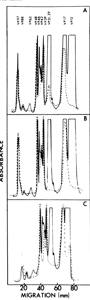
Densitometertracings
ofanau-toradiogramof suchagel (Fig. 6A)showed that
the iodine label waslocalized atpositions
cor-responding to VP17, VP39, VP42,
VP48,
andVP97. Ofthesepolypeptides,VP17wasthe
ma-jor component. The smallamount of label
as-sociated with
nucleocapsid proteins
VP29 and VP31probably
resulted fromsomeenvelope
dis-ruption duringpreparation of enveloped nucleo-capsids for iodination.NP-40 treatment of the iodinated
enveloped
nucleocapsids
provided
further evidence that these five structuralpolypeptides
wereenvelope
proteins.Viralproteinsthatweresolubilizedby
NP-40wereprecipitatedwith 25%
trichloroace-tic acid and resolved in
polyacrylamide
gels.Autoradiography revealed that the label mi-gratedwithelectrophoretic mobilities character-istic-ofVP17,VP39, VP42, VP48,and VP97(Fig.
6B).The viral proteinswhichwerereleasedby
NP-40 were also examined after their
electro-phoi;esis
inSDSdiscontinuousgelsand staining with Coomassiebrilliantblue. As shown inFig. 6C,theproteinsthatwerevisualizedagainwere VP17, VP39, VP42, VP48, and VP97. A small amountof VP12wasalso observed in thesegels.Its presence mayindicate thatsome
nucleocap-sid disruption occurred during detergent treat-ment.
on November 10, 2019 by guest
http://jvi.asm.org/
Modifications of GV structural
polypep-0. tides.(i)Glycosylation.Asensitivetechnique W W,n40, _: N C for detection of carbohydrate moieties on
poly-> > > >>>>> > > peptides consists of radiolabeling with
[3H]so-diumborohydride after neuraminidase and
ga-lactose oxidase treatment (3, 9). Thisprocedure
was used to determine whether any of the struc-turalpolypeptides ofP.interpunctellaGVwere glycosylated. Even afterprolonged exposure (4
months) ofgelscontaining radiolabeled virus to
X-ray
film,
noradioactivity
wasdetectedinany of the polypeptides associated with granulin,enveloped nucleocapsids,
ornucleocapsids.
Un-a
: derthe same conditionsoflabeling andexposure,
thetwostructuralglycoproteins of Sendaivirus were extensively labeled. A labeling procedure
specific
for sialic acid residues(3)
also failedto .s, .revealanyglycosylationin GV..,,.
Glycosylation
ofviralpolypeptides
was alsoinvestigated by SDS gel electrophoresis and
-Ai--
"fluorography
ofviralcomponents
isolatedfrominfected larvaethat were injected with
['4C]fu-cose
(15
Ci/mmol,
NewEngland
Nuclear)
orB['4C]glucosamine (30
Ci/mmol,
NewEngland
Nuclear). Again, noincorporation ofisotope by anyof theGV structural proteinswasobserved. (ii) Phosphorylation. Preliminary
experi-ments wereconductedtodeterminewhetherGV
uW
structuralpolypeptides
were phosphorylated.32P-labeled
viruswasproduced by injecting
1I1
'
(25
mCi/ml)
of[32P]orthophosphoric
acid(neu-,a tralizedto
pH
7.4with1MTris-hydrochloride)
co W . 3into GV-infected larvae. Afterpurification ofthe
virus from the larvae, the granulin, enveloped
0 I' 'l
||nucleocapsids,
andnucleocapsids
wereisolated,
dissociated withSDSandmercaptoethanol,and
electrophoresed
onSDS-polyacrylamide
gels.
The association of radioactive
phosphate
withviral
polypeptides
wasdeterminedby
compari-^,,,C FIG. 6. IdentificationofGVenvelopeproteins. (A)
Surface-iodinated enveloped
nucleocapsidswere dis-sociated with SDS and mercaptoethanol, and theproteins
were resolvedby
SDSgel
electrophoresis.
.*h
I X,Viralpolypeptides
wereidentified by comparing den-sitometer tracings of autoradiograms of the gels*M1 s 1
1,
(---)tothoseof
Coomassie blue-stainedgels
( )..*,%1
4,
(B andC)Iodinated
orunlabeledenvelopednucleo-capsids
were incubated in 1% Nonidet P-40for 30min at roomtemperature. The solubilizedproteins
were isolatedas shown inFig. 4 andprecipitated
I
I ' V i U 1. with 25% trichloroacetic acid. Theprecipitateswere
washed withethanol,dissociated with SDS and mer-a
1''.t1 1'captoethanol, and resolved by SDS gel
electrophore-sis.
Envelope proteins
were identified bycomparing densitometer tracings of autoradiograms (B) or of l-e;k- i stainedgels (C) containing the solubilized proteins
->2t-\j (---) to those ofstainedgels containing enveloped
..*| nucleocapsids( ).
MIGRATION
(mm)
VOL. 33,1980
on November 10, 2019 by guest
http://jvi.asm.org/
[image:7.514.64.252.67.692.2]884 CONSIGLI
son of densitometertracingsofautoradiograms
of each gel to those of the samegelstained with
Coomassiebrilliant blue. Themajorconstituent
of theproteinmatrix, granulin,wasobserved to
be phosphorylated (Fig. 7A). Of the proteins
present in enveloped nucleocapsids, VP17,
VP29,VP39, VP44,and VP48 contained
signifi-cant amounts of 32P-label (Fig. 7B). This
re-sultedfromincorporation of theisotope bytwo ofthe envelope proteins, VP17 and VP39. The remaining three phosphorylated species, VP29, VP44, and VP49, wereassociated with nucleo-capsidpreparations
(Fig.
7C).
DISCUSSION
The techniques described in this paper
pro-vided forthe separation of the GV ofP.
inter-punctellainto
protein
matrix,
enveloped
nucleo-capsid, nucleocapsid, and solubilized envelopecomponents. By isolation of each viral
compo-nentandanalysis ofitspolypeptide composition, the number andorganization ofthe
major
pro-teins in the structurallycomplex
GV could be Luelucidated. Thepredominantconstituent of the z
proteinmatrixhadamolecular
weight
of28,000. ,Inthisproperty,it resembled thematrixproteins ci
that have beencharacterized inother baculovi- l
ruses (6, 11, 16,22).The
high-molecular-weight
0
proteins associated withthegranulin appear to i
be unique to the protein matrix, since their < electrophoretic mobilities were different from
that of the polypeptides found in either the
enveloped nucleocapsids or nucleocapsids. McCarthy and Liu (16) have also observed a
high-molecular-weight
protein in the proteinmatrix prepared from the NPV of Porthetria
dispar.The distribution oftheseminorproteins
within the protein matrix, aswell as their
bio-chemical properties andimportancetothe viral
infectionprocess, remains to bedetermined.
The number ofpolypeptides observedin the
enveloped nucleocapsidsandnucleocapsidswas dependent on the resolving power ofthe SDS gel inwhich theywereelectrophoresed. Gener-ally, 15polypeptideswerepresent inenveloped nucleocapsids and 8 were found in
nucleocap-FIG. 7. Identificationofphosphorylated GV struc-turalpolypeptides. Proteinmatrix (A), enveloped
nu-cleocapsids (B),andnucleocapsids (C) were isolated as described from GV grown in the presence of
[32P]orthophosphate.
Eachviral component wassol-ubilizedby boilingin SDS and mercaptoethanol, and itspolypeptideswere resolved by SDS gel
electropho-resis.Phosphorylatedviralpolypeptideswere
identi-fied by comparing densitometertracings of
autora-diograms of eachgel (---) to those of the same gel
stained withCoomassiebrilliantblue ( ).
J.
MIGRATION (mm)
on November 10, 2019 by guest
http://jvi.asm.org/
[image:8.514.279.457.65.660.2]sids. The minor protein species associatedwith these viralcomponents mayrepresent cleavage
products or modified (phosphorylated or
acet-ylated) forms ofthe major viralproteins.
Two-dimensional gel electrophoresis is currently
being used to investigate these possibilities as
wellas toseparate the polypeptides of enveloped
nucleocapsids and nucleocapsids into more clearlydistinguishable proteins.
Iodination studiesshowed that at least five of the proteins found in enveloped nucleocapsids were located on the outer surface of the viral envelope. Previous studies comparingthe poly-peptide compositions of GV andNPVenveloped nucleocapsids and nucleocapsids have impli-catedcertainproteinsaspossible viral envelope components (5, 7, 11). The lactoperoxidase la-beling procedure used in this work provided a direct means of identifying these proteins as VP17,VP39, VP42, VP48, and VP97. Nonidet
P-40 treatment of the enveloped nucleocapsids
provided further evidence for localization of theseproteins in the viral envelope. These poly-peptides were readily released from the viral
structure during solubilization of the envelope
by thedetergent.
Therewere afewadditional viralpolypeptides
(VP38,VP16, andVP46) which were present in
enveloped nucleocapsids butnot in
nucleocap-sids. Although this observation suggests that
these proteins are envelope proteins, they did
notappear tobe labeledby theradioiodination
procedure. Itispossible that these proteins are located moreinternally inthe viral membrane than the other envelopeproteins, and, as a re-sult,areunexposedtothelactoperoxidase.They
mayalsobe componentsofanintermediate layer
which has been proposed by Kawamoto et al. from electron microscopic observations (12) to existbetweentheenvelopesandnucleocapsids. This intermediatelayermaybe analogousto the "membrane" (M) proteins associated with the envelopes of rhabdoviruses, orthomyxoviruses, and paramyxoviruses which are speculated to
serve as recognition sites for assembly of the
nucleocapsids into envelopes (17).
The number and relative amounts of struc-tural
polypeptides
observedinthe GVbasedonin vivo
incorporation
of14C-amino
acids intoviral proteins were very similar to those based
onintensityofCoomassiebrilliant bluestaining.
Theseresults indicate thatpreexistinghost
pro-teins are not incorporated to any great extent
intoGV duringits replicationandsuggest that the structural proteins are virus
specified.
The possibility isnotruled out, however, thatsomeof the proteins observed in the GV are
host-contributed polypeptides that are synthesized
after initiation of virus infection. Because the effect ofGV infection onhost protein synthesis
has notbeendetermined,the specific association
of cellular proteins with the structural
compo-nents of GV cannot be further evaluated at
present.
None ofthe GV structural polypeptides was
radiolabeled by invitrolabelingprocedures spe-cific for carbohydrateresidues on proteins. This mayindicate that:(i) none of the viral polypep-tides contains carbohydrate moieties, (ii) some of the structural polypeptides are glycosylated but with residues other than the sugars sialic acid,galactoseorN-acetyl galactosaminewhich the in vitro labeling procedureare specific for, or (iii) some of the polypeptides are glycopro-teinscontaining galactoseor N-acetyl galactos-amine residuesthat are sohighly substituted by other sugars such as fucose that they do not become labeled.Itisdifficult,atpresent, torule out any of these possibilities, particularly
be-causeviralpolypeptides synthesizedin larvae in
thepresence of
["C]fucose
or['4C]glucosamine
may not haveincorporated labelto adetectable
specificactivity. Theseresultsare
especially
sur-prising in thecaseof theenvelope proteins. All other well-characterized proteins found ontheoutersurface of animal cell membranesor
ani-mal virusenvelopes havebeen found to be gly-coproteins (9).
The 80 x
106-dalton
genomeofthe GV of P.interpunctella (24) is
genetically complex
enoughto codefor
approximately
150proteins having an average molecularweight of 42,000. To synthesize the total number of structural polypeptides identified so far in thisvirus,
it would benecessary to utilizeonly
20 to 25%of theDNA molecule. Thisindicatesthat theGVgenome probably also contains information to
code for nonstructural proteins having
regula-tory or
enzymatic
activities. Whethervirus-coded enzymes or
regulatory
proteins
such as earlyproteins
are involvedin the viralreplica-tionprocess hasnotbeen
investigated.
Comparison
ofthepolypeptide composition
of the GV ofP.interpunctella
tothatof the GVsof T.ni, S.
frugiperda,
andP. brassicaerevealsseveral
differences,
basedonelectrophoretic
mo-bilities, in the structural
proteins
of thesevi-ruses. Thesedifferences appeartobemore
pro-foundfor the
proteins
of theenveloped
nucleo-capsids than for those of thenucleocapsids,
in-dicatingthatamajor
divergence
between theseGVsmay be due to their
envelope
proteins.
Ifthe envelope
proteins
areresponsible
for thetissue and host
specificity
exhibitedby
theGV,
changes in their
composition
may account forintrinsic differences observed in the
biological
33,
on November 10, 2019 by guest
http://jvi.asm.org/
activity of these viruses. From these
observa-tions, the importance of determiningthe number
and structural position of theviralpolypeptides
forelucidating their biologicalfunctionbecomes
obvious. This information isalso necessary for
thedevelopment ofimmunologicalreagents
pre-ciseenoughtoprovidesatisfactoryidentification
ofbaculoviruses as well as to analyze specific
steps in the infectionprocess ofthese complex viruses.
ACKNOWLEDGMENTS
This workwassupported byPublic Health Servicegrant ES02036fromtheNational Institute ofAllergyand Infection-Environment Safety. K.A.T. was supported bya research
associateshipfrom the U.S. GrainMarketingResearch Labo-ratory,U.S. DepartmentofAgriculture,Science and Educa-tion AdministraEduca-tion and from the KansasAgricultural Exper-iment Station.
We thankKimberly Osborne, Diane Potts,andViolaHill for theirexcellent technical assistance.
LITERATURE CITED
1. Beaton, C. D.,and B. K. Filshie. 1976. Comparative ultrastructural studies of insectgranulosesand nuclear polyhedrosesviruses. J. Gen. Virol. 31:151-161. 2. Bergold, G.1947. DieIsolierungdesPolyeder.Virus und
die Natur derPolyeder.Z.Naturforsch.26:122-143. 3. Blumenfeld,0.O.,P. M.Gallop,and T. H.Liao.1972.
Modification and introductionofaspecificradioactive
label into theerythrocytemembranesialoglycoproteins. Biochem.Biophys. Res. Commun.48:242-251. 4. Bonner,W.M.,and R. A.Laskey.1974. A filmdetection
method fortritium-labeled proteinsand nucleicacidsin polyacrylamide gels.Eur.J. Biochem. 46:83-88. 5. Brown, D. A., H. M. Bud, and D. C. Kelly. 1977.
Biophysical properties of the structuralcomponentsof
agranulosis virusisolated from thecabbage white but-terfly (Pierisbrassicae). Virology 81:317-327. 6. Cibulsky,R.J.,J. D.Harper, and R. T.Gudauskas.
1977. Biochemicalcomparison ofpolyhedral proteins from five nuclearpolyhedrosis viruses infecting Plusiine larvae (Lepidoptera:Noctuidae). J. Invertebr. Pathol. 29:182-191.
7. Cibulsky,R.J.,J. D.Harper, and R. T.Gudauskas. 1977.Biochemical comparisonof virionproteins from five nuclearpolyhedrosis virusesinfectingPlusiine
lar-vae(Lepidoptera:Noctuidae).J. Invertebr. Pathol.30:
303-313.
8. Crozier, G., and G. Meynadier. 1975.Lesproteines des
corps d'inclusion des baculovirus. Entomophaga 20: 421-432.
9. Gahmberg, C. G., and S. Hakamori. 1973. External labelingof cellsurfacegalactose and galactosaminein
glycolipid andglycoprotein of human erythrocytes.J.
Biol. Chem. 248:4311-4317.
10. Harrap,K. A.1972.Thestructureof nuclearpolyhedrosis viruses. I.The inclusion body. Virology50:114-123. 11. Harrap, K.A., C. C. Payne, and J. S. Robertson.1977.
Thepropertiesofthree baculoviruses fromclosely
re-lated hosts. Virology79:14-31.
12. Kawamoto,F.,N.Kumada,and M.Kobayashi.1977. Envelopment ofthe nuclearpolyhedrosis virus of the Oriental tussock moth, Euproctis subflava. Virology 77:867-871.
13. Kawanishi, C. Y., K. Egawa, and M. D. Summers. 1972.Solubilization ofTrichoplusia ni granulosis virus proteinic crystal. II. Ultrastructure. J. Invertebr. Pa-thol. 20:95-100.
14. Krywienczyk, J.,andG.H.Bergold.1960.Serological relationship ofviruses from somelepidopterous and hymenopterousinsects.Virology10:308-315. 15. Laemmli, U. K. 1970. Cleavage ofstructural proteins
during the assemblyof the head ofbacteriophage T4. Nature(London)227:680-685.
16.McCarthy,W.J.,and S.Liu.1976.Electrophoretic and serological characterization of Porthetria dispar poly-hedronprotein.J.Invertebr. Pathol.28:57-65. 17. Mountcastle, W. E., R. W.Compans,andP. W.
Chop-pin. 1971. Proteins and glycoproteinsof
paramyxovi-ruses: acomparisonof simianvirus 5,Newcastle disease virus, and Sendaivirus.J. Virol.7:47-52.
18. Obijeski,J.K.,D. H.L.Bishop,F.A.Murphy, and E. L. Palmer. 1976. Structuralproteinsof LaCrossevirus. J. Virol. 19:985-997.
19. Philips,D.R.,and M. Morrison. 1970. Thearrangement
ofproteinsin the humanerythrocytemembrane. Bio-chem.Biophys.Res. Commun.40:284-289.
20. Sangar,D.V.,D. J.Rowlands,D.Cavanagh,and F. Brown. 1976. Characterization of the minor polypep-tides in thefoot-and-mouth disease particle. J.Gen. Virol.31:35-46.
21. Summers,M.D.,and D. L. Anderson. 1973. Character-ization of nuclearpolyhedrosis virusDNAs. J.Virol. 12:1336-1346.
22. Summers,M.D.,and G. E. Smith. 1978.Baculovirus structuralpolypeptides. Virology84:390-402. 23. Tweeten,K.A., L.A.Bulla,Jr.,and R. A.Consigli.
1977. Isolation and purification ofa granulosis virus
frominfected larvaeofthe Indianmealmoth,Plodia interpunctella. Appl. Environ. Microbiol.34:320-327. 24. Tweeten,K.A.,L. A.Bulla,Jr.,and R.A.Consigli. 1977.Supercoiledcircular DNA ofaninsectgranulosis
virus.Proc.Natl.Acad.Sci. U.S.A.74:3574-3578. 25. Tweeten, K.A., L.A.Bulla,Jr.,and R. A.Consigli.
1978. Characterization ofanalkaline protease associ-ated withagranulosisvirus of Plodiainterpunctella. J.
Virol.26:702-711.
26. Weber, K., and M.Osborne. 1969. Thereliability of molecular weight determinations by dodecyl sulfate-polyacrylamide gel electrophoresis. J. Biol.Chem.244: 4406-4412.
27. Witte,0.N.,I.L.Weissman, andH. S.Kaplan.1973.
Structural characteristics ofsomemurine RNAtumor
viruses studied by lactoperoxidase iodination. Proc. Natl.Acad. Sci. U.S.A. 70:36-40.

![FIG. 7.fiedstaineddiograms[32P]orthophosphate.resis.turalcleocapsidsasubilizedits described polypeptides Identification ofphosphorylated GV struc- polypeptides](https://thumb-us.123doks.com/thumbv2/123dok_us/1500473.102777/8.514.279.457.65.660/fiedstaineddiograms-orthophosphate-turalcleocapsidsasubilizedits-described-polypeptides-identification-ofphosphorylated-polypeptides.webp)