JOURNAL OF VIROLOGY, Oct. 1975,p.913-926 Copyright0 1975 AmericanSociety tor Microbiology
Vol. 16,No.4 Printed inU.S.A.
Morphogenesis
of
Sindbis
Virus in
Cultured
Aedes
albopictus Cells
JEFFREY B. GLIEDMAN,1 JONATHAN F. SMITH, AND DENNIS T. BROWN* Institutfar Genetik, Universitat zuK6ln,5Koln 41, Weyertal 121, Federal-Republic ofGermany
Received forpublication23May 1975
Cultured mosquito cells were found to produce Sindbis virus nearly as
efficiently as BHK-21 cells at 28C. In virtually all of the cells observed in the
electron microscope, virus morphogenesis was found to occur within complex vesicular structures which developed after viral infection. Viral nucleocapsids
were first seen in these vesicles and appeared to be enveloped within these
structures. The process ofenvelopment within these inclusions differed in some
respects fromthe process previouslydescribedfortheenvelopment of
nucleocap-sids at the plasma membraneofvertebrate cells. Free nucleocapsids were only
rarely seen in the cytoplasm of infected mosquito cells, andbudding of virus from
the cell surfacewasdetectedsoinfrequentlythat this process of virus production
could not account for the amount ofvirus produced by theinfected cells. The
vast majority of extracellular virus was produced bythe fusion of the
virus-con-taining vesicles with the plasma membrane releasing mature virions and
membrane nucleocapsid complexesinvarious stages ofdevelopment.
Togaviruses are perpetuated in nature by
their ability to infect and replicate within the
cells ofboth vertebrate andinvertebrate hosts.
The morphogenesis of these viruses in
verte-bratetissue-cultured cells has beenextensively
investigated byelectronmicroscopy (4,6, 8, 12,
15).Alpha togavirusmorphogenesis takes place
in two distinct and separate processes in
cul-tured vertebrate cells. (i) Virus nucleocapsids
are assembled in the cellcytoplasm from virus
capsid protein and RNA. (ii) The completed
nucleocapsid is enveloped by budding through
theplasma membraneofthe infected cell after
thatmembranehasbeenmodifiedbythe
inser-tion ofvirus-specific glycoproteins. The
devel-opment of Flavitogaviruses (groupB) in
verte-bratecells may differ from the above scheme in
that envelopment by budding ofthe
cytoplas-mic nucleocapsid does not take place at the
plasma membrane but rather occurs through
thelimiting membraneofinternal vesicles(12).
The development of stable lines of cultured
mosquito cells by Singh (21) has facilitated a
limited number ofinvestigationsof the
morpho-logical processesrelated totogavirusproduction
in cultured cells of the invertebrate vector.
Studiesofthegrowthof anumberoftogaviruses
in cultured mosquito cells have revealed that
although the onset of virus production, rate of
virus
synthesis,
and yield of virus is similar to 'Present address: University of Utah College of Medicine, DepartmentofPathology, Salt Lake City, Utah 84111.that found in cultured vertebrate cells, the
virus-infected mosquito cells do not show the
acute cytocidal effects observed in vertebrate
host cells (5, 16, 22). Togavirus-infected
mos-quito cells establish a stateofchronic infection
in which the infected culture continuously
produces virus for periods of several months
(16).
Filshie andRehacek(8)studied the
morphol-ogy of Aedesaegypticells infected withMurray
Valley encephalitis and Japanese encephalitis
viruses(both Flavi togaviruses) and concluded
that theprimarysite of virus developmentwas
theendoplasmic reticulum of the infected cell.
The cisternae of the endoplasmic reticulum
were found to contain many mature virions,
suggesting thatenvelopmentofthe virions took
placethrough this intracellular membrane.
Al-though these authors saw large numbers of
mature virionsassociated with theoutersurface
ofthe cell, no budding of virions through the
plasmamembrane could be detected.
The production of alpha togaviruses in
cul-turedA. albopictus was examined
morphologi-cally by Raghowet al. (18, 19). These authors
described electron-dense vesicles which
con-tained viral capsids and mature virions in
infected mosquito cells. Although the authors
concluded that the capsids were probably
en-veloped within the vesicles, they discredited
thesestructures asasource ofprogeny virus as
theyfeltthat these inclusionsfusedwith
lysoso-913
on November 10, 2019 by guest
http://jvi.asm.org/
914 GLIEDMAN, SMITH, AND BROWN
mal vesicles,thus destroying the contents.
Rag-how and co-workers further concluded that
extracellular viruses were produced bybudding
of nucleocapsids through the plasma
mem-brane, a process similar tothat which hasbeen
described in the vertebrate cell system. We have investigated the development of the
alpha togavirus Sindbis in cultured A.
albopic-tus cells in an attempt to further understand
the morphogenesis of the togavirus in
inverte-brate cells and the establishment of the non-cytopathic persistent state of infection.
(A summary of this work was presented at the
annual meeting of the American Society for
Microbiology, 27April-2 May 1975, NewYork,
and at the 4thInternationalCongresson
Inver-tebrate Tissue Culture, 5-8 June 1975. Mqont
Gabriel, Canada.)
MATERIALS AND METHODS
Virus, cells, and media. Sindbis virus was do-nated by Elmer Pfefferkom (Dartmouth Medical School) and maintainedbypassage at low multiplic-ity on chicken embryo fibroblasts or BHK-21 cells. Titrations of virus werecarriedout in BHK-21bythe methodofPfefferkornand Clifford (17).
A.albopictuscells wereprovided by SonyaBuckley (Yale Arbovirus Research Unit) and the American TypeCultureCollection.The cells werepropagatedas monolayers inthe medium describedbyMitsuhashi and Maramorosch (M + M) (14)supplemented with 20% fetal calf serum and adjusted to pH 7.2 by addition of sodium bicarbonate. The growth of ar-boviruses intissue-cultured mosquito cells has been described(5, 16, 22).A.albopictuscells were infected with a small volume ofEagle medium (7) containing sufficient virus toproducea multiplicityof infection of 50 to 100 PFU per cell. After 1 h at room temperature, thecellswerewashed once with M + M media, given newmedia, and incubatedat 28C. For comparison, similar procedures were used to infect BHK-21 cell cultures grown inEaglemedium.
Electron microscopy. Negative staining was car-ried out by the procedure of Anderson (2). Phos-photungstic acidwaspreparedas a 2% solution and adjustedtopH7.2with NaOH.
For ultrathin sectioning, cells were fixed with ice-cold 2% glutaraldehyde in phosphate-buffered saline. After fixation for 1 h, the cells were washed
in phosphate-buffered saline and postfixed for i h
with 1% osmium tetroxide in the phosphate buffer of Millonig (13). The fixed cells were embedded in Epon 812bytheprocedureofLuft(10),and ultrathin sectionswere cutwithaReichartOMU-2microtome.
Freeze-etching of virus-infected cells was carried outby the procedure described byBrownetal. (4).
Allspecimens werephotographedin aSiemens101 electron microscope, the magnification ofwhichwas calibrated withacarbongratingreplicahaving 2,160 linesper mm (ErnestFullamCo.;no. 1002).
The micrographs of freeze-etched specimens pre-sented herewereproducedfrom contactcopiesofthe originalphotographic platessothat themetalsource
J. VROL.
appears as the source of illuminaition; the shadows containing nometal are dark.
RESILTS
Growth of Sindbis virus in vertebrate and invertebrate cells. Figure 1 shows the growth kinetics of Sindbis virus in both BHK-21 cells
and A. albopictus cells at 28C. The onset of
virusproduction occurs soon after infection in
bothcell systems. The production of virus in the
mosquito cells lags somewhat behind that in
BHK cells. At about 14 to 18 h after infection,
production of virus in the BHK cells is com-plete, and gross cytopathic effects are observed
microscopically at about 20 h postinfection.
Maximum virusproduction in the mosquito cell
system isobtainedat 18 to 20h after infection,
but is not accompanied by observable
cyto-pathic effects. In the invertebrate system a
decreaseinvirus titer isobservedatabout 24 to
26 h after infection. By 40 to 42 h, virus
production is reduced by approximatelyonelog
(not shown) and is maintained at this lower
level foraperiod of weeks.
Morphology of Sindbis virus-infected
mos-quito cells. Figures2and3showat low
magnifi-cation uninfected (Fig. 2) and Sindbis
virus-infected cells at 6 h postinfection (Fig. 3).
Infected cellsdifferedfromuninfected or
mock-infected cells primarily by the presence of a
number of vesicular structures in the infected
cell cytoplasm. Many of the vacuoles seen at
those earlytimes were found to contain
aggre-gates ofmembranes which seemed to traverse
the vesicle from side to side in a complex
"stacked"organization(Fig.3and 4). Ithasnot
been possible to determine the origin of these
vesicular structuresor toestablishfromwhat, if
any, pre-existing cellular structures they were
derived. In some instances layered membranes
ofthe typeseen inthevesicles could be detected
in the cell cytoplasm without a surrounding
limiting membrane (Fig. 4), suggesting that such a structure might be a precursor to the
membrane-limited vesicles. Thevacuoles
con-tained much amorphous electron-dense
mate-rial and ribosomes, suggesting that some
cyto-plasmic constituents became entrapped in the
vacuoles during theirformation.
Freeze-etching of these early membranous
vesicles at 4 to 8 h after infection (Fig. 5)
revealed typical cleavageplanes inthe stacked
membranes and the presence ofinterior
mem-brane beads(presumablyglycoprotein [11,
23]).
Freezeetching furthersuggested that, although
the stacked membranes approach the limiting
membrane ofthevesicle, they are not
continu-ous with it, and therefore do not represent an
on November 10, 2019 by guest
http://jvi.asm.org/
SINDBIS MORPHOGENESIS IN MOSQUITO CELLS 915
invagination of the limiting membrane. The
freeze-etched vesicles also revealed the presence
ofsmaller closed membrane structures within
7-=6
o5-A /
X, 0
5) ~~~~~~~0
c3- i
/
1-2 6 10 14 18 22 26 30
Hoursafter Infection
FIG. 1. Growth of Sindbis virusat28Cincultured A.albopictus (0) andBHK-21 (0) cells.
the vesicle itself. Viral nucleocapsids or mature
virions were not found associated with the
infected cells at this time.
At later times after infection (8 to 12 h), the cytoplasmic vesicles were found to contain many viral nucleocapsids and mature virions
(Fig. 6). Occasionally, partially enveloped
cap-sids were seen. Cells examined by thin
section-ing contained few nucleocapsids in the cell
cytoplasm and no mature virions associated
with the cell surfaces.
At 14 to 20 h postinfection the cells contained
a largenumber of very electron-dense vacuoles
(Fig. 7a) which were found to contain amor-phous electron-dense material and mature
vi-rions whenexamined at high magnification. In
many instances the virions in the inclusions
appeared to be packed in a paracrystalline array
having hexagonal symetry (Fig. 7b). Cells
hav-inglarge numbersofthese virus-rich inclusions
showed no evidence of virus production at the
plasmamembraneby a classical budding
proc-ess (Fig. 7a).
-.
?'z.
.,-FIG. 2. Thin sectionof anuninfected cultured A. albopictus cell. No contaminating endogenous virus-like
structures are seen. x18,500. VOL. 16,1975
on November 10, 2019 by guest
http://jvi.asm.org/
[image:3.493.42.228.119.303.2] [image:3.493.40.439.332.661.2]916 GLIEDMAN, SMITH, AND BROWN
VP
[image:4.493.65.460.76.436.2]~~~bN
FIG. 3. Thin sectionofA.albopictuscell 6 hafterinfection with Sindbis virus. Arrows point to membranous vesicles. x18,500.
The vesicles appeared to release their
con-tents into the surrounding medium by fusing
with the plasma membrane (Fig. 8). As release
from the cell occurred the uniform
electron-densecontentsofthe vesicle appearedtobreak
up releasing virions with much of the dense
material associated with thevirussurface (Fig.
8). This material did not seem to be tightly
bound tothe virions asnegative stainingof the
virionsreleasedintothe mediabefore andafter
density gradient purificationrevealed thevirus
to be free of bound surface contaminants. At
these later times all ofthe describedvesicular
structures were present in the infected cells.
This suggests that if indeed some sequential
process occurs in the development of these
structures,thesystem is notsynchronousatthis time.
Images of freeze-etched cells at later times
after infection revealed an alteration in the
morphologyofsomeofthe vesicularmembranes
(Fig. 9). These vesicles contained some
mem-braneswhich,whencleaved, containedahigher
than normal density of interior membrane
beads, and others which, after cleaving,
ap-peared to be completely free of interior
mem-braneparticles. Other membranes in thesame
cells contained a normal distribution of
mem-brane particles, suggesting that the atypical
situation viewed in some of the intravesicular
membranes mayresultfromtopological
altera-tions in the membranes accompanying virus
development. That the production of mature
virions fromthevesicularmembranes is
accom-panied by loss of the interior membrane
parti-cles is supported by the observation that the
freeze-cleavedenvelopes ofSindbisvirions
pro-duced frommosquitocells lackparticles in the
intramembrane space (Fig. 10).
Although the vast majority ofinfected cells
J. VIROL.
on November 10, 2019 by guest
http://jvi.asm.org/
SINDBIS MORPHOGENESISIN MOSQUTrO CELLS 917
produced virus exclusively in the above
de-scribed vesicular structures, a few cells have
been found whichhadnucleocapsidslocated in
the cytoplasm and mature virions associated
with the outer surface of thecell(Fig. 11). These
cells occasionally possessed structures which
suggested envelopment of nucleocapsids by
budding through the plasmamembrane.
How-ever,thesmallamountof virusassociated with
the surface ofthe infected cells and the
infre-quency of budding at the plasma membrane
suggest that few if any of the extracellular
virions were produced by this route in these
experiments.
Electron microscopy of cell-free
virus-re-lated structures. The possibilitythat the total
contents of the vesicles are released into the
extracellular fluid prompted us to examine by
negative staining the structures present inthe
media of infected cultures. One might expect
thatiftheentire contents ofthese vesicleswere
dischargedfromthe infected cells, intermediate
structures in virus maturation would also be
released.
Examination of the extracellular material
revealed largenumbersof intact mature virions
ofnormal size and the morphological variants
described
by
BrownandGliedman (3). Inaddi-tion to these "normal particles" a number of
interesting aberrant virus structures were also
observed. These structures were found in
un-treated growth media and could be purified
away from cell debris by isopycnic
centrifuga-tion inpotassium tartrategradients. The
abber-rant structures were less dense than the main
virus band which appeared at a density of 1.2
g/cm3. Membraneswere found inthe'mediato
which viral capsids appeared firmly attached
(Fig. 12a). The capsids attached to the
illus-trated membrane fall into three different size
classes, correspondingto the threecapsid sizes
predicted for the Sindbis morphological
vari-ants (3). In some ofthe capsids it is possible to
discem the presence of capsomeres (Fig. 12a,
insert). These capsomeres have a
center-to-cen-ter spacing of about 7.0 nm, as described by
Brownet al. (4) forthe normal-size
nucleocap-sidproduced fromvertebrate cells.
The membrane to which the nucleocapsids
are attached (Fig. 12a) does not show the
presence of spikes on its surface opposite the
nucleocapsids. This suggests that either the
topological alteration in the membrane
result-ing in the appearance of spikes has not taken
FIG. 4. Highermagnification ofthe membrane-rich vesiclesseenininfectedmosquitocellsat4to6hafter infection. Thevesicles containribosomes in additiontothe stacked membranes. Onesetof membranes(arrow) appears notyettobe enclosed inalimiting membrane. x50,OOO.
VOL. 16, 1975
on November 10, 2019 by guest
http://jvi.asm.org/
[image:5.493.50.436.372.641.2]918 GLIEDMAN, SMITH, AND BROWN
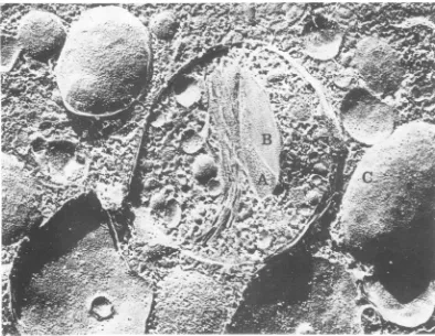
FIG. 5. Freeze-etchreplicaofamembranous vesicleinanA.albopictuscell 6 hafter infection with Sindbis virus. The arrow indicates the point of contact ofthe stacked membrane with the surrounding limiting membrane.Thecleaved membraneinteriors have thetypicaldistribution of interiormembrane beads. x95,000.
placeorthatthesestructures werelost from the
membrane prior to examination. Other large
membranous structures with associated
nu-cleocapsids could also be found in the culture
media (Fig. 12b and c). These structures
dif-feredfromthat illustratedinFig.12ainthat the
viral nucleocapsids appear to be partially
en-veloped in the membrane. Some areas of the
membrane are contoured as though they
sur-rounded nucleocapsids which were later lost from the structure (Fig. 12b).The entire outer surface ofthemembrane appears tobe covered
with spike-like structures similar to those seen
in intact virions. The presence of the spikes on
this membrane surface suggests that the
topo-logical rearrangement of themembrane
accom-panying its conversion to the viral envelope is
complete andthat this membrane representsa
moreadvancedstateof virusdevelopment than
that shown inFig. 12a. Theculture mediaalso
contained large numbers ofclosedspiked viral
membranes surrounding many nucleocapsids
(Fig. 12c).
Examination of ultrathin sections of
virus-infected mosquito cells suggested that viral
nucleocapsidswere enveloped within the
vesic-ular structures by interaction with the in-travesicular membranes (Fig. 6). Further evi-J. VIROL.
on November 10, 2019 by guest
http://jvi.asm.org/
SINDBIS MORPHOGENESIS IN MOSQUITO CELLS 919
dence for such a process of envelopment was
obtained by examination ofextracellular viral
structures (Fig. 13). Many membrane
frag-ments were found with attached single virions
in the final stages of envelopment. The
nu-cleocapsids of such viruses were enveloped to
the extentthat only a small area of attachment
remained. In some instances the membrane of
the viral envelope was continuous with the
membrane from which it was derived. Such
.4
Z*-S a m
1.
.^ as. ..
A-,*/ .1P . .
V. ;
', Str
1. It
.5V~~~~~~~.
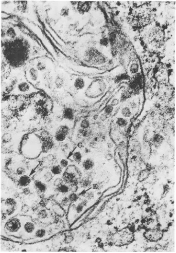
FIG. 6. Thinsectionofavirus-inducedvesicleat 12hpostinfection.A largenumberofmaturevirions(V) can beseeninadditiontoviralnucleocapsids (C);onevirion ispartiallyenveloped (E). x135,000.
VOL. 16, 1975
.J
on November 10, 2019 by guest
http://jvi.asm.org/
[image:7.493.63.425.141.659.2]920 GLIEDMAN, SMITH, AND BROWN J. VIROL.
91
_,9-.,b.vEe,,/~~~~~21
'NJJ9,~~~~~~~~~~~~~~~~~~~~~Y;.fi.4
0$;p'
;*4;'
't*,,ob
<;,
r
~~~~~~~~~~~~~~~~~~~~~~~~~~~~~..ii
FI.
.A.aboitu
el t
0h
otifcto.
a)Lw
aniiato
o
cl
soin
nmero
eletrn-ens
iclsios
anW8)whchwee
fun t
cotan
arg
nmbrsof iron. Bddngof
irs ro
the~~~~~~~~~~~~~~~~~~~~~
plsm
mebaeiTo
vdn.x800
b
ihmgiiaino
n
fteeeto-es
eils
Viin7r
akd
nahxgnllatc.x1400
on November 10, 2019 by guest
http://jvi.asm.org/
SINDBIS MORPHOGENESISINMOSQUITO CELLS
FIG. 8. Athin section ofacytoplasmic vesiclecontaining many virions(unmarked arrows). Thevesicleis close to theplasmamembrane(Pm)and it appears thatthelimitingmembraneofthevesiclehasfusedwith the plasma membrane at thepointindicatedbythearrow(A). Theuniform appearanceofthevesicle (seen inFig. 7b) has been lost. The virionsarecoatedwith thecontentsof the vesicle. x120,000.
structures are very similar to the last stage of envelopemembranesinto aclosedsphereatthe
envelopment ofSindbis virus from the plasma point ofattachment.
membranes of vertebrate cells previously
re-ported (4). This suggests that as in vertebrate DISCUSSION
cells, the final release of the virions from the AlthoughthegrowthkineticsofSindbisvirus
parent membrane occurs with fusion of the aresimilarinBHK-21andA.albopictuscellsat
921 VOL. 16,1975
on November 10, 2019 by guest
http://jvi.asm.org/
[image:9.493.55.429.72.556.2]922 GLIEDMAN, SMITH, AND BROWN
-FIG. 9. Freeze-etchreplicaofavirus-induced vesicleat18hpostinfection.Someofthe stackedmembranes have a very high density of interior membrane particles (A), whereas others are smooth (B). Normal distribution of the interior membraneparticles can beseen onother membranesoutside of the vesicle (C). x80,000.
11
FIG. 10. Freeze-etchreplica of pelletedvirions pro-duced from A. albopictus cells. The interiorof the viral envelope is free of interior membrane beads. Arrows indicate thecross-fractured outerleafletof the viral membrane. x180,000.
28 C, the pattern of virus morphogenesis was
found to be very different in the two cell
systems.Theproductionofmaturevirions from
vertebrate cells occurs by the budding of free
cytoplasmic nucleocapsids through the plasma
membrane of infected cells (1, 4). Thisprocess
is accompanied by gross cytopathic effects and
cell death within 20 h postinfection.
Mor-phogenesis of Sindbis virus in A. albopictus
cellsoccurredprimarilyincytoplasmic vesicles;
buddingofvirions through plasma membranes
and accumulation ofcytoplasmic nucleocapsids
occurred only in a very small percentage (less
than 1%) oftheobserved cells. The infection of
the invertebrate cellsby Sindbis virus doesnot
result in noticeable cytopathic effect or in a
detectable reduction in therateofcell growth.
The observation ofvirus development in
in-ternal vesicles presented here is similarto the
report of Raghow and co-workers for twoother
alpha togaviruses (18, 19).Unlike theseauthors,
however, we did not find any evidence for the
destructionof thevirus-rich inclusionsbyfusion
withlysozome-like bodies. We suggest that the
J. VIROL.
on November 10, 2019 by guest
http://jvi.asm.org/
[image:10.493.64.458.78.383.2] [image:10.493.68.251.434.629.2]SINDBISMORPHOGENESIS INMOSQUITO CELLS 923
vesicles release matured virions into the
sur-rounding medium by fusion with the plasma
membrane. Examination of cells persistently
infected with Sindbis virus forperiods of time
upto 5days after infection revealed the
contin-ued presence of virus-containing vesicles inthe
cytoplasm ofthe infected cells, althoughfewer
in number than seen in Fig. 7a. With the
exceptionofthe reduction innumberofvesicles
the general morphology of the persistantly
in-fectedcellswasfoundtobesimilartothat of the
cells at about 20 h after infection when the
logarithmic stage of virus production is
com-pleted. It is not yetclear whether the apparent
sequestering of virus morphogenesis in this
mannerenables mosquito cells tosurvive
infec-tion.
Theoriginofthenucleocapsids which appear
in the cytoplasmic vesicles described here and
by Raghow et al. (18, 19) is unclear. The
presence of free nucleocapsids within the
vesi-cles implies that the nucleocapsids are either
assembled in the vesicles, transported into the
vesicles afterassemblyinthe cellcytoplasm, or
that the vesicles are developed around the
nucleocapsids which are first assembled in
re-stricted areas ofthe cytoplasm. The last two
possibilities seem less likely to us considering
the scarcity of free cytoplasmic nucleocapsids
and the fact that we have never observed
nucleocapsids attached to or budding into the
cytoplasmic vesicles. However, it is possible
that transport of free nucleocapsids into the
vesicles or elsewhere occurs very rapidly and
that the small percentage of cells having
cyto-plasmic nucleocapsids may result from a
slow-ing or an interruption inthis transport process.
We arepresentlyconductingastudy of infected
cellsby pulse-chase autoradiography with
pro-tein and RNA precursors in an attempt to
localize RNA and proteinsynthesis and the fate
of these products in infected mosquito cells.
Theenvelopment of the nucleocapsids in the
vesicles presumably takes place through
in-teraction of the nucleocapsids with the
mem-branes present in the vesicles and appears to
possesssomedifferencesfromthe interaction of
nucleocapsids with the plasma membrane that
occurs during "budding" inthevertebrate cell
system (1, 4).
The attachment of the viralnucleocapsidsto
themembranes found within the vesiclesofthe
infected mosquito cells seems tobefollowedby
a topological change in the entire membrane
structure asenvelopment occurs.This
general-ized morphological reorganization ofthe
mem-branes was seen in negatively stained
prepara-tions of premature nucleocapsid-membrane
.v.
---.N.
.?
a
_q4
,
,'
-FIG. 11. A.albopictus cells withsurface-associated virions. (a) Twoinfectedcellswithanumber of mature completed virionsintheextracellular space. Thecytoplasm ofthe lower cellcontainsnucleocapsids ofwhich oneisassociated with theplasma membraneasthoughin anearlystageofbudding(arrow). x160,000.(b)The surfaceofaninfected cellwithvirions inwhatmay bealatestageofbudding. x160,000.
VOL. 16, 1975
t..q.s
;g. 1.
on November 10, 2019 by guest
http://jvi.asm.org/
[image:11.493.61.423.388.638.2]924 GLIEDMAN, SMITH,AND BROWN
ai-a
[image:12.493.63.454.80.609.2]b9_.--w
c
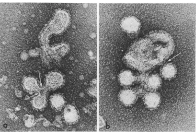
FIG. 12. Negative stain of extracellular nucleocapsid-membrane complexes. (a)Alargespikeless membrane fragment withattached normal size (A), medium size (B), and small size (C) Sindbis nucleocapsids. The inset at highermagnificationshowsthree of the attached nucleocapsids in which capsomeres (arrows) are visible. x320,000;insertx597,000.(b)Alarge fragment ofamodifiedmembrane withexternallylocatedspikesandone or two envelopednucleocapsids (A). Otherareasof the membrane appear tohavelost capsid structures (B). x179,000.(c)Membranefragment containingseveral virusnucleocapsids.This membrane isapparentlyclosed and hasspikeson itsoutersurface. x 179,000.
J. VIROL.
on November 10, 2019 by guest
http://jvi.asm.org/
SINDBISMORPHOGENESIS INMOSQUIrO CELLS 925
FIG. 13. Negative stainofextracellularcomplexescontainingmodifiedmembranesandmaturingvirions.In some areas the viral envelopes are still continuous with the modified membrane from which they have developed (arrows). x180,000.
complexes (Fig. 12 and 13) and in freeze-etch
images of the interior regions of the
intravesicu-larmembranes (Fig. 9). The total alteration of
the internal and external
morphology
of themembrane, includingregions of the membrane
whichare notbeing wrappedaround
nucleocap-sids, is very different from the localized
topo-logical change described for the vertebrate
cellsystems(4).
The presence ofthe
multi-nucleocapsid-con-tainingenvelopedstructureswhich arefound in
preparations from invertebrate cells
(Fig.
12b,12c, and 13) suggests that the process of
en-velopment within the vesicles is not asspecific
asthe individualnucleocapsid packaging
proc-ess seen attheplasma membrane invertebrate
cells. It appears that once the modification of
the membranes in the invertebrate vesicles is
complete, the membranes close around the
attached nucleocapsids, occasionallyproducing
the aberrant multicapsid structures. The
re-lease ofpartially enveloped nucleocapsidsfrom
the infected cellsraises the possibilitythatthe
process ofenvelopmentmay continue even after such structures are released from thehostcells.
An exhaustive search for the budding of
virions from the plasma membrane (as de-scribed for virus maturation invertebrate host
cells [1, 4]) utilizing freeze etching and
ultra-thin sectioning of Sindbis-infected mosquito
cells during the period of maximum virus
pro-ductionwasgenerally negative. Thepresence of
large numbers ofintact virionsonthe surfaceof
the infected cells as described by other
inves-tigators (8, 18, 19) was also not seen. The
presence ofsurface-associated virus is of itself
not an indication that budding of virus has
taken place, asunder appropriate conditions of
glutaraldehyde fixation one would expect a
certainamountof freevirustobecross-linkedto
the cell (9,24). In nostudy of togavirus
produc-tion ininvertebrate cells has extensive budding
at the plasma membrane been reported. The
few images of what appears asbuddingvirions
produced here (Fig. 11) andinother studiesare
infrequent compared to the ease with which
they are obtained in thevertebrate cellsystem
underconditions producing similaramountsof virus. The few incidences ofapparent budding
produced inthis study could actuallyrepresent
stages in penetration during reinfection by
ex-tracellularvirus.
It is possible, however, that the occasional
budding figures and the infrequently observed
free cytoplasmic nucleocapsids represent an
alternative process of virus maturation. This
VOL. 16, 1975
on November 10, 2019 by guest
http://jvi.asm.org/
926 GLIEDMAN, SMITH, AND BROWN
process occurs in a small percentage of the
infected cells and reflects eitheraheterogeneity
in the cell population or an alteration in the
physiological condition of a few of the infected
cells. It is also possible that both processes of
virus production can occur in a particular cell
and whether onepathway orthe other prevails
is determined by factors effecting the
physiol-ogy of the cell. Support for this latternotionhas
been obtained by experimentsconductedinthis
laboratory in which large numbers of
cytoplas-mic nucleocapsids were accumulated in
Sind-bis-infected mosquito cells after treatment with colcemid (to be published elsewhere).
Treat-ment with colcemid did not increase the
num-berofbudding virions observed and reduced the
amount of virus produced.
ACKNOWLEDGMENTS
Weacknowledge the technicalassistance of Donald Filtzer andDoris Renz.
This research wassupportedbytheDeutsche Forschungs-gemeinschaftSFB74.
LITERATURE CITED
1. Acheson, N. J., and I. Tamm. 1967. Replication of Semliki Forest virus: an electron microscopic study. Virology32:128-143.
2. Anderson, T. F.1962. Negative staining and its use in the study of viruses and their serological reactions, p. 251-262. InR. J. C. Harris(ed.),Theinterpretation of ultrastructure, vol. 1.SymposiumoftheInternational SocietyforCellularBiology. Academic Press Inc., New York.
3. Brown, D. T., and J. B. Gliedman. 1973.Morphological variants ofSindbis virus obtained from infected mos-quito tissue culturecells. J. Virol. 12:1534-1539. 4. Brown, D. T., M. R. F. Waite, and E. R. Pfefferkorn.
1972.Morphology andmorphogenesis of Sindbis virus as seen with freeze-etching techniques. J. Virol. 10:524-536.
5. Buckley, S. M. 1969.Susceptibilityofthe Aedes albopic-tus and A. aegypti cell lines to infection with ar-boviruses. Proc.Soc.Exp. Biol. Med. 131:25-30. 6. Bykovsky, A.F.,F. I.Yershov, and V. M. Zhdanov. 1969.
Morphogenesis of Venezuelan equine encephalomyeli-tis virus. J.Virol. 4:496-504.
7. Eagle, H. 1959. Amino acid metabolism inmammalian cellcultures.Science 130:432-437.
8. Filshie, B. K., and J. Rehacek. 1968. Studies on the morphology ofMurray valley encephalitis and Japa-neseencephalitis viruses growing in cultured mosquito cells.Virology 34:435-443.
J.VIROL.
9. Hopwood, D. 1969. A comparison of the cross-linking abilities of glutaraldehyde, formaldehyde and
a-hydroxyadipaldehyde with bovine serumalbumin and casine.Histochemie 17:151-161.
10. Luft, J. H. 1961.Improvements in epoxy resin embedding methods. J. Biophys. Biochem. Cytol. 9:409. 11. Marchesi. V. T., R. L. Jackson, J. P. Segrest, and I.
Kaltane. 1972. Molecular features of the major glyco-protein ofthe humanerythrocytemembrane in mem-branes and mechanisms of hormone action. Fed.Proc. 32:1833-1837.
12. Matsumura, T., V. Stollar, and R. W. Schlesinger. 1971. Studies on the nature of Dengue viruses. V. Structure anddevelopment of Dengue virus in vero cells. Virology 46:344-355.
13. Millonig, G. 1961. Advantages of a phosphate buffer for 0,4solutions in fixation. J.Appl. Physiol.32:1637. 14. Mitsuhashi, J., and K. Maramorosch. 1964. Leafhopper
tissue culture: embryoni, nymphal and imaginal tis-sues from aseptic insects. Contrib. Boyce Thompson Inst. 22:435.
15. Morgan, C., C. Howe,and H. M. Rose. 1961. Structure and development of viruses as observed in the electron microscope. V. Western equine encephalomyelitis vi-rus. J.Exp. Med. 113:128-143.
16. Peleg, J. 1969. Inapparent persistent virus infection in continuously grown Aedes aegypti mosquito cells. J. Gen.Virol. 5:463-471.
17. Pfefferkorn, E. R., and R. L.Clifford. 1964. The origin of theproteinsofSindbisvirus.Virology23:217-223. 18. Raghow, R.S.,M. W. Davey, and L.Dalgarno. 1973. The
growth of Semliki Forest virus in cultured mosquito cells: ultrastructural observations. Arch. Gesamte Vi-rusforsch. 43:165-168.
19. Raghow, R.S., T. D. C.Grace,B. K.Filshie, W. Bartley, and L. Dalgarno.1973. Ross River virusreplication in culturedmosquitoandmammalian cells: virusgrowth andcorrelated ultrastructural changes. J. Gen. Virol. 21:109-122.
20. Sefton,B.M., and B. J.Gaffney.1974.Effect of the viral proteins on the fluidity of the membrane lipids in Sindbisvirus. J. Mol. Biol. 90:343-358.
21. Singh, K. R. P. 1967. Cell cultures derived from larvaeof
Aedesalbopictus (Skuse)andAedesaegypti (L). Curr. Sci. 36:506-508.
22. Stevens, T. M. 1970. Arbovirus replication in mosquito cell lines (Singh) grown in monolayer orsuspension cultures (34793). Proc. Soc. Exp. Biol. Med. 134:356-361.
23. Tillack, T. W., R. E. Scott, and V. E. Marchesi. 1972. The structure of erythrocyte membranes studied by freeze-etching. II. Localization of receptors for phyto-hemagglutinin and influenza virus tothe intramem-branousparticles. J. Exp. Med.135:1209.
24. Waite, M. R. F., D. T. Brown, andE. R. Pfefferkorn.
1972. Inhibition of Sindbis virus releasebymedia of low
ionicstrength: an electronmicroscopestudy. J.Virol.
10:537-544.
on November 10, 2019 by guest
http://jvi.asm.org/