0022-538X/93/031493-10$02.00/0
Copyright (C 1993, American Society forMicrobiology
Cell Fusion
by the Envelope Glycoproteins of Persistent Measles
Viruses
Which Caused Lethal
Human
Brain Disease
ROBERTOCATTANEOt* ANDJOHN K. ROSE
Department of Pathology andDepartmentof Cell Biology, Yale University Medical
School,
NewHaven,
Connecticut 06510Received31 August 1992/Accepted 10December 1992
Measles virus (MV) rarely induces lethal diseases ofthe human central nervous system characterized by reduced expression of the viral envelope proteins and by lack of viral budding. TheMVenvelope containstwo
integral membrane proteins, termed fusion (F) protein and hemagglutinin (H) protein, and a
membrane-associatedmatrix(M) protein. Previously, analysis ofMVgenesfromautopsymaterial indicatedthat the M protein and the F protein intracellular domainareoften drastically altered by mutations. Here, wepresent evidencethattruncation oftheF protein intracellular domain doesnotimpairfusion function, andwesuggest that this alteration interfereswith viral budding. Unexpectedly, certain combinations of functional F andH proteins were unable to induce syncytium formation, an observation suggesting that specific F-H protein
interactions are required for cell fusion. We also found that three of four H proteins of persistent MVsare
defective in intracellular transport, oligosaccharide modification, dimerization, and fusion helper function. Thus, MVs replicating in the brainatthe terminalstage of infectionaretypically defective in M protein and in the two integral membrane proteins. Whereas the M protein appears dispensable altogether, partial
preservationofF-protein functionand H-protein functionseemstoberequired,presumablytoallow local cell fusion. Certain subtle alterations of the F and H proteinsmaybeinstrumentalfor disease development. Measles virus(MV)isoneof theprimarycausesof infant
death in Third World countries (5), and a recent MV epi-demic has caused morethan 100 deaths inNorth American
inner cities(46). Onrareoccasions, MVpersistence induces lethal syndromes of the central nervous system known as subacute sclerosing panencephalitis (SSPE) and measles inclusion body encephalitis (MIBE) (57). SSPE, which
oc-curs5to10yearsafteranacuteMVinfection, isoneof the
most thoroughly studied persistent viral infections of the human centralnervoussystemandserves as amodel for the
studyofpersistentinfections byRNAvirusessuspected to
cause other human syndromes (26). MIBE shares many
characteristics with SSPE but occurs in immunocompro-mised patients and hasashorterincubation time.
MVsreplicatingin the brains ofSSPE and MIBEpatients
arecharacterized bydefectiveexpression of theirenvelope
proteins. Infectious virus is not detectable in the brains of SSPE or MIBE victims, but occasionally cell-associated defective viruses can be isolated by cocultivation of brain cells with fibroblasts(62).Most of thesecell-associated MVs aredefective inexpressionof thematrix(M) protein,which lines the inner surface of the viralenvelope (18, 61).
More-over, the M protein, as well as the two viral integral
membrane proteins, termed fusion (F) protein and hemag-glutinin (H) protein, often cannot be detected in brain autopsy material (2, 35). Reduced production of the viral envelope proteinsinhumanbraininfectionscanbe ascribed partly to a steepgradient oftranscription resulting in low-levelexpressionof thedistallylocatedgenesforM, F,and H proteins (9).
On the otherhand, in MVs replicatingin brains of SSPE and MIBEpatients, numerous mutational alterations of the
*Correspondingauthor.
tPresent address: Institut fir Molekularbiologie I, Universitat
Zurich, Honggerberg, CH-8093Zurich, Switzerland.
envelope genes have been identified (3). Characteristically, certain mutations alter drasticallythe entire M protein and the F protein intracellular domain (11, 48), whereas other, more-subtle mutations distinguishthe extracellulardomains of the F and Hproteins (12). Here,westudied theexpression and function of F and H proteins in cultured cells. These proteins were produced from cDNAs of MV mRNAs ob-tained frombrainautopsymaterial oftwoSSPEpatients and
aMIBEpatientand fromacell line derived from braincells of another SSPE patient. Wepresent evidence that, in one
instance, aberrantprocessing oftheF protein extracellular domain resultsinstrongfusion inhibition. We also foundthat three of four Hproteins ofpersistent MVs are significantly or completely defective in intracellular transport, oligosac-charide modification, dimerization, and fusion-helper func-tion and thatspecific F-H protein interactions arerequired for efficientcell fusion.
MATERIALS AND METHODS
Cells and viruses. HeLa cells were obtained from Ari Helenius (Yale University, NewHaven, Conn.),andHeLa T4 cells were obtained from the American Type Culture Collection (Rockville, Md.). Both types of cells were
pas-sagedinDulbecco's modifiedEagle'smedium with5% fetal
calf serum. MV Edmonston (MV strain E) was obtained
from Stephen A. Udem (Universityof Medicine and Den-tistryof NewJersey, Newark)andpropagatedasdescribed
elsewhere(58). Propagationandpurificationof the
recombi-nant vacciniavirus-encoding T7 polymerase (vTF7-3) (16)
was performed with HeLa cells essentially as described previously(29),exceptthatthe final step ofsucrosegradient
centrifugationwasomitted.
Plasmidsandexpressionsystem.Full-lengthcDNAs of MV
genes wereproduced accordingtothe method of Schmid et
al. (47). Clones of MV strain E(15), fromSSPEpatientsA and B and MIBE patient C (11), and of SSPE cell line 1493
on November 9, 2019 by guest
http://jvi.asm.org/
four F protein genes used in this study (from plasmids paFl, pbFll, pcF5, and pi62F1) was substituted for the strain E insert of this vector by using the HpaI site situated 52 nucleotides downstream of the first F protein gene AUG and a SacIl site situated in the polylinker, downstream of the MV insert. The subclones were named paF, pbF, pcF, and piF. For expression of H proteins, full-length H protein genes were subcloned in plasmids named peHl-SK, paH2, pbH4-SK, pcH4-SK, andpi62H1-SK. The vaccinia virus T7 RNA polymerase system (16) was used by following the protocol established by Whitt et al. (63). Plasmid transfec-tion in HeLa or HeLa T4 cells was performed with the cationic lipid TransfectACE (Life Sciences, Inc.), as de-tailed by Rose et al. (44).
Antibodies and immunofluorescence staining. Polyclonal goat antiserum raised against total MV was a kind gift of Stephen A. Udem (University of Medicine and Dentistry of NewJersey, Newark). Monoclonal antibody 129, recogniz-ing an H protein epitope common to many different MV strains (52), was a kind gift of Erling Norrby (Karolinska Institute, Stockholm, Sweden). Polyclonal rabbit antiserum raised against a fragment of the F1 protein expressed in Escherichia coli was a kind gift of Timothy C. Wong (University of Washington, Seattle). The procedures for indirect immunofluorescence staining were performed as describedpreviously (42) with a 1:200 dilution of monoclonal antibody 129. Fluorescein-conjugated goat anti-mouse im-munoglobulin G antibody (Cappel Laboratories, Cochran-ville, Pa.) was used in a 1:100 dilution as a secondary antibody.
Fusionassays. Fusion assays were performed initially with HeLa cells and then with HeLa T4 cells. Fusion assays performed by using the same F and H proteins with the two cell lines yielded similar results, but formation of large syncytia on HeLa cells was often inhibited by cytopathic effects and cell rounding. Vaccinia virus infection of HeLa T4 cells produced only limited cytopathic effects and cell rounding, and thus, fusion assays were more reproducible (38). Initially, different ratios ofFto Hplasmid DNA were tested for theirfusion effects. Ratios ranging from 1:1 to 1:4 were found to induce similar fusion levels, and a 3:7 ratio was chosen for subsequent fusion assays. Confluent 3.5-cm dishes of HeLa T4 cells were cotransfected with 1.5 p,g of DNA of a plasmid expressing an F protein and 3.5 ,ug of DNAof a plasmidexpressing an H protein.
Metaboliclabelingandimmunoprecipitation. Labeling with
[35S]methionine
wasachieved by usingTrans35Slabel (ICN Biomedicals, Inc.) in methionine-free, serum-free Dulbec-co'smodified Eagle's medium for the times indicated in the figure legends. Chases were in Dulbecco's modified Eagle's medium containing 0.5% fetal calf serum and 2 mM methi-onine. Immunoprecipitation and digestion with endo-p-N-acetylglucosaminidase H (endo H) were performed as de-scribedpreviously (43). Surface immunoprecipitations were performedasdescribed by Machamer and Rose (28) by using eithermonoclonal antibodyI29 or anti-MV goat antiserum. Labeled proteins were resolved on polyacrylamide gels0
553 537 3 529
Edmonston virus SSPE case A SSPE case B MIBE case C SSPE cellline I
FIG. 1. Characteristics of the F and H proteins of MVs analyzed in this study deduced from the sequences of the corresponding genes. The predicted primary translation products of F and H protein genes, 553 and 617 amino acids long, respectively, are represented by vertical lines. The plasma membrane is shown as a stippled region. Amino acids differing from the F and H protein consensus sequences, defined on the basis of the sequences of six lytic and persistent viruses (12), are shown as circles. A frameshift in theintracellular domain of the F protein from patient A (case A), caused by a 1-nucleotide deletion, is indicated by a small open box. One potential glycosylation site situated in the presumptive F protein signal peptide has not been indicated. Note that the subclon-ing strategy used resulted in the substitution of the first 17 amino acids of the F protein signal peptide of each protein with the correspondingamino acids of strain E; in three F protein genes, the sequences werecompletely identical to the E strain sequence, but for the F protein from patient C two amino acids differed.
containing sodium dodecyl sulfate as described elsewhere (27) and visualized by fluorography (6). For analysis of nonreduced proteins, ,B-mercaptoethanol was omitted from the loading buffer. Quantification of signals on gelswasdone with aPhosphorlmager (Molecular Dynamics).
RESULTS
Wehave compared theFand Hproteins expressed from cloned cDNAsproduced from brain autopsy material of two SSPE patients (patients A and B) and a MIBE patient (patient C) and from acell line derived frombrain cells of another SSPEpatient (cell lineI) with the F and H proteins of the lytic strain E. The MV F protein (Fig. 1), a type I transmembrane protein with anextracellular amino terminus and an intracellular carboxyl terminus, is produced as a precursor,
Fo.
The signal peptide is first removed, and a subsequentcleavage yields a 20-kDa F2 protein and a 40-kDa F1 protein, covalently linked by one disulfide bridge (41). The Hprotein is a type IItransmembrane protein with the opposite orientation.Previous cDNA sequence analysis revealed several differ-ences in the predicted sequences of the F and H proteins expressedinthis study from the F and H proteinconsensus sequences(12) (these differences are shown by small circles in Fig. 1). Two point mutations and a 1-nucleotide deletion nearthe carboxyl-terminal coding region of the three SSPE Fproteins result in the deletion or alteration of a majority of the 33-amino-acid F protein intracellular domain (12) (Fig. 1). Differences are also clustered around the first leucine (position 466) of the zipper structure essential for fusion (7), including athreonine-to-isoleucine change at position 464 in the F proteins from patients A and B and
on November 9, 2019 by guest
http://jvi.asm.org/
[image:2.612.315.551.72.224.2]a
b
cF x eH
++
iF x eH
-FIG. 2. Syncytium formation in HeLa T4cells afterF and Hproteincoexpression. HeLa T4 cells were infected withvTF7-3 and then cotransfected with theplasmids indicated.Cellswerephotographed 12 haftertransfection. (a) Coexpressionof the Fprotein from patient C (cF) with theHprotein ofstrain E(eH). ++,extensivesyncytium formation. (b)Coexpressionof theFprotein of cell line I(iF) withthe same Hprotein(eH)asinpanela. -, nosyncytiumformation.
serinechangesatposition465 in theFproteinfrompatientA andatposition468in the FproteinsfrompatientAand cell lineI. In addition, inthe Fproteinfrompatient C,there isa glycine-to-valine changeatposition 127, within the fusogenic peptide located in the amino-terminal region of the F1 subunit (60). Asshownbelow, theFproteinsofpatientsA, B, and Cwerefunctional,andthus, these differences didnot
have negative consequences.
Although mutations of the H protein genes of persistent MVs do not result in striking alterations of the reading frames, the Hproteins accumulatemorechanges than the F proteins(Fig. 1) (46). TheHprotein of the vaccine strain E has sixpositionsatvariancewith the consensus, whereas the H proteins from the four persistent strains have 8 to 25 differences from the consensus. Two of the five potential glycosylation sites are eliminated in the H protein from patient A. In the H protein from patient B, a potential glycosylation site is added. The H protein of cell line I, exhibiting stronglyreducedhemadsorption activity (50, 59), shows acluster of 16 amino acidchanges dueto a hypermu-tationevent (8).
Afunctionaltestof F and Hproteins: afusion assaybased on avacciniavirus T7 RNApolymeraseexpression system. A vaccinia virus-based expression system has recently been usedtostudycellfusioninducedbyMVproteins,leadingto
the conclusion that fusion requires the expression of both MV F and MV H proteins (56, 64). We have used the vaccinia virus T7 RNApolymerase (vv-T7) expression sys-tem tostudytheinteractions of the F and Hproteinsnotonly of the lytic strain E but also of the four persistent MVs described above.
Coexpression
ofcertain combinations of F andHproteins in HeLa T4 cells resulted inmore orlessefficientcell fusion as visualized by syncytium formation (Fig. 2a), but other combinations of Fand Hproteins were not fusogenic (Fig. 2b). In our fusion assay, relative values were generally similar among different transfection series, but absolutevaluesvaried. For this reason, eachexperiment was done in quadruplicate, aspresented in Table 1. The standards used to evaluate fusion efficiency are indicated in footnote a to Table 1; at least six fields were examined in each experi-ment.
TheFproteins of the lytic strain E and ofthree of the four persistent MVs showed high fusionefficiencywhentested in combination with the H protein of strain E(Table 1).The F protein of cell line I was only marginally functional in combination with the H protein of strain E (Table 1). In addition, the fusion efficiency of the five F proteins was tested in combinations with the H proteins of the four persistent MVs. Interestingly, the F protein from strain E wasless fusogenic than the F proteins from patients A, B, andC when tested incombination with the H proteins from patients A, B, and C (Table 1; compare positions 2 to 4 in column 1 with thecorresponding positions in columns 2 to
4).
The fusion assays presented in Table 1 can also be interpreted with respect to the Hproteins. The H proteins from strain E and patient C cooperated efficiently with differentFproteinstoinduce cell fusion. TheHprotein from patient B induced lower levels of cell fusion when coex-pressed with three different Fproteins. The Hprotein from patient A wasweaklyactive when coexpressedwithtwo F
proteins, andthe H protein from cell lineI was completely inactive in fusion helper function. As demonstrated below, the fusionhelper functionofthe different Hproteinsroughly correlated with theefficiency of theirexpression at the cell surface.
Homologous combinations of F and H proteins (under-lined in Table 1), with the exception of that forpatient A, showedhigherfusionactivitythanmostoftheheterologous combinations. Unexpectedly, certain combinations of func-tional proteins were unable to induce efficient fusion. For example, the Hprotein frompatient Ccouldnot cooperate with the Fprotein of strain Etoproduce cellfusion, in spite
on November 9, 2019 by guest
http://jvi.asm.org/
[image:3.612.133.496.61.281.2]B -,-,-,- (+) 7+), (+) ++, +,+ -,-,(+),(+) -,,, 0.7
C -,-,-,- +,(+),+,+ + + ++ + + +++ + -, -, -, - 1.3
Cell lineIP _ ,_ ,_ ,__ _ ,__ ,_ 0
aData are fromquadruplicate experiments.In eachexperiment, severalfields were evaluated. Symbols fordatafor anaveragefield(about 2,000cells): ++,
severalsyncytia with more than 10 nuclei(sometimesup to half of thenucleiwere found in syncytia); +, one ortwosyncytiawithmore than 10nuclei (andmany
moresmaller syncytia);(+),several4- or5-nucleussyncytia and,occasionally,larger ones; -, two or fewer 4- or5-nucleus syncytia (background). Homologous
combinations areunderlined. Fusion coefficientsare given in brackets.Pointswereassignedasfollows:-,0;(+),1; +, 2; + +, 3. Coefficients werecalculated
byadding the points and dividing the total bythenumber of fusionassays.Strain E is lytic; theotherMVs arepersistent.PatientsA and B hadSSPE,patient
C had MIBE, andcellline I was derived frombrain cellsof anotherSSPEpatient.
bOur failure to observefusion helper activitywith the Hproteinof celllineIismostlikelydue to theextremelyreduced surfaceexpression.
of the fact that the Hprotein from patient C was active as a helper in combinations with the Fproteins from patients A, B, and C, and the F protein of strain E was fusogenic in combinations with the H protein of strain E and the H protein from patient A. This and other results, which cannot be explained by reduced cell surface expression of the corresponding proteins (see below), suggest that specific interactions between F and H proteins are required for efficient fusion.
TheFprotein of cell lineIisprocessed aberrantly. Inspite ofthe fact thatpreliminary experiments suggested that only asmall fraction of the five Fproteins examined reached the cell surface (data not shown), the functional analysis pre-sented above indicated that four of the five F proteins can
efficientlyinduce cell fusion. Only theFprotein of cell lineI is functionallyimpaired. To address the cause of this defect, we analyzed the expression of this protein, as well as the expression of the other fourF proteins, by immunoprecipi-tation withtwodifferentantisera.
Immunoprecipitations with an antiserum recognizing pref-erentially the unfolded precursor, F0, indicated that similar amountsof
Fo
precursors are produced by all five plasmids (Fig. 3a, 0-h chase; the upper band corresponds to the completelyglycosylated precursor, and the lower two bands correspond to forms not completely glycosylated). As ex-pected, aftera3-h chasealarge fraction of the precursor,F0,
disappeared (Fig. 3a).
An antibody recognizing preferentially the native, pro-cessed form of the Fproteinwasusedtodetect the
F,
and F2 subunits,whichappearedas expectedforproteinsof strain Eand frompatients A, B, and C only after a 3-h chase (Fig. 3b). In all cases, only a fraction of theFo
precursor was chased in theF1 and F2subunits. Note also that the serum againsttotalMVrecognizes the native F protein from patient Bpoorly; this protein is precipitated at a level comparable to the level ofthe corresponding protein of strain E or that from patientCbytheantiserumpreferentially recognizingFo
(Fig. 3a). The largest fraction of the F protein of cell line I, however,wasdetected as a broad 33- to 34-kDa band, which is 6 to 7 kDa shorter than the weak but sharp band corre-spondingtotheF1protein (Fig. 3b and c, 3-h chase). Thus, we conclude that the poor functionality of the F protein of cell line I correlates withits aberrant processing.Half ofthesamples immunoprecipitated with the antibody againstnativeFprotein were digested with endo H (Fig. 3c). This enzyme cleaves high-mannose oligosaccharides that are
a
anti-denaturedF
0hours 3 hours
E A B C ri FASCC Irn.
- 65
. up
* M3we
:
_FI
36
b ive
0hours
C
3 hours cnose 3 hours (+endoH)
EA B C n. E A SCrB !. Foorteii EA s CrJn
- ...__ ...I0
-* 200c- .
- 0
65
F~ _,,
36-F2
-~ ~ ~ ~ 3
FIG. 3. Pulse-chase and endo H analyses of the expression of different Fproteinsin HeLa cells. Transfected HeLacells express-ing the Fproteinof strain E(lanes E),Fproteins from SSPE patients Aand B and MIBEpatientC(lanesA, B,andC,respectively),and the Fprotein of the SSPE-derived cell line I (laneI)were labeled with[35S]methionine for1 hand then chased in mediumcontaining unlabeled methionine for the indicated times.(a) Immunoprecipita-tion with antibodies againstanF1proteinfragmentexpressedin E. coli(anti-denatured F); (bandc)immunoprecipitationwith antibod-iesagainstnative Estrain MV(anti-nativeF).Thesamplesshown in panelc weretreated with endo H. Molecularweights (inthousands) and thepositionsof the different forms of theFo,F1,andF2proteins
are indicated. The positions of the molecularweight markers are
indicated bydots. n., negative controls, in whichno plasmidwas
used for transfection.
on November 9, 2019 by guest
http://jvi.asm.org/
[image:4.612.65.553.84.188.2] [image:4.612.334.534.275.582.2]surface
EA
total
FIG. 4. Immunofluorescence analysisof H proteinexpressionin HeLa cells. Transfected HeLa cells expressing the H protein of strain E (panels E),Hproteins from SSPE patientsAandBandMIBE patient C (panels A, B, and C, respectively), the H protein of the SSPE-derived cell line I(panels I),and anegativecontrol (not shown) were stained with monoclonal antibody I29, recognizing an H protein epitope common tomanydifferent MV strains. Five hours after transfection, cellswerefixed with3%paraformaldehyde. (a) Analysis of surface H protein expression inintact cells;(b) analysis oftotal H protein expression in cells permeabilized with 1% Nonidet P-40.
added in the endoplasmic reticulum, but it does not cleave oligosaccharides which have been modified by the Golgi enzymesN-acetylglucosaminyltransferase and mannosidase II (25). SinceN-acetylglucosaminyltransferase is located in the medial Golgi compartment, the acquisition of endo H resistanceby glycoproteinsmarks their arrival at themedial Golgi compartment (14). As expected,when the F proteins weretreated with endo H, areduction in molecular weight wasobservedfor the F2 but notforthe F1 subunit, which is notglycosylated (51).In the threecases in which there was enoughmaterialtodetect theF2 subunit, equivalent amounts oftwobandsmigrating3and 5 kDa faster than the untreated F2subunitwere detected(Fig. 3c, lanes E, A, and C). This result indicates that after a 3-h chase about half of the F2 molecules acquired processed glycans and, thus, had reached the medialGolgi orasubsequent compartment.
The three SSPE H proteins are defective in intracellular transport,oligosaccharidemodification, and dimerization. To determinewhether Hproteinsareexpressedandtransported to the cell surface,we analyzed their intracellular distribu-tion in intact HeLa cells by immunofluorescence (Fig. 4a). Clearcellsurface stainingwasdetected for the Hprotein of strain E and the H protein from patient C, but for the H proteins from patients A and B and cell line I, surface stainingwasbelow the detection level (Fig. 4a).
Toconfirm thatthe Hproteins from patientsAandBand cell line Iwereexpressed, immunofluorescenceanalysiswas
performedon
permeabilized
cells.Indeed,for the Hproteins frompatient Aand cell line I, internal stainingwasclearly visible, but surface stainingwas againbelow the detection level(Fig. 4b). In contrast, for the Hproteinofstrain E and theHproteinfrompatient C,which showed strong internal staining, superimposed weak surface staining could be de-tected. The H protein from patient B was intermediate; whereas internal stainingwas also strong, the edges of the cellswerelessevident.To further investigate the intracellular transport of the
different H proteins, we studied their glycosylation and oligomerization. A pulse-chase labeling experiment was car-riedoutwith transfected HeLa cells as shown in Fig. 5. Half of the samples were analyzed directly after immunoprecipi-tation (Fig. 5a), whereas the other half of the samples were digested with endo H (Fig. Sb). Immediately after pulse labeling, asingle, approximately 80-kDa form was detected forallfiveHproteins expressed (Fig.Sa, lanes 1to5). Strain
E protein and the protein from patient C were always
a
i i 06ods %
5hors T~-1-.<
b
3 hours
- -/ \ m - ~A C
234 5 6 ;._
'
8 19202222324FIG. 5. Pulse-chase and endoH analysis of the expression of differentHproteinsin HeLa cells.TransfectedHeLacells express-ing the H protein of strain E (lanes E), H proteins from SSPE patients A and B and MIBE patient C (lanes A, B, and C, respectively), and the H protein of the SSPE-derived cell line I (lanes I) were labeled with [35S]methionine for 30 min and then chased in medium containing unlabeled methionine for the times indicated. Smallamountsof endoH-resistantmaterial from patient Aarehighlighted by arrowheads. The molecular weight markersare indicatedbydots in the center laneof eachpanel, andtheir sizes (in thousands)areshown between thepanels.n., negative controls,in which no plasmid was used for transfection. See Fig. 8 for a pulse-chase analysisin whichalargerfraction oftheHprotein from patientAhas received theprocessed glycans. Exposure times,6and 24hforpanelsaandb, respectively.
on November 9, 2019 by guest
http://jvi.asm.org/
[image:5.612.60.557.62.279.2] [image:5.612.314.557.466.581.2]additional potential glycosylation site, was also about 10 kDa. Thus, we conclude that the additionalpotential glycos-ylation site present in the H protein from patient B is not used.
After a 3-h chase, two different forms of H protein each weredetected for strain E and patient C (Fig.5a, lanes 7 and 10). The lower band migrated slightly fasterthan the single band detected immediately after the pulse (Fig. 5a, lanes 1 and 4), whereas the upper band was more diffuse and its electrophoretic migration was slightly slower. A longer exposure ofthis gel indicated the presence ofaweakupper band for the Hproteins from patients A and B also. These new forms presumably resulted from oligosaccharide cessing. To determine whether the proteins contained pro-cessed glycans, we digested the 3-h samples with endo H. FortheHprotein ofstrain E and the H protein from patient C,glycanswerecompletely removed from about two-thirds of theproteins while the remainder contained endo H-resis-tant glycans (Fig. 5b, two upper bands in lanes 19 and 22). Small amounts of endo H-resistant material were detected also for the Hprotein from patient B (Fig. 5b, lane 21); even smaller amounts of endo H-resistant material detected for the Hprotein from patient A are highlighted by arrowheads (Fig. Sb, lane 20). The presence of2 or 3 bands containing resistant glycans indicates heterogeneous processing of oli-gosaccharides. No processing of glycans on the H protein of cellline Iwas detected (Fig. 5b, lane 23). These results are consistent with what we observed by immunofluorescence microscopy and indicate relatively efficient transport of the Hprotein of strain E and the H protein from patient C, poor transport of the H proteins from patients A and B, and no detectable transport of the H protein of cell line I.
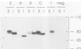
Endo H-resistant glycans are acquired in the Golgi com-partment, and proteins containing these glycans are gener-ally transported rapidly to the cell surface (39). To confirm that this was also true for the MV H protein, selective immunoprecipitation of cell surface H proteins was per-formed(Fig. 6). Indeed, for strainEand patient C, the more slowly migrating form of the H protein was detected in the surface fraction whereas the majority of thefaster-migrating formwasin the internalfraction. Small amounts of the more slowlymigrating form of the H protein from patient B were also detected at thecell surface together with larger amounts of the migrating form. We believe that the faster-migratingHprotein is a contaminant of the surface fraction and that thiscontamination is caused by lysis of some cells before or during the first incubation or by antibody ex-change. With a longer exposure of this gel, a weak cell surface signal was also detected for the H protein from patientA, whereas cell surface expression of the H protein ofcell line I wasalways below thedetection level. Note that low levels of surfaceHproteins detected with this assay (for example,Hproteins from patients A and B) reflect not only inefficient cell surface expression but also limitations in the efficiency of our immunoprecipitation assay. In summary, the Hprotein of strain E and the H protein from patient C, which contain processed glycans, were readily detected at
FIG. 6. Cell surface expression ofdifferent H proteins. Trans-fectedHeLa cells expressing the H protein of strain E (lanesE), H proteins from SSPE patients A and B and MIBE patient C (lanes A, B,and C,respectively), and the H protein of the SSPE-derived cell line I(lanes I["I" in top row]) were labeled with[35S]methioninefor 1 hand then chased in mediumcontaining unlabeled methionine for 3 h. Cells were then detached from the plate with EDTA and incubated withspecific antibodies at0°Ctolimitendocytosis. After extensivewashing to remove unbound antibodies, cells were lysed and the antibody-antigen complexes were collected as surface (S) material. The supernatant was then reprecipitated with antibodies, and theantibody-antigen complexes were collected as internal (I) material. The positions of the molecular weight markers are indi-cated by dots, andtheir sizes (inthousands)areshown on the right. neg.,negative controls, in which noplasmidwasused for transfec-tion.
the cell surface. The H protein of cell line I, devoid of processed glycans, was not detected at the cell surface. The Hproteins from patients A and B showed intermediate levels ofprocessed glycans and reduced but detectable cell surface expression.
Oligomerization is an obligatory step in the intracellular maturation of the H proteins of MV and related viruses. Initially, disulfide-linked dimers and, then, tetramers and othernoncovalentlyassociated oligomers are formed (17, 34, 40). Tocompare the dimerization of the five H proteins, we analyzed a fraction of the samples used for the gel shown in Fig. 5a on another gel in the absence ofareducing agent in the sample buffer (Fig. 7). About one-third each of the H protein from strainEandtheHprotein from patient C was detected as a dimer immediately after the pulse (O h). After a3-h chase, the large majority of the H protein from the E strain and of the H protein from patient C was in the dimeric form.The same was true for about half of the H protein from patient B and for some of theHprotein from patient A, but no material from cell line I was in the dimeric form. Thus, the levels of H-protein dimer formation correlated with the level of glycanprocessing and surface expression.
H protein expression: interference experiments. Defective MVshave been shown tointerfere with lytic MVs in mixed infections(19, 23). To investigate whether the expression of defective MVHproteins caninterfere with the expression of functional MV H proteins, we coexpressed the H protein from patientA and the Hprotein of strain E. The forms of these proteins without processed glycans have different electrophoretic mobilities and, thus, can be separated. Fig-ure8shows anexperiment in which the H protein of strain E and the H protein from patient A expressed alone or coexpressed at the amounts of plasmid DNAs indicated above the gel were labeled and then immunoprecipitated after either no chase (Fig. 8, lanes 1 to 6) or a 3-h chase (Fig. 8, lanes 7to 12). No clear interference effect was observed; theHprotein from patient A did not retain the H protein of strain E in the endoplasmic reticulum, as judged by the acquisition of processed glycans, and the H protein of strain Edid notpromote more-efficient transport of the H protein
on November 9, 2019 by guest
http://jvi.asm.org/
[image:6.612.368.504.73.156.2]FUNCTIONAL ANALYSIS OF GLYCOPROTEINS OF PERSISTENT MVs 1499
a
_,~~~~~~~~~~~~~
b
.,. , ..._._,,_,.._.-..-,r-..-. rl%
,ecc.e_esce
230-,-
-K.~~~~~~~~~~~~~~~~~~~~~~~~~~~~~~~~~~~~~~~~~~
FIG. 7. Dimerization of different H proteins in HeLa cells. Transfected HeLa cells expressing the H protein of strain E (lanes E),H proteins fromSSPE patients Aand B and MIBE patientC (lanes A, B, and C, respectively), and the H protein of the SSPE-derivedcell line I(lanes I)werelabeledwith[35S]methionine
for 30minandthen chased inmediumcontaining unlabeled
methi-onine for thetimes indicated.Samples wereseparated on a
nonre-ducing gel. Thepositions ofthe Hproteinmonomers, dimers,and
aggregates areshownon theright. The molecular weightmarkers
are indicatedby dots in thecenter lane, and their sizes (in thou-sands) areindicatedontheleft. n., negative controls, in whichno
plasmidwasused for transfection.
frompatientA. The fact that theHprotein frompatientA failedto retain the Hproteinofstrain E in theendoplasmic reticulum is probably related to its inability to dimerize efficiently (22, 45).
Four of thefive potentialglycosylation sites of the H protein are used. To determine how many of the five potential glycosylation sites on the H protein of strain Eand ofthe three sites on the H protein from patient A are used, we
performedapartialendo Hdigest.The time ofdigestionwas varied sothatthe numberofpartially digested bands could be counted. Figure 9 shows the results of this analysis,
c s e S . .T.
LiOD -z_E. , 2 _2
,
FIG. 8. The H protein from patient Adoes not retain the H
protein of strain E in the endoplasmic reticulum. Proteins from transfected HeLa cells labeled with [35S]methionine for 1 h were
immunoprecipitatedafter eithernochase(lanes1to6)or a3-h chase
(lanes7to12)inmediumcontainingunlabeled methionine.The H
proteinof strain E and the HproteinfrompatientAwereexpressed
alone (lanes 1, 5, 7, and 11) or coexpressed at the amounts of
plasmidDNAs indicatedatthe top(lanes2to4and 8to10).The H
protein of strain E and the H protein from patient A without
complex sugars migrate at different positions (lanes 1 and 5), as indicated onthe left, but theproteinswith processed glycansare
almostexactly superimposed (lanes7 and11).
FIG. 9. Four of the five potential glycosylation sites of the H proteinareused.Proteins fromtransfected HeLa cells labeled with [35S]methioninefor 1 h wereimmunoprecipitatedanddigested with endo H. Partial endo H digestions of the H protein of strain E (a) and of the H protein from patient A (case A) (b) are shown. The positions of the 80,000- and 65,000-molecular-weight markers are indicated by dots in the center lane. The length (in minutes) of the endo H treatmentforeach sample isindicated above each lane. Four bandsinadditiontothe completely deglycosylated protein band can be detected for the H protein of strain E, and two bands can be detected for the H proteinfrom patient A. Edm., Edmonston strain (strainE).
indicating that four bands in addition to the completely deglycosylated protein are detected for strain E(Fig. 9a) and that twobands are detected for the Hprotein from patient A (Fig. 9b). We conclude that four sites in the H protein of strain E and two sites in the Hprotein from patient A are used. Moreover, the analysis presented in Fig. 5 suggests that either one or twoof theprimary glycans are processed when theHproteins transit through the Golgi complex.
DISCUSSION
Intracellular transportof MV envelope proteins. Tostudy the F and HproteinsofpersistentMVs,weused the vv-T7 systemand HeLa cells(16).As astandard, weexpressed the vesicular stomatitis virus G protein and found, asexpected (63), that 80 to 90% of the G protein contained processed glycans after a 1-h chase (data not shown). The rate of acquisition of processed glycans by the MV E strain H protein was considerably slower. In different experiments, 20to60% of the H protein received processed glycans after a3-h chase. InlyticMV Estraininfections of HeLacells, 40 to80% of theHproteinhadacquiredprocessedglycans after a 3-h chase (7a), a rate similar to the one reported for the samesystemby Kohamaetal. (24)but slower than therate recently measured by Ogura et al. (36). Thus, glycan pro-cessingin the vv-T7 systemis slightlyslowerthan inalytic MVinfection. Inafinding similartothat for the Hproteinof strainE, 30to60%of the HproteinfrompatientCacquired processed glycans after a 3-h chase. In contrast, after the same chase period, only 5 to 15% of the H protein from patientB,2 to10% of the HproteinfrompatientA, and less than 1% of the Hprotein of cell line I acquired processed glycans.
It is noteworthy that the H protein from patient A, in which two of the four glycosylation sites used have been eliminated by mutation, is not dimerized efficientlyand is poorlytransportedtothecell surface. Onespecific carbohy-drate chain in the hemagglutininneuraminidase of the other paramyxovirus,simian virus5,playsamajorrolein promot-ingcorrect folding, and anotherchain,notnecessary in the initial folding, plays a role in the aggregation ofoligomers (33). It is likelythat individual carbohydrate chains of the MV Hprotein havesimilar functions.
SSPE istypicallycharacterizedbyalterations in the short Fprotein cytoplasmicdomain of the persistent MV
(48).
It has been observed previously that alterations of the cyto-VOL.67, 1993on November 9, 2019 by guest
http://jvi.asm.org/
[image:7.612.96.272.70.249.2] [image:7.612.362.517.76.136.2] [image:7.612.97.261.529.611.2]extracellulardomain
H Stronglyreduced cell Reducedcell surface None Extremelyreducedcellsurface
expres-surfaceexpression expression sion
aReferences aregivenin parentheses.PatientsAand B hadSSPE, patient ChadMIBE,andcell line Iwasderivedfrom brain cells of another SSPEpatient.
plasmic domain of another typeIintegral membrane protein capable ofinducingfusion, the vesicular stomatitis virus G protein, bring about two effects: the transport rate and cell surface expression of G protein arereduced (43), and pro-teins are notincorporated efficientlyinto virusparticles(63). On the otherhand, humanimmunodeficiencyvirus types 1 and 2 carrying truncated transmembrane proteins retain infectivity and cytopathogenicity, at least in selected cell lines (32, 53, 65). MV F proteins witha truncated intracel-lular domain appear as efficientin fusion asproteins with a complete intracellular domain, suggesting that truncated F proteins may be transportedtothe cell surface asefficiently
asFproteinswith an intact intracellular domain, asituation moresimilar to the one observed with the human immuno-deficiency virus type 1 and 2 envelope proteins than to that with the vesicular stomatitis virus envelope protein. We suggest that truncation of part of the Fprotein intracellular
domain,
whichishighly conserved among all morbilliviruses (48), mayreduce functional interactions with theMproteinor nucleocapsid and, thus, impede efficient viral assembly andbudding.
Specific interactions of F and H proteins are required for efficient fusion of HeLa cells. The roles of the F and H proteins of different paramyxoviruses in the cell fusion process have recently been studied intensively (20, 21, 30, 31). The F proteins of different viruses exhibit various degrees of fusion activity independently of H proteins in differentexpression systems, but the formation of syncytia can generally be enhanced by the coexpression of the homologousHproteins(20). Inthe vv-T7 system and HeLa or HeLa T4 cells, fusion by the MV F protein was com-pletely
dependent
on the coexpression ofHprotein. Unex-pectedly,weobserved that not everycombination of func-tional F and Hproteins induced efficient fusion. Our data extendthe observationsofHu et al. (21) that only homolo-gouspairsofFand Hproteins of different viruses are ableto triggercellfusion. Inview of thesignificant homology of the F and H proteins of different MV strains (amino acid identity, 97to 99%), our data suggest very specific cooper-ative interactions during fusion. It should be possible to define the individual amino acids influencing cell fusion efficiencyby constructing hybrid proteins.The fusion tests presented here, carried out with an efficient transient expression system in HeLa T4 cells, cannotbedirectlyextrapolated to thefusion function of the same F and H proteins in resting brain cells. In neurons, certain viralsurface glycoproteins are transported to specific cellularcompartments (13), and it is conceivable that the MV envelope proteins are present locally at concentrations suf-ficienttoinduce cellfusion,evenwhen the reduced transport
rate of H proteinsdefined here and the reduced transcription of the MVenvelope genes in brain tissue (9, 49) are taken into account. Toobtain more information on thepathogenic significance of the functional defects defined in this study, it will benecessary to use a systemapproximatingapersistent infection of human brain tissue.
DefectiveMVenvelope proteins and lethal human diseases. Before the role ofdefective MVenvelope proteins in disease is discussed, several facts should be recalled. First, we cannot be certain that the selected F and H cDNAs are representative of the mRNApopulation of agiven brainor cell line, because MV genomes replicating in persistent infections aremoderately heterogeneous (3, 48, 55,66).For the rest of thisdiscussion, however,wewillassumethat the randomly chosensingle cDNAsreflect theproperties of the majority of the viral mRNAs from the same source. Second, itshould be stressed that SSPE(patientsAandB)is distinct from MIBE (patient C) in two aspects: it has a longer incubation time andastrongerimmune response, which has to be evadedby thespreading MVs (4, 37).Third, since MV mutations do occur during propagation of persistently in-fected cell lines (66), not all defects detected in the SSPE-derived cell line I are necessarily disease related.
Although it might notbe statistically relevant,wenoted a correlation between thelength of theMVpersistentinfection andthe number of detected defects in theenvelopeproteins. Table2 shows that onedefectwasidentified for the MIBE patient, twoandthreedefects, respectively, were identified forSSPEpatientsAandB, andfour defectswereidentified for the SSPE cell line I. Are these defects accumulating fortuitously during persistent infections or are they instru-mental for theestablishment of brain disease?
Aplausible scenario for the role of mutational alterations ofthe MVenvelope proteins in thedevelopment of SSPEor MIBE is thefollowing. During acute disease, a fully func-tional MV maycrossthe blood-brainbarrier.Then, a muta-tionimpairing viral assemblyoccurs.This mutation maybe aprerequisiteforlethalMVpersistence, since all character-ized MVs from SSPE and MIBE patients show mutations throughout the entireMprotein, the cytoplasmic domain of the F protein, or both. It appears likely that additional, more-subtle mutations, like those in the extracellular do-mains of the F and H proteins, are necessary to allow a defective virusnotonlytopersist inonecellorinalocalized brain areabut to spread throughout the entire brain, over-growing thewild-type virus(19, 67).
Supportfor thehypothesisthat mutationsareinvolved in diseasedevelopmentcomesfrom thefact thatMVgenomes found in diseased brains are of clonal origin and contain mutationsnot compatiblewith lytic replication. For
on November 9, 2019 by guest
http://jvi.asm.org/
[image:8.612.59.561.88.190.2]ple,ahypermutationeventchanging 132 of 266 Uresiduesin the M protein genewasidentified for all MVgenomesfrom
the brain of MIBE patient C analyzed (11). Moreover, recently, in the M protein gene of patient B, sequentially
selected hypermutation events have been characterized(1).
In summary, during persistent infection, many mutations
which are neutral or which favor viral propagation only slightlymayoccurinall viralgenes.However, one or afew
mutations, causing defective viral assembly and possibly reduced cell surface expression of the envelope proteins, seem to beinstrumental in the establishment of lethal MV persistence.
ACKNOWLEDGMENTS
Wethank StephenA. Udem,Erling Norrby, Timothy C.Wong, and AkikoHirano for gifts of antibodies; Stephen A. Udem and
MichaelWhitt for giftsof viruses; Knut Baczko for communicating
unpublished results; Fritz Ochsenbein forthephotographs; Martin
A. Billeter, Pius Spielhofer, Bruce Crise, Randall J. Owens, and
Panayiotis Zagouras fordiscussions; andLisaChong, KarinKalin, andMartin A. Billeter for careful readingof themanuscript.
This work was supported by grants 31-29343.90 (START) and
31-30885.91 from theSchweizerische Nationalfondsand bygrantAl
24345 from the National Institutesof Health. REFERENCES
1. Baczko, K., J.Lampe,U.G. Liebert, V. terMeulen, I.
Pardo-witz, H. Budka, S.L.Cosby,S.Isserte, and B. K. Rima. Clonal
expansion of hypermutated measles virus in an SSPE brain.
Submitted for publication.
2. Baczko,K., U.G.Liebert, M. Billeter, R. Cattaneo, H.Budka,
and V.terMeulen. 1986.Expression ofdefective measles virus
genes in brain tissues of patients with subacute sclerosing
panencephalitis. J. Virol. 59:472-478.
3. Billeter, M.A., and R. Cattaneo. 1991. Molecular biology of
defective measles viruses persisting in the human central
ner-voussystem,p.323-345.In D. Kingsbury (ed.), The
paramyxo-viruses.Plenum Press, New York.
4. Billeter,M. A., R. Cattaneo, A. Schmid,D. Eschle, K.Kaelin, G.
Rebmann, S. A. Udem, R. D. Sheppard, K. Baczko, U. G.
Liebert, S. Schneider-Schaulies, U. Brinckmann, and V. ter Meulen.1989. Host andviral features in persistentmeasles virus
infections of the brain,p. 356-366. In B. W. J. Mahy and D.
Kolakofsky(ed.), Genetics andpathogenicity of negative strand viruses. Elsevier,Amsterdam.
5. Bloom, B. R. 1989. Vaccines for the Third World. Nature
(London) 342:115-120.
6. Bonner, W. M., and R. A. Laskey. 1974. A film detection methodfortritium-labelled proteins and nucleic acids in
poly-acrylamide gels. Eur. J. Biochem. 46:83-88.
7. Buckland,R.,E.Malvoisin,P. Beauverger, and F. Wild. 1992. A
leucine zipper structure present in the measles virus fusion proteinisnotrequired for itstetramerization but is essential for fusion. J. Gen. Virol.73:1703-1707.
7a.Cattaneo,R.1992.Unpublished observations.
8. Cattaneo, R., and M. A. Billeter. 1992. Mutations and A/I hypermutations in measles virus persistent infections. Curr. Top. Microbiol. Immunol. 176:63-74.
9. Cattaneo,R.,G.Rebmann, K.Baczko, V.terMeulen, and M. A.
Billeter. 1987. Altered ratios of measles virus transcripts in diseased humanbrains. Virology 160:523-526.
10. Cattaneo, R., A. Schmid, M. A. Billeter, R. D. Sheppard, and S. A. Udem. 1988. Multiple viral mutations rather than host
factors cause defective measles virus gene expression in a subacutesclerosingpanencephalitis cell line. J. Virol.
62:1388-1397.
11. Cattaneo,R.,A.Schmid, D.Eschle, K.Baczko, V.terMeulen, and M. A. Billeter. 1988. Biased hypermutation and other genetic changes in defective measles viruses in human brain infections. Cell55:255-265.
12. Cattaneo,R., A. Schmid, P.Spielhofer, K.Kaelin, K. Baczko, V. terMeulen, J. Pardowitz, S. Flanagan, B. K. Rima, S. A. Udem, and M. A. Billeter. 1989. Mutated and hypermutated genes of persistent measles viruses which caused lethal human brain diseases. Virology 173:415-425.
13. Dotti, C. G., and K. Simons. 1990. Polarized sorting of viral glycoproteinstotheaxon and dendrites of hippocampal neurons in culture. Cell62:63-72.
14. Dunphy, W. G., and J. E. Rothman. 1985. Compartmental organizationofthe Golgi stack. Cell 42:13-21.
15. Eschle, D. 1988. Diploma thesis. University ofZurich. Zurich, Switzerland.
16. Fuerst, T. R., E. G. Niles, F. W. Studier, and B. Moss. 1986. Eukaryotictransient-expression system based on recombinant vaccinia virus that synthesizes bacteriophage T7 RNA poly-merase. Proc. Natl. Acad. Sci. USA83:8122-8126.
17. Graves,M. C.1981. Measles viruspolypeptides in infected cells studied by immune precipitation and one-dimensional peptide mapping. J. Virol. 38:224-230.
18. Hall, W. W., R. A. Lamb, and P. W. Choppin. 1979. Measles and subacute sclerosing panencephalitis virus protein: lack of antibodies to the M protein in patients withsubacutesclerosing panencephalitis. Proc.Natl.Acad. Sci. USA 76:2047-2051. 19. Hirano, A. 1992. Subacute sclerosing panencephalitis virus
dominantly interferes with replication of wild-type measles virus in a mixed infection: implication forviralpersistence. J. Virol.66:1891-1898.
20. Horvath, C. M.,R.G.Paterson,M. A.Shaughnessy, R.Wood, and R. A. Lamb. 1992. Biological activity of paramyxovirus fusion proteins: factors influencing formation of syncytia. J. Virol.66:4564-4569.
21. Hu, X., R. Ray, and R. W. Compans. 1992. Functional interac-tions between the fusion protein and hemagglutinin-neuramini-dase of human parainfluenza viruses. J. Virol. 66:1528-1534. 22. Hurtley,S. M., and A.Helenius.1989. Proteinoligomerizationin
theendoplasmic reticulum. Annu. Rev. Cell Biol.5:277-307. 23. Ju, G., M. Birrer, S. Udem, and B. R. Bloom. 1980.
Comple-mentation analysis of measles virus mutants isolated from persistently infected lymphoblastoid cell lines. J. Virol. 33: 1004-1012.
24. Kohama, T., T. A. Sato, F. Kobune, and A. Sugiura. 1985. Maturation of measles virushemagglutinin glycoprotein. Arch. Virol. 85:257-268.
25. Kornfeld, R., and S. Kornfeld. 1985. Assembly of asparagine-linkedoligosaccharides. Annu. Rev. Biochem.54:631-664. 26. Kristensson, K., and E. Norrby. 1986. Persistence of RNA
viruses in the central nervous system. Annu. Rev. Microbiol. 40:159-184.
27. Laemmli,U. K.1970. Cleavage ofstructuralproteins during the assembly of the head ofbacteriophage T4. Nature (London) 227:680-685.
28. Machamer, C. E., and J. K. Rose. 1988. Influence of new glycosylation sites on expression of the vesicular stomatitis virus G protein at the plasma membrane. J. Biol. Chem. 263:5948-5954.
29. Mackett, M., G. L. Smith, and B. Moss. 1985. Theconstruction andcharacterizationofvacciniavirusrecombinants expressing foreign genes, p. 191-211. In D. Rickwood and B. D. Hames (ed.), DNAcloning,vol. 2. IRL Press,Washington, D.C. 30. Morrison, T., C. McQuain, and L. McGinnes. 1991.
Comple-mentation between avirulent Newcastle disease virus and a fusion protein geneexpressed from a retrovirus vector: require-ments formembranefusion. J. Virol. 65:813-822.
31. Moscona, A., and R. W. Peluso. 1991. Fusionproperties of cells persistently infected with human parainfluenza virus type 3: participation ofhemagglutinin-neuraminidase in membrane fu-sion. J. Virol.65:2773-2777.
32. Mulligan, M. J., G. V. Yamschchikov, G. D. Ritter, F. Gao, M. J. Jin, C. D. Nail, C. P. Spies, B. H. Hahn, and R. W. Compans. 1992. Cytoplasmic domain truncation enhances fu-sion activity by the exterior glycoprotein complexof human immunodeficiencyvirus type 2 in selected celltypes. J. Virol. 66:3971-3975.
on November 9, 2019 by guest
http://jvi.asm.org/
berg. 1985. Measles virus matrixprotein detected by immune fluorescencewithmonoclonalantibodiesinthebrainof patients withsubacutesclerosing panencephalitis. J.Virol. 56:337-340. 36. Ogura, H., H. Sato, S. Kamiya, and S. Nakamura. 1991. Glycosylation ofmeasles virus haemagglutinin protein in in-fectedcells.J. Gen. Virol. 72:2679-2684.
37. Oldstone,M. B.A. 1989. Viralpersistence. Cell56:517-520. 38. Owens,R.J. 1991. Personalcommunication.
39. Pfeffer, S. R., and J. E. Rothman. 1987. Biosynthetic protein transportandsorting by the endoplasmic reticulumandGolgi. Annu. Rev.Biochem.56:829-852.
40. Ray, R., L. Roux, and R. W. Compans. 1991. Intracellular targeting andassemblyofparamyxovirusproteins,p. 457-479. In D. Kingsbury (ed.), The paramyxoviruses. Plenum Press, New York.
41. Richardson, C., D. Hull, P. Greer, K. Hasel, A. Berkovich, G. Englund, W. Bellini, B. Rima, and R. Lazzarini. 1986. The nucleotidesequenceofthemRNAencoding the fusionprotein ofmeasles virus (Edmonston strain): a comparison offusion proteins from severaldifferentparamyxoviruses. Virology 155: 508-523.
42. Rose, J.K., and J. E. Bergmann. 1982.Expressionfrom cloned cDNAof cell-surfaceand secretedformsof theglycoproteinof vesicularstomatitisvirus in eucaryotic cells. Cell 30:753-762. 43. Rose, J. K., and J. E. Bergmann. 1983. Altered cytoplasmic
domainsaffectintracellulartransportof thevesicularstomatitis virusglycoprotein. Cell 34:513-524.
44. Rose, J. K., L. Buonocore, and M. A. Whitt. 1991. A new cationic liposome reagentmediating nearly quantitative trans-fection ofanimalcells.BioTechniques 10:520-525.
45. Rose, J. K., and R. W. Doms. 1988.Regulationofprotein export fromthe endoplasmic reticulum. Annu. Rev. Cell Biol. 4:257-288.
46. Rota, J. S., K. B. Hummel, P. A. Rota, and W. J. Bellini. 1992. Geneticvariability ofthe glycoprotein genes of current witd-typemeasles isolates. Virology188:135-142.
47. Schmid, A.,R.Cattaneo,and M.A.Billeter. 1987. Aprocedure for selective fulllengthcDNAcloningofspecificRNA species. NucleicAcidsRes. 15:3987-3996.
48. Schmid, A., P. Spielhofer, R. Cattaneo, K. Baczko, V. ter Meulen, and M.A. Billeter. 1992. Subacute sclerosing panen-cephalitis istypically characterized by alterationsinthe fusion protein cytoplasmic domain of the persisting measles virus. Virology 188:910-915.
49. Schneider-Schaulies, S.,U.G. Liebert, K. Baczko, R.Cattaneo, M. Billeter, and V. ter Meulen. 1989. Restriction ofmeasles virus gene expression in acute and subacute encephalitis of Lewisrats. Virology 171:525-534.
65.
53. Shimizu, H.,F.Hasebe,H.Tsuchie,S.Morikawa,H.UshiJima, and T.Kitamura. 1992.Analysisof ahumanimmunodeficiency virustype1isolatecarryingatruncated transmembrane glyco-protein.Virology189:534-546.
54. Spielhofer,P. 1990.Diplomathesis.UniversityofZurich, Zur-ich,Switzerland.
55. Steinhauer,D.A.,andJ.J.Holland. 1987. Rapidevolution of RNAviruses. Annu. Rev. Microbiol. 41:409-433.
56. Taylor, J.,S.Pincus,J.Tartaglia, C.Richardson, G.Alkhatib, D.Briedis, M. Appel, E. Norton, and E. Paoletti. 1991. Vaccinia virus recombinantsexpressingeither the measles virusfusionor
hemagglutinin glycoproteinprotectdogs against canine distem-perviruschallenge.J.Virol. 65:4263-4274.
57. ter Meulen, V., J. R. Stephenson, and H. W. Kreth. 1983. Subacutesclerosing panencephalitis. Compr. Virol. 18:105-159. 58. Udem,S. A. 1984.Measles virus:conditions for thepropagation and purification of infectious virus in high yield. J. Virol. Methods 8:123-136.
59. Udem,S. A. 1989. Personal communication.
60. Varsanyi, T. M., H. Jornvall, and E. Norrby. 1985. Isolation and characterization of the measles virusFlpolypeptide: compari-son with otherparamyxovirus fusion proteins. Virology 147: 110-117.
61. Wechsler, S. L., and B. N.Fields. 1978. Differences betweenthe intracellular polypeptides ofmeasles and subacute sclerosing panencephalitisvirus.Nature(London)272:458-460.
62. Wechsler,S.L., and H. C. Meissner.1982. Measles and SSPE viruses: similaritiesanddifferences.Prog. Med. Virol.28:65-95. 63. Whitt, M. A., L. Chong, and J. K. Rose. 1989. Glycoprotein cytoplasmicdomainsequencesrequiredfor rescueof a vesicu-larstomatitisvirusglycoproteinmutant.J.Virol. 63:3569-3578. 64. Wild, T. F., E. Malvoisin, and R. Buckland. 1991. Measles virus: both the haemagglutinin and the fusion glycoprotein are re-quiredforfusion.J.Gen. Virol. 72:439-442.
65. Wilk, T., T. Pfeiffer, and V. Bosch. 1992. Retained in vitro infectivityandcytopathogenicity ofHIV-1despite truncationof theC-terminaltail oftheenvgeneproduct.Virology 189:167-177.
66. Wong, T. C., M.Ayata, A. Hirano, Y. Yoshikawa, H. Tsuruoka, and K. Yamanouchi. 1989. Generalized and localized biased hypermutation affecting the matrix gene of a measles virus strain thatcausessubacutesclerosing panencephalitis. J.Virol. 63:5464-5468.
67. Wong, T.C.,M.Ayata,S.Ueda, and A. Hirano. 1991. Roleof biasedhypermutationinevolutionof subacutesclerosing panen-cephalitis virus from progenitor acutemeasles virus. J. Virol. 65:2191-2199.


![FIG. 9.ofpositionsbandsbeendoindicateddetectedendoprotein(strain[35S]methionine the detected Four of the five potential glycosylation sites of the H are used](https://thumb-us.123doks.com/thumbv2/123dok_us/1302801.83435/7.612.97.261.529.611/fig-ofpositionsbandsbeendoindicateddetectedendoprotein-strain-methionine-detected-potential-glycosylation-sites.webp)

