0022-538X/88/082817-06$02.00/0
Copyright ©1988, AmericanSocietyforMicrobiology
Determination of the Rate of Base-Pair Substitution
and Insertion
Mutations
in
Retrovirus Replication
JOSEPH P. DOUGHERTYt AND HOWARDM. TEMIN*
McArdle Laboratory, UniversityofWisconsin, Madison, Wisconsin 53706 Received 24 March 1988/Accepted 26 April 1988
We recently described a protocol for determination of retrovirus mutation rates, that is, the mutation frequency inasingle cycle of retrovirus replication (J. P.Dougherty and H. M.Temin,Mol.Cell. Biol. 6:4378-4395, 1987; J. P. Doughertyand H. M. Temin,p. 18-23, in J. H. Miller and M. P.Calos,ed., GeneTransfer
Vectorsfor Mammalian Cells, 1987). We used this protocol to determine the mutation rates for defined mutations ina replicatingretrovirus by usinga spleennecrosis virus-based vector. Wedeterminedthat the mutationrateforasingle base pairsubstitution during replication of this avian retrovirus is 2x
l0-5
perbasepairperreplication cycle and the insertionrateis
l0-7
perbasepairperreplication cycle. It will be possibletousethis protocoltodeterminemutationratesforotherretroviruses.
Retroviruses are RNA viruses that replicate through a
DNA intermediate, the provirus. The provirus integrates into the target cell genome, where it is stably maintained.
Spleen necrosis virus (SNV) is an avian retrovirus thatcan
alsoinfectsomemammalian cells, suchasratand dog cells. Previously, we described a protocol that can be used to
determine retrovirus mutation rates (3, 4). Using an
SNV-based vector, we showed that the mutation rate leadingto
expression ofa suppressedgene is 5 x
10-3
perbase pair(bp) per replication cycle. However, the nature of the mutationsleadingtoexpression of the suppressedgene were
not characterized. They could have been point mutations, deletions, insertions,orinversions (3, 4, 14).
In the present paper, we describe the determination of
retrovirusmutation rates for basepair substitutions and for insertions. We found that the base pair substitutionrate is 2
x
10-5
perbpperreplication cycle and the insertionrateis10-7
perbpperreplicationcycle. We also studied apparentgene conversion ofa provirus and found that after 15 cell
generations the apparent gene conversion frequency was
10-3.
MATERIALS ANDMETHODS
Nomenclature. Mutation rate means the mutation
fre-quency persingle cycle of virus replication. hygro and neo
refer to genes, while neor and hygror refer to resistance phenotypes. Plasmids have a small p before their names,
while viruses derived from these plasmids donot.
Plasmidconstructions.Theconstruction ofpJD216NeoHy
was previously described (3, 4). pJD216Neo(Am)Hy was
derived from pJD216NeoHy by substituting a PvuII
frag-mentcontaining the amber codonfor thewild-typePvuIIneo
fragment (6).
Cells. The D17 cell line is an osteosarcoma-derived dog
cell line which is permissive for SNV infection. The C321 and.2G celllinesarehelpercell lines derived fromD17cells. C321 cells have already been described (22). The only
* Correspondingauthor.
tPresent address: DepartmentofMolecular Genetics and
Micro-biology, Robert Wood Johnson Medical School, University of
MedicineandDentistryof NewJersey, Piscataway,NJ08854.
difference betweenthe C321 and .2G cell lines is that C321 cells contain wild-type copies of neo (in pSV2neo), which
confers resistancetoG418, whereas .2G helper cells contain copies of a mutant dihydrofolate reductase gene, which
confers resistancetomethotrexate (17). D17 andhelper cells
were grown as previously described (22). Selection for
hygromycin-resistant cellswasdone in thepresenceof 50to
100 ,ug of hygromycin per ml. Selection forG418-resistant
cells was done in the presence of 400 ,ug of G418 per ml.
Selection for methotrexate-resistant cells was done in the
presence of 90ngof methotrexateperml.
Transfections and virus infections. Transfections were
done by the calcium phosphate method (7, 23). Infections
weredoneaspreviously described (3). Virus collected from
helper cells was clarified by centrifugation followed by
immediateuse orstorage at -70°C. Virus titerswere
deter-minedby infecting 2 x 10⁵ D17 cells in60-mm-diameter petri dishes with 10-fold serial dilutions of virusfrom each helper cell cloneinthepresenceof 100,ugofPolybreneperml in 0.4 ml of medium, followed by selection forhygromycin resis-tanceor G418 resistance. Titersaregiven in hygroror neor
transforming units(TU)per0.2 ml of virusstock. TUarethe
number ofphenotypically transformed cell colonies formed after infection and selection, multiplied by the dilution.
Polymerase chainamplification andDNAsequencing.
Poly-merase chain amplification reactions were performed with 1.5jigofgenomicDNA in75-,J reactions containing 67mM Tris hydrochloride (pH 8.8), 6.7 mM MgCI2, 16.6 mM
ammonium sulfate, 10 mM
P-mercaptoethanol,
6.7 mM EDTA, 0.4 mM deoxynucleoside triphosphates, 4.5 pLg of each primer, 4.5 U of Taq polymerase, and 10% dimethyl sulfoxide. The first stepwas to heat the polymerase chain reaction (PCR) samples to 90°C for 2 min, followed by 30 cycles of incubation at 70°Cfor 5 min,followed by incuba-tion at 90°C for 1 min. The samples were thenelectropho-resed in an 8% acrylamide gel and stained (11). After electrophoresis, amplified DNAwas electroeluted from the acrylamide gel, followed by 32P end labeling with T4 poly-nucleotide kinase and [_y-32P]ATP, followed by digestion with BglII, electrophoresis in an 8% acrylamide gel, and electroelution of the largerend-labeled fragment. The puri-fiedend-labeledfragmentwasthensequenced bythe Maxam
and Gilbertprotocol (12).
2817
on November 10, 2019 by guest
http://jvi.asm.org/
k
Infection
Reverse
Transcription
PROVIRUS
Target Cell
CTTGG
GAACC
pJD216NeoHy
8S SS
amber
CTf
GAATC
SS as
CTNAG GANTC
FIG. 1. Single cycleof retrovirusreplicationand vectors.Single
roundof retrovirusreplication. Theopenrectangles represent long
terminalrepeats,the horizontal linesrepresentviralsequences,the
openandfilled boxesrepresentinsertedgenes,and thejaggedlines
representchromosomalsequences.(B)Vectors used.ss,Splicesite.
The solid boxes represent hygro gene coding sequences, and the
open boxes represent neo gene coding sequences. The asterisk
represents anamber codon introduced into theneogene (6). The
amber codoncreatesaDdeI restriction site(CTTAG).The inverted
triangleandsequenceabove the neogeneofpJD216NeoHy
repre-sents the wild-type neo sequence where the point mutation was
introducedto createanamber codon andaDdeI restrictionenzyme
cleavage site. The inverted triangle over the neo sequence of
pJD216Neo(Am)Hyshows thechangethatwasmade. RESULTS
Single round of retrovirus replication. Retrovirus helper
cells provide trans-acting functions required for retrovirus
vector replication. Superinfection of such helper cells is
blockedbyafactor ofover100as aresult ofsuperinfection
interference (data not shown), and vector virus cannot
spread inthetargetcellsbecause it isdefective andthere is
no helper virus to supply viral proteins. Therefore, going
fromadefectiveretrovirusvectorprovirusinahelpercell to
adefectivevectorprovirusinatargetcell isasingle cycleof
replication. Sucha single cycleinvolvesoneroundof RNA transcription and one round of reverse transcription (Fig.
1A).Growthofacloneofcells theninvolvesmultiplerounds
ofcellreplication.
JD216NeoHyexpresses both theneoand hygrogenes. For
our studies ofretrovirus mutation rates, we used splicing
vectors JD216NeoHy and JD216Neo(Am)Hy (Fig. 1B). A
[image:2.612.64.300.74.367.2]splicing vector is a vector in which two genes can be expressedfromasingle longterminal repeat promoter,one from unspliced viral RNA and the otherfrom spliced viral RNA. JD216NeoHy contains both the neo gene, which
TABLE 1. neoandhygrotitersproducedbyJD16NeoHy provirusesin .2GorC321 helpercellsa
Titer(TU)ofb:
Clone
neo hygro
.2G clones
1 110 x 102 130 x 102
2 51 x 102 47x 102
3 34 x 102 39x 102
4 31 x 102 27 x 102
5 17 x 102 18x 102
6 47 x
101
83 x101
7 75 x 101 61 x 101
8 65 x 10' 56x 10'
9 51 x 10' 55 x 10'
10 36 x
101
43x101
C321 clones
1-1 190 x 102 180x 102
1-2 60 x 102 60x 102
1-3 14 x 102 17x 102
aTo establish .2G and C321 helper cells with a single JD216NeoHy
provirus, C321 cells were transfected withpJD216NeoHy, transfected cells
wereselected for hygromycin resistance, and virus was harvested fromhygror
C321 cells and used to infect either .2G or C321 helper cells at a low
multiplicity of infection. Infected .2G or C321 cells were selected for
hy-gromycin resistance, and individual cell clones were picked and grown. Virus
titers for each helper cell cloneharboring aJD216NeoHy provirus were
determinedasdescribed in Materials andMethods afterfreezing and thawing
atleast oncebefore use.
b For .2Gclones, the overall neo and hygro titerswere27x 103 and 29 x
103TU,respectively, and for C321 clones the corresponding values were both
26x 103.
confers resistance to G418 (8), and the hygro gene, which confers resistance to hygromycin B (9). We used two D17 (dog cell line)-based SNV helper cell lines, C321 and .2G. C321 has already been described (22). The only difference between the C321helper line and .2G helper line is that .2G contains a mutantdihydrofolatereductase gene, which con-fers resistance to methotrexate (17), while C321 contains
wild-type copies ofthe neo gene. As apositive control, we established 10 .2Ghelper lines and 3 C321 helper celllines,
each containing a JD216NeoHy provirus. We harvested virusfrom each cellline, infected fresh D17 cells, selected for G418-resistant (neor) or hygromycin-resistant (hygror)
cellcolonies,andobtained neo andhygrotitersexpressedas TU(Table 1). JD216NeoHy was able to form equal numbers of neor andhygror cell clones.
Rate of base pair substitutions. To determine the rate of basepair substitutions, we constructedpJD216Neo(Am)Hy,
which differs from pJD216NeoHy by a single base pair
change resultingintheintroduction ofanamber codon into the 5' coding regionof neo(Fig. 1B)(6). Weestablishedby
infection 43 .2G helper cell lines, each containing a JD216Neo(Am)Hy provirus. We harvested virus from each cellline, infected freshD17cells,selected forneor or hygror cell colonies, and obtained neo and hygro TU titers. The
neor/hygror ratio obtained with the 10 clones giving the
highest overall titers was 2 x 10⁻⁵ (Table 2). The sum of
neor/hygror colonies obtained for all 43 clones was 129/ 5,916,000 or 2.2 x 10-. The neor colonies represent the number of vector mutants thatexpress theneogene, while
thehygror colonies represent the overall vector titer.
There-fore, theneor/hygror ratiorepresents the minimal mutation
frequency (see below). The mutation rate is the mutation
frequency per replication cycle. Since only one round of virusreplicationoccurred, 2 x 10⁻⁵is the mutation rate per
replication cycle.
A virus
0
virus
production
Transcription
PROVIRUS
Helper Cell
B
pJD216Neo(Am)Hy
Ddelrestriction site
on November 10, 2019 by guest
http://jvi.asm.org/
[image:2.612.312.556.92.272.2]TABLE 2. Rate of base pair substitutions for virus from .2Gcells"
Titer (TU)of":
Clone
neo hygro (104)
1 9 68
2 17 67
3 10 44
4 4 43
5 1 23
6 4 22
7 7 19
8 6 15
9 2 15
10 3 14
a.2Gcellclones containinga JD216Neo(Am)Hy provirus were established
in the same manner as the cell lines described in Table 1, except that the
original transfectionswere done with pJD216Neo(Am)Hy. Virus titers were
determinedas described in Table 1, footnote a, except that the virus stocks
from the cell clones were not frozen and thawed but only clarified by
centrifugation. The titers in this experiment are higher than those in Table 1
because this viruswas not frozen and thawed before assay.
b The overallneo and hygro titers were 63 and 330 x104,respectively, and
themutationrate was determined as follows:neo NUlhygroNU =63/330 x
10 =2x 10⁻⁵.
To test whether the fluctuations in the neo/hygror ratios obtained with individual cell clones were clonally
distrib-uted, we applied the chi-square goodness-of-fit test to the results obtained with 31 of these clones. We found that the
differences were not significant, indicating that the
propor-tions were equal and the distribution was not clonal. To prevent anypossiblevirus spreadduring growth of the cell clones, we performed the same experiment with 12 .2G helper cell lines, each harboring a JD216Neo(Am)Hy pro-virus, except that we grew the cell clones in the presence of a neutralizing antibody to SNV proteins (3). The antibody
wasremovedfrom the cell cultures 24 h before harvest of the virus. The number of neor/hygror colonies obtained was 28/ 1146 x
103
or2.4 x10'
and was the same as thatobtained when the .2G clones were grown in the absence ofneutral-izing antibody.
DdeI site lost in most ofthe cases tested.Introduction ofthe
amber codon into pJD216Neo(Am)Hy also resulted in cre-ation ofa DdeI restriction site CTNAG (Fig. 1B). A base
changein position2or3 ofthis amber codon results in loss
of the DdeI restriction site (Fig. 1B). If the neo revertants obtainedin the experimentdescribed in the legendtoTable 2werethe resultofbasepairsubstitutions atposition2 or3
of this ambercodon,then theneo revertants should contain proviruses that lost the DdeI restriction site. To test this
hypothesis, we grew 17 neor D17 cell clones, obtained as described in Table 2, footnote a, isolated
genomic
DNA from these clones, digested thegenomic
DNA with DdeI, electrophoresedthe DNA ina1.2%agarosegel,blotted the DNAtonitrocellulose, andhybridized
it withaneo-specific
probe, followed by autoradiography(Fig.
2). In the three casesshown,theDdeIsitewaslost. Inall,
wefound that15 of17 clones had lost the DdeI site.Reversion frequency with C321 helpercells. We also
per-formed an
experiment
with theprotocol
describedin Table 2,footnotea,with C321helpercells,whichcontainmultiple
wild-type copies of neo in pSV2neo (17). That
is,
weestablished 10 C321 helper cell
lines,
eachcontaining
a JD216Neo(Am)Hyprovirus.
We harvested virus fromthese cell lines, infected fresh D17cells,
selected for G418 or hygromycin resistance, and obtained neo andhygro
TU1 2 3M
A
I
Dd~~~~~~~~~~.
eDde
I~
4
.>
Ddel
pJD216Neo(Am)Hy |
ss Ss
FIG. 2. AnalysisofJD216Neo(Am)Hy proviruses in neorcells. Three individual neorD17 cell clones described in Table 2 were
grown. GenomicDNAwas isolated, and 10
p.g
wasdigestedwith DdeI,followedby electrophoresisina1.2%agarosegel andblotting of the gel to nitrocellulose. The blot was then hybridized with a32P-labeled
neo-specific probe, followed by autoradiography (19). GenomicDNAsfrom the cloneswere runin lanes1to3. Lane Mis acontrolofplasmidDNAin which theDdeI site inneoispresent. DNAsfrom suchaclone and fromonewithout theDdeI sitewere run in allgels. Atthe bottom is a diagram ofJD216Neo(Am)Hy DNAwith theDdeIrestrictioncleavagesites in theneogeneshown (arrowheads).ss,Splicesite. The asteriskindicatesanambercodon introduced intotheneogene(6).titers. Thereversion
frequency
weobtainedwasio-3 (Table
3), which is 50 timeshigher
than that obtained with .2Ghelper cells,
which do not containendogenous copies
ofwild-type
neo.We also tested whether the
proviruses
in the neo rever-tants obtained in theexperiment
described in Table3,
footnote a, had lost theDdeIrestriction site. In all of the 19 cases
tested,
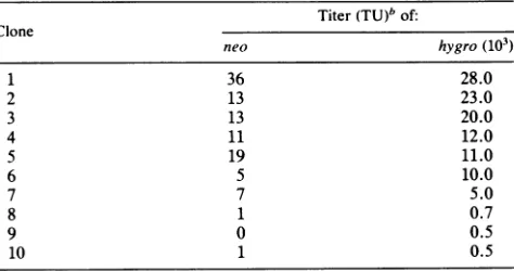
theDdeI restriction site was lost. We suggestTABLE 3. Reversionfrequencyof virusgrown inC321 helpercells"
Titer(TU)" of:
Clone
neo hygro (103)
1 36 28.0
2 13 23.0
3 13 20.0
4 11 12.0
5 19 11.0
6 5 10.0
7 7 5.0
8 1 0.7
9 0 0.5
10 1 0.5
"C321 cell clonescontaining a JD216Neo(Am)Hy provirus were
estab-lished in thesame mannerasthecell linesdescribedin Table1,footnotea,
except that theoriginal transfections were done withpJD216Neo(Am)Hy.
C321helpercell clonesweregrownforapproximately15celldoublingsbefore
viruswasharvested. Virus titersweredeterminedas described in Table2,
footnotea.
"Theoverallneoandhygrotiterswere106and 110.7 x 10³,respectively,
and the reversion frequency was determined as follows: neolhygro =
106/110.7 x
io0
=1 X10-3.on November 10, 2019 by guest
http://jvi.asm.org/
[image:3.612.323.560.527.652.2]1
2
3
MA
264 bpa
B
I
EI
I
G T G T
G + C G + +
t.7 x g
_
~ ~
E~
| 242
bp
GG
W . c
GC D
AA d
C
I...a=-I
T
AG
TA
AA
G
C
Gc
A
T
FIG. 3. Genomic sequencingof neo revertants. (A)Acrylamide
gel ofamplified DNA obtained with three separate neor D17 cell
clonesinfected withvirus from .2G helpercell clones. Mrefers to
molecularweightmarkers(MspI-digested pBR322). (B)
Autoradio-gramsobtained from DNA sequencingof two clones. The bracket
labeledDdeI indicates theposition oftheDdeI sitein the original JD216Neo(Am)Hyvector. InI there wasa 4-bpinsertion, CCGA,
indicated attheleft.
that thehigherreversion frequencyobtained withthe C321 helpercellsistheresultofgeneconversion(see Discussion).
Genomicsequence analysis ofneo revertants by PCR
am-plification. The exactnature ofthe changesgiving the neor phenotype was analyzed by direct sequencing of
PCR-amplifiedgenomicDNAfrom13 revertants(13,16, 25).Two
20-nucleotideoligomers weresynthesized andusedas
prim-ersto amplify a264-bp regionthatspans the ambercodon. Thirtysequentialcyclesofprimer annealing,DNA
polymer-ase extension (with DNA polymerase from Thermus
aqua-ticus), and denaturation were performed. Amplified DNA
was purified in an 8% polyacrylamide gel (Fig. 3A) and
sequencedbytheMaxamandGilbertprotocol(Fig.3B)(12).
Figure 3B shows amplified genomic DNA from three neor
D17 cell clones originally infected with JD216Neo(Am)Hy
virus harvestedfrom .2G helper cell clones. Typically, we
obtained 1to5 ,ugofamplifiedDNAfrom1.5jigof genomic
DNA, indicatingapproximately 108-fold amplification.
We performed this amplification on DNAs from seven
neor D17 cell clones originally infected with JD216Neo
(Am)Hyvirusfrom.2Ghelper cellclonesand onsix DNAs
fromcells originally infected with JD216Neo(Am)Hy virus
from C321 helper cell clones (containing endogenous neo
genes), all of which had lost the DdeIsite (Fig. 2). In all
casesinwhichtheDdeIsitewaslost,base pair4oftheDdeI
site
(corresponding
to base 2 of the ambercodon)
wasconverted from A-Tto
G-C
(Fig.
3B, II).
Two neor D17 cell clones
originally
infected withJD216Neo(Am)Hy
virusfrom.2G helper
cell clones didnot lose their DdeI sites. DNAsfromthesetwocloneswereused astemplates
foramplification
andsequencing.
Thesequence obtainedfor one clone is shown inFig.
3B, I.
Asexpected,
theDdeI site wasmaintained. Therewas nochange
fromT toC.However,
therewas an insertion of fourbases(CCGA)
seven bases downstream from the amber codon. The inser-tion is a
duplication
of theadjacent
CCGA. Thomas andCapecchi
have shown that+1, +4,
+7,
etc., frameshift mutationsjust
downstream of this amber codoncan restore neo function (seeDiscussion) (21).
Sequencing
ofthe other neo revertant that did not lose theDdeI
site confirmed that theDdeI
site was maintained withoutchange,
butwe wereable to find no
compensating
mutations within 40 basessurrounding
the amber codon.DISCUSSION
In this paper we describe the direct measurement of mutationrates
during
retrovirusreplication
for both asingle
base
pair
substitution at a defined locus and insertions in adefinedarea.
Base
pair
substitution mutation rate. The basepair
substi-tution rate that we obtained was 2 x10⁻⁵
perbp
perreplication cycle.
Initially
we thought thatmutations would occur at any of the three bases ofthe amber codon(TAG)
unless they produced another stop codon. However,
ge-nomic
sequencing
ofPCR-amplified
DNA showedthat sub-stitutions were limited to asingle change
atthe second baseofthe amber
codon,
thatbeing
atransition fromA-TtoG-C
which restores theoriginal
wild-type sequence. Thewild-typecodonisTGG, which isthe tryptophancodon. All other
base substitutions at the amber codon would result in
changestocodons fordifferent amino acids. It seems
possi-ble that we were limited tothischange because when the 5' end of the wild-type
neo
gene productis present, there may be an absolute requirement for tryptophan at the locus we were studying. Thus, the value we obtained for the rate of base pair substitution is a minimal one, because there may have been selection against other mutations. Experimentswith other mutations (in progress) will test this hypothesis. The base pair substitution rate is similar to that
deter-mined with cell-free systems (10). Furthermore, the muta-tion rate we observe is similar to that calculated for two other RNA viruses, poliovirus type 1 (less than 10'- for the VP1 gene) and influenza virus (8 x
10-5
for the NS seg-ment), and higher than that for a third, Sindbis virus (2 x10⁻⁷)
(5, 15, 18). These other RNA viruses utilize different types ofpolymerases than doretroviruses.Mutations in our system could have occurred at either the RNAtranscription step or the reverse transcription step. We are not sure at which step the mutations occurred. It is
possible toseparate these two steps physically, which may allow determination of the mutation rate at each step.
Insertion mutation rate. Twoneorevertant D17 cell clones originally infected with JD216Neo(Am)Hy virus from
.2G
helper cells did not lose theDdeI
site. Genomic sequencing with PCR-amplified DNA revealed that one clone retained the DdeI site and the amber codon (Fig. 3B,I),
but there was a 4-bp insertion 7 bp downstream from the amber codon. Thomas and Capecchi have found that +1 frameshift muta-tions within an areaof 11 bp downstream of the amber codon cancompensate for the stop codon (21). Translation can beI
on November 10, 2019 by guest
http://jvi.asm.org/
[image:4.612.62.299.72.385.2]initiated at an AUG in the -1 reading frame upstream of the amber codon, allowing readthrough of the amber codon which is in the 0 reading frame. The + 1 frame shift then allows the ribosome to regain the proper phase. The target size for selectable insertions is about 11 bp downstream from the amber codon (21). Since the only insertions that score as revertants are +1 frameshift mutations, our effective target is the equivalent of a single codon. The insertion mutation rate per codon is 1/17 times the base substitution mutation rate. Therefore, the insertion mutation rate at this site is about
10-7
per bp per replication cycle.The other clone that retained the
DdeI
site also retained the amber codon. We discovered no compensating muta-tions (20 bases 5' or 20 bases 3' to the amber codon) to account for its neor phenotype. It is possible that there is a compensating mutation outside of the area we sequenced or that there is a cellular mechanism by which this cell clone can suppress the amber codon. (We have not been able to recoverneor-transforming
virus by superinfection of cells carrying this provirus, even though the superinfecting virus replicated well [unpublished data]).If the retroviral mutation rates we measured are true for the entire genome, we can roughly estimate how many replication cycles different retrovirus isolates have under-gone since divergence from a cell or other virus isolate. Reticuloendotheliosis virus strain T (REV-T) contains the oncogene v-rel. Comparing v-rel from REV-T coordinates 4290 to 4675 with its proto-oncogene, c-rel, we found that v-rel and c-rel differ by 6 bp and an insertion of 6 bp in v-rel (24). The changes in this area of v-rel are not important for the transforming ability of v-rel (20), and so we consider them silent. Therefore, from the time the c-rel sequence was transduced into REV-T until the REV-T provirus was cloned (2), REV-T underwent approximately 800 replication cycles (number of replication cycles = 6 bp substitutions/2 x
10-5
substitutions per bp per replication cycle x 385 bp). A similar calculation can be made for other retroviruses, if we assume that the mutation rates we measured also apply to them and the observed differences in nucleotide sequence are primarily silent. Thus, we can calculate that the observed substitution rate of lentiviruses (visna and human immuno-deficiency viruses) of approximately
10-3
per nucleotide per year (1, 26) indicates approximately 50 replication cycles per year since the divergence of different isolates.Gene conversion. We also used the procedure describedin
Fig. 1A with C321 helper cells, which contain multiple wild-type copies of neo in pSV2neo. The reversion fre-quency that we obtained with C321 cells was
10-3,
which is 50 times higher than that obtained with .2G helper cells, which do not contain endogenous copies ofneo. Since there is no other difference between C321 and .2G cells, we suggest that the higher reversion frequency is the result of gene conversion which occurred either during growth ofthe C321 helper cells containing the JD216Neo(Am)Hy provi-ruses or during retrovirus replication or both. It is also possible that this high reversion frequency was a result ofrecombination. However, because the wild-type neo RNA contains no retrovirus sequences, it would only be packaged at a very low frequency, and a double crossover would be necessary to recover a wild-type neo virus. As expected, all revertants obtained with virus from C321 cells contained the wild-type sequence.
ACKNOWLEDGMENTS
We thank S. Coe, J. Couch, S. Hinz, J. Rein,and R. Wisniewski for technical assistance; K. Thomas and M. Capecchi for a gift of
pRH4-14/TK; H. S. Kim and 0. Smithies for help with PCR amplification; Tom Leonard for help with the chi-square goodness-of-fit test; and RalphDornburg,Celine Gelinas, Wei-Shau Hu, Mark Hannink, Antonito Panganiban, and Bill Sugden for helpful com-ments on the manuscript.
This research was supported by Public Health Service research grants CA-22443 and CA-07175 from the National Institutes of Health. J.P.D. was supported by Public Health Service research award CA-09075 from the National Institutes of Health. H.M.T. is an American Cancer Society research professor.
LITERATURE CITED
1. Braun, M. J., J. E. Clements, and M. A. Gonda. 1987. The visna virus genome: evidence for a hypervariable site in the env gene andsequence homology amonglentivirusenvelopeproteins. J. Virol. 61:4046-4054.
2. Chen,I.S. Y., T. W. Mak, J. J. O'Rear, and H. M. Temin. 1981. Characterization of reticuloendotheliosis virus strain T DNA and isolation of a novel variant of reticuloendotheliosis virus strain Tbymolecular cloning. J. Virol. 40:800-811.
3. Dougherty, J. P., and H. M. Temin. 1987. Highmutationrate of a spleen necrosis virus-based retrovirus vector. Mol. Cell. Biol. 6:4387-4395.
4. Dougherty, J. P., and H. M. Temin. 1987. Determination of retroviral vector mutation rates and a promoterless retroviral vector, p. 18-23. In J. H. Miller and M. P. Calos (ed.), Gene transfer vectors for mammalian cells. Cold Spring Harbor Laboratory, Cold SpringHarbor, N.Y.
5. Durbin, R. K., andV. Stollar. 1986. Sequence analysisof the E2 gene of a hyperglycosylated, hostrestricted mutant of Sindbis virus and estimation of mutation rate fromfrequency of rever-tants. Virology 154:135-143.
6. Folger, K. R., K. Thomas, and M. R. Capecchi. 1985. Nonre-ciprocal exchanges of information between DNA duplexes coinjected into mammalian cell nuclei.Mol. Cell. Biol. 5:59-69. 7. Graham, F. L., and A. J. van der Eb.1973. A newtechniquefor the assayof infectivity of humanadenovirus 5 DNA. Virology 52:456-467.
8. Gritz, L., and J. Davies. 1983. Plasmid-encoded hygromycin B resistance: the sequence ofhygromycin B phosphotransferase geneandits expression. Gene25:179-188.
9. Jorgensen, R. A., S. J. Rothstein, andW. S. Reznikoff. 1979. A restriction enzyme cleavage mapofTn5 and location of aregion encoding neomycinresistance. Mol. Gen. Genet. 177:65-72. 10. Kunkel, T. A., F. Eckstein, A. S. Mildvan, R. M. Koplitz, and
L. A. Loeb. 1981. Deoxynucleoside [1-thio]triphosphates pre-vent proofreading during in vitro DNA synthesis. Proc. Natl. Acad. Sci. USA78:6734-6738.
11. Maniatis, T., E. F. Fritsch,and J. Sambrook. 1982. Molecular cloning: a laboratory manual. Cold Spring HarborLaboratory, N.Y.
12. Maxam, A. M., and W. Gilbert. 1980. Sequencing end-labeled DNA with base-specificchemical cleavages. MethodsEnzymol. 65:499-560.
13. McMahon, G., E. Davis,and G. N.Wogan. 1987. Characteriza-tion of c-Ki-rasoncogene alleles by direct sequencingof enzy-matically amplified DNA from carcinogen-induced tumors. Proc. Natl. Acad. Sci. USA 84:4974-4978.
14. Miller, C. K., J. E. Embretson, and H. M. Temin. 1988. Transforming viruses spontaneously arise from nontransform-ing reticuloendotheliosis virus strain T-derived viruses as a result of increased accumulation ofspliced viral RNA. J. Virol. 62:1219-1226.
15. Parvin, J. D., A. Moscona, W. T. Pan, J. M. Leider, and P. Palese. 1986. Measurement of the mutation rates of animal viruses: influenza A virus and poliovirus type 1. J. Virol. 59: 377-383.
16. Saiki, R. K., S. J. Scharf,F. Faloona,K. B. Mullis,G. T.Horn, H. Erlich, and N. Arnheim. 1985. Enzymatic amplification of beta-globin genomic sequences and restriction siteanalysis for diagnosis of sickle cell anemia. Science230:1350-1354. 17. Simonsen, C. C., and A. D. Levinson. 1983. Isolation and
expression of an altered mouse dihydrofolate reductase cDNA.
on November 10, 2019 by guest
http://jvi.asm.org/
Proc.Natl. Acad. Sci. USA80:2495-2499.
18. Smith, D. B., and S. C. Inglis. 1987. The mutation rate and variability of eukaryotic viruses: ananalytical review.J. Gen.
Virol.68:2729-2740.
19. Southern P. J., and P. Berg. 1982.Transformation of
mamma-lian cells to antibiotic resistance with a bacterial gene under
control of theSV40 early regionpromoter.J.Mol. Appl. Genet. 1:327-341.
20. Sylla, B. S.,and H. M.Temin.1986. Activation ofoncogenicity ofthe c-relproto-oncogene.Mol. Cell. Biol. 6:4709-4716. 21. Thomas, K. R., and M. R. Capecchi. 1986. Introduction of
homologous DNA sequences into mammalian cells induces
mutations inthecognate gene.Nature (London)324:34-38. 22. Watanabe, S., and H. M.Temin. 1983. Construction ofahelper
cellline for avian reticuloendotheliosis virus cloning vectors. Mol. Cell.Biol. 3:2241-2249.
23. Wigler, M.,R.Sweet,G. K.Sim,B.Wold, E. Lacy,T.Maniatis, S.Silverstein,and R.Axel.1979. Transformation of mammalian cells withgenes fromprocaryotes to eucaryotes. Cell 16:777-785.
24. Wilhelmsen, K. C., K. Eggleton, and H. M. Temin. 1984. Nucleic acid sequencesof the oncogenev-rel in reticuloendo-theliosis virus strain T and its cellular homolog, the
proto-oncogenec-rel. J.Virol. 52:172-182.
25. Wong, C., C. E. Dowling, R. K. Saiki, R. G. Higuchi, H. A. Erlich, and H. H. Kazazian. 1987. Characterization of beta-thalassaemia mutations using direct genomic sequencing of amplified singlecopyDNA. Nature(London) 330:384-386. 26. Yokoyama, S.,and T. J. Gojobori. 1987. Molecular evolution
andphylogeny of the human AIDS viruses LAV, HTLV-III, and ARV.J. Mol. Evol. 24:330-336.