JOURNAL OFVIROLOGY, May1987, p. 1712-1716 Vol.61, No. 5 0022-538X/87/051712-05$02.00/0
Copyright©D 1987,American Society forMicrobiology
Expression of
Polyomavirus
Large T
Antigen by Using
a
Baculovirus Vector
WILLIAMC.
RICE,1t
HEATHER E.LORIMER,2
CAROLPRIVES,2AND LOIS K.MILLER'3*
Department
of Bacteriology and
Biochemistry, University of Idaho, Moscow,
Idaho838431;
Department of
BiologicalSciences,
Columbia
University, NewYork,
New York100272;
andDepartments of Entomology
andGenetics,
Universityof Georgia, Athens, Georgia
306023*Received 16 October1986/Accepted 4 February 1987
Ageneencoding the large T antigen of
polyomavirus
wasinserted intothe baculovirusAutographa
californica
nuclear polyhedrosis virus so that gene
expression
wasunder the control of thestrong,
very late polyhedrin gene promoter.Singnificantly
morelarge
Tantigen
wasproduced
in recombinant virus-infected insect cells than was observed inpolyomavirus-transformed
mouse cells. The insect-derived Tantigen
exhibitedpolyomavirus origin-specific
DNAbinding.
Thebaculovirusexpression system
provides
aconvenientsourceof T antigen for in vitro studies.Polyomavirus
large Tantigen (PyTAg) is
aspecific
DNA-binding
protein
(3-5, 24)which is involved in
theregulation
of viral
geneexpression (6,
8),
theinitiation of
viral DNAreplication
(7, 20), and theoncogenic transformation
ofprimary
ratembryo cells (23).
Theprotein is
present inlow
quantities in both lytically
infected
andtransformedcells.
Toobtain
higher quantities of the
protein
for
invitro
studies,
helper-independent adenovirus
vector systems havebeen
developed which produce approximately fivefold
more PyTAgthan do wild-type
polyomavirus-infected
cells (13, 14).A
helper-independent baculovirus
vectorsystem has beendeveloped for
thehigh-level expression of foreign
genes in aeucaryotic cell environment
(11,16-19,
22,27).
It wasof
interest
todetermine
whetherthis insect
virus vector system canefficiently
expressbiologically active PyTAg.
We now report that abaculovirus
expression
system iscapable of
providing sufficient quantities of biologically active
Tantigen
for in vitro studies.
A
recombinant baculovirus
expressing
the PyTAg gene wasconstructed
by
allelic replacement
of wild-typebaculovirus polyhedrin
gene sequences with sequences of arecombinant
plasmid,pEV51LT,
whichwas constructed asoutlined in
Fig.
1.Anintronless
constructof
thePyTAg gene was obtained from plasmid pspLT5 (28) byBamHI
andXhoI
digestion, purified
by gelelectrophoresis, and inserted into KpnI- andXhoI-digested
pEV-51 (16), a plasmid whichfacilitates
theinsertion
offoreign genes into the polyhedrinregion
ofthe
baculovirus Autographa californica nuclearpolyhedrosis virus
(AcNPV). Cotransfection of the resultingplasmid
pEV51LT
with wild-type AcNPV DNA into apermissive
host cell line, Spodopterafrugiperda
IPLB-SF21,
resulted in allelic replacement of the wild-typepolyhedrin
gene of AcNPV with the PyTAg gene under the controlof
the abundantly expressed polyhedrin promoter.Recombinant virus plaques were visually selected by their
occlusion-negative
phenotype (16, 25), resulting frompolyhedrin
replacement with the PyTAg gene region of pspLT5. A stock ofrecombinant virus was developed, andthe
viral
DNA was isolated as previously described (16). The*Corresponding author.
tPresent address: U.S. Department of Agriculture, Columbia, MO 65205.
nature of the recombinant virus DNA was confirmed
by
digestion withEcoRI, PstI, and EcoRI-PvuII and by South-ernblottingwith labeled pEV51LTasahybridization
probe.
Therestriction map of the EcoRI-Iregion of the recombinant virus and the predicted nucleotide sequence of the leader region of the resulting fusion are shown at the bottom of
Fig.
1. The BglII site of pEV-51 is positioned within the polyhedrin leader sequence 22 base pairs upstream of the polyhedrin translational start site (approximately 33 base pairs downstream of the 5' end of the transcriptional start
site),
and the XhoI site of thePyTAg
gene is 24 base pairs upstream of theinitiating
ATGofPyTAg.
The recombinant AcNPVis heretofore referred to as vEV51LT.To monitor thesynthesis of PyTAg in vEV51LT-infected cells, S. frugiperda cells were infected with either
wild-type
orrecombinant virus and
pulse-labeled
with[35S]methionine
at various times
postinfection (p.i.).
Proteins frominfected cell lysates were analyzed by sodium dodecyl sulfate-polyacrylamide gel electrophoresis (SDS-PAGE). Theaccu-mulation
ofpolyhedrin
protein is visible inwild-type-infected cells
beginning
18h
p.i.
by Coomassie brilliant
bluestaining
of thegels (data
notshown).
Increasedsynthesis ofthis protein,
beginning
at 18 hand
extending through
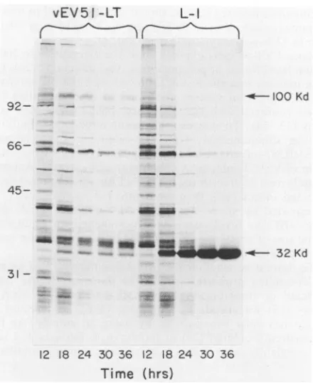
36 h p.i., is observed by use ofautoradiography (Fig. 2). This is a normal pattern forpolyhedrin synthesis (22).
Polyhedrinsynthesis
is not observed invEV51LT-infected cells
(Fig.
2); anotherproteinexpressed
earlier in infectioncomigrates
in the32-kilodalton
(kDa) region of the
gel (22). A 100-kDaprotein is observed in
both wild-type
lanes and vEV51LT lanes at 12 hp.i.
by
autoradiography (Fig.
2), but a strongersignal
is observed in the vEV51LT lanes from 18through 36 hp.i.
Thisobservation is consistent
with the interpretation that PyTAgsynthesis begins
at 18 h in vEV51LT-infected cells andcomigrates with
anotherprotein ofsimilar
size that decreases after 18 h.Unlike
polyhedrinsynthesis
in wild-type (L-1)-infected cells, the 100-kDa protein does not accumulatethrough
36 hp.i., asindicated by the absence ofsignificant Coomassie
blue staining in this region (data notshown).
In contrast to the expression usually noted forforeign
genesexpressed
underpolyhedrin promotercontrol,
theamountof
100-kDa proteinsynthesized
during the pulse ismaximal
at 18 h p.i. and declines through 36 h invEV51LT-infected
cells (Fig. 2).1712
on November 10, 2019 by guest
http://jvi.asm.org/
NOTES 1713
vEV51 -LT
L-1
Q100
Kd92-
-66- =__ -SMIND
45--- Mr m m
a mIWo MMa "
--- 32 Kd
31
-5'
ATAAATAAGTTTTCGTAACAGTTTTAGA1CTCGAQ
TTTCAGCCTCACCACCATC7TG
Polyhedrinleader 8 Xh()I -e--PolyomaT-ogleader
_-EcoRI EcoRYr EcoRI
Ec PVU Xhol SalI Pvu PvuI31I
I
I1,
i
3
I-ni9.l7
!P
2 3 4 5 7 9
[image:2.612.65.296.63.434.2]i IKb
FIG. 1. Construction of the transplacement plasmid pEV51LT,
which containsthe PyTAggeneunder AcNPV polyhedrinpromoter
control, and a map of the EcoRI-I fragment of the recombinant
baculovirus vEV51LTcontaining thePyTAggene.ThePyTAggene
()wasremoved fromplasmidpspLT5(28) and inserted(12)into
themulticloning site(MCS)ofpEV-51 (16). Theresultingplasmid, pEV51LT, containingthePyTAggenein theappropriateorientation and position with respect to polyhedrin promoter control, was
selected withampicillin intransformed Escherichia coli JM83. To
facilitate allelicreplacement of pEV51LT sequenceswith the
cor-responding regions of homology in the AcNPV genome (m2), pEV51LT was digested with
Sfi1
and Notl before cotransfectionwith wild-type AcNPV DNAintoS.frugiperda cells(16). Plaques
withocclusion-negative phenotypes were selected and replaqued,
and virus stocks were prepared. The restriction pattern of the
recombinant AcNPV EcoRI-Ifragment (bottomoffigure) andthe
predictednucleotidesequenceatthe XhoI fusionsitearepresented.
Kb, Kilobasepairs.
Correspondence of the 100-kDa protein with PyTAg is shown by immuno
precipitation
of the protein from vEV51LT-infected butnotfrom wild-type-infected24-hcell lysates by using apolyclonal anti-PyTAg antibody (Fig. 3).Celllysateswerepreparedfrommonolayersof S.
frugiperda
cells (1.5 x
107
cells per 1,000-mm-diameter dish) whichwere infected at a multiplicity of infection of 20 with vEV51LT or wild-type (L-1) virus and pulse-labeled with [35S]methionine (22) at24h p.i. The mediumwasremoved
12 18 24 30 36 12 18 24 30 36
Time
(hrs)
FIG. 2. Autoradiogram of proteins synthesized in recombinant and wild-type virus-infected S. frugiperda cells. Cells (108 per 35-mm-diameterdish) were infected (20 PFU per cell) with either recombinantvEV51LTor wild-type (L-1) AcNPV andpulse-labeled with[35S]methionineat selected times p.i. as previously described (22). The cells were harvested, pelleted, and suspended in water, and an equal volume of solubilization solution (2% SDS-2% 2-mercaptoethanol-1 M urea) was added. The proteins were separated bySDS-PAGE (10) and visualized by autoradiography. Molecular-weight (103) markers are indicated on the left; PyTAg (100 kDa) and the
polyhedrin
protein (32 kDa) are indicated on theright.
from
thedishes,
thedishes
wereplaced
onice,
and the cells were washed twice with 5 ml of coldphosphate-buffered
saline per
plate.
A0.75-mlsample of
lysis buffer (50
mMTris[pH
8]-150
mMNaCl-1% Nonidet P-40-0.1% aprotinin
[Sigma
ChemicalCo.])
was distributed over the cellsur-faces.
Thecells
werescraped into cold
microcentrifuge
tubes and
kept
at0°C for
30 min. Thetubes
werecentrifuged
at
15,000
x gfor
2min, and
thesupernatants
(cell extracts)
were
stored
inliquid
N2 until use. Extracts wereincubated
with anti-PyTAg
antiserum, and antibody-antigen complexes
were
precipitated, washed,
andsuspended
aspreviously
described
(22).
To
quantitate
the amount ofPyTAg
made invEV51LT-infected
cells, PyTAgs from
lysates
of apolyomavirus-transformed
mouse cellline, PYT-54,
and vEV51LT-infected S.frugiperda
cellscollected
24 hp.i.
wereimmunoprecipitated
with
amonoclonal
antibody.
Theim-munoprecipitated
proteins
wereseparated
by
SDS-PAGE andvisualized
by
silverstaining
(Fig. 4A).
On the basisof Lowryprotein determinations
andquantitation
ofsilver-stained
PyTAg
bandsonSDS-polyacrylamide gels,
approx-imately 10-fold
morePyTAg
wasproduced
permilligram
of infected insectcell
extract than PYT-54 cell extract. The amountof
PyTAg
produced
in the recombinant virus-infected cellscorresponds
toapproximately
15,ug/107
in-fected S.frugiperda
cells. The size ofPyTAg produced
inVOL.61, 1987
on November 10, 2019 by guest
http://jvi.asm.org/
[image:2.612.329.549.65.334.2]1714 NOTES
baculovirus-infected cells is similar ifnotidenticaltothat of mammalian cell-derived PyTAg, onthe basis of their migra-tion in SDS-polyacrylamide gels (Fig. 4).
Since PYT-54 cells expresslarge Tantigen at70 to 100% of the level foundinpolyomavirus lytic infections (24), the dataindicate that the levels achieved with the baculovirus-based expression system are equal to or higher than the levels achieved with the adenovirus-based expression sys-tems (13, 14). To address this point directly, we purified PyTAg
simultaneously
from the adenovirus recombinant AdSVR587-infected CV1(monkey kidney)cell extracts and fromvEV51LT-infected S. frugiperdacell extracts. Approx-imately two to five times more PyTAg was obtained frominfected
insect cells than from infectedmonkey cells,
as determined by silver staining ofSDS-polyacrylamide
gels(Fig.
4B); the levels were then normalized for equivalent quantities of protein, asdeterminedbyLowry protein anal-yses of initial cellextracts.To determine whether the insect cell-derived
PyTAg
has DNA-bindingproperties
similar to those ofPyTAg
frominfected
ortransformed
mousecells,
a modified McKay assay (15) was used. PyTAgfrom vEV51LT-infected
S. frugiperda cells was purified by using an anti-PyTAgim-munoaffinity
column (20)of monoclonal
antibody
F-4 (21, 24)covalently cross-linked
toanti-mouse
immunoglobulin
GvEV51LT
L-1
d
IGI
I0
Kd -. -'U_
-
--
spolyhedrin
FIG. 3. Immunoprecipitation ofPyTAgfrom vEV51LT-infected
cells.Immunoprecipitatedproteins(I)from[35S]methionine-labeled
vEV55LT-orwild-type(L-1)-infected cell extracts (seetext) were
comparedwithtotalproteins(C) in the cellextractsbySDS-PAGE.
Anautoradiogram of theresulting gel is presented;the positions of
thepolyhedrin and PyTAg(100kDa) proteinsarenoted.
(2b m
93v-N
- *N
.4
-4 [image:3.612.319.553.66.227.2] [image:3.612.73.285.327.660.2]..A.
...xi--1
a
00 M.-:I,:A~-93
-65
-46
B
FIG. 4. Polyacrylamide gel analysis of immunoprecipitated PyTAg from vEV51LT-infected cells, polyomavirus-transformed PYT-54cells, and AdSVR587-infected CV1 cells. (A) PYT-54cell
extract (2 mg) wasprecipitated with monoclonal control pab419T
(lane a) or with anti-PyTAg monoclonal antibody F-4 (lane b).
Extracts(0.8 mg) of S. frugiperda cells infected with wild-type (lane
c)orrecombinant(lane d) AcNPVwerepreparedasdescribed inthe
legendtoFig. 3andwereprecipitated with antibody F-4. Thecrude
extract of vEV51LT-infected cells (lane e) is also shown. (B)
Extracts of wild-type-infected S. frugiperda cells (lane a),
vEV55LT-infected S.frugiperda cells (lane b), and AdSVR587-infected CV1 cells (lane c) were immunoprecipitated with F-4 antibody andanalyzed by SDS-PAGE. In both panels, proteinbands
were visualized by silver staining; the arrowheads indicate the
position of PyTAg. Proteinsize markers(lanes m) of 92.5, 65, and 46
kDa areindicated.
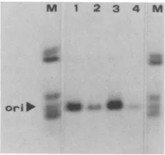
agarose beads. Fourimmunopurified PyTAg fractions were incubated with 32P-labeled, HinfI-digested p37.3A2 DNA,a plasmid which contains the entire polyomavirus genome inserted into the plasmid pAT153 (3); Hinfl digestion pro-duces 23 fragments ranging in size from 1,801 to 18 base pairs. The nature of the DNA in PyTAg-DNA complexes precipitated with F-4monoclonal antibody wasdetermined by gel electrophoresis. Specific precipitation of the 604-base-pairHinflfragment containing the origin of replication of the polyomavirus genome wasobserved by autoradiography of gelscontaining theimmunoprecipitated DNA (Fig. 5). Thus, the insect-derived PyTAg possesses specific DNA-binding properties.
Insect baculovirus expression vector systems have been shown to produce moderate to high levels of a variety of differentforeigngenes(1,9, 11, 16, 22, 26,27). TheAcNPV vector system can produce low quantities of PyTAg. Nev-ertheless, the levels of PyTAg produced in vEV51LT-infected insect cells are significantly higher than those ob-tained in polyomavirus-infected cells or -transformed cells. Baculovirus-mediated PyTAg expression compares favor-ably with
adenovirus-mediated
expression (13). The helper-independent vEV51LT is easily propagated, reaches high titers in themediaofinfected cells (>108 PFU per ml),and is stable. A safety feature of the baculovirus expression system is the barrier to entry andexpression of baculovirus genesin mammalian cells (2).The AcNPV expression system, using the polyhedrin promoter, usually produces much higher levels of foreign gene products (1, 9, 16, 22, 26, 27) than those we have observed forPyTAg. The low level of expression of PyTAg may be related to the observation that synthesis of the protein appears to decrease from 18 to 36 h p.i., whereas J. VIROL.
on November 10, 2019 by guest
http://jvi.asm.org/
M 1 2 3 4 M
ori' ***
FIG. 5. Specific binding of immunoaffinity-purified baculovirus-derived PyTAg to the polyomavirus DNA origin of replication. PyTAg from infected cells was purified from cellular extracts by
immunoaffinity
column chromatography by using goat anti-mouse agarose coupled to F-4monoclonal antibody as previously described (20). Samples were tested for specific polyomavirus DNA binding in a nitrocellulose filter-binding assay (see below). Peak fractions of HS and EG eluates containing greater(EG1, HS1) and lesser (EG2,HS2)
DNA-binding activity were pooled, dialyzed (20), and stored at-20°C.
The DNA-binding immunoassay was a modification of procedures described by Cowie and Kamen (3) in whichimmunoaf-finity-purified
PyTAg (5 p.1) was incubated with 20 ng of32P-labeledHinfl-digested
p37-3A2
DNA at room temperature for 1 h, followed by incubation with F-4 monoclonal anti-PyTAg antibody (30min). Immune complexes were precipitated with immunoglobulin G sorb-ent. The DNA fragments were released and analyzed on agarose gels as previously described (24). Lanes 1 to 4 are DNA fragments bound by immunopurified fractions of PyTAgHS1, HS2, EG1, and EG2, respectively. Outer lanes (M) are total 32P-labeled Hinfl fragments of p37.3A2. The fragment containing the polyomavirus DNAreplication
origin (ori) is indicated.expression of proteins under
polyhedrin
control generally increases and accumulates during this time (22). It is possible that the T antigen is exerting some negative effect on virus expression. A contributing factor to lowexpression may be the useof the pEV-51 vectorinstead of pEV-55 (16); pEV-51 lacks 22 base pairs of the polyhedrin mRNA leader, whereas pEV-55includes the entire leader (16).The PyTAg produced inbaculovirus-infected insect cells is biologicallyactive with respect toits origin-specific DNA-binding properties. Ongoing experiments show that the PyTAgproduced in this systemis active in initiating DNA replication in vitro and possesses ATPase activity (Y. Murikami, C.
Priv'es,
and J. Hurwitz, work in progress). Since T antigens undergo extensive posttranslational modi-fications in mammalian cells, it will beinteresting to deter-minewhat types of posttranslationalmodifications are pres-entin insect-derived protein and to correlate modifications with the numerousfunctionsof this complexprotein.Plasmids pEV51, pspLT5, and p37.3A2 were kindly provided by D. W. Miller and R. Kamen, Genetics Institute, Cambridge, Mass. Monoclonal anti-PyTAg antibody-producing cell line F-4 was a gift of Ed Harlow.
This research was supported in part by Public Health Service grantAl 23719 (to L.K.M.) from the National Institute of Allergy and Infectious
Diseases,
by institutional grant IN-119G (to W.C.R.) from the American Cancer Society, and by Public Health Service grant CA26905 (to C.P.) from the National Institutes ofHealth.LITERATURE CITED
1. Baum, J. A., R. Geever, and N. H. Giles. 1987. Expression of qa-1Factivator protein: identification of upstreambinding sites in the qa gene cluster and localization of the DNA-binding domain. Mol. Cell. Biol. 7:1256-1266.
2. Carboneil, L. F., M. J. Klowden, and L. K. Miller. 1985. Baculovirus-mediated expression ofbacterial genesindipteran
andmammalian cells. J. Virol. 56:153-160.
3. Cowie, A., and R. Kamen. 1984. Multiple binding sites for polyomavirus large T antigen within regulatory sequences of polyomavirusDNA. J. Virol.52:750-760.
4. Cowie, A., and R. Kamen. 1986. Guanine nucleotide contacts within viral DNA sequences bound by polyomavirus large T antigen. J. Virol.57:505-514.
5. Dilworth,S.M.,A.Cowie, R. Kamen, and B. E.Griffin. 1984.
DNAbinding activity ofpolyomavirus large T antigen. Proc. Natl. Acad. Sci. USA81:1941-1945.
6. Farmier, W. G., and W. R. Folk. 1984.Regulation of polyoma-virustranscription by large tumor antigen. Proc. Natl. Acad. Sci.USA81:6919-6922.
7. Franke, B., and W. Eckhart. 1973. Polyoma gene function requiredfor viralDNAsynthesis. Virology55:127-136. 8. Kamen, R., P.Jat, R.Treisman, J. Favaloro, andW. R. Folk.
1982. 5'Termini ofpolyomavirus early region transcripts syn-thesized in vivo by wild-type virus and viable deletionmutants.
J. Mol. Biol. 159:189-224.
9. Kuroda, K., C. Hauser, R. Rott, H.-D.Klenk,and W. Doerfler. 1986.Expression of theinfluenza virushaemagglutininin insect
cells by abaculovirus vector. EMBO J. 5:1359-1365.
10. Laemmli,U.K. 1970.Cleavage of structuralproteins duringthe assembly of the head ofbacteriophage T4. Nature (London) 227:680-685.
11. Maeda, S., T. Kawai, M. Obinata, H. Fujiwara,T.Horiuchi,Y.
Saeki, Y. Sato, and M. Furusawa. 1985. Production of human alpha-interferon in silkworm using a baculovirusvector. Nature
(London) 315:592-594.
12. Maniatis, T., E. F.Fritsch, andJ. Sambrook. 1982. Molecular cloning: a laboratory manual. Cold SpringHarbor
Laboratory,
Cold Spring Harbor, N.Y.13. Mansour, S. L., T. Grodzicker, and R. Tjian. 1985. An adeno-virus vector system used to express polyoma virus tumor
antigens. Proc. Natl. Acad. Sci. USA 82:1359-1363.
14. Massie, B., Y. Gluzman, and J.A.Hassell.1986. Construction of a helper-free recombinant adenovirus that expresses
polyoma-virus large T antigen. Mol. Cell. Biol. 6:2872-2883.15. McKay, R. D. G. 1981. Binding ofanSV40 T
antigen-related
protein to DNA. J. Mol. Biol. 145:471-488.16. Miller, D. W., P. Safer, and L. K. Miller. 1986. An insect baculovirus host-vector system for
high-level
expression
of foreign genes, p. 277-298. In J. K. Setlow and A. Hollander (ed.), Genetic engineering, vol. 8. Principles and methods. Plenum Publishing Corp., NewYork.17. Miller, L. K. 1981. A virus vector for
genetic
engineering
in invertebrates, p. 203-224. In N. J. Panopolous(ed.),
Genetic engineering in the plant sciences.PraegerPublishersNewYork. 18. Miller, L. K., D. W. Miller,and M. J.Adang. 1983. Aninsect virus for genetic engineering: developing baculoviruspoly-hedrinsubstitution vectors, p. 89-97.In P. F.
Lurquin
andA.Kleinhofs (ed.), Genetic engineering in
eukaryotes.
Plenum Publishing Corp., New York.19.Miyamoto, C., G. E. Smith, J. Farrell-Towt, R.
Chizzonite,
M. D.Summers, andG.Ju. 1985. Production ofhumanc-mycprotein in insect cells infected with a baculovirus
expression
vector. Mol. Cell. Biol.5:2860-2865.
20. Murakami, Y., T. Eki,M. Yamada, C.Prives, andJ. Hurwitz. 1986. In vitro synthesis ofDNA
containing
thepolyoma
virusreplication origin and requiring the
polyoma
Tantigen.
Proc. Natl.Acad. Sci. USA83:63474351.21. Pallas, D. C., C. Schley,M. Mahoney,E.
Harlow,
B.S. Schaff-hausen, and T. M. Roberts. 1986.Polyomavirus
smalltantigen:
overproduction in bacteria, purification, and utilization for monoclonal and polyclonal
antibody
production.
J. Virol. 60:1075-1084.
22. Pennock, G. D., C.Shoemaker, and L. K.Miller. 1984.
Strong
andregulated expressionof Escherichia coli,B-galactosidase
in insect cells with a baculovirus vector. Mol. Cell. Biol. 4: 399-406.23. Rassoulzadegan, M., Z.
Naghashfar,
A.Cowie,
A. Carr, M.Grisoni,R.Kamen,and F.Cuzin. 1983. Expressionof thelarge T protein of polyoma virus promotes the establishment in
on November 10, 2019 by guest
http://jvi.asm.org/
[image:4.612.122.238.69.178.2]1716 NOTES
culture of "normal" rodent fibroblast cell lines. Proc. Natl.
Acad. Sci. USA80:4354-4358.
24. Scheller, A.,andC. Prives. 1985.Simian virus 40 and
polyoma-virus large tumor antigens have different requirements for
high-affinity sequence-specific DNA binding. J. Virol. 54:
532-545.
25. Smith, G.E., M. J. Fraser, and M. D.Summers. 1983.
Molec-ular engineering of the Autographa californica nuclear
poly-hedrosis virusgenome:deletion mutations within the polyhedrin
gene. J.Virol. 46:584-593.
26. Smith, G. E., G.Ju, B. L.Ericson,J.Moschera, H.-W. Lahm,
R. Chizzonite, and M. D. Summers. 1985. Modification and secretion of human interleukin 2produced in insect cells bya
baculovirus expressionvector.Proc. Natl. Acad. Sci. USA 82: 8404-8408.
27. Smith,G.E.,M.D.Summers,and M.J.Fraser.1983. Produc-tion of human beta interferon in insect cells infected with a
baculovirus expressionvector. Mol. Cell. Biol.3:2156-2165.
28. Zhu, Z.,G. M.Veldman,A.Cowie, A. Carr, B.Schaffhausen,
and R.Kamen. 1984.Construction and functional
characteriza-tion ofpolyomavirusgenomesthat separatelyencode the three
early proteins. J.Virol. 51:170-180.
J. VIROL.