Copyright ©) 1992, American Society for Microbiology
Mutations in the SH3 Domain of the
src
Oncogene
Which
Decrease
Association of Phosphatidylinositol 3'-Kinase Activity with
pp60v-SrC
and
Alter Cellular Morphology
DAVIDS. WAGES, JEFFREY KEEFER, THOMAS B. RALL, AND MICHAEL J. WEBER* Departmentof Microbiology, Health Sciences Center, UniversityofVirginia, Charlottesville, Virginia 22908
Received 20 August 1991/Accepted 20December 1991
To analyze thesignalingpathways utilized in malignant transformation bypp6Ov.sc,wehave isolated and
characterizedsrcmutantswhichpossessnormal levels of protein tyrosinekinase
activity
but whichcauseonly apartially transformed phenotype. Our hypothesis isthat such mutantsare partially defective fortransfor-mation because they are defective in their ability to activate specific components of the cellular signaling machinery while still activating others. In this communication,wereport onthe molecular and biochemical
characterization ofonesuchmutant,CU12 (D.D.Anderson,R. P. Beckmann, E. H. Harms, K. Nakamura, and M. J. Weber, J. Virol. 37:455-458, 1981). Cells infected with this mutant are capable of
anchorage-independent growth, but rather than exhibiting the rounded and refractile morphology characteristic of wild-type-infected cells, they display an extremely elongated, fusiform morphology. The morphological
properties of thismutantsrccould be accounted forentirelybyasingle mutationin theSH3domain(lysine106 toglutamate).Other mutationswereconstructed in thisregion byinvitromutagenesis, bothina v-srcandin anactivatedc-srcbackground,and several ofthem also inducedafusiformmorphology. Allofthe mutations inducing fusiform morphology also resulted in decreased association of pp6Osc with phosphatidylinositol 3'-kinaseactivity.Inaddition,association ofpp6G"" withsometyrosine-phosphorylated proteinswasaltered.
Weproposethat the SH3 domain participates (alongwith the SH2 domain)inthe interaction ofpp6Os'cwith cellularsignaling proteins,andwespeculatethat the association withphosphatidylinositol 3'-kinase playsan
importantrole in theregulation of cellular morphology.
pp60-SrC,theproductof thesrconcogene,isanonreceptor
protein tyrosine kinase whoseenzymatic activityis essential to its ability to transform cells (reviewed in reference 52). Despiteconsiderable effortoverthepast10years,knowledge isfragmentary concerning thesignaling pathways utilized in cell transformation by pp6Ovsrc. It is clear thatthe tyrosine kinase activity and membrane association of pp60osrc are
bothnecessaryfortransformation,but neither is sufficient. In
no case has phosphorylation ofa specific cellular substrate proteinbeenproventobe essential for transformation.
Oneapproach toidentifying potential functionally signifi-cant substrates for pp60vsrc utilizessrc mutantswhich are
completelyorpartiallydefective intransforming ability (2, 3,
14, 37-39, 57, 58, 67, 68). In this approach, tyrosine phos-phorylation of individual proteins is correlated withspecific manifestations oftransformation, with theexpectation that proteinswhosephosphorylationis associated with transfor-mationmaybecausallyinvolved inoncogenesis.The
recep-tor for insulinlike growth factor I (46, 47) and a 120-kDa
protein (46)were identified as candidate functional targets forpp6ovsrcin thisway,andsimilarlythetyrosine
phosphor-ylations of calpactin (14, 50) and vinculin (4, 43, 44) were
shownnottobenecessary for transformation.
Theuseofsrcmutants toanalyzetransformation also has helped elucidate functional domains ofpp6Ovsrc whichare
importantfor substrate recognitionand transformation (52). The amino terminus ofpp60ovs? beginswithamyristylation
signal sequence, andmyristylation is necessaryfor
appro-priatemembrane association ofpp60vsrcand for transforma-tion (40, 41). Followingthemyristylation signalsequence is
*Corresponding author.
a unique domain of approximately 70 amino acids, whose
function is unknown. The SH3 domain extends from resi-dues85to137ofpp6Qsrc, and similar SH3 domainsarefound in othernonreceptortyrosinekinasesaswellasinthe v-crk
oncogene,
fis-I
ofSaccharomyces cerevisiae andtwoneu-trophil oxidases (48). Little is known about the function of the SH3 domain, although mutations in this domain can
activate thec-srcproto-oncogene, implyingthat this domain participatesinthenegative regulationof thepp6oc-srckinase activity (35, 36, 54).Residues 137to241encompasstheSH2 domain. There is recent evidence that this domain partici-pates inmultiprotein complexformation that is involved in intracellular signaling by tyrosine phosphorylation (11, 45). The carboxy-terminal half of pp6ovsrc has the tyrosine kinase domain.
Considerable current interest has been focused on
phos-phatidylinositol (PI) 3'-kinase as a functionally significant target forpp6o-src and other tyrosinekinases. This
enzy-maticactivitywas firstreportedto associate with polyoma-virus middleT/pp6Ocsrc complexes (70).It hassubsequently been foundassociated with othertyrosine kinases, including pp60v-src, productsof
the_frn
andyesoncogenes,andrecep-tortyrosinekinasessuchasreceptorsforepidermal growth factor, platelet-derived growth factor, colony-stimulating factor 1, and insulin(5-8, 11, 13, 20, 21, 24-27, 31, 32, 59, 63, 64). Associationwith somanyproteins involved ingrowth control suggests PI 3'-kinase .activity may play a role in
growth signaling.
PI 3'-kinase can phosphorylate inositol on the D-3
hy-droxyl position, thereby producing PI 3-phosphate (PI-3-P), PI3,4-bisphosphate (PI-4,5-P2),andPI3,4,5-triphosphate (5, 6, 11, 17).These productsofPI 3'-kinase aredistinct from those of theclassicPIturnoverpathwaybecausetheydonot
1866
on November 9, 2019 by guest
http://jvi.asm.org/
appear to be substrates of phospholipase C--y (60). PI 3'-kinaseactivity has beenpurified(12),andthe kinaseactivity
appears to reside in a heterodimer consisting of 85- and 110-kDa proteins. The 85-kDasubunit has been cloned and sequenced and found to contain one SH3 domain and two SH2domains(22,51,61). Byitself,the 85-kDa subunit does not have PI 3'-kinase activity.
In this report, we show that immunoprecipitates of the pp60-src encoded by a partially transforming src mutant, CU12 (2), display decreased association with PI 3'-kinase activity. CU12-infected cells assume astrikinglyneedlelike, fusiformmorphology, and both themorphological properties
and the decreased association with PI 3'-kinase can be accounted for by a single point mutation at residue 106 in the SH3 domain ofsrc. Other SH3 mutations which caused a fusiform morphology also reduced association with PI 3'-kinase, raising the possibility that the association with PI
3'-kinaseisimportantfor fullmorphological transformation.
Immunoprecipitates of these mutant pp60`rc proteins dif-fered fromthose of the wild type also with respectto some ofthe tyrosine-phosphorylatedproteins which were associ-ated withpp6"rc. These results show that the SH3 domain is important for association of pp60src with cellular proteins
such as PI 3'-kinase, although they do not determine
whetherSH3directlyparticipates in these interactions. Our results also suggest that these protein-protein associations are necessary for pp60`rc to induce a fully transformed
morphology.
MATERLALSANDMETHODS
Vector construction and mutagenesis. DNA from cells infected with CU12 was digested with EcoRI, and DNA in the 3-kb size range (corresponding to the size of the Rl fragment of src from Rous sarcoma virus) was cloned into AgtlO. Colonies were screened with a nick-translated v-src probe, and the insert from positive colonies was recloned into a Rous sarcoma virus expression vector, using an approach similar to that described by Cross et al. (15, 16). After ensuring the cloned CU12 induced morphological features andgrowth-stimulatorypropertiesidenticaltothose induced by the partially transforming mutant virus, the v-src genewas cloned into M13mpl8 forsequencing and further
manipulation. The mutant v-src was sequenced with the
Sequenasekit from United States Biochemical Co.
(Cleve-land, Ohio). Oligonucleotides for sequencing were gener-ously supplied by the laboratory of J. T. Parsons or were synthesized inourcore facility.
Todetermine which of the point mutations found in CU12 wasresponsible for its partially transforming phenotype, we
took advantage oftheMluI site which bisects the src gene
and constructed chimeras consisting of the 5' half of CU12 with the 3' half ofwild-type virus, orthe converse. Plasmids were transfected onto chicken embryo fibroblasts (CEFs),
and morphological properties and growth in soft agar were
assessed.
Mutagenesiswas performedwith the Promega (Madison,
Wis.)Altered Sites kit and appropriate 25-mer
oligonucleo-tides. The presence of the desired mutationwas confirmed by sequencing.
Plasmids containing c-src G2A/Y527F and d192/Y527F
clones weregifts from the laboratory of J. T. Parsons. Cell culture.CEFswerepreparedfromgs-(group-specific
antigen-negative) embryos from Spafas Co. (Norwich,
Conn.) andweremaintained in Dulbecco's modified Eagle's
medium supplemented with 0.2% heat-inactivated chicken
serumand 4% fetal calf serum. Penicillin Gwasaddedto50 U/ml, and streptomycin was added to 50 ,ug/ml. The cells were grown at 37°C with 7.5% CO2.
Kinase assays. The PI 3'-kinase assay was similarto that described by Whitmanetal. (69, 70). Anti-pp6Qsr immuno-precipitations for PI 3'-kinase assaysand autokinase assays were performed essentially the sameway. Immunoprecipi-tationsweregenerallyfrom celllysatescontaining800,ug of protein, using4 ,u ofEC10 monoclonal antibodytopp6O0 (53). This procedure was followedbya 1-h incubation with protein A-Sepharose or fixed Staphylococcus aureus (Im-munoprecipitin; Bethesda ResearchLaboratories, Gaithers-burg, Md.). Lysatesand washes were similar for bothlipid andprotein kinase assays exceptthat after the last washthe protein A-SepharoseorImmunoprecipitinwasresuspended in50,u of20 mMpiperazine-N,N'-bis(2-ethanesulfonic acid (PIPES; pH 7.2)-10mMMnCl2for theproteinkinase assay. Then 5 ,Ci of [y-32P]ATP was then added, and the tubes wereplacedat37°C for 2 min. The reactionwasstopped by
adding 50
RI
of 2x electrophoresis sample buffer, boiled for 2 min, and then subjected to sodium dodecyl sulfate-poly-acrylamide gel electrophoresis (SDS-PAGE).RESULTS
Decreased PI
3'-kinase
associated with an SH3 mutant ofpp60v5rc.
Apreliminaryscreenof thepartiallytransformingRoussarcomavirusmutantsreported by Andersonetal. (2) showed that immunoprecipitates of pp6ovs?' from cells
infectedwith the CU12 src mutant, which had an extreme
fusiform morphology, had reduced PI 3'-kinase activity, whereas the other partially transforming mutants showed associated PI 3'-kinaseactivity comparable to that of wild-typeinfected cells (60a). These results indicated a correla-tion between the partially transformingproperties of CU12 andPI 3'-kinase.
To further analyze the molecular basis of the defect in
CU12, the mutant src DNA was sequenced and found to
contain two mutations: E at amino acid 106 (in the SH3
domain)had been mutated toK(E106K),and Vatposition
461(inthe kinasedomain)hadbeen mutatedtoM(V461M).
Bytakingadvantageofthe MluI sitenearthemidpointof the
srcgene, we wereabletoformchimeras between wild-type andmutant srcDNAswhich each containedonlyoneof the mutations found in CU12. We found that the fusiform
morphologyof CU12 could be accounted forentirelyby the
E106K mutation (Fig. 1) and that the V461M mutation by itselfwaswithout effect(28;datanotshown). Therefore,the E106Kmutantwasusedin subsequentexperiments.
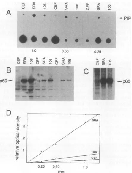
Wenextestablished thelinearityof the PI3'-kinase assay as afunction ofamountoflysate.Assayswereperformedon three different amounts of lysate from uninfected CEFs, cells infected with wild-type virus, and cells infected with
the E106K mutant. Over the three different amounts of
lysate used, the amount ofPI-3-P producedwas less in the
E106K immunoprecipitates than in the wild-type immuno-precipitates (Fig. 2A). The E106K immunoprecipitates
showed only slightly more PI-3-P produced than did the uninfected CEFnegative control. Western immunoblot
anal-ysis revealed that comparable amounts of
pp60vsrc
were present in lysatesfromwild-type- and mutant-infected cells (Fig. 2B). Thewild-type and mutantpp6ovsrc
immunopre-cipitates also displayed comparable amounts of protein
kinase activity (Fig. 2C). Previousresults have shown that the overallamount andpattern oftyrosinephosphorylation in CU12-infected cells didnotdiffersignificantlyfrom results
on November 9, 2019 by guest
http://jvi.asm.org/
CEF
wild
type
CU12
E106K
L.L <4 < U
0 Co- C~~~~~~) C,,~~~ 0 W
A
(n - 0O
n 0 EoA
*.
**0*S
*
6
0
la
e *0 o
1.0
B
p60-D
0.50
,*_aF0C -,
._ ..
cn
-o
-it
C-,
0
.2_
coU,)
K1
04E
K1
04E
/
V461
M
[image:3.612.65.301.75.484.2]di
1
05
G2A /
El
06K
FIG. 1. Morphology of cells expressing SH3 mutants of src.
Wild-type, Schmidt-Ruppin strain subgroup A. CU12, partially transformingmutantcontainingEtoKatamino acid 106and VtoM
atamino acid 461. Othermutantsare asdesignated.
forwild-type infected cells, as detected by antiphosphoty-rosineimmunoblottingof whole celllysateswhichhad been separatedbySDS-PAGE (46). Therefore, underconditions controlling for the amount and activity ofpp6Osrc, E106K anti-pp6Ov-s immunoprecipitates have less PI 3'-kinase
ac-tivity associated with them than dowild-type anti-pp60vs?r immunoprecipitates. Quantitation of these data is shown in Fig. 2D.
Assays comparing PI 3'-kinase activityassociatedwiththe wild type with that associated with E106Kwere repeated
ninetimes. Theresultswerequantitated bydensitometryof the PI-3-P spots and/or by scraping the thin-layer chroma-tography platesandcountingthe silica inaliquidscintillation counter.When the amountofPI-3-Pwasnormalizedto the amount of pp6O'rc present in the lysate as determined by
immunoblotting, a significant difference (P < 0.05) was
found between theactivityassociatedwiththe wildtypeand that associated withE106K. Onaverage,fivetimesmorePI
0.25 0 50 1.0
mg
FIG. 2. Decreased PI 3'-kinase activity associated with E106K
mutant pp6ovsrc. (A) PIP produced in immunoprecipitates of pp6ovsrc, indicated by the arrow. Radioactivity at the origin is
variable isotope carriedoverintheCHCI3-methanol extractions. (B
and C) Western blots and autokinase activities, respectively, of pp6Ovsrc in theimmunoprecipitates showninpanelA.(D)
Quanti-tationofPIPproductionshown inpanel A, determined by densito-metric scanningwith a BioImage Visage. The ordinate is relative arbitrary optical densityunits.SRA,wild-typesrc(Schmidt-Ruppin
strainsubgroup A); 106,E106Ksrcmutant.
3'-kinase activity was associated with wild-type pp60v-src
than withE106K.
Nonmyristylated 106 mutants ofpp6Osc. The amino acid change that occurs in E106K, a lysine substituting for glutamate, could affect the association ofpp6Ovsrc with PI 3'-kinase intwodifferentways. E106K pp60vsrccould have
analtered intracellular distribution and therefore might not be abletobindPI3'-kinase because thesrcproteinwasinan
inaccessible location.Alternatively, the 106 mutation could affect the PI 3'-kinase binding site of pp60v-sC and thus reduce association between the proteins. To distinguish between these twopossibilities, we took advantage of the fact that myristylation ofpp6Orc is necessaryforits
mem-brane association (40, 41). We made a nonmyristylated
version of E106K, which we designate G2A/E106K, by
replacingwith alanine the amino-terminal glycine to which myristicacid isnormallyattached.
Mutants of pp6ovsrc which are nonmyristylated but are
otherwise wildtypearenontransforming (40, 41).
Nonmyri-stylated pp6Orc associates withPI3'-kinase, indicatingthat while PI3'-kinase association maybe necessaryfor
trans-formation, it is not sufficient (24, 31). We reasoned that if decreased association ofPI3'-kinase with theE106Kmutant was aconsequenceofthemutantpp6O being sequesteredin
-PIP
0.25
cLJ Er
C w
_-p60
,f....
on November 9, 2019 by guest
http://jvi.asm.org/
[image:3.612.331.553.78.369.2]A. A.
CEF wildtype G2A E106K 2A/106K d105
, 0
-s--PIP
*0 * * *
w = eC -
-0 3 Ca cNb
CEF SRA E106K K104E 104E/461M
a
s +
t$*
t4
-t*PIP
t
0 *
**
**
*
*
B. 1 < e w s>s
0 cllwy
-FIG. 3. PI 3'-kinase activity in immunoprecipitates ofpp60ovrC SH3mutants.(A) PI3'-kinaseassay.Duplicate samplesareshown
(except for CEF); (B)anti-pp6Q' Westernblot forthisexperiment. Note that the elevated levels of pp6VSrC in cells infected with nonmyristylated mutantsare commonly observed but that
associ-atedPI 3'-kinase levelsareneverthelesslowerincells infectedwith
the2A/106Kmutant.
an inaccessiblemembrane location,thenfreeingthemutant pp6Ofromthemembraneby blockingitsmyristylationwould allowittobindPI3'-kinase(whichisalsocytosolic)asmuch
asdoesG2App6ov-src.Therethusshouldbeequalamounts ofPI3'-kinaseactivityinthenonmyristylatedwild typeand in the double mutant. If, on the other hand, the E106K
mutation affects more directly the binding site of PI
3'-kinase, therewould belessPI 3'-kinase activity associated withG2A/E106K pp6ovsrc immunoprecipitates thaninG2A pp6Ovsrc immunoprecipitates.
Our results indicate that theE106K mutation affects the bindingofpp60V-s? toPI3'-kinaseevenwhenthepp6Qv-srcis
nonmyristylated andcytosolic. When cells are infected by
G2A/E106K, theyhaveanontransformedphenotype(Fig. 1)
similar tothat ofG2App6O-s?C (data not shown). In three separateexperimentswithassaysperformedinduplicate,PI 3'-kinase activity associated with immunoprecipitates of G2A/E106K pp6oV-Srcwas seen tobe less than that associ-ated with G2A pp6ovsrc (Fig. 3). Therefore, we conclude
thatresidue 106 in the SH3 domainofpp6o0-src plays arole inPI 3'-kinaseassociationby altering thebinding site for PI 3'-kinase ratherthan by alteringthelocation ofpp60v-scin thecell.
Mutation ofglycine 105 decreases PI 3'-kinase association with pp6O'C. Much of the SH3 domain is strongly
con-served among nonreceptor tyrosine kinases and in a wide variety of otherproteinsasdiverseasphospholipase C--y and myosin 1B. Theresidues nearglutamate 106ofpp6O5rc are
amongthemoststronglyconserved.Whilesomedeletions of
theSH3 domainhave beenmade(35, 36, 54), few mutations have beenreportedinthisconservedareaof the SH3domain nearresidue 106.
To investigate this region further, we made mutants of wild-typepp60vsrc bydeleting the glycineatposition105and by altering the charge of the residue at position 104 by changing the lysine to a glutamate. We reasoned that the
glycine might be important in its potential role as a helix
breakerand thatadeletionwouldalterthe structureof this
- Elba. --_
pp60src
FIG. 4. PI3'-kinase activityin immunoprecipitates of
pp6ovsrC
SH3mutants.(A) PI3'-kinase activity; (B)anti-pp6O" Western blot for thisexperiment. SRA,Schmidt-Ruppin strainsubgroupA.part of the SH3 domain. We made the charge change at
residue 104 to investigate the possibility that PI 3'-kinase
binds to pp60 by alternating charged residues, that is to
say,byhavingalysineat104and thenan
oppositely
chargedglutamate atresidue 106.We also madea104E/461Mmutant reminiscent of CU12(2) todetermine whether the
carboxy-terminal half of
pp6Q"
could affect this SH3mutation. Wedesignated the glycine deletion mutant dllO5 and the 104
mutants K104E and
K104ENV461M,
respectively.Whenwe transfected CEFs with K104E,
K104ENV461M,
and dllO5, we found that both K104E and
K104ENV461M
produced aroundedmorphologyindistinguishable fromthe
wild-typemorphology (Fig.1).In contrast,deletion of amino
acid 105 caused an extreme fusiform morphology
indistin-guishable from thatofE106K(Fig. 1).TheassociationofPI
3'-kinase activity with pp6o-src immunoprecipitates
corre-lated with the morphological changes: both K104E and
K104E/V461M displayed associated PI 3'-kinase activity
comparable towild-type levels (Fig. 4), while thevalue for
immunoprecipitateofdllO5wasverysimilartothe low level
ofE106K whennormalizedtotheamountof
pp6Q"
protein(Fig. 3).
PI 3'-kinase association with activated (Y527F) c-src
mu-tants.Activationofpp6o-srC resultsin increasedassociation with PI3'-kinase (13),andwethereforewishedtodetermine the effects of SH3 mutations on the association with acti-vated forms of the proto-oncogene. To do this, we made
mutations in c-src and the activated
c-src/Y527F
(inwhichY527, whichis thesite ofnegativeregulatory
phosphoryla-tion, is replaced by F) similar to those described above in
v-src. That is, we altered the charge at residue 104 by
replacing
lysine
with glutamate, deleted glycine 105, orreplaced glutamate 106 with alysine, makingthe activated c-src equivalentof E106K.
Whenplasmidswith the104, 105, and 106 mutations in a
normal c-src background were transfected onto CEFs, no
morphologicalchangeswere noted (datanotshown).Thus,
these mutations do not activate c-srcwith respect to
mor-phologicaltransformation. However, when cells were
trans-fected withplasmids carryingthesec-srcmutations in com-bination with the activating Y527F mutation, a variety of
morphologies were seen (Fig. 5). Cells transfected with
qp
*
f#.
0
il
on November 9, 2019 by guest
http://jvi.asm.org/
[image:4.612.323.555.80.261.2] [image:4.612.61.303.80.263.2]4.."
Y527F
Y527F
El 06K / Y527F
K1 04E
/
Y527F
dl
1
05 /
Y527F
:. &a u u rr'w es. '7'7c-.. ---caw 't*-'
d192
/
Y527F
G2A / Y527F
FIG. 5. Morphology of cells expressing SH3 mutants of acti-vatedc-src.
K104E/Y527F
looked as completely transformed as thosewhich had received Y527F. Thus, the charge mutation at
position104hadnodetectable effectsonc-src/Y527F,c-src,
or v-src despite the fact that K at this position is highly
A. 2A, dl921 2A/dl92 .
GEF 527F 527F 527F 527F
conserved inSH3 domains oftyrosine kinases.Surprisingly,
theE106K mutation also did not cause a fusiform morphol-ogy in theactivated c-src background even though it had a pronounced effect in the v-src context. Therefore, the pres-ence ofsecondary mutations in wild-type v-src is required for106K to cause a fusiform morphology in infected cells. Nevertheless, this portion of the SH3 domain isimportant
forregulating morphology, because deletion of glycine 105 in
c-srcI527Fcaused an extreme fusiform morphology, as did
thedllO5 and E106K mutations in the v-src context. Thus, glycine 105 has similar effects in both pp6Oc-src/527F and pp6Ovsrc. Infection with an activated c-src with residues 92 to95deleted(d192IY527F) (54, 55) yielded cells that werenot asrounded as cells infected with wild-type src but did not have theextremeneedlelike, fusiform morphology seen with
dlO15.
When the 104-106 series of activated pp60c-s' mutants was tested for associated PI 3'-kinase, the results followed the morphological correlations seen in the wild-type mu-tants: fusiform cells had lowered PI 3'-kinase activity as-sociated with pp6X'r". Thus, we found equivalent associa-tion of PI 3'-kinase activity with
pp60'csrc/527F
and in the fully transforming mutantspp60C-s?r1O6K/527F
(Fig. 6) and pp6OcsrclO4E/527F
(data notshown), whereas deletion ofgly-cine105 resulted in reducedassociation of PI 3'-kinase (Fig. 6), even when the lower level ofpp6W'S was accounted for. The
d192/Y527F
mutant displayed little if any reduction in p60-associated PI 3'-kinase activity. Interestingly, the un-myristylated version of this mutant (G2A/dl92/Y527F)showed very little associated PI 3'-kinase activity; perhaps membrane association can compensate for some of the struc-tural defects in this mutant p60.
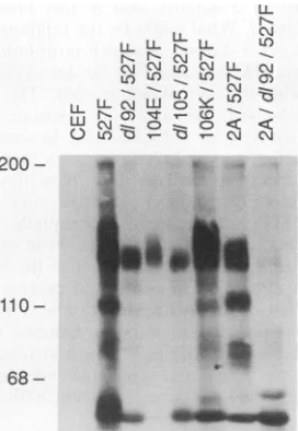
Associationofp130 and pllO with SH3 mutants ofpp6W'. Because partof the PI 3'-kinase enzyme complex is approx-imately 110 kDa (12),we examinedpp6Qsrc immunoprecipi-tatesof these SH3 mutants todetermine whether the pres-ence of the 110-kDa phosphotyrosine protein studied by Parsons and associates (42, 55) correlated with pp60src-associated PI 3'-kinase activity. We also tested for the
106K dl105/
527F CEF 527F 527F
0:9:'iPs
t~~~~~~~~~~~~~~~~~~~~~~~~.
....B.
LL N N
Ns
NN a)
LL
N
U) C\M
U0)
LU kn\ B
_O__4U_lmIuSw4
pp60src pp60src _ "NFIG. 6. PI3'-kinaseactivityinimmunoprecipitatesof activatedpp60C-rCSH3mutants.(A)PI3-kinase activity; (B)anti-pp60srcWestern blot for thisexperiment.
on November 9, 2019 by guest
http://jvi.asm.org/
[image:5.612.59.298.74.380.2] [image:5.612.143.470.490.704.2]LL
N-LL LL C%
XL N CD
C)InO
LL
N-LLL
LL r,-LL U') rC C\Jr L
--N) cC L c\
'tV CD o _ o < <
v 5:sc\j c\
200
110
68-FIG. 7. Tyrosine-phosphorylated cellular proteins associated
withSH3mutantsof activatedpp60c-s?. pp6Owas immunoprecipi-tated from infectedcells, the immunoprecipitatewasseparated by
SDS-PAGEandwasthenprobedwithantiphosphotyrosine
antise-rum.Sizesareshowninkilodaltonsatthe left.
presence of a 130-kDa tyrosine-phosphorylated protein
which associates with pp60vsrc. Immunoprecipitates of pp6Osrc were separated by SDS-PAGE and then
immuno-blotted withantiphosphotyrosine antibodiesto detect
asso-ciated p1lOandp130 thatarephosphorylatedon tyrosine.
As reported previously (42, 55), immunoprecipitates of
pp60c-srcI527F
contained associated tyrosine-phosphorylated proteins of 110 and 130 kDa (Fig. 7), whereas the SH3 mutantd192/Y527F associated withp130 but notp110.This pattern also was observed for the transforming K104E/ Y527F and the fusiform d11O5/Y527F. However, the trans-forming E106K/Y527F mutant appeared to associate withp110, albeit to a somewhat reduced degree. Because d192/
Y527F and K104E/Y527F have PI 3'-kinase activity
asso-ciated with their immunoprecipitates to a degree
com-parable to that of pp6Oc-src/527 yet have little to no
as-sociatedtyrosine-phosphorylated ppllO,theseresults would imply, but not prove, that the pllO seen here is not the 110-kDa subunitofPI3'-kinase.
Analysisoftyrosine-phosphorylated proteins which coim-munoprecipitate with the wild-typeSchmidt-Ruppin strain of pp6Ov-srcusedbyushaspreviouslybeen lesscompletethan
forthePraguestrainpp6OvsrCorpp6ocs/527F.Interestingly,
the pattern oftyrosine-phosphorylated p60-associated
pro-teins differs between thetwo strains. We found little
asso-ciation ofpllOwithSchmidt-Ruppin pp60vsrc (Fig. 8),even
though cells infected with this strain are fully transformed
and thepp6ovsrc associates with PI 3'-kinase activity. The
one exception is that the unmyristylated mutant displayed associationwith p110. Similar results were obtained when
the immunoprecipitateswere blotted with antibody against pllO (42) (datanotshown). However, this strain of pp60vsrC displayedassociation withtyrosine-phosphorylated proteins of 130 and 145 kDa. The SH3 mutants which induced a
fusiform morphology displayed reduced association with these tyrosine-phosphorylated proteins, most dramatically in the case of the dllO5 mutant. The fact that a different pattern of coimmunoprecipitating proteins is seen with
LU <CD °) l <
-c)coL
LL<Crg0 \j
C) CDw '6:b 0 coi
200
-110
-FIG. 8. Tyrosine-phosphorylated cellular proteins associated with SH3 mutants of Schmidt-Ruppin strain subgroup A (SRA) pp6Ov-s.Conditionswere asforFig. 7.Sizesareshownin kilodal-tons attheleft.
Schmidt-Ruppin strainsrccomparedwith Praguestrainsrc
and c-src derivatives may be a consequence of the three additional SH3 domain mutations found in the Schmidt-Ruppin strain (52).
DISCUSSION
Workpresentedinthis report demonstrates that mutations
inthe SH3 domaincanresult in reducedamountsofactive PI 3'-kinase inimmunoprecipitatesofpp6Osrc.Thesemutations also cause an altered, fusiform cell morphology. Although
this connection between lowered PI 3'-kinase activity and alteredmorphologyiscorrelational,thesefindingssuggesta
potential functionfor the SH3 domain and raise the
possi-bilitythat PI 3'-kinaseactivityplaysaroleinmorphological
transformation bypp6O"".
Itisimportanttoemphasizethatwhiletheseexperiments
show that mutations in the SH3 domain ofpp6O'" canalter the PI3'-kinase activity associated with
pp6Q",
theexper-imentsdo notdistinguish between effects onthe binding of
thePI 3'-kinaseprotein topp60' andeffectsonthe activa-tion of the PI3'-kinase enzyme.
The mutationdiscoveredtoberesponsiblefor the fusiform
morphology ofcells infected with CU12, glutamate 106 to
lysine, is in a particularly conserved region of the SH3
domain. Despite the conserved nature of these residues,
Hirai and Varmusfound that alteration of nearby residue 109
hadlittleeffectontransformingability (35, 36).Similarly,we
found that changing adjacentresidues could have
dramati-cally differenteffects. Inwild-typepp60vsrC, residue104can
undergoacharge change withnoeffectonphenotype, while
deletion of glycine 105 or a charge change at residue 106
causes a fusiform morphology. Mayer et al. report that a derivative ofmutantPA101 which has a substitution muta-tionatresidue105, alongwithother amino-terminalchanges,
hasafusiformmorphology(49).Ourresults suggest that the
change at position 105 may be sufficient for the fusiform
morphology causedby PA101.
Although the SH3 sequences inc-src and v-src arevery
similar,the 106 mutation hasnoobvious effect whenplaced
in the
c-srcI527F
background. This indicates thatsomeof the other mutations in v-src interact with the SH3 domain. Iton November 9, 2019 by guest
http://jvi.asm.org/
[image:6.612.108.244.76.273.2] [image:6.612.375.496.76.244.2]would appear, however, that glycine 105 isa critical struc-tural residue because its deletion in c-src/527F or in v-src causes the fusiform phenotype. These findings along with those reported by Hirai and Varmus (35, 36) and those discussed in Parsons and Weber (52) indicate that the SH3 domain has complex interactions with the rest of thepp6Qsrc protein. This is underscored by the fact that changing residues 105 and 106 leads to the same pattern for PI
3'-kinase association: all of the SH3 mutations inducing a fusiform morphology also caused a reduced association with
PI 3'-kinase. PI 3'-kinase association studies on mutants
made by Hirai and Varmus have not been reported. Fukui et al. studiedPI3'-kinase association withpp6ovsrc
by using a variety of mutants(24-27).Of particular relevance to our work was their finding that PI 3'-kinase associates with the SH3 deletion mutant NY311, in which residues 15 to 149 of the v-src protein are deleted. Fukui et al. concluded that this region was probably not directly involved in binding PI 3'-kinase. Conversely, small deletions in the SH2 domain were found to reducePI 3'-kinase association, and such SH2 mutants induced a fusiform morphology (15). Similarly, Liu et al. (48a) have found that asrcSH2 domain expressed as a bacterial fusion protein can bind to PI 3'-kinase but that the equivalent construct containing only an SH3 domain cannot. Thus, it seems likely that the SH2 domain is directly involved in binding toPI3'-kinaseand that the SH3 domain is neither necessary nor sufficient for such binding.
How can these reports be reconciled with our finding that a point mutation or a single amino acid deletion within the SH3 domain nonetheless reduces association with PI 3'-kinase activity? We interpret our results as pointing to an interaction between the SH3 domain and the otherregions of
pp6Ov-src(in particularSH2) which are involved in binding of
PI 3'-kinase. Mutations in the SH3 domain which result in decreasedPI3'-kinase association could cause intramolecu-lar interactions which block theaccessibility ofPI 3'-kinase to the remainder of this binding site, whereas mutations which do not decrease PI 3'-kinase association could leave the binding site accessible.
These results and the complex nature of the mutations reported by Hirai and Varmus (35, 36) and Wang and Parsons (66) suggest the SH3 domain has interactions both with other parts of the pp60 protein and also with proteins that associate with pp6o0rc. Depending on the location and typ of mutation, lesions in the SH3 domain appear to either
act>.ate or inhibit tyrosine kinase activity (52). Results in
thi, report also show that different changes in the SH3 domain have diverse effects on proteins that coimmunopre-cipitate withpp6Q"rc. The SH3 mutants described here show varying associations with the 110- and 130-kDa phosphoty-rosine proteins described by Reynolds et al. (42, 55). In an activated c-src background, K104E, dllO5, and E106K all associate withp130; K104EanddllO5 do not associate with
ap110,whileE106K, which isphenotypically
indistinguish-able from K104E, does associate with p110. Moreover
K104E has PI 3'-kinase activity associated with it to the
same degree as does the activated c-src/Y527F without additional mutations.
Cellmorphology, SH3 domains and PI 3'-kinase: a specu-lation. Work presented in this report provides the first instance in which association ofPI3'-kinase activity with an oncogene product correlates with a specific parameter of transformation, namely, cellular morphology: all of the mutants examined by us and by Fukui et al. (24-27) which displayed reduced association with PI 3'-kinase induced a fusiform morphology. Only one fusiform mutant, NY311
(25), bound to PI 3'-kinase, and in this mutant the SH3 region was deleted. What might be the relationship between SH3 domains, PI 3'-kinase, and cell morphology? All pro-teins containingSH3 domains thus far discovered have some connection with the cytoskeleton (30). The nonreceptor tyrosine kinases with SH3 domains associate with the cy-toskeleton when they are activated (34). In some cases, SH3 proteins are actually cytoskeletal proteins themselves, such as myosin or spectrin; in other cases, they may beenzymes thatactively modulate the cytoskeleton, such as S. cerevi-siae ABP1 (18). In the case of neutrophilic p47 and p67 oxidases, upon activation they move from cytosol to the cytoskeleton, in a manner reminiscent of the conversion of pp60csrc to a cytoskeleton-associated protein upon muta-tional activation (48). Thus, SH3 domains may serve to regulate the association of various proteins with the cy-toskeleton. The location ofpp6Ovsrc at cell adhesion plaques (10, 56) and its rapid and profound effects on cellular morphology (1, 9, 57, 65) are consistent with this specula-tion.
PI 3'-kinase may be a component of this cytoskeletal signaling complex. Recently it has been shown that an intermediate in the classic PI cycle, PI-4,5-P2, can regulate the actin-binding protein profilin (29, 30, 48). Profilin is a low-molecular-weight protein which can function as an ac-tin-sequestering agent
(23,
33, 62). Releasing profilin from actin would allow actin monomers to polymerize into fila-ments. Thus, a connection between PI metabolism and cytoskeletal control has been established. However, there are some problems with the idea that PI-4,5-P2 is the agent regulating profilin. Conditions associated with morphologi-calchanges and actin polymerization can beaccompanied by a decrease in PI-4,5-P2 levels (19, 23). However, activation of PI 3'-kinase and/or increased PI-3-P metabolites have been described duringagonist-induced actin polymerization and surfaceruffling (3, 19). Therefore,it isquite possible that the physiological regulator ofprofilin is PI-3-P or one ofits metabolites.Thus, we speculate that SH3domains serve to regulate the formation of a ternary complex between pp60vsrc, the cy-toskeleton, and PI 3'-kinase and that the products of PI 3'-kinase regulate local actin polymerization by inhibiting profilin. Theavailability of molecular and chemical reagents tostudyPI 3'-kinase and its products will make itpossible to testthis speculation.
ACKNOWLEDGMENTS
This work was supported by NIH grantsCA39076, CA40042,and CA 47815 to M.J.W.D.S.W. was apredoctoraltraineesupportedby 5-T32-GM 07267.
We thank Tony Pawsonand Jone Liu forcommunicating results prior to publication. We are grateful to J. Thomas Parsons and Bradley Cobb for a great deal of technical advice and insightful suggestions.
REFERENCES
1. Ambros, V. R., L. B. Chen, and J. M. Buchanan. 1975. Surface ruffles as markers for studies of cell transformation by Rous sarcoma virus. Proc. Natl. Acad. Sci. USA72:3144-3148. 2. Anderson, D. D., R. P.Beckmann,E. H.Harms, K. Nakamura,
and M. J. Weber. 1981. Biological properties of "partial" transformation mutants of Rous sarcoma virus and characteri-zation of theirpp6jSrc kinase. J. Virol. 37:445-458.
3. Anderson, S. K., and D. J. Fujita. 1987.Morphfmutantsof Rous sarcoma virus: nucleotide sequencing analysis suggests that a
class of morphf mutants was generated through splicing of a
crypticintron. J. Virol. 61:1893-1900.
on November 9, 2019 by guest
http://jvi.asm.org/
4. Antler, A., E. Greenberg, G. M. Edelman, and J. Hanafusa. 1985. Increasedphosphorylationoftyrosineinvinculin doesnot
occur upon transformation by some avian sarcoma viruses.
Mol. Cell. Biol.5:263-267.
5. Auger, K. R., and L. C. Cantley. 1991. Novel polyphosphoi-nositides in cell growth andactivation. Cancer Cells3:263-270. 6. Bansal, V. S., and P. W. Majerus. 1990. Phosphatidylinositol derivedprecursors andsignals. Annu. Rev. Cell Biol. 6:41-67.
7. Berridge, M. J., and R. F. Irvine. 1989. Inositol phosphate and cell signaling. Nature (London) 341:197-205.
8. Bjorge, J. D., T.Chan, M. Antczak, H. Kung, and D.F. Fujita. 1990.ActivatedtypeIphosphatidylinositol kinase is associated withthe epidermal growthfactor(EGF)receptorfollowing EGF
stimulation. Proc. Natl. Acad. Sci. USA87:3816-3820. 9. Boschek, C. B., B. M. Jockusch, R. R. Friis, R. Back, E.
Gurndmann, and H. Bauer. 1981. Early changes in the distribu-tion and organizadistribu-tion of microfilament proteins during cell transformation. Cell 24:175-184.
10. Burridge, K., K. Fath, T. Kelly, G. Nickolls, and C. Turner.
1988. Focal adhesions: transmembrane junctions between the
extracellular matrix and the cytoskeleton. Annu. Rev.Cell Biol. 4:487-525.
11. Cantley, L.C., K. R. Auger, C. Carpenter, B. Duckworth, A.
Graziani, R. Kapeller, and S. Soltoff. 1991. Oncogenes and signaltransduction. Cell 64:281-302.
12. Carpenter, C. L., B. C. Duckworth, K. R. Auger, B. Cohen,
B.S. Schaffhausen, and L. C. Cantley. 1990. Purification and
characterization of phosphoinositide 3-kinase fromratliver. J.
Biol. Chem.265:19704-19711.
13. Chan, T., A. Tanaka, J. D. Bjorge, and D. F. Fujita. 1990.
Association oftype I phosphatidylinositol kinase activity with mutationally activated forms of human pp60c-sl. Mol. Cell.
Biol. 10:3280-3283.
14. Cooper,J., K. D. Nakamura, T. Hunter, and M.J.Weber.1983.
Phosphotyrosine-containing proteins and expression of
trans-formation parameters in cells infectedwith partial
transforma-tion mutants ofRoussarcoma virus. J. Virol.46:15-28.
15. Cross,F. R., E. A. Garber, andJ. Hanafusa. 1985. N-terminal
deletions in Rous sarcoma virus p6Q"r: effects on tyrosine
kinase and biological activities and on recombination in tissue
culturewith the cellularsrcgene.Mol. Cell. Biol. 5:2789-2795.
16. Cross,F. R., and H. Hanafusa. 1983. Local mutagenesis of Rous
sarcomavirus: the majorsites of tyrosine and serine
phosphor-ylation of p60!rc aredispensable for transformation. Mol. Cell.
Biol. 4:1834-1842.
17. Cunningham, T.W., D. L. Lips, V. S. Bansal, K. K. Caldwell,
C. A. Mitchell, and P. W. Majerus. 1990. Pathway for the
formation of D-3 phosphatecontaining inositol phospholipidsin
intacthuman platelets. J.Biol. Chem. 265:21676-21683.
18. Drubin, D. G., J. Mulholland, Z. Zhu, and D. Botstein. 1990.
Homology ofayeastactin-bindingproteintosignaltransduction
proteins and myosinI. Nature (London) 343:288-290.
19. Eberle, M., A. E. Trayner-Kaplan,L. A.Sklar,and J. Norgauer.
1990. Is there a relationship between phosphatidylinositol
triphosphate and F-actin polymerization in human neutrophils?
J. Biol. Chem. 265:16725-16728.
20. Endemann, G., K. Yonezawa, andR. A. Roth. 1990.
Phosphati-dylinositol-3-kinase or an associated protein is a substrate for
theinsulinreceptortyrosinekinase. J. Biol. Chem.265:396-400.
21. Escobedo,J., D. R. Kaplan, W. M. Kavanaugh, C. W.Turck, and L. T. Williams. 1991.Aphosphatidylinositol-3 kinase binds to platelet-derived growth factor receptors through a specific
receptorsequencecontaining phosphotyrosine. Mol. Cell.Biol. 11:1125-1132.
22. Escobedo, J. A., S. Navankasattusas, W. M. Kavanaugh, D.
Milfay,V. A. Fried, andL. T. Williams. 1991. cDNA cloningof
a novel 85 kd protein that has SH2 domains and regulates
binding of P13-kinasetothe PDGFP-receptor.Cell65:75-82. 23. Forscher,P.1989.Calcium and polyphosphoinositidecontrol of
cytoskeletal dynamics.Trends Neurosci. 12:468-474.
24. Fukui, Y., and H. Hanafusa. 1989. Phosphatidylinositol kinase
activity associates with viral p60slc protein. Mol. Cell. Biol. 9:1651-1658.
25. Fukui, Y., and H. Hanafusa. 1991. Requirement of phosphati-dylinositol-3 kinasemodification for its association withp6O'".
Mot. Cell. Biol. 11:1972-1979.
26. Fukui, Y., S. Kornbluth, S.-M. Jong, L.-H. Wang, and H. Hanafusa. 1989. Phosphatidylinositol kinase type I activity associates with various oncogene products. Oncogene Res. 4:283-292.
27. Fukui, Y., A. R. Saltiel, and H. Hanafusa. 1991. Phosphatidyl-inositol-3 kinase is activated in v-src, v-yes, and
v-fps
trans-formed chicken embryo fibroblasts. Oncogene 6:407-411. 28. Garber, E. A., B. J. Mayer, R. Jove, and H. Hanafusa. 1987.
Analysis ofpp60vsrC mutants carrying lesions involvedin
tem-perature sensitivity. J. Virol. 61:354-360.
29. Goldschmidt-Clermont, P. J., J. W. Kim, L. M. Machesky,S. G. Rhee, and T. D. Pollard. 1991.Regulation ofphospholipaseC--yl by profilin and tyrosine phosphorylation. Science 251:1231-1233.
30. Goldschmidt-Clermont, P. J., L. M.Machesky,J. J. Baldassare, and T. D. Pollard. 1990. The actin-binding proteinprofilinbinds to PIP-2 and inhibits itshydrolysis byphospholipase C. Science 247:1575-1578.
31. Gorga, F. R., C. E. Riney, and T. L. Benjamin. 1990. Inositol trisphosphate levels in cells expressing wild-type and mutant polyomavirus middle T antigens: evidence for activation of phospholipase C via activation ofpp60c-srC.J. Virol.64:105-112. 32. Gutkind, J. S., P. M. Lacal, and K. C. Robbins. 1990. Thrombin-dependent association of phosphatidylinositol-3 kinase with p60C-src andp59fl in human platelets. Mol. Cell. Biol. 10:3806-3809.
33. Haarer, B. K., and S. S. Brown. 1990. Structure and function of profilin. Cell Motil. Cytoskel. 17:71-74.
34. Hamaguchi, M., and H. Hanafusa. 1987. Association ofp6OY' with triton X-100-resistant cellular structure correlates with morphological transformation. Proc. Natl. Acad. Sci. USA 84:2312-2316.
35. Hirai, H., and H. E. Varmus. 1990. Mutations in src homology regions 2 and 3 of activated chicken c-src that result in prefer-ential transformation of mouse or chicken cells. Proc. Natl. Acad. Sci. USA 87:8592-8596.
36. Hirai, H., and H. E. Varmus. 1990. Site-directed mutagenesis of the SH2- and SH3-coding domains of c-src produces varied phenotypes, including oncogenic activation of p60CSrC. Mol. Cell. Biol. 10:1307-1318.
37. Jove, R., E. A. Garber, J.Iba, and H. Hanafusa. 1986. Biochem-ical properties of p60vsPC mutants that induce different cell transformation parameters. J. Virol. 60:849-857.
38. Jove, R., B. J. Mayer, H.Iba, D. Laugier, F. Poirier, G. Calothy, T. Hanafusa, and H. Hanafusa. 1986. Genetic analysis ofp6ovsrC domainsinvolved in the induction of different cell transforma-tion parameters. J. Virol. 60:840-848.
39. Kahn, P., K. Nakamura, S. Shin, R. E. Smith, and M. J. Weber. 1982. Tumorigenicity of partial transformation mutants of Rous sarcoma virus. J. Virol. 42:602-611.
40. Kamps, M. P., J. E. Buss, and B. M. Sefton. 1985. Mutation of NH2-terminus glycine of p6O"c prevents both myristolation and morphological transformation. Proc. Natl. Acad. Sci. USA 82:4625-4628.
41. Kamps, M. P., J. E. Buss, and B. M. Sefton. 1986. Rous sarcoma virus transforming protein lacking myristic acid phosphorylates known polypeptide substrates without inducing transformation. Cell45:105-112.
42. Kanner, S. B., A. B. Reynolds, H. R. Wang, R. R. Vines, and J. T. Parsons. 1991. TheSH2andSH3domains ofpp60'cdirect stable association with tyrosine-phosphorylated proteinsp130 andp110. EMBO J. 10:1689-1697.
43. Kellie, S., B. Patel, A. Mitchell, D. R. Critchley, N. M. Wiggles-worth, and J. A. Wyke. 1986. Comparison of the relative importance of tyrosine-specific vinculin phosphorylation and the loss of surface-associated fibronectin in the morphology of cells transformed by Rous sarcoma virus. J. Cell Sci. 82:129-142.
44. Kellie, S., B. Patel, N. M.Wigglesworth, D. R. Critchley, and J. A. Wyke. 1986. The use of Rous sarcoma virus
on November 9, 2019 by guest
http://jvi.asm.org/
tion mutants withdiffering tyrosine kinase activities to study the relationships between vinculin phosphorylation, pp6o-srC loca-tion and adhesion plaque integrity. Exp. Cell. Res. 165:216-228. 45. Koch, C. A., D. Anderson, M. F. Moran, C. Ellis, and T. Pawson. 1991. SH2 and SH3 domains: elements that control interactionsof cytoplasmic signaling proteins. Science 252:668-674.
46. Kozma, L. M., A. B. Reynolds, and M. J. Weber. 1990. Glycoprotein tyrosine phosphorylation in Roussarcoma virus-transformed chicken embryo fibroblasts. Mol. Cell. Biol. 10: 837-841.
47. Kozma, L. M., and M. J. Weber. 1990.Constitutive phosphor-ylation of the receptor for insulinlike growth factor I in cells transformedby thesrconcogene.Mol. Cell. Biol.10:3626-3634. 48. Leto,T. L., K. J. Lomax, B. D. Volpp, H. Nunoi, J. M. G. Sechler,W. M. Nauseeef, R. A. Clark,J.I. Gallin,and H. L. Malech.1990.Cloning ofa67-kD neutrophiloxidasefactor with similarityto anoncatalytic region ofp60csrC.Science 248:727-730.
48a.Liu,J., T. Pawson, et al.Personal communication.
49. Mayer, B. J., R. Jove, J. F. Krane, F. Poirier, G.Calothy, and H. Hanafusa. 1986. Genetic lesions involved in temperature sensitivityofthe src gene productsof fourRous sarcoma virus mutants.J. Virol. 60:858-867.
50. Nakamura, K. D., and M. J. Weber.1982.Phosphorylation ofa 36,000-Mr cellularprotein in cells infectedwithpartial transfor-mationmutantsofRous sarcomavirus. Mol. Cell Biol. 2:147-153.
51. Otsu, M., I. Hiles, I. Gout, M. J. Fry, F. Ruiz-Larrea, G. Panayotou, A.Thompson, R.Dhand, J. Hsuan, N.Totty,A. D. Smith,S. J. Morgan, S. A.Courtneidge, P. J. Parker, and M. D. Waterfield. 1991. Characterization oftwo 85 kd proteins that associate with receptor tyrosine kinases, middle-T/pp60c<r` complexes, andP13-kinase.Cell 65:91-104.
52. Parsons, J. T., and M.J.Weber.1989.Genetics ofsrc:structure
andfunctionalorganization ofaproteintyrosine kinase. Curr. Top.Microbiol. Immunol. 147:80-127.
53. Parsons, S.J.,D.J.McCarley,C.M.Ely,D.C.
Benjamin,
and J. T. Parsons. 1984. Monoclonal antibodies to Roussarcoma viruspp60' reactwithenzymatically active cellularpp6lYrcof avian andmammalianorigin.J.Virol. 51:272-282.54. Potts, W.M.,A. B.Reynolds,T.J.Lansing,andJ.T.Parsons. 1988. Activation of pp60C< transforming potential by
muta-tions altering the structure ofanamino terminal domain
con-taining residues 90-95. Oncogene Res. 3:343-355.
55. Reynolds, A.B., S.B.Kanner, H. R.Wang,andJ.T. Parsons. 1989.Stable association of activatedpp60 withtwo tyrosine-phosphorylated cellularproteins. Mol. Cell.Biol.9:3951-3958. 56. Rohrschneider, L. 1980. Adhesion plaques of Rous sarcoma
virus-transformed cells contain the src gene product. Proc. Natl. Acad. Sci. USA 77:3514-3518.
57. Rohrschneider, L., and S. Reynolds. 1985.Regulation of cellular morphology by the Rous sarcoma virus src gene: analysis of fusiform mutants. Mol. Cell. Biol. 5:3097-3107.
58. Rohrschneider, L., and M. J. Rosok. 1983. Transformation parameters and
pp6O'rc
localization in cellsinfected with partial transformationmutantsofRous sarcomavirus.Mol. Cell. Biol. 3:731-746.59. Ruderman, N. B., R. Kapeller, M. F.White,and L. C. Cantley. 1990. Activation of phosphatidylinositol 3-kinase by insulin. Proc. Natl. Acad. Sci. USA87:1411-1415.
60. Serunian, L. A., M. T. Haber, T. Fukui, J. W. Kim, S. G. Rhee, J. M. Lowenstein, andS. G.Rhee.1989.Polyphosphoinositides produced byphosphatidylinositol 3-kinaseare poorsubstrates forphospholipases Cfrom rat liver and bovine brain.J. Biol. Chem. 264:17809-17815.
60a.Sisk,S., and M. J. Weber.Unpublished data.
61. Skolnick,E.Y., B.Margolis, M. Mohammadi, E. Lowenstein, R. Fischer, A. Drepps, A.Ullrich,andJ.Schlessinger. 1991. Clon-ing of PI3 kinase-associated p85 utilizClon-ing anovel method for expression/cloning of target proteins forreceptor tyrosine ki-nases.Cell 65:83-90.
62. Stossel,T. P.1989.Fromsignaltopseudopod: howcells control cytoplasmic actin assembly.J. Biol. Chem. 264:18261-18264. 63. Ullrich, A., andJ. Schlessinger. 1990. Signal transduction by
receptors withtyrosine kinase activity.Cell 61:203-212. 64. Varticovski, L., B. Druker, D. Morrison, L. Cantley, and T.
Roberts. 1989.Thecolony stimulating factor-1receptor associ-ates with and activates phosphatidylinositol-3-kinase. Nature (London) 342:699-702.
65. Wang,E., and A. R.Goldberg.1976.Changes in microfilament organization and surface topography upon transformation of chickembryo fibroblastswith Rous sarcomavirus.Proc. Natl. Acad. Sci. USA 73:4065-4069.
66. Wang, H.-C. R., andJ.T. Parsons.1989. Deletions and inser-tions within an amino-terminal domain ofpp6o-src inactivate transformation and modulate membranestability. J. Virol.63: 291-301.
67. Weber, M.J.1984.Malignant transformation byRoussarcoma
virus: fromphosphorylation tophenotype. Adv. Viral Oncol. 4:249-268.
68. Weber,M.J.,and R. R. Fris.1979.Dissociation of transforma-tionparametersusingtemperature-conditionalmutantsof Rous sarcomavirus.Cell 16:25-32.
69. Whitman, M., C. P. Downes, M. Keeler, T. Keller, and L. Cantley.1988.TypeIphosphatidylinositolkinase makesanovel phospholipid, phosphatidylinositol-3-phosphate. Nature (Lon-don) 332:664-666.
70. Whitman, M.,D. R.Kaplan,B. Schaffhausen,L. Cantley,and T. M. Roberts.1985.Associationofphosphatidylinositolkinase activitywithpolyoma middle Tcompetent fortransformation. Nature(London) 315:239-242.