International Journal of Innovative Technology and Exploring Engineering (IJITEE) ISSN: 2278-3075, Volume-8 Issue-12, October 2019
Melanoma Segmentation and Classification using
Deep Learning
R. D. Seeja, A. Suresh
Abstract: Melanoma is the most destructive form of skin cancer. Early diagnosis of melanoma can be curable. At the same time accurate diagnosis is very essential because of the similarities of melanoma and benign lesions. Hence computerized recognition approaches are highly demanded for dermoscopy images. The main purpose of this research is to develop an automatic system to improve the classification performance of melanoma.The effectiveness of this framework is evaluated on ISBI 2016 Skin Lesion Analysis towards Melanoma Detection Challenge dataset. Initially deep learning based U-Net algorithm is used to segment the lesion region from the nearby healthy skin and then extract discriminate features with the help of Convolutional Neural Network. VGG16 Net algorithm is used to classify every lesion in a dermoscopic image as a Benign or Melanoma. Results are found from classification with and without segmented images. Classification with segmented images produces accuracy of 83.18%, Sensitivity of 95.53%, and specificity of 96.22%. Based on these values the deep learning based classification with segmented images produces better result and it helps to improve the diagnosis performance. The proposed method would constitute a valuable support for physicians in every day clinical practice.
Keywords : Melanoma, Deep learning, Dermoscopy, Convolutional Neural Networks, Lesion Segmentation.
I. INTRODUCTION
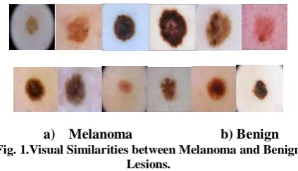
Melanoma is one of the most harmful type of skin cancer that begins in the pigment cells (melanocytes) of the skin. Due to the excess revelation of ultraviolet radiation from the sun, the skin cells are damaged and can affect the resistant capacity [1]. Quick diagnosis and treatment can result in a very high possibility of melanoma survival [2]. At the same time, due to the similarities of skin lesion types, a correct diagnosis is essential.The visual difference between melanoma and benign skin lesions can be very elusive even for trained medical professionals under naked eye observation. Figure 1 shows the images of melanoma and benign both pigments look like to be similar.
[image:1.595.63.275.586.707.2]a) Melanoma b) Benign
Fig. 1.Visual Similarities between Melanoma and Benign Lesions.
Revised Manuscript Received on October 05, 2019.
R. D. Seeja*, Research Scholar,Department of Computer Science, Periyar University,Salem-7, Tamil Nadu, India.
Email: [email protected].
Dr. A Suresh,Principal, Siri PSG Arts and Science College for women, Sankagiri, Salem-637301.,Tamil Nadu,India.
Dermoscopy method was developed to improve the diagnostic presentation of melanoma. Dermoscopy also called as skin surface microscopy is a non-invasive diagnosis technique which is used in the evaluation and variations of suspicious melanocytic lesions from melanoma and benign. It increases the clarity of exact spots on the skin surface and provides more details of skin lesions by 49%[3]. Medical experts use dermoscopy for diagnosis. However, the manual assessment made by dermatologists from dermoscopy images is a lengthy process and error-prone. Hence automated algorithms have become a necessity to classify melanoma which is assist for early diagnosis and improve accurate diagnosing performance.
This study aims that to develop an automatic diagnosis system of melanoma using Deep learning methods. For this purpose, initially the skin lesions were segmented using deep learning based U-Net algorithm. From the segmented images, deep features were extracted using Convolutional Neural Networks (CNN’s). Then the extracted deep features were fed into the VGG16 Net classifiers for classification.
II. RELATED WORK
Some of the research works on melanoma classifications have reported in the literature. By applying Principal Component Analysis (PCA) algorithm, 13 optimal features are selected among 187 low level features based on color, texture, border and asymmetry. Then classify melanoma using SVM classifier with accuracy 82.2% was presented by [4].
Discrete Wavelet Transform for feature extraction, texture analysis and these extracted features were the input to Stack Auto Encoders (SAEs) for training and testing the lesions as malignant or benign by [5].
Two supervised classification systems are proposed by [6], Binary and Multi-class classification: In a binary classification method classified melanoma into thin or thick and the three-class classification method classified melanoma into thin, intermediate and thick. The performance of several nominal classification methods are compared with Logistic regression using Initial variables and product Units (LIPU). Both of the methods LIPU produce highest performance with accuracy 77.6%.
Different combinations of parameters were used to analyze the performance of classifier for each feature.Classification process uses Ada boost, KNN, and
SVM classifiers. The evaluation result showed that color feature dominate
texture feature and local features with BoF provides slightly better result.
In paper [8], uses Region based Statistical Region Merging (SRM) algorithm is used for segmentation. Vector based Speeded Up Robust Features (SURF) technique is adopted for feature extraction and texture analysis. It uses Hessian matrix approximation for feature point detection and Haar-wavelet response for feature descriptions. Multi-Support Vector Machine classifier is used for classification provides 86.37% accuracy.
Many of the previous studies extract low level features from skin lesion images as a stage before classification, while convolutional Neural Networks are proficient of learning all the deep features without the necessity of other algorithms. Recently, CNN based algorithms have exposed successful results in segmentation and classification of skin lesion images.
Hybrid system for melanoma classification was proposed by [9]. Convolutional Neural Networks (CNNs), Support Vector Machine (SVM) and Sparce Coding were integrate to classify melanoma.
Multi scale integration approach for segmentation and proposed three classification approaches multiclass classification, Binary classification, and ensemble model was introduced by [10].
Romero and others [11] applied VGG net convolutional neural network architecture for two class classification for melanoma and benign lesions, and also uses the Transfer learning model.A sensitivity of 78.66% was achieved for classification process.
Linear classifier to classify 10 different skin lesions was proposed by [12]. Fully Convolutional Neural network is used to extract multi scale features. This test was conducted with 1300 non dermoscopic images taken from standard camera. An accuracy of 81.8% was achieved for 10 different types of skin lesion images.Several automated approach has been reported for melanoma classification with some hand crafted feature extraction methods and without segmentation process. To overcome the gaps in melanoma classification problems, the best deep learning based segmentation and classification methods are applied to increase classification accuracy.
III. METHODOLOGY
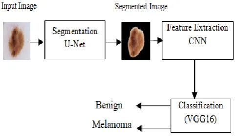
[image:2.595.310.552.98.241.2]In this section, the proposed technique of melanoma classification is described. Figure 2 shows a block diagram of the proposed method. As an initial step, the skin lesion region is segmented from the neighboring healthy skin by using deep learning based U-net algorithm and then extracts discriminate features with the help of Convolutional Neural Network. VGG16 Net algorithm is applied to classify each skin lesion in a dermoscopic image as a Melanoma or Benign.
Fig. 2. Block Diagram
A. Lesion Segmentation
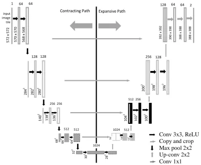
Segmentation is the process of splitting an image into multiple segments, set of pixels. Segmentation is needed to improve the image contrast and reduce the noise levels. There are various forms of network architectures that can have performed segmentation. Here U-net algorithm has been adopted. The U-Net has CNN architecture for fast and accurate segmentation of images. This network combines Convolutional network architecture with a de-convolutional architecture to produce the segmentation. It is a grouping of De-convolutional network and Fully Connected Network (FCN) [13].
a)Contracting:
Figure.3 reveals the U-Net Architecture which consists of contracting path and an expansive path. The contraction section contains many convolutional blocks. Each block receive an input and applies two convolutional layers of size 3x3 and followed by a Rectified Linear Unit (ReLU) and a max-pooling layer of size 2x2. The number of channels or feature maps are doubles after each block so U-Net design can learn the complex images effectively. The bottom layer intermediates contraction and Expansion layers.
b)Expanding
International Journal of Innovative Technology and Exploring Engineering (IJITEE) ISSN: 2278-3075, Volume-8 Issue-12, October 2019
[image:3.595.64.477.96.433.2]U-Net:
Fig. 3. U-Net Architecture
B. Feature Extraction
Feature extraction is a method to extract the unique feature of the skin lesion from segmented image. In some articles small set of features or hand crafted features were extracted but there is a chance to missing some accurate features. Convolutional neural networks automatically extract high level features effectively without the need of manual feature extraction task. In medical imaging there has been a high demand for deep learning methods [14]-[15].
a) CNN Architecture
Convolutional Neural Network (CNN) is a best standard and dominant algorithm for deep learning in areas such as image recognition, segmentation and classification [16]. It consists of an input layer, an output layer and several hidden layers in between.Hidden layers in which the features are extracted. It performs three operations: Convolution, Pooling and Rectified Linear Unit (ReLU). In Convolutional layer several convolutional filters are applied the input images; each filter activates certain features from the image. Pooling layer reduces the number of parameters by simplifying the output performance of nonlinear down sampling. Rectified Linear unit (ReLU) helps for faster and effective training by changing negative values to zero and retaining positive values. Because of that the above three operations are repeated over hundreds of layers, with each layer learning to identify different features.
Classification layer: After all features were detected, the CNN architecture transferred to classification. Fully connected layer is used for actual classification task. It will be able to predict the number of classes that the network has present.
C. Classification
a) VGG16-Net Architecture:
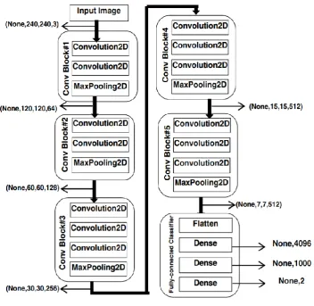
The VGG16 network is a convolutional neural network model comprises 16 convolutional layers with 3x3 filter size, five max-pooling layers with 2x2 window size. A heap of convolutional layers is followed by three fully connected layers. Figure 4 shows architecture of VGG 16 network architecture. It consists of five convolutional blocks; first two blocks have two convolutional layers and one max-pooling layer. Remaining three blocks have three convolutional layers and one max-pooling layer. The Input of first convolutional block is 240x240x3, because of RGB image the channel size is 3. Output of first block is 120x120 x 64 that is 64 channels. This feature map is fed into the input of second block and so on. For the coming blocks output is reduced half the size and the channel size is doubled. The final feature map produced by fifth block of size 7x7x512.
Contracting Path Expansive Path
input image tile
1 64 64
5 7 2 x 5 7 2 57 0 x 5 7 0 5 68 x 5 68
128 64 64 2
output segmentatior map 3 9 0 x 3 90 3 9 2 x 3 92 3 8 8 x 3 88 3 8 8 x 3 88
128 128 256 128
284 2 282 2 280 2 200 2 198 2 196 2
256 256
140 2 138 2 136 2
512 256
104 2 102 2 100 2
512 512
68 2 6 6 2 6 4 2 512 1024 56 2 5 4 2 5 2 2 32 2 30 2 28 2 1024
Fully connected classifier has 3 layers and the first fully connected (Dense) layer have 4096 outputs, second fully connected (Dense) layer have 1000 outputs, the third (Dense) layer performs classification and it contains 2 output which is melanoma or Benign.
that the test indicates the patient who has a specific disease. TN means that the test indicates the non-disease patient has
a disease. Likewise, FP means the test falsely indicates the non-disease patient has a disease, and FN means that the test falsely indicates the actual patient has no disease.
[image:4.595.123.476.167.510.2]Sensitivity shows the ability to absolutely identify the patients with malignant melanoma that is the proportion of patients who are diagnosed exactly as patients.
Fig. 4. Architecture of VGG16 Network
IV. EXPERIMENTAL RESULTS
A. Dataset
Experiments are achieved to evaluate the method on a public challenge dataset of skin lesion Analysis towards Melanoma detection released with ISBI 2016 [17]. This dataset is released by the International Skin Imaging Collaboration (ISIC).The challenge consists of three parts Part 1, part 2 and part 3. The challenge was further divided into sub challenges for segmentation, feature extraction and classification.
The experiment is carried out with 900 dermoscopic images taken from part1 as training image for segmentation with ground truth. Another 900 images from part3 is held out as test dataset for segmentation. From the 900 segmented images 90% of the images are used as training data for classification. Remaining 10% images held out as test data to predict the result.
B. Performance Metrics:
For performance measurement, the proposed work is evaluated using Accuracy, Sensitivity and Specificity[18].
These metrics are defined in terms of the number of True Positives (TP), True Negatives (TN), False Negatives (FN) and False Positives (FP). True Positive(TP) means
Sensitivity =
Specificity shows that the non-disease patient has a negative result that is the percentage of non-melanoma people who are identifiedexactly as healthy.
Specificity =
Accuracy shows the possibilityof the correct determination that is the percentage of patients and healthy people who are diagnosed correctly.
International Journal of Innovative Technology and Exploring Engineering (IJITEE) ISSN: 2278-3075, Volume-8 Issue-12, October 2019
C. Results and Analysis:
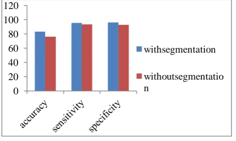
The classification performance of the proposed system is assessed by VGG-16 network algorithm. Twoexperiments are implemented using the same dataset.First classification experiment is executed with the original dataset with segmentation using U-Net algorithm.
[image:5.595.49.288.186.267.2]Second classification experiment is executed with the same dataset without segmentation. Both tests are conducted using VGG-16 network algorithm. Table.1 shows the computed classification performance results.
Table I: Classification Result for the proposed Method With
segmentation
Without segmentation
Accuracy 83.18 76.08
Sensitivity 95.53 93.45
Specificity 96.22 92.96
By comparing the values listed in this table, it is observed that VGG-16 Net Classifier with U-Net based segmentation achieves much better result than VGG-16 Net classifier without segmentation. This is because unsegment image size is very large and artifacts in images. Deep Learning based segmented images can generate more discriminate features for better recognition. VGG-16 classifier with U-Net based segmentation predicts accuracy of (83.18%), Sensitivity of (95.53%) and Specificity of (96.22%). VGG-16 classifier without segmentation predicts accuracy of (76.08%) , sensitivity of (93.45%) and specificity of (92.96%). Figure 5 describes that deep learning based classification with segmented image produce better accuracy, sensitivity and specificity than the classification without segmented image.
Fig. 5. Comparison Chart V. CONCLUSION
In this paper, a U-Net based segmentation and CNN based classification to achieve the challenges of automated melanoma classification in dermoscopy images, which consists of three steps: Segmentation, Feature Extraction and Classification. First the lesion regions are classified using VGG 16 algorithm with U-net based segmentation process and discriminate features are extracted using CNN. Further the same process is executed without segmentation process. Finally, it compares the classification result of both segmentation values and without segmentation values. Experiments conducted on the open challenge dataset of Skin Lesion Analysis towards Melanoma
Detection on ISBI 2016, based on the Accuracy, Sensitivity and Specificity. Result found with segmentation process produce better result compared to the other method. It is believed that the proposed deep learning based melanoma segmentation and classification system can be used as a part of more complex background for skin lesion analysis. In future, this concept can be extended to include integrating probabilistic graphical models into this network to produce more applications.
REFERENCES
1. Narayanan DL, Saladi RN, Fox JL., “Ultraviolet radiation and skin cancer”, International Journalof Dermatology, vol 49,no 9, pp. 978-986, 2010.
2. American Cancer Society. 2016, Atlanta, GA USA: American Cancer Society, 250 William Street, NW, Atlanta GA.
3. Kittler H, Pehamberger H, Wolff K, Binder M., “Diagnostic accuracy of dermoscopy”, Lancet Oncology, vol 3,no 3,pp.159-165, 2002. 4. Ramezani M, Karimian A, Moallem P., “Automatic detection of
malignant melanoma using macroscopic images”, Journal of Medical Signalsand Sensors,vol 4,no 4, pp. 281-290, 2014.
5. 5.Arasi MA, El-HorbatyES M, Salem AB M, El-Dahshan ES A., “Stack Auto encoders approach for Malignant Melanoma diagnosis in Dermoscopy Images”. 2017 IEEE International Conference on Intelligent Computing and Information systems, pp. 403-409, 2017 6. 6.Saez A, Sanchez-Monedero J, Gutierrez P A,Hervas-Martinez C.,
“Machine Learning methods for binary and multiclass classification of melanoma thickness from dermoscopic images”, IEEE transaction on Medical Imaging, vol 35,no 4,pp1036-1045, 2016.
7. 7.Blum A, Luedtke H, Elwanger U, Schwabe R, Rassner G, Garbe C.,” Digital image analysis for diagnosis of cutaneous melanoma- development of a highly effective computer algorithm based on analysis of 837 melanocytic lesions”, British Journal of Dermatology ,vol 151,no 5, pp.1029-1038, 2004.
8. 8. Thompson F, Jeyakumar MK, “Vector based classification of dermoscopic images using SURF”. International Journal of Applied Engineering Research”,vol 12,no 8,pp.1758-1764, 2017.
9. 9.Codella N, Cai J, Abedini M, Garnavi R, Halpern A, Smith J.R., “ Deep learning, Sparse Coding, and SVM for Melanoma Recognition in Dermoscopy Images”, International workshop on machine learning in medical imaging”, Springer,vol 9352. pp.118-126,2015.
10. 10.Bi L, Kim J, Ahn E, Feng D., “Automatic skin lesion analysis using large scale dermoscopy images and deep residual networks”, arxiv:1703.04197v2. 2017.
11. 11. Romero Lopez A, Giro-i-Nieto X, Burdick J, Marques O,“Skin lesion classification from dermoscopic images using Deep Learning Techniques”,13th IASTED International Conference on Biomedical Engineering,pp 49-54, 2017.
12. 12.Kawahara J, BenTaieb A, Hamameh G., “ Deep features to classify skin lesions” , 2016 IEEE 13th International Symposium on Biomedical Imaging (ISBI), pp. 1397-1400, 2016.
13. 13.Ronneberger O, Fischer P, Brox T., “ U-Net Convolutional networks for Biomedical image segmentation”, International Conference on Medical Image Computing and Computer Assisted Intervention(MICCAI), Springer, LNCS, vol 9351: pp.234-241, 2015. 14. 14.Dhungel N, CarneiroG, Bradley AP., "Automated Mass Detection in Mammograms Using Cascaded Deep Learning and Random Forests," 2015 International Conference on Digital Image Computing: Techniques and Applications (DICTA).,pp. 1-8, 2015.
15. 15. Suk HI, Shen D, “Deep learning-based feature representation for AD/MCI classification”,Medical Image Computing andComputer Assisted Intervention-MICCAI, vol. 16(pt 2), pp.583-590, 2013. 16. 16.Lecun Y, Bengio Y, Hinton G.,” Deep Learning”, Nature, vol.521,
pp.436-444,2015.
17. 17.GutmanD, Codella N, Celebi ME, Helba B, Marchetti M, Mishra N, Halpern A., “ Skin Lesion Analysis toward Melanoma Detection: A Challenge at the International Symposium on Biomedical Imaging (ISBI)” 2016 International Skin Imaging Collaboration (ISIC), arXiv : 1605.01397v1, 2016.
18. 18.Zhu W, Zeng N, Wang N., “Sensitivity, specificity accuracy associated confidence interval and ROC analysis with practical SAS implementations”, Northeast SAS User Group proceedings, Section of Health Care and Life Sciences., pp.1-9, 2010.
0 20 40 60 80 100 120
withsegmentation
[image:5.595.45.280.457.597.2]AUTHORS PROFILE
R.D.Seeja is a Senior Lecturer, Department of Computer Engineering, Moderator Gnanadason Polytechnic College, Nagercoil, TamilNadu, India. She is doing research under the guidance of Dr. A. Suresh, Principal, Siri PSG Arts and Science College for women, Sankagiri, TamilNadu,India. She received BSc degree in physics from ManonmaniamSundaranar University, Tirunelveli, also MCA and MPhil degree in Computer Science from ManonmaniamSundaranar University, Tirunelveli, TamilNadu, India. Her area of interest in research is Image Processing.